Original Article
Under-expression of LKB1 is associated with
enhanced p38-MAPK signaling in human
hepatocellular carcinoma
Liang Sha1*, Fang Lian2*, Kezhi Li1, Chuang Chen1, Yinnong Zhao1, Jianbo He1, Shan Huang1, Guobin Wu1
1Department of Hepatobiliary Surgery, Tumor Hospital of Guangxi Medical University, Nanning, P. R. China; 2 De-partment of Physiology, Guangxi Medical University, Nanning, P. R. China. *Equal contributors.
Received August 4, 2018; Accepted August 26, 2018; Epub November 1, 2018; Published November 15, 2018
Abstract: The tumor suppressor liver kinase B1 (LKB1), a highly conserved and ubiquitously expressed protein kinase, plays a critical role in tumorigenesis. LKB1 has recently been identified in tumorigenesis of several cancers including lung cancer, breast cancer, and pancreatic cancer. However, the role of LKB1 in hepatocellular carcinoma (HCC) remains unclear. Herein, we examined the expression levels of LKB1 in HCC patients and cell lines by quan -titative real-time PCR (qRT-PCR) and western blot analysis. Furthermore, LKB1 protein expression was analyzed in archived paraffin-embedded HCC tissues using immunohistochemistry (IHC), and its association with overall sur -vival was shown in statistical analysis. In vitro assays, including RNAi studies, were performed to further explore the role of LKB1 in tumor progression in HCC cell lines. Our results revealed that the expression of LKB1 was lower in HCC tissue and cell lines than in corresponding adjacent normal tissue and normal human liver cell line (HL7702). Moreover, HCC patients with low LKB1 expression had advanced clinical stage and worse prognosis than those with higher LKB1 expression. Furthermore, siRNA-mediated knockdown of LKB1 resulted in enhanced cell proliferation, migration, and invasion of HCC cells. Additionally, the expression level of LKB1 positively correlated with E-cadherin levels, wherein siRNA-transfected cells exhibited significantly decreased levels of E-cadherin, while phosphorylated p38 and vimentin levels were enhanced. Inhibition of p38 MAPK signaling was capable of reversing E-cadherin up-regulation and vimentin down-up-regulation. In all, our results indicate that LKB1 acts as a tumor suppressor gene, which may inhibit EMT through the p38 MAPK signaling pathway involved in HCC progression.
Keywords: LKB1, hepatocellular carcinoma, invasion, migration, p38-MAPK signaling
Introduction
Hepatocellular carcinoma (HCC) is the fifth
most common malignant tumor and the third
leading cause of cancer-related mortality world
-wide [1]. Currently, surgery is still one of the most effective treatments for liver cancer patients. However, because of the high rate of recurrence and metastasis after radical sur
-gery, the long term survival rate of patients with
liver cancer is only 25-39% [2]. Furthermore, the molecular mechanisms underlying invasion
and metastasis of liver cancer are not very clear; therefore, there is an urgent need to
understand HCC tumor invasion and metasta-sis in order to improve diagnometasta-sis, treatment
options, and prognosis of HCC.
Serine/threonine protein liver kinase B1 (LKB1)
is classified as a tumor suppressor that acti
-vates diverse downstream kinases, thus regu-lating multiple biologic processes, including energy metabolism, tumor progression, cell cy-
cle arrest, proliferation, and cell polarity. LKB1
inactivation has been associated with Peutz-Jeghers syndrome (PJS), an inherited autoso-mal disease characterized by gastrointestinal polyps, mucocutaneous melanin pigmentation, and multi-organ cancer susceptibility [3]. The
incidence of cancer among patients with PJS has been estimated to be 18-fold higher than in
the general population [4]. Mice with oncogenic
KrasG12D mutant and loss of LKB1 have sig
-nificantly shortened latency, increased tumor
burden, and increased lung cancer invasion and distant metastasis [5]. Several studies
have indicated that the under-expression of LKB1 might contribute to the progression of
car-cinoma [8], and liver cancer. Moreover, low
expression of LKB1 in HCC may be a poor prog
-nostic factor [9]. LKB1 directly activates AMPK
and AMPK-related kinases to regulate cell
metabolism, proliferation, and cell polarity [10]. Low expression of LKB1 altered cell polarity
and cell adhesion, which enhanced the
trans-formation of normal cells and the metastasis of
tumor cells [11]. Moreover, Li et al. observed that cell migration increased upon LKB1 knock-down in breast cancer [7]. Although LKB1
dis-plays some common features of tumor sup -pressor genes, it is unknown whether LKB1 is a liver cancer suppressor gene. To ascertain its
role, our study aimed to elucidate the function of LKB1 in the growth, proliferation, invasion, and migration of HCC. Specifically, we exam
-ined LKB1 regulating mechanisms of EMT in
HCC, which are currently poorly understood.
TGF-β is a key molecule that promotes EMT in
tumor cells. Previous studies have indicated a
positive correlation between the expression of TGF-β and the metastatic ability of tumor cells [12, 13]. TGF-β signaling involves either the
Smad-dependent pathway or the Smad-inde- pendent pathway, such as p38 mitogen-activat-ed protein kinase (p38 MAPK) and phos-phoinositide-3 kinase (PI3K) [14]. In addition, p38 MAPK activation can inhibit the expression
of E- cadherin [15]. LKB1 acts as an upstream regulator of AMPK and inhibits the activation of the TGF-β signaling pathway [16]. In breast cancer, LKB1 was shown to be a regulator of the p38 MAPK pathway [17]. Therefore, we
spe-culate that LKB1 may play an important role in p38 MAPK pathway-mediated EMT in HCC.
In the present study, we examined the
expres-sion of LKB1 in HCC and investigated its clinical significance and biologic functions. First, we investigated the expression of LKB1 in HCC tis -sues and cell lines by using qRT-PCR and west-ern blot. Second, we analyzed its correlations with clinicopathologic characteristics in order
to determine the clinical signature of LKB1 in HCC. We found that LKB1 was underexpressed
in most HCC cell lines and tissues, a pattern associated with poor prognosis in HCC. In
addi-tion, we used the RNA interference to knock -down the LKB1 expression level in the HCC cell lines and explored the impact on E-cadherin or
vimentin. Our data suggested that LKB1 under-expression may partially facilitate activation of
the p38 MAPK pathway and thus contribute to
EMT and progression of HCC. In all, a better understanding of the molecular mechanism of LBK1 may propel its identification as a novel
diagnostic biomarker and therapeutic target in HCC.
Materials and methods
Patients and surgical specimens
This study enrolled 79 HCC tissues samples for
HCC patients who underwent hepatectomy at
Guangxi Tumor Hospital from January 2014 to December 2014. None of the patients received chemotherapy, radiotherapy, radiofrequency
ablation, target therapy prior to the operation.
The exclusion criteria were a combination of another cancer and a history of liver transplan
-tation. The 79 samples of HCC and the corre
-sponding adjacent tissue were from the same patients. The diagnosis was confirmed by histo
-logic examination. All of the specimens were fixed in 10% formalin and embedded in paraffin for immunochemistry. However, only 49 pair of fresh HCC tissue and ANT (adjacent noncancer
-ous tissue), which was for quantitative PCR analysis, could be obtained from the Tumor Tissue Bank of Guangxi Tumor Hospital. Clini-copathologic data were collected from all pa-tients, including sex, age, HBV, AFP (alpha feto
-protein), maximum size of tumors, number of
tumors, tumor capsule, and tumor thrombus. In
this study, each patient gave informed consent and it was approved by the ethics committee of
Guangxi Tumor Hospital. Immunohistochemistry staining
For assessment of the expression levels of
LKB1 in HCC tissue, immunohistochemical
analysis (IHC) was carried out in samples from all patients (n=79). The sections were deparaf
-finized in xylene and rehydrated through a grad
-ed series of ethanol. Then the sections was placed at 100°C for 3 min in 10 mM sodium citrate buffer (pH=6.0) for antigen retrieval. The
slides were incubated with peroxidase blocking
agent for 10 min to block endogenous peroxi -dase activity and blocked with normal non-immunized animal serum according the
manu-facturer’s protocol. The sections were then
incubated with anti-LKB1 antibody (1:100
dilu-tion) for 14 h at 4°C. The primary antibody was
Evaluation of immunohistochemical staining
LKB1 is predominantly staining in the cyto-plasm. We used semi-quantitative analysis to
analyze the immunostaining intensity of LKB1, which was determined by the percentage of
positive cells and the staining intensity. Each slide was independently observed by two
expe-rienced pathologists. The proportion of positive cells was grade as follows: 0 (no positive tumor
cells), 1 (1-25% positive tumor cells), 2 (26-50% positive tumor cells), 3 (51-75% positive tumor cells), 4 (76-100% positive tumor cells). Staining
intensity was scored using four grades: 0 (nega -tive), 1 (weakly tive), 2 (moderately posi-tive), 3 (strongly positive). The staining index
(range from 0 to 12) was calculated by multiply
-ing proportion of positive cells score with score of staining intensity. Low expression of LKB1 group was for scores from 0 to 5. The ones with scores from 6 to 12 were defined as high expression of LKB1.
Cell lines
Human normal liver cell line (HL7702) and human HCC cell lines BEL-7404, Hep-G2, SSMC-7721, MHCC-97-H, Huh-7, were purch-
ased from the cell bank of the Chinese Aca-demy of Science. All cell lines were cultured in Dulbecco’s modified Eagle’s medium (DMEM, Gibco) with 10% fetal bovine serum (Gibco), 0.5% penicillin, and 0.5% streptomycin (Life
Technologies Corporation, Carlsbad, CA, USA). The cell lines were cultured in a sterile
incuba-tor maintained at 37°C with 5% CO2. Reagents and antibodies
Rabbit anti-human LKB1 monoclonal antibody
was purchased from Abcam PLC (Cambridge,
UK). Rabbit monoclonal antibody against hu- man p38 MAPK, Phospho-p38 MAPK, E-cad-
herin, β-actin were purchased from Cell
Sig-naling Technology, Inc. (Danvers, MA, USA). Rabbit monoclonal antibody against human
Vimentin was purchased from Santa Cruz Biotechnology. SB202190 was obtained from Sigma-Aldrich (St. Louis, MO, USA).
SiRNA and transfection
Three small interfering RNA sequences target
-ing different regions of human LKB1 mRNA were designed to exclude nonspecific effects. A
scrambled siRNA was employed as a negative control (Gene Pharma, Shanghai, China). The
siRNA targeting the LKB1 sequences
(LKB1-siRNA1 sense 5’-CCUGCUGAAAGGGAUGCU-UTT-3’ and antisense 5’-AAGCAUCCCUUUCA-GCAGGTT-3’; LKB1-siRNA2 sense 5’-GGAUG-UGUUAUACAACGAATT-3’ and antisense 5’-UUCG-UUGUAUAACACAUCCTT-3’ LKB1-siRNA3 sense
5’-CCAACGUGAAGAAGGAAAUTT-3’ and anti
-sense 5’-AUUUCCUUCUUCACGUUGGTT-3’) and
the non-silencing sequence
(LKB1-scrambled-siRNA sense 5’-UUCUCCGAACGUGUCACGUTT-3’ and antisense 5’-ACGUGACACGUUCGGAGA-ATT-3’). About 24 h before transfection, cells were seeded at the density of 5 × 10*6 in six-well plates. When the cells reached confluence of 70-80%, cells were transfected with small interfering RNA following the manufacturer’s protocol. After six hours of transfection, the
medium was replaced by the DMEM containing
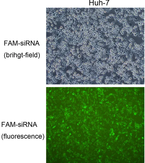
10% FBS. Transfection efficiency was evaluat
-ed by fluorescence microscopy (OLYMPUS
TH4-200, Figure S1) after transfection of FAM-siRNA for six hours. The silencing effect was detected by qRT-PCR and western blotting 48 h after transfection. The small interfering RNA
se-quence (LKB1-siRNA1) with better silencing
effect was selected for subsequent cell func
-tion experiments. For the cell prolifera-tion
assay, the Cell Counting Kit-8 (CCK-8) (Dojindo,
Kumamoto, Japan) was used. After 24 hours of transfection, the cells were collected by cen
-trifugation and resuspended. The cells were
counted using a hemocytometer and seeded in
a 96-well plate at a density of 5 × 10*3 cells/
well with six replicates. Cells were incubation in
DMEM containing 10% FBS for 6, 24, 48, and 72 h, respectively. At end of the time, the medi -um were added 10 µl CCK-8 reagent to each
well. The cells were cultured for 2 h at cell incu -bator. The absorbance was measured at a
wavelength of 450 nm.
Cell invasion assay
Matrigel (BD Biosciences, Franklin Lakes, NJ, USA) was incubated overnight at 4°C and di-
luted in serum-free DMEM medium. The
Tran-swell upper chamber is a polycarbonate mem-brane with 8 µm pore size coated with 60 µl the Matrigel and overnight at 37°C. The cells were
collected 24 hours after transfection. After the
digestion, the cells were resuspended in
serum-free medium and inoculated at 1 × 10*5/200 μl/well in the upper chamber. The chamber was
placed in a 24-well plate, seven hundred micro-liters DMEM containing 10% FBS added in the
cells were fixed in methanol and then stained
with Giemsa. The non-invading cells were
removed from the upper chamber, and cell on the lower surface were stained. Migrated cells were calculated in five random fields (100 × magnification) for each chamber under a light
microscope. Cell migration assay
For migration assay, 2 × 10*4 cells in 200 µl serum-free DMEM were added into the upper
Transwell chamber without Matrigel. The re- maining experimental steps were the same as the cell invasion assay.
Quantitative real-time PCR
QRT-PCR was used to research the LKB1 mRNA
levels of cell lines, HCC tissue and ANT. Total RNA was extracted from tissues and cell lines with TRIzol reagent (Invitrogen, Life Techno-logies, CA, USA) according to the manufactur
-er‘s instructions. The concentration of RNA was
AA-3’) and β-actin (F, 5’-AGTGTGACGTTGACAT-CCGT-3’ and R, 5’-GCAGCTCAGTAACAGTCCGC-3’). β-actin was used as an internal control. The
2-ΔΔCt method was used to calculate the fold
changes for LKB1 expression levels.
Western blotting assay
The LKB1 and E-cadherin protein levels of cells
were measured by western blotting. Cells were
lysed in RIPA buffer (50 mM Tris, PH 7.4, 150
mM NaCl, 1% TritonX-100, 1% sodium deoxy-cholate, 1% SDS) containing 1 mmol/l PMSF
and then centrifuged at 12000 rpm/min, 4°C for 10 min to collect the supernatant. BCA pro -tein assay kit (KGPBCA, Nanjing, China) was
used to measure the concentration of total pro
-tein in each sample. Fifty micrograms of total
protein were separated by SDS-PAGE and then
transferred to the polyvinylidene fluoride mem -branes. Subsequently, the membranes were
blocked in TBST solution containing 5% nonfat milk for 60 min, the membranes were incubat -ed with each primary antibody (LKB1, 1:100; E- cadherin, 1:1000; vimentin, 1:500; p38 MAPK,
1:1000; p-p38 MAPK, 1:1000; β-actin, 1:1000)
and then washed and probed with respective secondary peroxidase conjugated antibodies. The detection was implemented by ECL
detec-tion reagent (BOSTER, Wuhan, China).
Statistical analysis
All data processing was carried out using SPSS
19.0 software. The difference in LKB1 expres -sion between HCC tissues and adjacent tissues was analyzed by t test. The differences between
the groups were analyzed by one-way analysis
of variance (ANOVA) when there were more
[image:4.612.92.370.74.223.2]than two groups. The chi-square test was used Figure 1. Expression of LKB1 mRNA and protein in HCC cell lines (Huh7,
Hep-G2, SSMC-7721, MHCC-97H, and BEL7404) and HL-7702 were exam-ined by qRT-PCR and western blot.
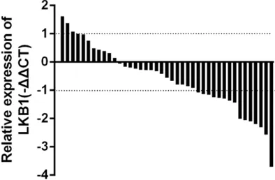
Figure 2. Waterfall plot shows expression of LKB1 in human HCC tissue (n=42), -ΔΔCt=(CtLKB1-Ctβ-actin) ANT-(CtLKB1-Ctβ-actin) HCC, Fold change=2-ΔΔCt.
measured at A260 and A280 by Nano drop 2000 (Thermo
Scientific, Waltham, MA, USA). Approximately 1 µg of RNA
was used to prepare cDNA using ReverTra Ace qPCR RT
kit (TOYOBO, Osaka, Japan) according to the manufactur
-er’s instructions. Q-PCR was
conducted using a THUNDER-
BIRD qPCR Mix (TOYOBO,
Osa-ka, Japan) in an Applied Bio- systems 7500 (Applied Bio-
systems). The following prim -ers were used to detect the
[image:4.612.91.288.285.414.2]5’-CCCTTCCCGATGTTCTC-ble 1). The remaining data suggested no corre-lation between LKB1 and age (P=0.493), gen-der (P=0.198), BCLC stage (P=0.146), number
of tumors (P=0.422), hepatitis B (P=0.791),
Child-Pugh stage (P=0.503), tumor embolus (P=0.075). Because LKB1 was closely related
to maximum size of tumors, tumor capsule and
histological grade, it can be deduced that LKB1 may be involved in HCC progression.
LKB1 is predictive of poor outcomes in HCC
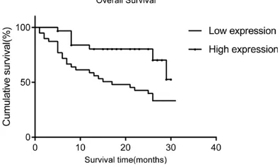
In order to evaluate whether LKB1 affected the survival of patients with HCC, we performed
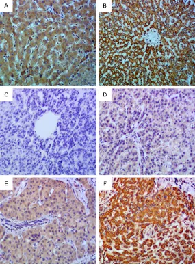
[image:5.612.90.375.73.462.2]survival analysis in relation to LKB1 expression. Kaplan-Meier analysis showed that patients Figure 3. LKB1 protein showed decreased expression in HCC tissues. A.
Immunohistochemical demonstration of LKB1 expression in normal livers. B. Positive staining of LKB1 in adjacent surrounding non-tumor tissues. C. Negative staining of LKB1 in HCC specimens. D. Weak staining of LKB1 in HCC specimens. E. Moderate staining of LKB1 in HCC specimens. F. Strong staining of LKB1 in HCC specimens.
BEL-7404, Hep-G2 SSMC-77- 21, and MHCC-97H. Compar- ed to the normal liver cell line
(HL-7702), we found that the mRNA and protein levels of LKB1 were significantly lower
in most HCC cell lines with
the exception of Huh-7 cells,
which showed higher
expres-sion of LKB1 (Figure 1). Sub-
sequently, we verified the ex-pression of LKB1 mRNA in
HCC tissues. The results sh- owed that the expression level
of LKB1 mRNA was lower in
most HCC tissues than in the corresponding adjacent tis-sues (Figure 2).
Association of LKB1 expres-sion with clinicopathological features of HCC
We used IHC to examine the
expression level of LKB1 in 79 cases of HCC samples. IHC
data showed that the LKB1 protein was mainly localized in the cytoplasm in tissues (Fi- gure 3). The enrolled patients were divided into two groups
(low and high expression of
LKB1) according to the IHC
results of LKB1. Our data
sh-owed that patients in the low
expression of LKB1 group we-re 54.4% (43 out of 79), and
LKB1 IHC staining was corre-lated with tumor size (P= 0.005) and histologic grade (P=0.024) in HCC patients (Ta-
to analyze the relationship between LKB1 expression and clinicopathologic characteris-tics. The association between LKB1 expression and overall survival, as well as the prognostic
value of LKB1, were estimated by Kaplan-Meier
and Cox regression analysis, respectively. P<
0.05 was considered significant.
Results
LKB1 is under-expression in HCC cell lines and HCC tissues
We used qRT-PCR and western blot analyses to
measure the expression levels of LKB1 mRNA
with low LKB1 expression had a significantly
shorter overall survival than those with high
LKB1 expression (χ2=6.244, P=0.012) (Table
2). Univariate analysis indicated that tumor
size (P=0.026), number of tumors (P=0.008),
histologic grade (P=0.025), LKB1 expression
(P=0.012) were closely related to unfavorable
outcomes in HCC patients. Multivariate
analy-sis showed that number of tumors (HR 2.456;
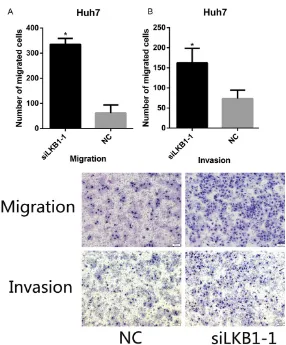
We used transwell assays in order to study
the regulation of LKB1 on the invasion and migration of HCC cells. We observed that the migration and invasion of cells were sig-nificantly increased after LKB1 knockdown
compared to the negative control groups. These results indicated that LKB1 may inhibit
the migration and invasion of HCC cells (Figure 7).
95% CI 1.194, 5.051; P= 0.015), LKB1 expression (HR 0.405; 95% CI 0.188, 0.875; P=0.021) were independent prognostic predictors. In gen-eral, these results indicated
that low expression of LKB1
was likely an independent
indi-cator of poor outcome in HCC
(Table 2; Figure 4).
LKB1 knockdown increase the proliferation of Huh7 cells
To further examine the mecha -nism underlying the potential
tumor suppressor role of LKB1
in HCC, siRNA transfection was utilized. Successful siR
-NA-LKB1 transfection was confirmed by qRT-PCR and
western blot analyses; the re- sults showed that the
expres-sion of LKB1 mRNA decreased significantly after transfection of LKB1-siRNA in Huh7 cells,
and siLKB1-1 decreased more
significantly (P<0.05 vs. nega -tive control). The expression
of LKB1 protein was also sig
-nificantly inhibited; therefore, siLKB1-1 was selected for sub -sequent studies (Figure 5).
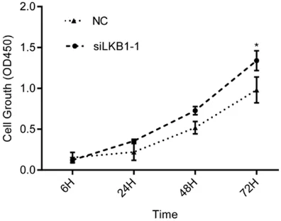
Next, we examined the impact
of LKB1 on the proliferation of
HCC cells via the Cell Count- ing Kit-8 (CCK-8) assay. The results showed that siLKB1
significantly promoted prolif
-eration of Huh7 cells (P<0.05)
(Figure 6).
[image:6.612.90.373.97.594.2]Under-expression of LKB1 enhanced HCC cell migration and invasion in vitro
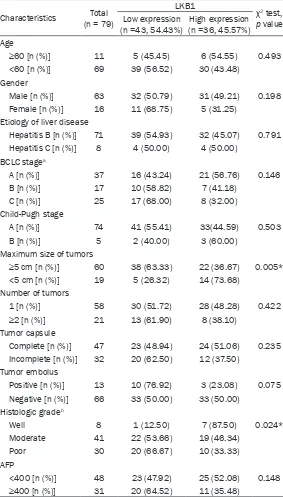
Table 1. Correlations between LKB1 expression and
clinicopatho-logical characteristics of HCC patients
Characteristics (n = 79)Total
LKB1
χ2 test,
p value Low expression
(n =43, 54.43%) (n =36, 45.57%)High expression Age
≥60 [n (%)] 11 5 (45.45) 6 (54.55) 0.493
<60 [n (%)] 69 39 (56.52) 30 (43.48) Gender
Male [n (%)] 63 32 (50.79) 31 (49.21) 0.198
Female [n (%)] 16 11 (68.75) 5 (31.25) Etiology of liver disease
Hepatitis B [n (%)] 71 39 (54.93) 32 (45.07) 0.791 Hepatitis C [n (%)] 8 4 (50.00) 4 (50.00)
BCLC stagea
A [n (%)] 37 16 (43.24) 21 (56.76) 0.146
B [n (%)] 17 10 (58.82) 7 (41.18)
C [n (%)] 25 17 (68.00) 8 (32.00)
Child-Pugh stage
A [n (%)] 74 41 (55.41) 33(44.59) 0.503
B [n (%)] 5 2 (40.00) 3 (60.00)
Maximum size of tumors
≥5 cm [n (%)] 60 38 (63.33) 22 (36.67) 0.005*
<5 cm [n (%)] 19 5 (26.32) 14 (73.68) Number of tumors
1 [n (%)] 58 30 (51.72) 28 (48.28) 0.422
≥2 [n (%)] 21 13 (61.90) 8 (38.10)
Tumor capsule
Complete [n (%)] 47 23 (48.94) 24 (51.06) 0.235 Incomplete [n (%)] 32 20 (62.50) 12 (37.50)
Tumor embolus
Positive [n (%)] 13 10 (76.92) 3 (23.08) 0.075 Negative [n (%)] 66 33 (50.00) 33 (50.00)
Histologic gradeb
Well 8 1 (12.50) 7 (87.50) 0.024*
Moderate 41 22 (53.66) 19 (46.34)
Poor 30 20 (66.67) 10 (33.33)
AFP
<400 [n (%)] 48 23 (47.92) 25 (52.08) 0.148
≥400 [n (%)] 31 20 (64.52) 11 (35.48)
Effect of LKB1 on the Expression of E-cadherin and Vimentin in Huh7 cells
The expression levels of LKB1, E-cadherin and
vimentin were evaluated by western blotting to determine the correlation between LKB1 and
EMT in HCC cell lines. Our results indicated that down-regulation of LKB1 resulted in upregula
-tion of vimentin and down-regula-tion of
E-cad-herin protein expression in Huh7 cells (Figure 8A). These results indicated that LKB1 may play an important role in HCC progression and metastasis by regulating the EMT process. LKB1 regulates EMT through p38 MAPK sig-naling pathway
Thus far, our results suggested that downregu
-lation of LBK1 may be critical in promoting
EMT. To further investigate this hypothesis, we examined the activation of p38 MAPK signaling
pathway which has previously been suggested
to inhibit the expression of E-cadherin. The expression of phosphorylated p38 was signifi
-cantly increased following transfection of
si-LKB1 in Huh7 cells (Figure 8A). Therefore, we
hypothesized that LKB1 modulated the
expres-sion of EMT-related molecules through p38 MAPK signaling pathway. To further investigate the role of p38 MAPK in regulation of EMT, we blocked the activation of p38 MAPK by employ -ing SB202190, a strong and highly selective
inhibitor of the p38 MAPK pathway. Huh7 cells were treated with SB202190 (10 μmol/L) for 4 h, the levels of E-cadherin, vimentin, and phos -phorylated p38 are presented in Figure 8. We
observed that SB202190 treatment of siLKB1-transfected cells upregulated the expression of
E-cadherin while downregulating the
expres-sion of vimentin and p-p38 (Figure 8B). Taken together, our data suggested that inhibition
of LKB1 may affect EMTby the p38 MAPK
pathway.
Discussion
Hepatocellular carcinoma (HCC) is one of the
most common human malignant tumors. With
a high incidence of liver cancer in China, the number of new cases and deaths in the country accounted for about 50% of the number of new
cases worldwide [18]. Despite improvement in
[image:7.612.92.520.97.283.2]surgery for HCC treatment in recent years, late diagnosis or misdiagnosis of symptoms results Figure 4. The survival analysis of LKB1. Patients with
[image:7.612.90.288.318.436.2]low LKB1 expression in tumor were closely corre-lated with poorer overall survival than patients with tumor with higher LKB1 expression.
Table 2. Clinical variables correlated with overall survival in patients with hepatocellular carcinoma based on Cox univariate and multivariate models
Variables Univariate analysis Multivariate analysis
HR (95%CI) p HR (95%CI) p
Age: <60 vs. ≥60 0.914 (0.328-2.716) 0.914
Sex: male vs. female 0.869 (0.376-2.010) 0.743
Tumor size: <5 cm vs. ≥5 cm 3.034 (1.061-8.679) 0.038*
Number of tumors: single vs. multiple 2.518 (1.230-5.156) 0.012* 2.456 (1.194-5.051) 0.015#
Tumor capsule: complete vs. incomplete 1.015 (0.504-2.043) 0.966 Histological grade: well/moderate vs. poor 2.114 (1.066-4.190) 0.032*
Serum AFP (ng/ml): <400 vs. ≥400 1.491 (0.749-2.970) 0.256
HBsAg: positive vs. negative 0.960 (0.292-3.160) 0.946 Tumor embolus: positive vs. negative 0.957 (0.394-2.325) 0.923 BCLC stage: A vs. B vs. C 1.168 (0.800-1.706) 0.420
Child-Pugh: A vs. B 0.701 (0.168-2.929) 0.626
in rapid HCC progression. Typically, patients have advanced liver cancer or the emergence
of extensive liver metastasis at diagnosis, thus
limiting their comprehensive treatment options.
Furthermore, the prognosis of HCC patients after systemic treatment still remains poor
[19]. Tumor invasion and metastasis are major
causes for treatment failure; therefore, the ulti -mate goal is to determine the molecular
mech-anism of metastasis and target critical pathway inhibition of this process. Tumor metastasis
and invasion are complex biologic processes, which are impacted by both oncogene activa-tion and tumor suppressor gene inactivaactiva-tion.
Therefore, understanding the molecular mech
-anisms of tumor invasion and metastasis with
of EMT is the transformation of epithelial cells into mesenchymal cells. The expression of epi -thelial maker E-cadherin was down-regulated and mesenchymal maker vimentin was up-reg-ulated in the present study. The epithelial cells lose cell polarity and cell adhesion, and acquire mesenchymal-like characteristics in order to
migrate away from the original tissue.
E-cad-herin has been recognized as a very important
marker for EMT, and its under-expression has
been strongly associated with cancer progres-sion and poor prognosis [20]. Vimentin is an
intermediate filament protein is mainly expre-ssed in mesenchymal cells, and in a variety of
epithelial tumors. Vimentin is closely related to
the differentiation, invasion, and metastasis of
tumor cells, and can be used as a prognostic
indicator of malignant tumor. Increasing evi -dence suggests that EMT plays an extremely
important role during the process of the spread of cancer of HCC [21]. In agreement with the results of previous research, we found that
knockdown of LKB1 decreased E-cadherin and
increased vimentin expression, enabling in the
spread of HCC in distant metastatic sites.
In the current study, to interrogate the role of
LKB1 in HCC, we detected the expression
lev-els of LKB1 mRNA and protein in HCC tissues and cell lines. The expression of LKB1 mRNA in HCC tissues was significantly lower compared
to the adjacent tissues. Moreover, the relative
expression of LKB1 mRNA in most cell lines
was lower than in normal liver cell line (HL-7702), in all but one cell line (Huh-7). Fur-
thermore, we used IHC analysis of 79 cases
[image:8.612.92.372.72.222.2]and examined the relationship between LKB1 Figure 5. Knockdown of LKB1 enhanced cell proliferation in Huh7 cells. A,
B. The relative mRNA and protein expression level of LKB1 in Huh7 cells was significantly decreased by siLKB1 compared with si-NC group.
Figure 6. After transfection of 24 h, cell proliferation assays were conducted to determine the proliferation of Huh7 cells. Results are expressed as mean ± SD for three replicate determinations. Statistically sig -nificant differences between siLKB1 group and si-NC group were determined by Student’s t test.
the aim of exploring effective
drug targets and biomarkers
for the diagnosis and treat
-ment of cancer is of great significance.
Recent research shows that the invasion and metastasis
of cancer is a multistep pro
-cess. EMT, which was first
discovered at key transition steps during embryogenesis, is thought to be a key
pheno-typic change in the ability of
tumor cells to acquire invasion
and metastatic features and
[image:8.612.90.288.287.443.2]Figure 7. Knockdown LKB1 expression was enhanced cell migration and invasion of Huh7 cells. Trans-well assay showed Huh7 cells infected with si-LKB1 displayed significantly higher migration capacity compared with the si-NC group. Cell invasion assay showed Huh7 cells infected with si-LKB1 dis -played significantly higher invasion capacity compared with the si-NC group.
Figure 8. A. Effects of knockdown of LKB1 on expression of E-cadherin and vimentin and p-p38 in Huh7 cells. B. Effects of knockdown of LKB1 on p38 MAPK signaling and treated with SB202190.
expression and
clinicopatho-logic features. The results
sh-owed that the relative
expres-sion level of LKB1 was signifi -cantly correlated with tumor size and histologic grade in HCC patients. However, LKB1 expression was not associat-ed with age, gender, BCLC
stage, number of tumors, hep -atitis B, Child-Pugh stage, or tumor embolus. Furthermore, we demonstrated that low
expression of LKB1 protein exhibited a significant associ -ation with a shorter overall survival (P=0.021). These da- ta indicated that the low ex-
pression of LKB1 was a pos -sible independent prognostic
factor in HCC. Our study was
consistent with a previous
report that found that the expression of LKB1 was lower
in gastric tissue cell lines and tumor tissues (155 gastric cancer) compared to normal gastric cells and tissues (95 normal gastric epithelial tis-sue specimens) [22]. There-
fore, we speculate that LKB1
may also regulate the grow-
th and metastasis of HCC
cells.
To determine the biological
function of LKB1 in HCC, we
silenced its expression by
siRNA transfection. Following the inhibition of LKB1, the proliferation, invasion, and migration of cells were signifi -cantly enhanced compared with the control group. In
addition, the expression of
E-cadherin and vimentin was
examined, and we found that down-regulation of E-cadherin and increased expression of
vimentin in the siLKB1 group suggesting that
down-regula-tion of LKB1 in Huh7 cells may
induce EMT to regulate
[image:9.612.89.376.495.670.2]cells. We also found that phosphorylated p38 was significantly increased in the LKB1-si-lenced group. Because the activation of p38
MAPK signaling pathway can down-regulate the
expression of E-cadherin, we hypothesized that
LKB1 low expression leads to the progression
of tumor by modulating EMT by p38 MAPK sig
-naling pathways and thus affecting the malig -nant progression of HCC. Furthermore, we fo-und that with the inhibition of the p38 MAPK
pathway, phosphorylated p38 levels were
sig-nificantly decreased, E-cadherin expression
increased, whereas vimentin expression de- creased. These data suggested that the
activa-tion of p38 MAPK signaling pathway is one of
the mechanisms leading to tumor invasion and migration when LKB1 is silenced. However,
investigation of the detailed molecular regula
-tion mechanism requires further study.
None-theless, our data suggests that LKB1 may play an important role in the progression,
tumori-genesis, and development of HCC as a tumor
suppressor gene.
Our research has several limitations: our stud -ies on cell growth and metastasis were only
carried out in vitro, thus need to be verified in in
vivo studies. Secondly, only one HCC cell line
was employed in majority of this study; there
-fore, additional research in HCC cell lines that
overexpress LKB1 are required to support our
findings. Finally, the amount of clinical sample data was small (n=79), and follow-up time (32
months) was too short; thus, larger studies with
longer follow-up are needed to confirm our
views.
In conclusion, our results suggest that LKB1 is a tumor suppressor gene with low expression in hepatocellular carcinoma and may regulate
the progression of HCC through the p38 MAPK
signaling pathway. Further studies are needed to understand the mechanisms underlying the
effect of LKB1-induced cell polarity on liver
cancer metastasis and invasion.
Acknowledgements
This work was supported by grants from the National Natural Science Foundation of China
(Grant nos. 81460426 and 81360315), the
Natural Science Foundation of Guangxi
Pro-vince (Grant no. 2014GXNSFAA118191).
Disclosure of conflict of interest
None.
Address correspondence to: Dr. Guobin Wu, Depart- ment of Hepatobiliary Surgery, Tumor Hospital of Guangxi Medical University, Nanning 530000, P. R. China. E-mail: gb.wu@139.com
References
[1] Jemal A, Bray F, Center MM, Ferlay J, Ward E, Forman D. Global cancer statistics. CA Cancer J Clin 2011; 61: 69-90.
[2] Crissien AM, Frenette C. Current management of hepatocellular carcinoma. Gastroenterol Hepatol (N Y) 2014; 10: 153-161.
[3] Bardeesy N, Sinha M, Hezel AF, Signoretti S, Hathaway NA, Sharpless NE, Loda M, Carrasco DR, DePinho RA. Loss of the Lkb1 tumour sup -pressor provokes intestinal polyposis but resis-tance to transformation. Nature 2002; 419: 162-167.
[4] Hemminki A, Avizienyte E, Roth S, Loukola A, Aaltonen LA, Järvinen H, de la Chapelle A. A serine/threonine kinase gene defective in Peu -tz-Jeghers syndrome. Duodecim 1998; 114: 667-668.
[5] Ji H, Ramsey MR, Hayes DN, Fan C, McNamara K, Kozlowski P, Torrice C, Wu MC, Shimamura T, Perera SA, Liang MC, Cai D, Naumov GN, Bao L, Contreras CM, Li D, Chen L, Krishnamur-thy J, Koivunen J, Chirieac LR, Padera RF, Bron-son RT, Lindeman NI, Christiani DC, Lin X, Sha-piro GI, Jänne PA, Johnson BE, Meyerson M, Kwiatkowski DJ, Castrillon DH, Bardeesy N, Sharpless NE, Wong KK. LKB1 modulates lung cancer differentiation and metastasis. Nature 2007; 448: 807-810.
[6] Li F, Han X, Li F, Wang R, Wang H, Gao Y, Wang X, Fang Z, Zhang W, Yao S, Tong X, Wang Y, Feng Y, Sun Y, Li Y, Wong KK, Zhai Q, Chen H, Ji H. LKB1 inactivation elicits a redox imbalance to modulate non-small cell lung cancer plastic-ity and therapeutic response. Cancer Cell 2015; 27: 698-711.
[7] Li J, Liu J, Li P, Mao X, Li W, Yang J, Liu P. Loss of LKB1 disrupts breast epithelial cell polarity and promotes breast cancer metastasis and invasion. J Exp Clin Cancer Res 2014; 33: 70. [8] Morton JP, Jamieson NB, Karim SA, Athineos
D, Ridgway RA, Nixon C, McKay CJ, Carter R, Brunton VG, Frame MC, Ashworth A, Oien KA, Evans TR, Sansom OJ. LKB1 haploinsufficiency cooperates with Kras to promote pancreatic cancer through suppression of p21-dependent growth arrest. Gastroenterology 2010; 139: 586-597, e1-6.
Asian Pac J Cancer Prev 2013; 14: 1985-1988.
[10] Gan RY, Li HB. Recent progress on liver kinase B1 (LKB1) - expression, regulation, down-stream signaling and cancer suppressive func -tion. Int J Mol Sci 2014; 15: 16698-16718. [11] Nakano A, Takashima S. LKB1 and
AMP-acti-vated protein kinase: regulators of cell polarity. Genes Cells 2012; 17: 737-747.
[12] Guarino M. Epithelial-mesenchymal transition and tumour invasion. Int J Biochem Cell Biol 2007; 39: 2153-2160.
[13] Akhurst RJ. TGF-beta antagonists: why sup-press a tumor supsup-pressor. J Clin Invest 2002; 109: 1533-1536.
[14] Zhu J, Huang S, Wu G, Huang C, Li X, Chen Z, Zhao L, Zhao Y. Lysyl oxidase is predictive of unfavorable outcomes and essential for regu -lation of vascular endothelial growth factor in hepatocellular carcinoma. Dig Dis Sci 2015; 60: 3019-3031.
[15] Zohn IE, Li Y, Skolnik EY, Anderson KV, Han J, Niswander L. p38 and a p38-interacting pro-tein are critical for downregulation of E-cad -herin during mouse gastrulation. Cell 2006; 125: 957-969.
[16] Li NS, Zou JR, Lin H, Ke R, He XL, Xiao L, Huang D, Luo L, Lv N, Luo Z. LKB1/AMPK inhibits TGF-beta1 production and the TGF-beta signaling pathway in breast cancer cells. Tumour Biol 2016; 37: 8249-8258.
[17] Rhodes LV, Tate CR, Hoang VT, Burks HE, Gil-liam D, Martin EC, Elliott S, Miller DB, Buechlein A, Rusch D, Tang H, Nephew KP, Burow ME, Collins-Burow BM. Regulation of triple-negative breast cancer cell metastasis by the tumor-suppressor liver kinase B1. Oncogenesis 2015; 4: e168.
[18] Torre LA, Bray F, Siegel RL, Ferlay J, Lortet-Tieu-lent J, Jemal A. Global cancer statistics, 2012. CA Cancer J Clin 2015; 65: 87-108.
[19] Zhu AX. Systemic treatment of hepatocellular carcinoma: dawn of a new era. Ann Surg Oncol 2010; 17: 1247-1256.
[20] Thiery JP. Epithelial-mesenchymal transitions in tumour progression. Nat Rev Cancer 2002; 2: 442-454.
[21] Chen J, Liu WB, Jia WD, Xu GL, Ma JL, Huang M, Deng YR, Li JS. Overexpression of Mortalin in hepatocellular carcinoma and its relation-ship with angiogenesis and epithelial to mes-enchymal transition. Int J Oncol 2014; 44: 247-255.