Original Article
Effect of miR-146a on glioma cell line proliferation
in targeted-regulating MIF gene
Xiangdong Lu1,2, Feng Si2, Shouqing Zhang2, Zhenrui Liu2
1Department of Neurosurgery, Provincial Hospital Affiliated to Shandong University, Jinan 250021, Shandong,
China; 2Department of Neurosurgery, Laiwu City People’s Hospital, Laiwu 271199, Shandong, China Received October 29, 2015; Accepted December 25, 2015; Epub March 1, 2016; Published March 15, 2016
Abstract: It is reported that miR-146a is associated with various tumors. However, its specific effect on glioma has not yet been studied. This paper is to explore the effect of miR-146a on glioma. RT-PCR method was used to detect the expression of miR-146a in neurogliocytoma tissue and corresponding non-tumor normal tissue, as well as in neurogliocytoma cell strain and neurogliocyte in normal people. IHC was used to detect the expression of MIF in neurogliocytoma tissue and corresponding non-tumor normal tissue. Relationship between MIF and miR-146a was detected with the luciferase reporter gene. Cell jamming technology was adopted, by which, miR-146a mimics and miR-146a negative control (NC) were transferred into cell line of neurogliocytoma for detecting the expressions of MIF mRNA and proteins. MTT method was used to detect cell viability and cloning experiments to detect the pro-liferative potential of cells. The expression quantity of miR-146a in neurogliocytoma tissue was lower than that in corresponding non-tumor normal tissue. The miR-146a in cell line of neurogliocytoma was lower than that in normal neuroglia tissue and the expression quantity in U87 was appropriate. The expression of MIF proteins in neuroglio-cytoma tissue was higher than that in corresponding non-tumor normal tissue. And luciferase reporter gene had verified that MIF was the downstream target gene of miR-146a and transferred to U87 cell by liposome. miR-146a shows low expressions in neurogliocytoma tissue and cell line, while MIF shows over expressions in neurogliocytoma tissue. The miR-146a expression can be up-regulated by targeted inhibiting the MIF expression, accordingly, to in-hibit the proliferation of neurogliocytoma.
Keywords: miR-146a, MIF, glioma
Introduction
Glioma, a kind of malignant tumor, is one of the most harmful cancers to human health in 21st
century. Amount of studies have confirmed that
MicroRNA (miRNAs) shows abnormal
expres-sion in kinds of cancers and is closely related to the development and progression of multiple
tumor, in which the glioma is included [1]. miR-NAs is an endogenous small-molecule non-encoding RNA, which is highly conserved during evolution. Thereinto, miR-146a is located in chromosome 5 LOC285628 gene with its mature sequences at exon 2. Studies have
con-firmed that miR-146a content in patients with
glioma is lower than that in benign neuroglia tissue and the expression quantity in glioma cells is lower than that in normal nerve cells with low miR-146a expression and high TNM-staging. Low survival rate is closely related to lymph node metastasis [2-4]. It has shown that
miR-146a has close relation with the
develop-ment and progression of the gloma, but the specific mechanism is unknown. Macrophage migration inhibitory factor (MIF) is a kind of highly conserved protein, derived from T cell or
can by secreted by monocyte/macrophage and anterior pituitary cells. Several studies have
found that MIF is involved into the development and progression of gloma. Kamimura et al. [5] have researched the MIF over expression in
gloma tissue of gloma patients by IHC and
shown the close relation with prognosis. Several
research findings show that the expression degree of MIF in different patients with gloma was different [6]. It shows that both miR-146a
and MIF can show abnormal expressions in gloma and speculates that MIF may be one the
downstream target proteins of miR-146a. Therefore, on the above basis, this paper is respectively detecting the expressions of
IHC, verifying if MIF is the downstream target proteins of miR-146a by luciferase reporter gene analysis and transferring the miR-146a
mimics into glioma cell with the liposome
trans-fection method. This paper also aims at explor
-ing the effect of MIF expression on cell prolif -eration in gloma cells.
Materials and methods
Study subjects
Twenty cases of glioma tissue samples and 20 cases of corresponding non-tumor normal tis -sue samples which were removed surgically were collected between July 2014 to June 2015 in Laiwu City People Hospital. Among the cases
of glioma tissue samples, there were 12 cases that had suffered lymphatic metastasis, while
other 8 cases didn’t. Cases were divided into
different clinical stages in accordance with the TNM-staging standard of Union for International
Cancer Control (UICC) in 2009. The TNM-staging was shown below: 14 cases in Stage I-II, 6 cases in Stage III-IV. The cases above had
received chemoradiotherapy before operation and were pathologically confirmed.
Inclusion criteria: 1. Patients and family mem -bers must be agree to sign; 2. Cases report in detail; 3. Pathological diagnosis result in detail (Observed by microscope, tumor cells consist
of more mature astrocytes. Cells which are rich in gelatinous fibre are fibrous astrocytes and
cells which are rich in cytoplasm are protoplas-mic cells).
Regents and instrument
Glioma cell line of U251, U87 and C6 and nor
-mal astrocyte (RA) were purchased from the Cell Institute of China Science Academy in
Shanghai, miR-146a monoclonal antibody (Epitmics Company); rabbit-anti MIF antibody (Cell Signaling Technology); methyl thiazolyl
tet-razolium (MTT) (Gibco Company); fetal calf
serum, RPMI-1640 base (Hyclone Company);
Caspase 3 active detection kit (Nanjing KeyGEN
Biotech. Co., Ltd). Wild type MIF 3’untranslated
region (3’UTR) luciferase vector pGL3-MIF 3’UTR-Wt and mutant type MIF 3’UTR lucifer -ase vector pGL3-MIF 3’UTR-Mut (Invitrogen Company); and the synthetic reagent being ordered and compounded by RiboBio Co. Ltd. ChemiDocTM XRS Gel imaging system (Bio-Rad
Company); Luciferase activity detection system purchased from Promega Company and MTT powder purchased from Sangon Biotech.
Detecting U87 cell viability with MTT method
Glioma cell was inoculated into 96-well plates.
Transfection on miR-146a mimics and NC was implemented with the LipofectamineTM 2000
when cell confluence degree reached 50% and the transfection concentration was 50 nmol and 50 nmol respectively. After 48 h, 20 μl 5
mg/ml MTT was added and kept on cultivating
for 4 h, after then culture medium was aban
-doned. 150 μl DMSO was added into each well.
Then we shocked to make crystal substance
dissolved sufficiently, measured the OD value at 560 nm of microplate reader and calculated relative proliferative effect with the 630 nm wavelength as a reference [7].
Colony forming experiment
Glioma cell was inoculated into 6-well plates.
Transfection on miR-146a mimics and NC was implemented with the LipofectamineTM 2000
when cell confluence degree reached 50% and the transfection concentration was 50 nmol and 50 nmol respectively. After 48 h, contain
-ing 10% formaldehyde and 0.1% crystal violet-solution were used to stain fixedly and placed for 30 min in the room temperature. Swing to remove the staining fluid slightly, wash every
well with distilled water, invert the culture plate
on absorbent paper for drying and finally take
photo and analyze.
RT-PCR
Total RNA was extracted referring to the opera
-tion instruc-tion of trizol kit (Invitrogen). The
whole extraction process was under an envi-ronment without RNAase. Primers see Table 1. RNA was reversely transcribed into cDNA and
PCR amplified by One-step RT-PCR kit and got 5
[image:2.629.99.298.91.187.2]uL PCR product, which could be used in next
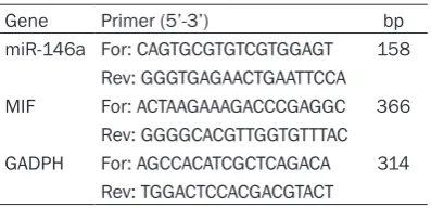
Table 1. RT-PCR primer
Gene Primer (5’-3’) bp
miR-146a For: CAGTGCGTGTCGTGGAGT 158 Rev: GGGTGAGAACTGAATTCCA MIF For: ACTAAGAAAGACCCGAGGC 366
Rev: GGGGCACGTTGGTGTTTAC GADPH For: AGCCACATCGCTCAGACA 314
step, 2% agarose gel, for detecting and taking
photos. Primers were respectively added into
25 μL PCR reaction system with reaction condi -tions were: 94°C degeneration 45 s, 59°C renaturation 45 s, 72°C extension 60 s, 35 cycles in total.
Immunohistochemical staining to detect the expression of MIF protein in lung tissue Samples were fixed by 10% formalin, embed -ded, sliced, dewaxed by xylene, dehydrated by
anhydrous, 95% and 80% ethanol, swashed by
running water, antigen repaired, antigen blocked by horse serum, primary antibody blocked, second antibody blocked, soaked by hematoxylin and hydrochloric-alcohol solution, swashed by running water to blue, dehydrated
by gradient ethanol, with hyaline xylene. After blow-dried, perform neutral balsam mounting
and microscopic examination. MIF positive
staining was faint yellow and claybank, posi -tioned on cytoplasm.
Luciferase reporter gene detection: plasmid co-transfection in glioma cells. Grouping as follow:
Result
Expressions of miR-146a and MIF in glioma tissues and glioma cells
RT-PCR results showed that the expression of miR-146a in glioma tissues was significantly
lower than that in corresponding non-tumor
normal tissues with the ratio of (0.68 ± 0.05) vs. (0.23 ± 0.00), which showed significant dif
[image:3.629.96.375.79.159.2]-ference (P < 0.05) (see Figure 1). As shown in
Figure 2, Q-PCR detection results showed that
the relative expressions of miR-146a in glio -blastoma cell lines U251, U87 and C6 were
sig-nificantly lower than those in normal astrocytes (RA), 0.20 ± 0.02, 0.24 ± 0.04, 0.51 ± 0.05, 0.52 ± 0.02) vs. (0.75 ± 0.02). Expression in U87 was moderate. Therefore, U87 was select -ed to be the study object. As shown in Figure 3, immunohistochemical results showed that
when compared positive staining of golden
area with corresponding non-tumor normal tis-sues, MIF positive expression in glioma tissues
[image:3.629.101.372.205.378.2]was higher (0.89 ± 0.02) vs. (5.49 ± 0.48), which showed significant difference (P < 0.01). Figure 1. Expressions of miR-146a in glioma tissue and adjacent tissue.
Figure 2. Expressions of miR-146a in glioblastoma cell lines and normal astrocyte cell lines.
146a mimics + Wt MIF, 146a mimics NC + Wt MIF, 146a mimics + Mut MIF, miR-146a mimics NC + Mut MIF.
Dual-luciferase detection system was used to detect the transfect
-ed luciferase activity. Steps as shown below: after PBS washed, lysate was added into. After fully
lysed, LAR solution was added
into. Then, read fluorescence
value on enzyme detector, add
stop buffer, read fluorescence
value once more, calculate
rela-tive fluorescence value. The computational formula was: rela
-tive fluorescence value = firefly luciferase value/ranilla lucifer -ase value.
Statistical analysis
RT-PCR results and Q-PCR, west-ern-blot results were counted by
Image-J software and all the data
were counted and analyzed with SPSS 17.0, repeated three times
or more for each experiment. It
was shown through t-test as
Luciferase reporter gene detection
MiR-146a mimics, NC and wild-type vector pGL3-MIF 3’UTR-Wt, as well as mutant-type
vector pGL3-MIF 3’UTR-Mut were transfected into U87 cell. The result showed that the fluo
-rescence signal intensity in cotransfection group of miR-146a mimics and wild-type vector
pGL3-MIF 3’UTR-Wt was obviously weaker than
that in other transfection group, which showed significant difference (P < 0.01). Whereas for mutant-type vector pGL3-MIF 3’UTR-Mut, fluo
-rescence intensity in different groups had no
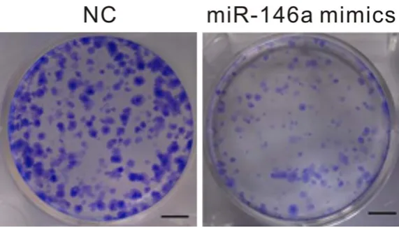
control group (P = 0.0074). As shown in Figure 7, after transfecting the miR-146a mimics with U87 cell, cloning experiment found that cell cloning ability in transfection group was signifi -cantly decreased when compared with that in
control group (68.34 ± 8.23) vs. (35.49 ± 3.65)
(P = 0.00032).
Discussion
The occurrence of tumor can be caused by many reasons. Plenty of evidence confirmed that miRNAs plays the role of a tumor suppres -Figure 3. Expressions of MIF in glioma tissue and adjacent tissue under the microscope.
Figure 4. Luciferase reporter gene expression analysis. Compared with that in NC group, **P< 0.01.
any difference (P > 0.05), see Figure 4.
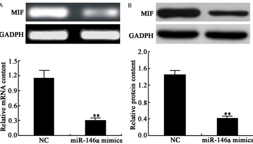
Effect of miR-146a mimics on MIF protein and mRNA expression in U87 cell
As shown in Figure 5, after
miR-146a mimics and NC
were transfected into U87
cell, MIF protein and mRNA expression quantity were
significantly decreased (P =
0.0045, P = 0.0041). Effect of miR-146a mimics on U87 cell viability
As shown in Figure 6, after transfecting the miR-146a
mimics with U87 cell, MTT
experiment found that cell viability in transfection group was significantly decreased
[image:4.629.99.534.79.260.2] [image:4.629.99.389.303.535.2]sor or a tumor promoter in different tumors and also plays an important role in the proliferation, apoptosis and signal transduction of tumor
cells, as well as in regulating the pathogenesis
of tumors [8]. As a kind of tumor suppressor
gene, miR-146a expression is down-regulated
in many types of cancer cells [9, 10]. Some
researches[11] verified that the expression quantity of miR-146a in lung carcinoma cell
was lower than that in normal lung cell, which
that it might be caused by invasive range among cell lines, and the miR-146a expression
in U87 was appropriate. In order to show no dif
-ference caused by expression quantity of too high and too low, therefore, miR-146a was selected as the follow-up experiments cell line.
The infinite proliferating tumor can lead the tumor cell to division and proliferation continu
[image:5.629.102.526.76.317.2]-ally, which makes the anabolism of protein in
Figure 5. Effect of miR-146a mimics on MIF protein and mRNA expression in U87 cell Compared with that in NC group, **P< 0.01. A: Effect of miR-146a mimics on MIF mRNA expression in U87 cell. B: Effect of miR-146a mimics on protein expression in U87 cell.
Figure 6. Effect of miR-146a mimics on U87 cell viability. Compared with that in NC group, **P< 0.01.
was fully illustrated that
miR-146a low expression occur- red in NSCLC and had close relation with the
develop-ment and progression of
NSCLC. The results revealed
for the first time that
miR-146a showed low expression in glioma. The miR-146a expression in glioma was lower than that in adjacent tissue. And the expressions in glioma cell line U251, U87 and C6 were lower than that
of normal astrocyte. Thought
the reason why the
expres-sion of miR-146a in cell lines differed was not discussed in
[image:5.629.101.389.384.564.2]tumor cell be higher than catabolism and
despoil products of normal protein metabolism to make the body in a state of cachexia and lead to the further deterioration. And research has identified that miRNAs can regulate the bio
-logical behavior of tumor cell by targeted-regu
-lating some molecules [12, 13]. Therefore, it is confirmed by luciferase reporter gene analysis that MIF is the downstream target gene of miR-146a. MIF is a kind of cytokine derived from T
lymphocyte, which can inhibit the macrophage migration and give rise to the aggregation and
infiltration of macrophage in delayed-onset hypersensitivity to participate in the inflamma
-tory response. The expressions of MIF in mela -noma, NSCLC, prostatic carcinoma and colorec-tal cancer, etc. are higher [14]. This research
has verified by immunohistochemistry that MIF
protein expression in glioma is higher than that in adjacent tissue. And another research [15] shows that MIF over expression can be achieved
by immunohistochemistry of lung tissue of glio -ma and be related to prognosis. Research has
found that overall survival of high expression MIF is lower than that of low expression [16].
Some studies indicate that high expression MIF in glioma tissue is positively correlated with
VEGF high expression in blood, and is signifi -cantly and positively correlated with the
microvessal density [17], which is fully illustrat -ed that MIF is over-express-ed in glioma, so
does in glioma as the downstream of miR-146a. Therefore, the over expression of MIF may be
closely related with the development and
pro-gression of glioma [18-20]. The subsequent
ex-periment results show that the over expression
of miR-146a has affected the cell proliferation, which has been confirmed by our subsequent
lates the multiplication capacity of cell by its downstream MIF protein or not. And no further
exposition shows regulation is made by which signal molecular mechanism, which still needs
to research further.
In conclusion, miR-146a shows low expression in glioma tissue and in cell line, while MIF shows
high expression in glioma tissue. After up-regu -lated, miR-146a expression can inhibit MIF
expression targeted to further inhibit the U87 tumor cell proliferation. Therefore, future exper
-iment can probe the effect of miR-146a on bio -logical behavior, such as glioma cell invasion
and metastasis, on a specific signaling
pathway.
Disclosure of conflict of interest
None.
Address correspondence to: Dr. Xiangdong Lu, Department of Neurosurgery, Provincial Hospital Affiliated to Shandong University, Jinan 250021, Shandong, China; Department of Neurosurgery, Laiwu City People’s Hospital, Laiwu 271199, Shandong, China. Tel: 634-6279185; Fax: +86-634-6279198; E-mail: [email protected]
References
[1] Reddy KB. MicroRNA (miRNA) in cancer. Cancer Cell Int 2015; 15: 38.
[image:6.629.99.385.78.241.2][2] Jia Y, Zang A, Shang Y, Yang H, Song Z, Wang Z, Ren L, Wei Y, Hu L, Shi H, Li H. MicroRNA-146a rs2910164 polymorphism is associated with susceptibility to non-small cell lung cancer in the Chinese population. Med Oncol 2014; 31: 194.
Figure 7. Effect of miR-146a mimics on U87 cell colony-forming ability.
colony forming experiment. At
the same time, MIF protein and mRNA expressions in
U87 cell are significantly
inhibited by up-regulating the miR-146a expression, which declares that MIF is the
down-stream target molecule of
miR-146a protein. So with the
above basis, we can further
probe the action mechanism
of miR-146a in regulating the proliferation of U87 cell by
targeted-regulating the MIF molecule. However, this re-
search does not confirm
regu-[3] Wu C, Cao Y, He Z, He J, Hu C, Duan H, Jiang J. Serum levels of miR-19b and miR-146a as prognostic biomarkers for non-small cell lung cancer. Tohoku J Exp Med 2014; 232: 85-95. [4] Kim TM, Huang W, Park R, Park PJ, Johnson
MD. A developmental taxonomy of glioblasto-ma defined and glioblasto-maintained by MicroRNAs. Cancer Res 2011; 71: 3387-3399.
[5] Fowler A, Thomson D, Giles K, Maleki S, Mreich E, Wheeler H, Leedman P, Biggs M, Cook R, Little N, Robinson B, McDonald K. miR-124a is frequently downregulated in glioblastoma and is involved in migration and invasion. Eur J Cancer 2011; 47: 953-963.
[6] Lim PK, Bliss SA, Patel SA, Taborga M, Dave MA, Gregory LA, Greco SJ, Bryan M, Patel PS, Rameshwar P. Gap junction-mediated import of microRNA from bone marrow stromal cells can elicit cell cycle quiescence in breast can-cer cells. Cancan-cer Res 2011; 71: 1550-1560. [7] Chen G, Umelo IA, Lv S, Teugels E, Fostier K,
Kronenberger P, Dewaele A, Sadones J, Geers C, De Grève J. miR-146a inhibits cell growth, cell migration and induces apoptosis in non-small cell lung cancer cells. PLoS One 2013; 8: e60317.
[8] Mawhinney L, Armstrong ME, O’Reilly C, Bucala R, Leng L, Fingerle-Rowson G, Fayne D, Keane MP, Tynan A, Maher L, Cooke G, Lloyd D, Conroy H, Donnelly SC. Macrophage migration inhibi-tory factor (MIF) enzymatic activity and lung cancer. Mol Med 2014; 20: 729-735.
[9] Labbaye C, Testa U. The emerging role of MIR-146A in the control of hematopoiesis, immune function and cancer. J Hematol Oncol 2012; 5: 13.
[10] Li L, Chen XP, Li YJ. MicroRNA-146a and hu-man disease. Scand J Immunol 2010; 71: 227-231.
[11] Cornett AL, Lutz CS. Regulation of COX-2 ex-pression by miR-146a in lung cancer cells. RNA 2014; 20: 1419-1430.
[12] Wang H, Yan C, Shi X, Zheng J, Deng L, Yang L, Yu F, Yang Y, Shao Y. MicroRNA-575 targets BLID to promote growth and invasion of non-small cell lung cancer cells. FEBS Lett 2015; 589: 805-811.
[13] Xu W, Hang M, Yuan CY, Wu FL, Chen SB, Xue K. MicroRNA-139-5p inhibits cell proliferation and invasion by targeting insulin-like growth factor 1 receptor in human non-small cell lung cancer. Int J Clin Exp Pathol 2015; 8: 3864-3870.
[14] Babu SN, Chetal G, Kumar S. Macrophage mi-gration inhibitory factor: a potential marker for cancer diagnosis and therapy. Asian Pac J Cancer Prev 2012; 13: 1737-1744.
[15] Shih HC, Huang MS, Lee CH. Polymorphonu- clear cell priming associated with NF-kB acti-vation in patients with severe injury is partially dependent on macrophage migration inhibito-ry factor. J Am Coll Surg 2010; 211: 791-797. [16] Paik JH, Jang JY, Jeon YK, Kim WY, Kim TM,
Heo DS, Kim CW. MicroRNA-146a downregu-lates NFkappaB activity via targeting TRAF6 and functions as a tumor suppressor having strong prognostic implications in NK/T cell lymphoma. Clin Cancer Res 2011; 17: 4761-4771.
[17] Zhao JL, Rao DS, Boldin MP, Taganov KD, O’Connell RM, Baltimore D. NF-kappaB dys-regulation in microRNA-146a-deficient mice drives the development of myeloid malignan-cies. Proc Natl Acad Sci U S A 2011; 108: 9184-9189.
[18] Paik JH, Jang JY, Jeon YK, Kim WY, Kim TM, Heo DS, Kim CW. MicroRNA-146a downregu-lates NFkappaB activity via targeting TRAF6 and functions as a tumor suppressor having strong prognostic implications in NK/T cell lymphoma. Clin Cancer Res 2011; 17: 4761-4771.
[19] Zhang S, Zis O, Ly PT, Wu Y, Zhang S, Zhang M, Cai F, Bucala R, Shyu WC, Song W. Down-regulation of MIF by NFkappaB under hypoxia accelerated neuronal loss during stroke. FASEB J 2014; 28: 4394-4407.