Original Article
MIR-194 is related to the pathogenesis of asthma by
regulating TLR4 expression
Kaijin Cai1, Jiaying Ke2, Yaoguo Wang1, Lisong Chen1, Liyong Shi3, Yiming Zeng3
Departments of 1Emergency, 3Respiratory, The Second Affiliated Hospital of Fujian Medical University, Quanzhou 362000, Fujian, China; 2School of Chemical and Biological Science, Quanzhou Normal University, Quanzhou 362000, Fujian, China
Received October 12, 2015; Accepted November 25, 2015; Epub February 1, 2016; Published February 15, 2016
Abstract: Objective: Activation of Toll-like receptors on immune surveillance cells in the lung has been confirmed to be implicated in the pathobiology of allergic asthma. It is reported that TLR4 activation with LPS induced significant
increases in IL-6 release and evoked pro-asthmatic like changes in the constrictor and relaxation responsiveness
of isolated airway smooth muscle (ASM) tissues. IKK mediated NF-κB activation is also involved in the responses of ASM to LPS. Methods: In this study, we first screened 10 candidate miRNAs in the plasma samples of the pa
-tients with asthma and found that the expression of miR-375 and miR-194 was reduced significantly. Subsequently, we confirmed 2 target genes of miR-375 that are related to the hypersensitivity of the NF-κB signaling. Results: We found miR-194 and miR-375 were reduced and miR-21 was significantly up-regulated in the plasma samples of asthma patients. The same condition was also confirmed in the LPS stimulated ASM cells except the expres -sion of miR-375. After prediction by bioinformatics tools; we noticed that TLR4 may be a direct target of miR-194.
Conclusions: Confirmed by dual luciferase assay and Western blot, we identified that the expression of TLR4 was repressed by miR-194. Meanwhile, reduced expression of miR-194 is correlate with over activated NF-κB signaling
and up-regulated cytokines from ASM cells when stimulated by LPS.
Keywords: microRNA, TLR4, asthma, cytokine, NF-κB
Introduction
Asthma is a chronic inflammatory disease of the airways which was mostly induced by inhaled antigens. Recently, several reports indi-cated that airway smooth muscle (ASM) can directly respond to various pro-asthmatic stim-uli [1, 2]. Meanwhile, it is well known that human ASM cells constitutively express TLR4 and TLR9 [3, 4]. After stimulation by LPS, acti-vated TLR4 potently elicits release of the pleio-tropic pro-inflammatory cytokine IL-6 and evokes significant pro-asthmatic like changes in rabbit ASM tissue, suggesting that ASM cells play important roles during the pathogenesis of asthma [5, 6].
MiRNA is a group of endogenous, short non-coding RNAs, which regulates genes expres-sion through targeting the 3’UTR of mRNA. MiRNAs have been found in various organisms,
and many of them are evolutionary conserved. Meanwhile, it is estimated that more than a half of all human protein-coding genes are poten-tially regulated by miRNAs [7]. MiRNA is a group of indispensable immuno-response regulator that modulates the homeostasis of the immune system. Disturbed miRNAs expression has been found to be related to many kinds of auto-immune diseases such as lupus erythematosus [8], rheumatoid arthritis [9], systemic sclerosis [10] and so on. Recently there are reports indi-cated that altered miRNA profile was existed in the asthma patients [11]. However, the role of miRNAs during asthma pathogenesis needs to be further unveiled.
A role of down regulated miR-194 in asthma
of miR-375 that are related to the hypersensi-tivity of the NF-κB signaling.
Materials and methods
Subjects
28 patients (17 males and 11 females) with bronchial asthma were recruited by the Respiratory Department, the Second Affiliated Hospital of Fujian Medical University. The inclu-sion criteria were as follows: 1) patients had previous history of paroxysmal wheezing, dys-pnea, chest distress, and/or coughing; 2) according to the Global Initiative for Asthma, patients had reversible airflow limitation as measured by an increase in forced expiratory volume in one second (FEV1) of at least 15% after inhalation of 200 μg salbutamol, or a decrease in FEV1 of over 20% after inhalation of <8 mg/mL acetylcholine; 3) skin prick tests showed patients were allergic to at least one of the following allergens: house dust mites, mixed grass pollens, mixed tree pollens, dog hair, feathers, cat hair, fine soft hair, cockroach -es, or mold; 4) patients had no upper and lower respiratory tract diseases within 2 months and had no chronic heart or lung disease; and 5) within at least 4 weeks, patients had not sys-temically used corticosteroids, theophylline, long-acting β2-agonists, leukotriene receptor antagonists, or antihistamines.
28 age and sex matched, healthy, non-asth-matic volunteers were recruited as control group. Normal volunteers were not allergic to anything or drugs investigated, had no respira-tory tract diseases, and had normal lung func-tion. This study met the relevant ethical require -ments for human research, approved by the Ethics Committee of the Second Affiliated Hospital of Fujian Medical University, and all subjects signed a written consent form.
Blood sample collection
10 ml blood were obtained by venipuncture from patients and controls and then centri-fuged at 1,000 g for 10 min at 4°C. 200 μl plas -ma samples were collected for RNA extraction.
Cell culture
Human ASM cells, isolated from bronchial smooth muscle of asthma patients, were pur-chased from Lonza (Allendale, NJ, USA) and maintained in SmGM™-2 Bullet Kit™.
HEK293T and PC-3 cells were cultured in Dulbecco’s Modified Eagle Medium containing 10% fetal bovine serum (Hyclone, Logan, UT, USA), 100 IU/ml penicillin and 10 mg/mL strep-tomycin. All cells were maintained at 37°C under an atmosphere of 5% CO2.
K562 cells were cultured in RPMI + 10% fetal bovine serum (Hyclone, Logan, UT, USA) supple-mented with penicillin/streptomycin and main-tained at 37°C throughout the experiment.
RNA extraction and qRT-PCR
Total RNA was extracted from plasma samples by using TRIzol LS (Invitrogen, Carlsbad, CA, USA) according to the manufacturer’s instruc-tions. The expression of miRNAs was detected by TaqMan miRNA RT-Real Time PCR. Single-stranded cDNA was synthesized by using TaqMan MicroRNA Reverse Transcription Kit (Applied Biosystems, Foster City, CA, USA) and then amplified by using TaqMan Universal PCR Master Mix (Applied Biosystems, Foster City, CA, USA) together with miRNA-specific TaqMan MGB probes (Applied Biosystems, Foster City, CA, USA). The expression of miR-16 was used for normalization. Each sample in each group was measured in triplicate and the experiment was repeated at least three times.
Dual luciferase assay
A 523 bp segment of TLR4 3’UTR containing the predicted target region of miR-194 were cloned into pmirGLO vector separately, down-stream of firefly luciferase coding region (Promega, Madison, WI, USA) to generate lucif-erase reporter vectors. For luciflucif-erase reporter assays, cells were seeded in 48-well plates. MiRNA mimics or inhibitors and luciferase reporter vectors were co-transfected into cells by lipofectamine 2000 (Invitrogen, Carlsbad, CA USA). Two days post transfection, cells were harvested and assayed with the Dual-Luciferase Assay (Promega, Madison, WI USA). Each treatment was performed in triplicate in three independent experiments. The results were expressed as relative luciferase activity (Firefly LUC/Renilla LUC).
Immunoblotting
electrophoresis, and the proteins in the gels were blotted onto PVDF membranes (Amersham Pharmacia Biotech, St. Albans, Herts, UK) by electrophoretic transfer. The membrane was incubated with rabbit TLR4 polyclonal anti-body (Abcam, Cambridge, MA, USA) or rabbit anti-p65 polyclonal antibody (Abcam, Cam- bridge, MA, USA) or mouse anti-β-actin mono -clonal antibody (Santa Cruz Biotechnology Inc., Santa Cruz, CA, USA) for 1 h at 37°C. The spe-cific protein antibody complex was detected by using horseradish peroxidase conjugated anti-rabbit or anti-mouse antibody. Detection by the chemiluminescence reaction was carried using the ECL kit (Pierce, Appleton, WI, USA). The β-actin signal was used as a loading control.
Supernatant protein analysis
TNF-α and IL-6 enzyme-linked immunosorbent assay. sandwich enzyme-linked immunosor-bent assays (ELISAs) for TNFα and IL-6 were carried out on cell culture supernatants accord-ing to the manufacturer’s instructions (R&D Systems Europe Ltd, Abingdon, Oxfordshire, UK). Supernatants were diluted with RPMI 1640 as appropriate for ELISA analysis.
Statistical analysis
[image:3.612.93.523.76.466.2]Data were analyzed by using SPSS Statistical Package version 15. Independent two group’s analyses are used t-test. P<0.05 was consid-ered statistically significant.
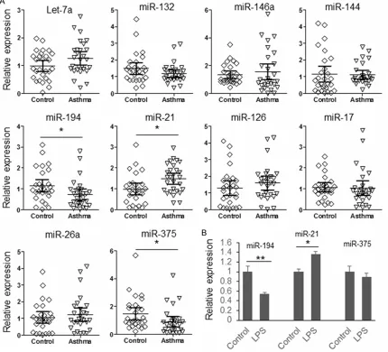
Figure 1. Expression of candidate miRNAs. A. The expression of 10 candidate miRNAs in the plasma sample of 28
patients with asthma and 28 paired controls were detected by qRT-PCR; B. Human ASM cells were treated with LPS for 30 min and the expression of miR-194, miR-21 and miR-375 was detected by qRT-PCR. The results were ana
A role of down regulated miR-194 in asthma
Results
To explore the roles of miRNAs during the pathogenesis of asthma, we first detected the
[image:4.612.94.522.75.591.2]expression of 10 candidate miRNAs in the plas-ma sample of 28 patients with asthplas-ma and 28 paired controls. These miRNAs were reported to have a disturbed expression in asthma Figure 2. miR-194 target 3’UTR of TLR4. A. Schematic diagram of predicted interaction between miR-194 and the
3’UTR of TLR4. Red letters represent the mutated nucleotides; B. Schematic diagram for reporter vector construc -tion; C. Dual luciferase assay. HEK293T cells were transfected with wild type or mutated TLR4 reporter vector and
miR-194 mimic or inhibitor, with sequence scrambled single or double strand RNAs as control. 48 hours after trans -fection, the cells were lysed and luciferases activities were detected. The results were analyzed by student’s t-test
patients or function as immune system regula-tors [12-14]. As shown in Figure 1A, compared with healthy controls, there are three miRNAs have significant changed expression in the plasma of asthma patients (miR-194 is down regulated; miR-21 and miR-375 are overex-pressed). To investigate whether there is the same condition in airway smooth muscle; ASM cells were treated with LPS for 30 min and then collected for RNA extraction. The expression of miR-21, miR-194 and miR-375 were detected by qRT-PCR. As exhibited in Figure 1B, only the expression of miR-21 and miR-194 was signifi -cantly changed after LPS stimulation. Since the up-regulated miR-21 in bronchial epidermal cells and plasma samples of asthma patients has been reported by other researchers, this study was focused on unveiling the function of miR-194 during the pathogenesis of asthma [15, 16].
To explore the biological function of miR-194, we first predicted the target genes of miR-194 by using online bioinformatics tool: TargetScan
A role of down regulated miR-194 in asthma
nificantly changed (P>0.05) when 3 nucleotides were mutated indicating that the predicted miR-194 target site is the real target site. Although miR-194 can bind with the 3’UTR of TLR4, it is still unknown whether miR-194 can regulate endogenous TLR4 expression. K562, PC-3 and ASM cells were transfected with miR-194 mimic or inhibitor in order to detect the effect of miR-194 on endogenous TLR4 expres-sion (Figure 3). Compared with miR-control, the expression of TLR4 was significantly reduced in the cells transfected with miR-194 mimic. Meanwhile, the expression of TLR4 was up-reg-ulated in the cells transfected with miR-194 inhibitor. These results further confirmed that TLR4 is a direct target of miR-194.
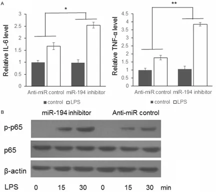
To further understand the function of miR-194 during TLR4 induced immune response, ASM cells were transfected with miR-194 inhibitor or anti-miR-control. 48 hours after transfection, the cells were treated with 500 ng/mL bacteri-al lipopolysaccharide (LPS) (Sigma Aldrich) for 30 minutes. The negative controls were treated by DMSO. The supernatant was collected for cytokines detection. As shown in Figure 4A, IL-6 and TNF-α levels were up-regulated higher in the miR-194 inhibitor transfected cells com-pared with negative control. Meanwhile, the phosphorylated p65 protein level in the ASM cells were also examined by immunoblotting. As exhibited in Figure 4B, the p-p65 level in the cells transfected with miR-194 inhibitor up-reg-Figure 4. LPS induced NF-κB activation was enhanced by miR-194 inhibitor. A. ASM cells were transfected with
miR-194 inhibitor or anti-miR-control. 48 hours after transfection, the cells were treated with 500 ng/mL LPS for 30
minutes. The concentration of IL-6 and TNF-α in the supernatant was detected by ELISA; B. The phosphorylated p65
[image:6.612.91.523.75.460.2]ulated to a higher level than the control cells, which confirmed the relation between repressed miR-194 and stronger immune response.
Discussion
Asthma is a chronic inflammatory disease of the airways which was mostly induced by inhaled antigens. Recently, it has been report-ed that disturbreport-ed miRNAs was detectreport-ed in the plasma and tissue samples of asthma patients [17-19]. However, the function of most of these miRNAs is not well understood. To unveil the functions of miRNAs during the pathogenesis of asthma, we first detected the expression of 10 candidate miRNAs in the plasma samples of 28 asthma patients and LPS treated ASM cells and found miR-194 and miR-21 have an altered expression. Predicted by bioinformatics tool and confirmed by dual luciferase assay and immunoblotting, we confirmed that miR-194 represses the expression of TLR4 by targeting 3’UTR. Meanwhile, we also confirmed a stron -ger activated NF-κB signal in the ASM cells. We unveiled the relationship between miR-194 repression and enhanced immune response in ASM cells for the first time and provide a poten -tial target for asthma treatment.
Unlike other well documented effects in the innate immunity system of cells, toll like recep-tors can stimulate a variety of non-immune cells types, such as endothelial cells, cardio-myocytes and pulmonary epithelial cells [4, 20-22]. Moreover, it has recently been demon-strated that ASM cells from different species, including humans, also express various TLRs [4]. TLR4 is constitutively expressed in the ASM cells and the activation of TLR4 by LPS was found to elicit release of IL-6, a pro-inflammato -ry cytokine. In this study, we construct the rela-tion between down regulated miR-194 and overexpressed TLR4 of ASM cells, which par-tially explained the hyper-sensitivity of asthma patients. However, our conclusion depends on in vitro study and clinical samples. Further in vivo research has to be employed to explore the roles of miR-194 in respiratory system.
In conclusion, we constructed the relationship between reduced miR-194 level and up-regu-lated TLR4 expression in ASM cells. This research partially explained the hyper-sensitivi-ty of asthma patients to pathogens and may provide a new target for clinical treatment.
Acknowledgements
This study was supported by Natural Science Funds of Fujian Province [No. 2015J01448], and Science and Technology Plan Projects Funds of Quanzhou City [No. 2015Z34].
Disclosure of conflict of interest
None.
Address correspondence to: Dr. Jiaying Ke, School
of Chemical and Biological Science, Quanzhou
Normal University, 398 Donghai Street, Quanzhou 362000, Fujian, China. Tel: +86-18959799175; Fax: +86-595-22770852; E-mail: kj_cai888@126. com
References
[1] Hakonarson H and Grunstein MM. Autologously up-regulated Fc receptor expression and ac-tion in airway smooth muscle mediates its al-tered responsiveness in the atopic asthmatic sensitized state. Proc Natl Acad Sci U S A 1998; 95: 5257-5262.
[2] Hakonarson H, Maskeri N, Carter C, Hodinka RL, Campbell D and Grunstein MM. Mechanism of rhinovirus-induced changes in airway smooth muscle responsiveness. J Clin Invest 1998; 102: 1732-41.
[3] Bachar O, Adner M, Uddman R and Cardell LO.
Toll-like receptor stimulation induces airway hyper-responsiveness to bradykinin, an effect
mediated by JNK and NF-kappa B signaling
pathways. Eur J Immunol 2004; 34: 1196-207. [4] Monick MM, Yarovinsky TO, Powers LS, Butler
NS, Carter AB, Gudmundsson G, Hunninghake
GW. Respiratory syncytial virus up-regulates TLR4 and sensitizes airway epithelial cells to
endotoxin. J Biol Chem 2003; 278: 53035-44.
[5] Savov JD, Brass DM, Lawson BL,
McElvania-Tekippe E, Walker JK and Schwartz DA. Toll-like receptor 4 antagonist (E5564) prevents the chronic airway response to inhaled lipopoly-saccharide. Am J Physiol Lung Cell Mol Physiol 2005; 289: L329-37.
[6] Shan X, Hu A, Veler H, Fatma S, Grunstein JS, Chuang S, Grunstein MM. Regulation of Toll-like receptor 4-induced proasthmatic changes in airway smooth muscle function by opposing actions of ERK1/2 and p38 MAPK signaling. Am J Physiol Lung Cell Mol Physiol 2006; 291: L324-33.
[7] Friedman RC, Farh KK, Burge CB and Bartel
A role of down regulated miR-194 in asthma
[8] Qu B, Shen N. miRNAs in the Pathogenesis of
Systemic Lupus Erythematosus. Int J Mol Sci 2015; 16: 9557-72.
[9] Singh RP, Massachi I, Manickavel S, Singh S, Rao NP, Hasan S, Mc Curdy DK, Sharma S,
Wong D, Hahn BH, Rehimi H. The role of miRNA in inflammation and autoimmunity. Autoimmun
Rev 2013; 12: 1160-5.
[10] Li Y, Huang J, Guo M and Zuo X. MicroRNAs Regulating Signaling Pathways: Potential
Biomarkers in Systemic Sclerosis. Genomics Proteomics Bioinformatics 2015; 13:
234-41.11.
[11] Liu Y, Yang K, Sun X, Fang P, Shi H, Xu J, Xie M, Li M. MiR-138 suppresses airway smooth mus-cle cell proliferation through the PI3K/AKT sig-naling pathway by targeting PDK1. Exp Lung Res 2015; 41: 363-9.
[12] Kabesch M and Adcock IM. Epigenetics in
asthma and COPD. Biochimie 2012; 94:
2231-41.
[13] Tay HL, Plank M, Collison A, Mattes J, Kumar RK and Foster PS. MicroRNA: potential bio-markers and therapeutic targets for allergic asthma? Ann Med 2014; 46: 633-9.
[14] Kabesch M. Epigenetics in asthma and allergy. Curr Opin Allergy Clin Immunol 2014; 14: 62-8. [15] Lu TX, Munitz A and Rothenberg ME.
MicroRNA-21 is up-regulated in allergic airway
inflammation and regulates IL-12p35 expres -sion. J Immunol 2009; 182: 4994-5002. [16] Wu XB, Wang MY, Zhu HY, Tang SQ, You YD and
Xie YQ. Overexpression of microRNA-21 and microRNA-126 in the patients of bronchial asthma. Int J Clin Exp Med 2014; 7: 1307-12.
[17] Wang Y, Yang L, Li P, Huang H, Liu T, He H, Lin
Z, Jiang Y, Ren N, Wu B, Kamp DW, Tan J, Liu G.
Circulating microRNA Signatures Associated with Childhood Asthma. Clin Lab 2015; 61: 467-74.
[18] Sawant DV, Yao W, Wright Z, Sawyers C, Tepper RS, Gupta SK, Kaplan MH, Dent AL. Serum
MicroRNA-21 as a Biomarker for Allergic Inflammatory Disease in Children. Microrna
2015; 4: 36-40.
[19] Comer BS, Camoretti-Mercado B, Kogut PC,
Halayko AJ, Solway J and Gerthoffer WT. MicroRNA-146a and microRNA-146b
expres-sion and anti-inflammatory function in human
airway smooth muscle. Am J Physiol Lung Cell Mol Physiol 2014; 307: L727-34.
[20] Andonegui G, Bonder CS, Green F, Mullaly SC,
Zbytnuik L, Raharjo E, Kubes P. Endothelium-derived Toll-like receptor-4 is the key molecule
in LPS-induced neutrophil sequestration into
lungs. J Clin Invest 2003; 111: 1011-20. [21] Frantz S, Kelly RA and Bourcier T. Role of TLR-2
in the activation of nuclear factor kappaB by oxidative stress in cardiac myocytes. J Biol
Chem 2001; 276: 5197-203.
[22] Cheng T, Liu Q, Zhang R, Zhang Y, Chen J, Yu R, Ge G. Lysyl oxidase promotes
bleomycin-in-duced lung fibrosis through modulating inflam