http://www.scirp.org/journal/jbm ISSN Online: 2327-509X
ISSN Print: 2327-5081
DOI: 10.4236/jbm.2017.510004 Oct. 24, 2017 34 Journal of Biosciences and Medicines
Obesity Indices Related to Obstructive Sleep
Apnea in Obese Adults
Jongwoo Kim, Seon Yeong Lee
*Department of Family Medicine, Inje University Sanggye-Paik Hospital, Seoul, South Korea
Abstract
Background: Obstructive Sleep Apnea (OSA) is prevalent in obese patients. OSA should be evaluated because it might increase mortality. We aimed to evaluate the risk for OSA in obese Korean adults, and to examine the obesity indices most strongly associated with OSA. Method: Anthropometric mea-surements, fat computed tomography (CT) and dual energy X-ray absorpti-ometry (DEXA) were performed in 30 obese patients. All patients were di-vided into low or high risk group of OSA using Berlin Questionnaire. We compared differences between the groups in obesity-related indices. Result: Eleven of the 30 patients (36.7%) were in the low OSA risk group and 19 (63.3%) were in the high OSA risk group. The correlation coefficients for BMI, neck circumference, and neck-to-height ratio × 100(%) in the high-risk group versus the low-risk group were 1.03, 1.96, and 4.04, respectively (P = 0.03). Conclusion: The obesity index most strongly associated with OSA was neck circumference to height ratio.
Keywords
Obesity, Sleep Apnea, Obstructive, Obesity Index, Neck Circumference to Height Ratio
1. Introduction
Obesity causes chronic disease including cardiovascular disease, diabetes and can-cer, and 40% - 90% of obese patients also exhibit signs and symptoms of sleep apnea [1]. Obesity-related obstructive sleep apnea (OSA) refers to repeated pha-ryngeal obstruction during sleep, which not only leads to negative consequences, such as fatigue, reduced cognitive ability, headache, depression and sexual dys-function, but also increases the risk for hypertension, cardiovascular disease, arrhythmia, and heart failure. OSA, therefore, is one of the most important How to cite this paper: Kim, J. and Lee,
S.Y. (2017) Obesity Indices Related to Ob-structive Sleep Apnea in Obese Adults. Journal of Biosciences and Medicines, 5, 34-43.
https://doi.org/10.4236/jbm.2017.510004
Received: September 29, 2017 Accepted: October 21, 2017 Published: October 24, 2017
Copyright © 2017 by authors and Scientific Research Publishing Inc. This work is licensed under the Creative Commons Attribution International License (CC BY 4.0).
http://creativecommons.org/licenses/by/4.0/
DOI: 10.4236/jbm.2017.510004 35 Journal of Biosciences and Medicines diseases to be assessed early in obese patients [2]. Risk factors associated with OSA include obesity, male sex, older age, neck circumference, facial dysmor-phism, genetic factors, and thyroid disease [2].
Body mass index (BMI) is widely used as a measure of obesity; however, be-cause it does not reflect the extent of obesity, it is difficult to use for diagnosing and evaluating the severity of OSA. Obesity and regional fat distribution, meas-ured by neck fat mass percentage using dual-energy X-ray absorptiometry (DEXA), might correlate with obstructive sleep apnea (OSA) severity in adults
[3], but Martin et al. reported the lack of correlation between DEXA measure-ments changes and the changes in the apnea hypopnea index [4]. There are still conflicting opinions among researchers regarding the factors that enable easy assessment of OSA, and a lack of research in this regard in Korea remains. Ac-cordingly, the present study aimed to evaluate the risk for OSA in obese Korean adults, and to examine the obesity indices most strongly associated with OSA.
2. Methods
2.1. Subjects
Obese adults who visited the obesity clinic at a single university hospital for an initial consultation between March and July 2013 were investigated. Thirty-four patients underwent an initial consultation during the study period; however, DEXA could not be performed in 4 individuals due to super-morbid obesity; therefore, 30 patients were included in the final analysis.
2.2. Anthropometric Measurements, DEXA, and Fat Computed
Tomography
Patient height, weight, neck circumference, and waist circumference were meas-ured at the first visit. Neck circumference was adjusted by separately calculating the neck-to-height ratio (neck circumference divided by height [both in cm]). Anthropometric measurements were each performed twice by a trained special-ist, and the average of the two measurements was used. Height was measured in cm and weight was measured in kg. Neck circumference was measured in cm, in the horizontal plane passing through the center of the cricothyroid membrane; waist circumference was measured in cm at the widest point below the navel.
DOI: 10.4236/jbm.2017.510004 36 Journal of Biosciences and Medicines the regional distribution of total body fat mass, the regional fat mass values measured using DEXA were divided by the total body fat mass and divided by 100 to obtain a percentage (%).
2.3. Berlin Questionnaire
The Berlin questionnaire was used to evaluate OSA risk [5]. This questionnaire consists of 10 questions divided into three categories. The first category consists of 5 questions on snoring and sleep apnea; answering positively to at least 2 questions is considered to be a positive result for the category. The second cate-gory consists of 4 questions on weekly tiredness; answering positively to at least 2 questions is considered to be a positive result for the category. The third cate-gory queries whether the subject has hypertension or a BMI of at least 30 kg/m2. Subjects with positive results in at least 2 of the 3 categories are classified as high risk for OSA, and other subjects are classified as low risk [1][5]. Because most of the subjects in the present study were severely obese, the BMI criterion was arbi-trarily set at 30 kg/m2.
2.4. Statistical Analysis
Patients in this study were divided into two groups according to OSA risk, and the Mann-Whitney test was used to compare differences between the groups in obesity-related indices. Correcting for age and sex, a logistic regression analysis was performed to examine the effects of obesity-related indices on OSA risk, and the regression coefficients and significance probabilities were calculated for each variable. SAS version 8.1 (SAS Institute, Cary, NC, USA) was used for statistical analysis and P < 0.05 was considered to be statistically significant.
3. Results
3.1. Comparison between Males and Females
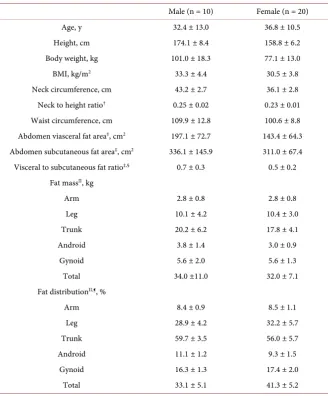
The 30 adult obese patients were divided according to sex, and the mean and standard deviation were calculated for each variable (Table 1). For males and females, respectively, the mean ages were 32.4 ± 13.0 years and 36.8 ± 10.5 years; the mean BMI was 33.3 ± 4.4 kg/m2 and 30.5 ± 3.8 kg/m2. The mean neck cir-cumferences for males and females, respectively, were 43.2 ± 2.7 cm and 36.1 ± 2.8 cm, the mean neck-to-height ratios were 0.25 ± 0.02 and 0.23 ± 0.01, and the mean waist circumferences were 109.9 ± 12.8 cm and 100.6 ± 8.8 cm. Abdominal visceral fat area, as measured by fat CT, was 197.1 ± 72.7 cm2 for males and 143.4 ± 64.3 cm2 for females. Body fat percentage measured using DEXA was 33.1% ± 5.1% in males and 41.3% ± 5.2% in females, the leg fat distribution was 28.9% ± 4.2% and 32.2% ± 5.7% in males and females, respectively, and the trunk fat distribution was 59.7% ± 3.5% and 56.0% ± 5.7%.
3.2. Comparison between High and Low OSA Risk Groups
DOI: 10.4236/jbm.2017.510004 37 Journal of Biosciences and Medicines
Table 1. Basic characteristics of subjects.
Male (n = 10) Female (n = 20)
Age, y 32.4 ± 13.0 36.8 ± 10.5
Height, cm 174.1 ± 8.4 158.8 ± 6.2
Body weight, kg 101.0 ± 18.3 77.1 ± 13.0
BMI, kg/m2 33.3 ± 4.4 30.5 ± 3.8
Neck circumference, cm 43.2 ± 2.7 36.1 ± 2.8 Neck to height ratio† 0.25 ± 0.02 0.23 ± 0.01 Waist circumference, cm 109.9 ± 12.8 100.6 ± 8.8 Abdomen viasceral fat area‡, cm2 197.1 ± 72.7 143.4 ± 64.3 Abdomen subcutaneous fat area‡, cm2 336.1 ± 145.9 311.0 ± 67.4 Visceral to subcutaneous fat ratio‡,§ 0.7 ± 0.3 0.5 ± 0.2
Fat massII, kg
Arm 2.8 ± 0.8 2.8 ± 0.8
Leg 10.1 ± 4.2 10.4 ± 3.0
Trunk 20.2 ± 6.2 17.8 ± 4.1
Android 3.8 ± 1.4 3.0 ± 0.9
Gynoid 5.6 ± 2.0 5.6 ± 1.3
Total 34.0 ±11.0 32.0 ± 7.1
Fat distributionII,¶, %
Arm 8.4 ± 0.9 8.5 ± 1.1
Leg 28.9 ± 4.2 32.2 ± 5.7
Trunk 59.7 ± 3.5 56.0 ± 5.7
Android 11.1 ± 1.2 9.3 ± 1.5
Gynoid 16.3 ± 1.3 17.4 ± 2.0
Total 33.1 ± 5.1 41.3 ± 5.2
Abbreviations: BMI, body mass index calculated as weight in kilograms divided by height in meters squared. †Neck to height ratio calculated as neck circumference in centimeters divided by height in
centi-meters. ‡Measurements by fat computed tomography. §Visceral to subcutaneous fat ratio calculated with
abdomen visceral fat area in centimeters squared divided by abdomen subcutaneous fat area in centimeters squared. IIMeasurements bydual energy X-ray absorptiometry scan. ¶Fat distribution calculated as each re-gional fat mass divided by total fat mass and multiplied by 100.
DOI: 10.4236/jbm.2017.510004 38 Journal of Biosciences and Medicines
Table 2. The comparison of variables according to OSA risk.
OSA low risk
(n = 11) OSA high risk (n = 19) P*
Number (male, female) 2, 9 8, 11
Age, y 31.0 ± 9.0 37.8 ± 12.0 0.13
Height, cm 161.8 ± 9.4 165.0 ± 10.5 0.35 Body weight, kg 78.0 ± 20.2 89.1 ± 16.9 0.04
BMI, kg/m2 29.3 ± 3.8 32.6 ± 4.0 0.02
Neck circumference, cm 35.9 ± 3.6 40.0 ± 4.1 0.01 Neck to height ratio† 0.22 ± 0.01 0.24 ± 0.02 <0.01 Waist circumference, cm 99.3 ± 11.3 106.2 ± 10.3 0.04 Abdomen viasceral fat area‡, cm2 125.3 ± 39.1 182.1 ± 77.3 0.03 Abdomen subcutaneous fat area‡, cm2 316.7 ± 114.8 320.8 ± 91.4 0.43 Visceral to subcutaneous fat ratio‡,§ 0.4 ± 0.2 0.6 ± 0.3 0.03
Fat massII, kg
Arm 2.5 ± 0.6 2.9 ± 0.8 0.16
Leg 10.7 ± 3.3 10.1 ± 3.4 0.85
Trunk 17.4 ± 5.1 19.3 ± 4.8 0.07
Android 2.9 ± 1.3 3.4 ± 1.1 0.06
Gynoid 5.7 ± 1.5 5.6 ± 1.6 0.85
Total 31.6 ± 8.4 33.3 ± 8.7 0.25
Fat distributionII,¶, %
Arm 8.1 ± 1.1 8.7 ± 0.9 0.11
Leg 33.8 ± 5.5 29.5 ± 4.8 0.03
Trunk 54.8 ± 5.3 58.6 ± 4.9 0.04
Android 9.1 ± 1.6 10.3 ± 1.5 0.04
Gynoid 17.9 ± 2.0 16.5 ± 1.6 0.06
Total 40.7 ± 3.9 37.4 ± 7.4 0.29
*P value from Mann-Whitney test. Subjects with positive results in at least 2 of the 3 categories of Berlin questionnaire were classified as high risk for OSA, and other subjects were classified as low risk. Abbrevia-tions: OSA, Obstructive sleep apnea. BMI, body mass index calculated as weight in kilograms divided by height in meters squared. †Neck to height ratio calculated as neck circumference in centimeters divided by
height in centimeters. ‡Measurements by fat computed tomography. §Visceral to subcutaneous fat ratio
calculated as abdomen visceral fat area in centimeters squared divided by abdomen subcutaneous fat area in centimeters squared. IIMeasurements by dual energy X-ray absorptiometry scan. ¶Fat distribution was cal-culated with each regional fat mass divided by total fat mass and multiplied by 100.
respectively, and the android fat distribution was 9.1% ± 1.6% and 10.3% ± 1.5%.
3.3. Regression Analysis
DOI: 10.4236/jbm.2017.510004 39 Journal of Biosciences and Medicines
Table 3. Logistic regression analysis of obesity indices on OSA risk.
Regression
coefficient P* 95% CI
Body weight, kg 1.07 0.12 0.98 - 1.16
BMI, kg/m2 1.03 0.03 1.03 - 2.03
Neck circumference, cm 1.96 0.04 1.02 - 3.75 Neck to height ratio†, % 4.04 0.03 1.20 - 13.65 Waist circumference, cm 1.09 0.14 0.97 - 1.21 Abdomen visceral fat area‡, cm2 1.02 0.16 0.99 - 1.04 Abdomen visceral to subcutaneous fat ratio‡,§ 8.93 0.35 0.91 - 874.21
Fat distributionII,¶, %
Leg 0.91 0.38 0.74 - 1.12
Trunk 1.06 0.61 0.86 - 1.30
Android 1.31 0.43 0.67 - 2.57
*P value from logistic regression analysis adjusted by age and sex. Abbreviations: OSA, Obstructive sleep apnea. BMI, body mass index calculated as weight in kilograms divided by height in meters squared. CI, Confidence interval. †Neck to height ratio (%) calculated as neck circumference divided by height and mul-tiplied by 100. ‡Measurements by fat computed tomography. §Visceral to subcutaneous fat ratio calculated as abdomen visceral fat area in centimeters squared divided by abdomen subcutaneous fat area in
centime-ters squared. IIMeasurements by dual energy X-ray absorptiometry scan. ¶Fat distribution was calculated
with each regional fat mass divided by total fat mass and multiplied by 100.
4. Discussion
In the present study, we used the Berlin questionnaire to examine 30 adult pa-tients with obesity at a single university hospital, and found that 19 (63%) were in the high OSA risk group. Compared with the low OSA risk group, the high-risk group exhibited a greater proportion of male patients and older age; however, these differences were not statistically significant. BMI, neck circumfe-rence, neck-to-height ratio, abdominal visceral fat area, abdominal visceral-to- subcutaneous fat ratio, trunk fat distribution, and android fat distribution were all significantly higher in the high OSA risk group.
DOI: 10.4236/jbm.2017.510004 40 Journal of Biosciences and Medicines In studies investigating OSA, BMI and neck circumference have been reported to be the strongest predictive factors for apnea-hypopnea [8] [9] [10] [11]. Meanwhile, other studies have reported that body fat and abdominal circumfe-rence are the factors that best reflect OSA risk [12][13]. In one investigation, the index most closely related to OSA risk was abdominal visceral fat in middle-age males and neck circumference in females [14].
Unlike previous studies, our study revealed no significant difference between the OSA risk groups in sex ratio or age; we believe this reflected the characteris-tics of the subject population. In one study [8], the mean age of the patients was the sixth decade of life (i.e., 50 s), and the majority were male. In our study, the mean age of the patients was the fourth decade of life (i.e., 30 s), and there were twice as many females as males. However, the previous study [8] involved tients who visited a sleep clinic for snoring, whereas our study focused on pa-tients visiting an obesity clinic for weight loss. Because most of the papa-tients at the sleep clinic were not obese, their complaints were more likely to be the result of anatomical issues in the head and neck, such as tonsillar hypertrophy or a small mandible, which may explain the difference in results.
Considering the nature of the obese patient group in the present study, we performed a regression analysis corrected for sex and age, which have been shown to affect OSA in previous studies. The variable that exhibited the largest significant correlation coefficient in the high OSA risk group was neck-to-height ratio × 100(%). The reason for converting neck-to-height ratio into a percentage (by multiplying by 100) is because, without this change, the correlation coeffi-cient was too large, in the order of 1060. Because the regression coefficoeffi-cient for the neck-to-height ratio (%) was 4, this means that an increase in the neck cir-cumference equal to 1% of the height will increase the likelihood of inclusion in the high OSA risk group by four-fold. One possible reason for patients with larger necks having an increased risk for OSA is the relationship with local fat accumulation. Because our study involved patients with a BMI of ≥25 kg/m2, accumulation of fat in the tongue and laryngopharynx could constrict the di-ameter of the trachea, potentially leading to obstruction during sleep [14]. We did not observe significant results for abdominal visceral fat area or regional fat distribution.
We attempted a stepwise regression analysis using variables other than age and sex; however, the results were not statistically significant. Consequently, it is difficult to determine which variables are the strongest predictors. Because other variables that are potential covariates are, in large part, obesity-related indices, there is some overlap, which can cause problems in the regression formula. For example, an individual with high body weight will likely have a broader abdo-minal fat area, while in DEXA results, trunk fat includes android fat. These is-sues will need to be analyzed in a study involving a larger sample size.
DOI: 10.4236/jbm.2017.510004 41 Journal of Biosciences and Medicines the results of our study, we confirmed that 60% - 70% of obese Korean patients are at high risk for OSA. Moreover, given that there is a close association be-tween high OSA risk and neck-to-height ratio, and that conducting polysomno-graphy in all obese patients is impractical, measuring neck circumference could be helpful in predicting OSA risk. Second, previous research reporting that ab-dominal obesity is the strongest predictive factor for OSA [5] used abdominal circumference and bioelectrical impedance analysis. However, in our study, we were able to analyze fat distribution more precisely using DEXA, which is con-sidered to be the standard method for body fat analysis, and fat CT, which can separately determine areas of abdominal visceral and subcutaneous fat. Third, in previous studies that used DEXA measurements to investigate fat distribution
[15][16], the distribution was calculated as regional fat weight/regional weight × 100(%), or as regional fat weight/total body weight × 100(%). In contrast, our study used regional fat weight/total body fat weight × 100(%). Calculating the regional fat distribution as a percentage of total body fat has the effect of norma-lizing according to total body fat, enabling meaningful comparisons of fat dis-tribution according to region, even among patients with the same total body fat. This differentiates our study from others and improves the validity of our re-sults.
Our study also had some limitations, the first of which were the small sample size and the low proportion of male subjects. Second, because this was a cross-sectional study, we were unable to clearly demonstrate causality. Third, we were unable to use polysomnography to accurately diagnose OSA; therefore, we used the Berlin questionnaire instead. Although the Berlin questionnaire is the best method to replace polysomnography for OSA screening, the effectiveness of the questionnaire for testing obese Korean patients has not yet been demon-strated, which limits the interpretation and applicability of our results. In addi-tion, because the Berlin questionnaire evaluates OSA risk as a categorical varia-ble (high risk versus low risk), we were only avaria-ble to perform a logistic regression analysis. If polysomnography had been used to obtain AHI values, it would be possible to derive more precise results using linear regression analysis. Because of these limitations, further research will need to be conducted in the future, studying more patients and over a longer duration. To investigate whether larger neck circumference in the high OSA risk group was actually due to fat distribu-tion in the neck, neck fat would need to be measured and analyzed using DEXA, similar to the study by Simpson et al.[14]. In addition, follow-up observation of the patients in this study to investigate changes in obesity-related factors with weight loss would help to verify whether this also reduces OSA risk.
examina-DOI: 10.4236/jbm.2017.510004 42 Journal of Biosciences and Medicines tions, and to evaluate the effects of intervention(s).
Conflict of Interest
We declare that we have no conflict of interest.
Ethical Approval
This study was approved by the Institutional Review Board of Inje University Sanggye Paik Hospital in Seoul, South Korea.
References
[1] Alan, R.S., Susheel, P.P., Alison, M.L., Vsevolod, P., Hartmut, S. and Philip, L.S. (2008) Obesity and Obstructive Sleep Apnea. Proceedings of the American Thoracic Society, 5, 185-192. https://doi.org/10.1513/pats.200708-137MG
[2] Neomi, S. and Francoise, R. (2009) The Relationship of Obesity and Obstructive Sleep Apnea. Clinics in Chest Medicine, 20, 455-465.
[3] Glicksman, A., Hadjiyannakis, S., Barrowman, N., Walker, S., Hoey, L. and Katz, S.L. (2017) Body Fat Distribution Ratios and Obstructive Sleep Apnea Severity in Youth With Obesity. Journal of Clinical Sleep Medicine, 13, 545-550.
https://doi.org/10.5664/jcsm.6538
[4] Martin, S.M., Roche, F., Thomas, T., Collet, P., Barthélémy, J.C. and Sforza, E. (2015) Association of Body Fat Composition and Obstructive Sleep Apnea in the Elderly: A Longitudinal Study. Obesity, 23, 1511-1516.
https://doi.org/10.1002/oby.21121
[5] Netzer, N.C., Stoohs, R.A., Netzer, C.M., Clark, K. and Strohl, K.P. (1999) Using the Berlin Questionnaire to Identify Patients at Risk for the Sleep Apnea Syndrome. Annals of Internal Medicine, 131, 485-491.
https://doi.org/10.7326/0003-4819-131-7-199910050-00002
[6] Kang, K., Park, K.S., Kim, J.E., Kim, S.W., Kim, Y.T., Kim, J.S. and Lee, H.W. (2013) Usefulness of the Berlin Questionnaire to Identify Patients at High Risk for Ob-structive Sleep Apnea: A Population-Based Door-to-Door Study. Sleep and Breath-ing, 17, 803-810. https://doi.org/10.1007/s11325-012-0767-2
[7] Kang, H.H., Kang, J.Y., Lee, S.H. and Moon, H.S. (2011) The Usefulness of the Ber-lin Questionnaire as a Screening for Obstructive Sleep Apnea in a Sleep CBer-linic Pop-ulation. Sleep Medicine and Psychophysiology, 18, 82-86.
[8] Han, S.J., Joo, E.Y., Kim, J.H., Kim, M.S. and Hong, S.B. (2004) Body Mass Index and Neck Circumference in Patients with Obstructive Sleep Apena-Hypopnea Syn-dromes. Journal of Korean Sleep Research, 1, 37-41.
https://doi.org/10.13078/jksrs.04016
[9] Yeh, P.S., Lee, Y.C., Lee, W.J., Chen, S.B., Ho, S.J., Peng, W.B., et al. (2010) Clinical Predictors of Obstructive Sleep Apnea in Asian Bariatric Patients. Obesity Surgery, 20, 30-35. https://doi.org/10.1007/s11695-009-9854-2
[10] Hari, L.P., Rajagopal, T., James, P. and Ravindran, C. (2012) Clinical Prediction of OSA in a Tertiary Care Setting. JCDR, 6, 835-838.
[11] Soriano-Co, M., Vanhecke, T.E., Franklin, B.A., Sangal, R.B., Hakmeh, B. and McCullough, P.A. (2011) Increased Central Adiposity in Morbidly Obese Patients with Obstructive Sleep Apnoea. Internal Medicine Journal, 41, 560-566.
DOI: 10.4236/jbm.2017.510004 43 Journal of Biosciences and Medicines
[12] Lovin, S., Bercea, R., Cojocaru, C., Rusu, G. and Mihăescu, T. (2010) Body Compo-sition in Obstructive Sleep Apnea Hypopnea Syndrome Bio-Impedance Reflects the Severity of Sleep Apnea. Multidisciplinary Respiratory Medicine, 5, 44-49. https://doi.org/10.1186/2049-6958-5-1-44
[13] Shinohara, E., Kihara, S., Yamashita, S., Yamane, M., Nishida, M., Arai, T., et al. (1997) Visceral Fat Accumulation as an Important Risk Factor for Obstructive Sleep Apnoea Syndrome in Obese Subjects. Journal of Internal Medicine, 241, 11-18. https://doi.org/10.1046/j.1365-2796.1997.63889000.x
[14] Li, Y., Lin, N., Ye, J., Chang, Q., Han, D. and Sperry, A. (2012) Upper Airway Fat Tissue Distribution in Subjects with Obstructive Sleep Apnea and Its Effect on Re-tropalatal Mechanical Loads. Respiratory Care, 57, 1098-1105.
https://doi.org/10.4187/respcare.00929
[15] Simpson, L., Mukherjee, S., Cooper, M.N., Ward, K.L., Lee, J.D., Fedson, A.C., et al. (2010) Sex Differences in the Association of Regional Fat Distribution with the Se-verity of Obstructive Sleep Apnea. Sleep, 33, 467-474.
https://doi.org/10.1093/sleep/33.4.467