Pharmacological inhibition of the transcription
factor PU.1 in leukemia
Iléana Antony-Debré, … , Gregory M.K. Poon, Ulrich Steidl
J Clin Invest.
2017;
127(12)
:4297-4313.
https://doi.org/10.1172/JCI92504
.
The transcription factor PU.1 is often impaired in patients with acute myeloid leukemia
(AML). Here, we used AML cells that already had low PU.1 levels and further inhibited PU.1
using either RNA interference or, to our knowledge, first-in-class small-molecule inhibitors
of PU.1 that we developed specifically to allosterically interfere with PU.1-chromatin binding
through interaction with the DNA minor groove that flanks PU.1-binding motifs. These small
molecules of the heterocyclic diamidine family disrupted the interaction of PU.1 with target
gene promoters and led to downregulation of canonical PU.1 transcriptional targets. shRNA
or small-molecule inhibition of PU.1 in AML cells from either PU.1
lomutant mice or human
patients with AML-inhibited cell growth and clonogenicity and induced apoptosis. In murine
and human AML (xeno)transplantation models, treatment with our PU.1 inhibitors
decreased tumor burden and resulted in increased survival. Thus, our study provides proof
of concept that PU.1 inhibition has potential as a therapeutic strategy for the treatment of
AML and for the development of small-molecule inhibitors of PU.1.
Research Article
Hematology
Therapeutics
Find the latest version:
Introduction
Acute myeloid leukemia (AML) is a cancer of the hematopoietic system characterized by the abnormal clonal proliferation of immature cells following various genetic and epigenetic alter-ations. Despite efforts to discover new therapeutic options, survival of patients with AML remains poor, with a 5-year over-all survival rate of 25% (1) and the worst outcomes for individ-uals over 60 years of age, who represent the vast majority of AML patients. In this age group in particular, clinical outcomes have not significantly improved in the past 4 decades. AML is a very heterogeneous genetic disease characterized by recur-rent genetic mutations that often occur in combination in indi-vidual patients (approximately 30 mutations recur in patients at a frequency of >1%), and, on average, patients with AML carry a combination of 3 to 5 driver mutations (2). One of the major challenges facing currently ongoing “precision oncolo-gy” efforts is the low frequency of a larger number of individ-ual mutations and their combinatorial occurrence. Instead of targeting specific genetic aberrations, an alternate strategy for AML treatment would be to target more commonly dysregulated
pathways that are implicated in various AML subtypes and in larger subsets of patients.
In the past 15 years, increasing evidence has shown the critical importance of PU.1, a lineage-restricted transcription factor of the hematopoietic system in AML. Functionally critical decreases in PU.1 levels have been described in FLT3–internal tandem dupli-cation (FLT3-ITD) (3), RUNX1-ETO (4), and promyelocytic leuke-mia (5), representing 24%, 7%, and 13% of all AMLs, respectively (cancer.sanger.ac.uk) (6, 7). Additionally, PU.1 loss-of-function heterozygous mutations or deletions have been described in AML and are found in approximately 10% of MLL-translocated AMLs (8–10). Homozygosity of a single nucleotide variant in an upstream regulatory element (URE) of PU.1, lowering PU.1 expression, has been described in AML with complex karyotype (11), and a study on highly purified stem cells from patients with AML showed reduced PU.1 levels in at least 40% of examined cases (12).
PU.1 is highly conserved between humans and mice, and its functions have been studied using a number of genetically engi-neered mouse models. PU.1 is important for myeloid and lym-phoid differentiation (13, 14), as well as hematopoietic stem cell (HSC) maintenance (15, 16). Its role in AML development has been firmly established through mouse models with reduced, but not completely absent, PU.1 expression. Homozygous knockout of an enhancer (URE) located –14 kb upstream of PU.1 leads to an 80% decrease in PU.1 expression and development of stem cell– derived AML between 3 and 8 months of age (12, 17). Enhancer haplodeficiency of PU.1 is not sufficient to induce leukemia by
The transcription factor PU.1 is often impaired in patients with acute myeloid leukemia (AML). Here, we used AML cells that already had low PU.1 levels and further inhibited PU.1 using either RNA interference or, to our knowledge, first-in-class small-molecule inhibitors of PU.1 that we developed specifically to allosterically interfere with PU.1-chromatin binding through interaction with the DNA minor groove that flanks PU.1-binding motifs. These small molecules of the heterocyclic diamidine family disrupted the interaction of PU.1 with target gene promoters and led to downregulation of canonical PU.1 transcriptional targets. shRNA or small-molecule inhibition of PU.1 in AML cells from either PU.1lo mutant mice or human patients with
AML-inhibited cell growth and clonogenicity and induced apoptosis. In murine and human AML (xeno)transplantation models, treatment with our PU.1 inhibitors decreased tumor burden and resulted in increased survival. Thus, our study provides proof of concept that PU.1 inhibition has potential as a therapeutic strategy for the treatment of AML and for the development of small-molecule inhibitors of PU.1.
Pharmacological inhibition of the
transcription factor PU.1 in leukemia
Iléana Antony-Debré,1 Ananya Paul,2 Joana Leite,1 Kelly Mitchell,1 Hye Mi Kim,2 Luis A. Carvajal,1 Tihomira I. Todorova,1
Kenneth Huang,2 Arvind Kumar,2 Abdelbasset A. Farahat,2,3 Boris Bartholdy,1 Swathi-Rao Narayanagari,1 Jiahao Chen,1
Alberto Ambesi-Impiombato,4 Adolfo A. Ferrando,4 Ioannis Mantzaris,5 Evripidis Gavathiotis,6,7,8 Amit Verma,5,7,8 Britta Will,1,7,8
David W. Boykin,2 W. David Wilson,2 Gregory M.K. Poon,2 and Ulrich Steidl1,5,7,8
1Department of Cell Biology, Albert Einstein College of Medicine, New York, New York, USA. 2Department of Chemistry, Georgia State University, Atlanta, Georgia, USA. 3Department of Pharmaceutical
Organic Chemistry, Faculty of Pharmacy, Mansoura University, Mansoura, Egypt. 4Institute for Cancer Genetics, Columbia University, New York, New York, USA. 5Department of Medicine (Oncology), Division
of Hemato-Oncology, Albert Einstein College of Medicine–Montefiore Medical Center, New York, New York, USA. 6Department of Biochemistry, 7Albert Einstein Cancer Center, and 8Ruth L. and David S.
Gottesman Institute for Stem Cell Biology and Regenerative Medicine, Albert Einstein College of Medicine, New York, New York, USA.
Authorship note: A. Paul, J. Leite, and K. Mitchell contributed equally to this work. D.W. Boykin, W.D. Wilson, G.M.K. Poon, and U. Steidl contributed equally to this work.
Conflict of interest: The authors have declared that no conflict of interest exists.
Submitted: December 27, 2016; Accepted: September 21, 2017.
The Journal of Clinical Investigation
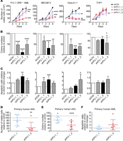
R E S E A R C H A R T I C L Ewell as apoptosis induction, were greater with the shRNA PU.1_2, leading to more efficient PU.1 knockdown (Supplemental Figure 1A). Knockdown of PU.1 in an immature murine hematopoietic cell line with normal levels of PU.1 (BaF3) did not have significant effects on proliferation or apoptosis (Supplemental Figure 1, C–F).
We next investigated the effect of PU.1 knockdown on human leukemic cell lines with different PU.1 levels (Supplemental Figure 1G). MOLM13 and Kasumi-1 cell lines harbor anomalies associ-ated with low PU.1 levels [FLT3-ITD mutation for MOLM13 and t(8;21) for Kasumi-1] (3, 4), while THP1 cells have higher PU.1 lev-els. Decreased PU.1 led to a strong inhibitory effect on the growth and clonogenic capacity of MOLM13 and Kasumi-1 cells, whereas it had no or very modest effects on THP1 cell growth (Figure 1, A and B). Accordingly, the apoptotic fraction was increased following PU.1 knockdown in MOLM13 and Kasumi-1 cells, but we observed no major effect in THP1 cells (Figure 1C). To confirm that this phe-notype was correlated with PU.1 levels, we overexpressed PU.1 in MOLM13 cells upon PU.1 knockdown with shPU.1_2 and found a rescue of the phenotype, with increased clonogenic capacity and decreased apoptosis (Supplemental Figure 1, H–J).
To determine whether PU.1 inhibition has an effect on primary cells from AML patients, we seeded mononuclear cells (MNCs) from AML patients in semisolid media for 2 weeks and assessed the number of colonies, the number of viable cells, and the propor-tion of apoptotic cells. Knockdown of PU.1 significantly decreased the number of viable cells (mean decrease of 18% for shPU.1_1 and 74% for shPU.1_2) (Figure 1D), as well as the colony-forming capacity of primary human AML cells (mean decrease of 27% for shPU.1_1 and 60% for shPU.1_2, compared with shCtrl) (Figure 1E). At the same time, the proportion of apoptotic cells increased on average by 2-fold upon knockdown of PU.1 in primary human AML cells with the more efficient shRNA (shPU.1_2) (Figure 1F).
Taken together, these data show that inhibition of PU.1 de creas-es cell growth and clonogenic capacity and increascreas-es apoptosis, in murine as well as human AML, and thus provide proof of concept for PU.1 inhibition as a possible therapeutic strategy in AML.
Development of small-molecule PU.1 inhibitors abrogating DNA binding by PU.1. Classical heterocyclic diamidines, typified by
furamidine (DB75), are highly selective and potent ligands for AT-rich DNA, a feature responsible for their success as inhibitors of kinetoplast DNA in trypanosomiasis (25, 26). As with other ETS family transcription factors, DNA site recognition by PU.1 involves contact with the major groove at the 5′-GGAA-3′ consen-sus, as well as an indirect readout of the minor groove backbone, where sequence-dependent geometry confers selectivity among ETS paralogs. PU.1 is distinguishable from other paralogs by the former’s strong preference for AT-rich sequences upstream of the 5′-GGAA-3′ consensus (27). We screened a number of designed AT-targeting heterocyclic diamidines and found that they effi-ciently competed with PU.1 for DNA binding. DNA footprinting experiments verified that the compounds targeted the flanking
sequences found in cognate PU.1 sites, using the λB motif of the
murine Igλ2-4 enhancer (5′-ATAAAAGGAAGTG-3′) (28) as a
model. On the basis of the general structural features of our initial PU.1 inhibitors (23), we designed and prepared a focused library of larger derivatives for screening using the solution and cellular analytical methods (vide infra). DB2115 and DB2313 were selected itself; how ever, it leads to myeloid bias in (preleukemic) stem
cells and MDS and AML development in combination with coop-erating events (18).
Overall, disruption of PU.1 expression or activity is present in more than 50% of patients with AML and is associated with a spe-cific transcriptional and epigenetic program (19, 20). Thus, target-ing PU.1 in AML could be an appealtarget-ing option for treatment. In the past, strategies to rescue PU.1 expression in AML cells have been explored. Overexpression of PU.1 is sufficient to trigger neutrophil differentiation in acute promyelocytic leukemia (APL) and leads to differentiation and apoptosis of various primary AML samples (5, 21). However, elevation of PU.1 levels or activity is difficult to achieve pharmacologically.
In this study, we used the inverse strategy. As complete loss of PU.1 leads to stem cell failure (15), we hypothesized that AML cells may be more vulnerable to further PU.1 inhibition in com-parison with normal hematopoietic cells. We used 2 alternative approaches to test this hypothesis: RNA interference and newly developed PU.1 inhibitors.
We have recently reported proof of principle for the ability to inhibit PU.1 by novel heterocyclic diamidines, which are deriva-tives of clinically tested compounds such as furamidine (22, 23). DNA recognition by PU.1 requires specific binding in the DNA major groove at consensus sites harboring a 5′-GGAA/T-3′ motif that typifies target sites for the ETS family. Selectivity for PU.1 is conferred through additional contacts with the minor groove of adjacent AT-rich tracks (24). We initiated a development and screening effort to find optimized compounds that would recog-nize a larger number of base pairs adjacent to a core ETS site as more specific PU.1 inhibitors. The PU.1 inhibitors we identified target the minor groove and lead to inhibition of PU.1 binding in the major groove via an allosteric mechanism.
Using RNA interference as well as our small-molecule inhibi-tors, we show that PU.1 inhibition is effective at inhibiting AML cell growth, including in murine and human cell lines and in primary AML patients’ cells in vitro and in vivo, and thus represents what we believe to be a fundamentally new strategy for the treatment of AML.
Results
PU.1 knockdown decreases cell growth and clonogenic capacity and increases apoptosis of murine and human AML cells. To determine
whether PU.1 inhibition may be a suitable strategy in AML, we used an established model of AML driven by reduced PU.1
lev-els, PU.1 URE–/– AML, in which PU.1 expression is reduced to
approximately 20% of normal levels by disruption of an upstream
enhancer (URE) (12, 17). The PU.1 URE–/– AML cell line has been
established from a leukemic mouse with homozygous deletion of the URE of the PU.1 gene, which has been previously described (17). We selected 3 shRNAs that decreased PU.1 expression in mouse and human cells (Supplemental Figure 1, A and B; sup-plemental material available online with this article; https://doi.
org/10.1172/JCI92504DS1). Knockdown of PU.1 in PU.1 URE–/–
AML cells by the 3 different shRNAs led to significantly decreased cell growth and colony formation (Figure 1, A and B). Likewise, the percentage of apoptotic cells was substantially increased upon
shRNA-mediated PU.1 knockdown in PU.1 URE–/– AML cells
narrow DNA minor groove, and can hydrogen bond with acceptor groups on AT base pairs (carbonyl of T, T=O, and N3 of A) at the floor of the minor groove. The benzimidazole-NH points into the minor groove and can also H-bond with AT base pair acceptors. In addition, all 3 groups (amidine, benzimidazole, and phenyl) as the 2 best compounds (Figure 2A). Both compounds harbor
[image:4.585.83.504.59.541.2]the amidine-benzimidazole-phenyl platform that provides excel-lent recognition for expanded AT sites. The concept for the units of these compounds is that the amidine cation imparts solubility, interacts favorably with phosphate groups that are close in the
fied, recombinant PU.1 ETS domain (Figure 2E). In the absence
of diamidine, PU.1 formed a 1:1 complex with the immobilized λB
motif with a dissociation constant of 5.4 nM, in agreement with solution fluorescence anisotropy measurements (31). Control titrations with the ETS domain of ETS1, which we have shown to be structurally superimposable with PU.1, showed a 3-log selectiv-ity of the λB site for PU.1 and attested to the specificity of our SPR
methodology. Immobilized λB DNA was loaded to greater than
95% capacity and then titrated with graded concentrations of each compound (32). Displacement of PU.1 was detected by the drop in SPR signal as a function of the compound concentration. All 3
compounds efficiently inhibited PU.1 binding, with IC50 values
in the 10–8 to 10–9 M range (Figure 2F). The rank order of the IC 50
values (DB2115 < DB2313 < DB1976) corresponded with the rank order of affinities, suggesting that the 3 compounds inhibited PU.1
binding to the λB motif via the same mechanism.
To gain structural insight into the mechanism by which the
diamidines inhibit PU.1 binding to the λB site, we performed a
molecular dynamics simulation of the DB2313-λB complex. The
λB-bound complex showed the expected binding mode, including
the typical water-mediated contacts between the 2 diamidinium-sand the DNA backbone (Supplemental Figure 2) (33). Compari-son of the equilibrated DB2313-DNA complex with the PU.1-DNA co-crystal structure (34) showed that the bound compound induced the double helix into a conformation that was incompat-ible with PU.1 binding. The co-crystal structure of the PU.1-DNA complex shows that extensive perturbations of the DNA groove structure occur in PU.1 binding. Specifically, compression of the minor groove in both width and depth at the AT-rich flanking sequences is required for indirect readout of the DNA backbone by PU.1. Crucially, insertion of DB2313 gives rise to a minor groove profile, in terms of groove width and depth, that deviates strongly from the requirement of PU.1 for binding (Figure 2G). The incom-patible effects of DB2313 and PU.1 on DNA structure therefore suggested that denial of PU.1 occupancy through conformational perturbations of the target DNA represents a major component of the efficacy of our compounds as PU.1 inhibitors. We confirmed experimentally that the diamidines induced incompatible DNA conformations for PU.1 binding by probing the drug-bound DNA with DNase I, a minor groove probe that is highly sensitive to local DNA structure (Figure 2H). Although each diamidine induced a unique DNase I footprint, as would be expected from their differ-ent structures, all were highly dissimilar to the characteristic foot-print produced by PU.1. In summary, computational and experi-mental evidence revealed mutually exclusive effects on DNA conformation as a fundamental mechanism of PU.1 inhibition by the identified diamidines.
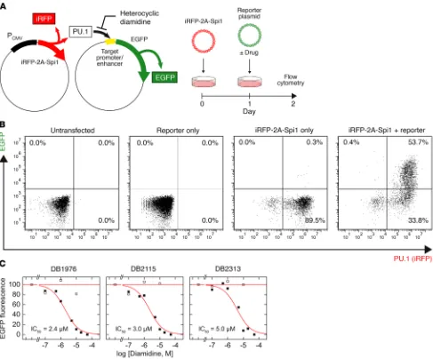
Functional inhibition of PU.1 by heterocyclic diamidines. Having
established the biophysical basis of PU.1 inhibition by DB2313, DB2115, and DB1976, we proceeded to define their cellular prop-erties and potential to inhibit PU.1-dependent gene transacti-vation. To assess the potential for functional inhibition of PU.1 transactivation by the 3 diamidines, we tested their effects on the expression of a cellular EGFP reporter under the control of a minimal PU.1-dependent promoter harboring a triple tandem
copy of the same λB enhancer site used in the SPR screen (Figure
3A). This cell-based system was designed such that ectopic PU.1 make excellent van der Waals contacts with the walls of the minor
groove and edges of AT base pairs at the floor of the groove. The compounds also have the appropriate curvature to match the DNA minor groove in AT sequences. Together, they make an excellent AT recognition module. Linking 2 of these modules, as in DB2115 and DB2313, provides a molecule for expanded and very strong AT site recognition. A docked structure shows that DB2313, for example, inserted itself precisely and deeply into the DNA minor
groove along the AT-track of the λB motif, interacting with the
DNA base pair edges at the minor groove floor over almost 10 con-secutive base pairs (Figure 2B). Thus DB2313 and DB2115, along with DB1976, which is the best compound from our initial studies of PU.1 inhibition (23), were evaluated in more detail.
We measured the affinity of these heterocyclic diamidines for the λB motif, a typical native DNA-binding site for PU.1, following our biosensor surface plasmon resonance (SPR) procedure (29, 30).
Briefly, duplex DNA harboring the λB motif was immobilized on a
streptavidin-functionalized sensor chip via a 5′-end–labeled biotin.
The affinity of each compound for the λB motif was determined
from the concentration-dependent increase in the SPR sensor-grams at steady state, as shown in Figure 2C for DB2313. The
spec-ificity of the compounds was probed by substituting the λB motif
with a model cognate site (5′-GCCGGAAGTG-3′, termed SC1) that is recognized by ETS1. As shown by DB2313, a very large excess of compound, at concentrations far in excess of those used to saturate the λB motif, failed to bind the SC1 site to detectable levels (gray sensorgram in Figure 2C). With the λB motif as a target, all 3 com-pounds bound their target DNA tightly, with 1:1 stoichiometry and dissociation constants in the 10–8 to 10–9 M range (Figure 2D).
To define the potential of the diamidines to inhibit cognate
PU.1 binding, we loaded the immobilized λB site with the
The Journal of Clinical Investigation
R E S E A R C H A R T I C L Eand reporter expression can be independently tracked by fluores-cence to filter out any background activation of the reporter. In PU.1-negative HEK293 cells, which express several endogenous ETS paralogs including ETS2, GABPA, and members of the ELF
subfamily (Human Protein Atlas), transfection of the λB-based
reporter was silent in flow cytometry, indicating specificity of
the λB enhancer to PU.1 (Figure 3B). All 3 compounds inhibited
PU.1-dependent transactivation of the reporter in a dose-
dependent manner, with similar IC50 values between 2 and 5 μM
(Figure 3C). The relatively high IC50 values should be considered
in light of the strong CMV promoter used to drive the expression of ectopic PU.1. To establish the specificity of diamidine action to the inhibition of PU.1, we tested whether the diamidines could inhib-it transactivation at an analogous minimal promoter in which the
λB enhancer sites were replaced by the SC1 sequence. Although
PU.1 can bind the SC1 site, it is not found in native PU.1 target genes (35) and is therefore functionally nonspecific. None of the diamidines significantly inhibited transactivation at the SC1-based enhancer (Figure 3C), consistent with the lack of PU.1 bind-ing to the SC1 site we observed by SPR (Figure 2C). Thus, hetero-cyclic diamidines appeared to maintain their strong AT selectivity in live cells and specifically inhibited PU.1 activity at functional DNA sites associated with PU.1 target genes.
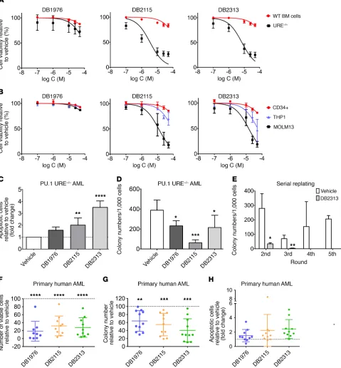
Small-molecule PU.1 inhibitors decrease cell growth and the clono-genic capacity of AML cells and lead to increased apoptosis. To
deter-mine whether our PU.1 inhibitors had functional effects on AML
cells, we treated murine PU.1 URE–/– AML and human MOLM13
cell lines with compounds at different concentrations and
com-Figure 3. Cellular properties and functional inhibition of PU.1 by expanded diamidines. (A) Ectopic PU.1 activity, as indicated by an iRFP marker, in live HEK293 cells was measured through expression of a fluorescent EGFP reporter, transfected with or without compound, under the control of a minimal PU.1-dependent, λB-based promoter. (B) Flow cytometric analysis of the PU.1 marker (iRFP) and EGFP reporter, in which the upper right quadrant rep-resents PU.1-dependent reporter activation. Note the lack of EGFP reporter activation by endogenous non-PU.1 ETS paralogs, indicating specificity of the
[image:7.585.40.527.55.459.2]ic capacity in the fourth and higher rounds of plating (Figure 4E). We also explored the effects of the small-molecule PU.1 inhib-itors on primary human AML cells and treated 13 AML patients’ samples (Supplemental Table 1) with PU.1 inhibitors. PU.1 inhib-itors led to a significant decrease in the number of viable cells (mean decrease of 81% for DB1976, 68% for DB2115, and 72% for DB2313) (Figure 4F) and clonogenic capacity (mean decrease of 36% for DB1976, 45% for DB2115, and 60% for DB23313) com-pared with vehicle-treated cells (Figure 4G). The apoptotic cell fraction increased on average by 1.5-fold with DB1976, 2.2-fold with DB2115, and 2.5-fold with DB2313 (Figure 4H). The apoptosis- inducing effect was also confirmed in short-term liquid culture for a few samples (Supplemental Figure 3E). Of note, we observed that PU.1 inhibition had an effect on the majority of samples, which harbored various genetic and cytogenetic anomalies.
Taken together, treatment with PU.1 inhibitors leads to decreased cell viability, colony formation, and increased apoptosis in PU.1lo-induced AML cell lines as well as in a majority of primary
AML cell samples from patients.
Inhibitors show on-target PU.1-inhibitory activity in AML cells.
To assess the on-target activity of our inhibitors in AML cells, we measured transcript levels of well-known PU.1 targets in PU.1 URE–/– AML cells. It has been shown that PU.1 positively regulates
CSF1R and JUNB and autoregulates itself, whereas it represses E2F1 (12, 16, 36, 37). In line with this, we found a decrease in
Csf1r, Junb, and PU.1 transcript expression and an increase in E2f1
expression upon treatment with DB2115 or DB2313 (Figure 5A, left). As Csf1r is one of the most sensitive PU.1 targets in myeloid cells, we assessed its expression at different time points (1 h, 4 h, 8 h, and 24 h) after drug treatment. Interestingly, Csf1r expression significantly decreased as early as 4 hours after treatment (Figure 5A, right), in line with a direct effect of the drugs on PU.1 transcrip-tional activity. Furthermore, treatment of BM MNCs isolated from a PU.1-GFP–knockin reporter mouse model (38) led to a decrease in GFP reporter expression after treatment with DB1976, DB2115, or DB2313, further confirming a direct effect of the compounds on PU.1 transactivation (Figure 5B and Supplemental Figure 4A) and consistent with PU.1 positive autoregulation (36). ChIP assays revealed that treatment of AML cells with DB1976, DB2115, or DB2313 decreased PU.1 occupancy on E2f1, Junb, and Csf1r pro-moters, confirming that the compounds were directly interfering with PU.1 binding to chromatin in vivo (Figure 5C).
To gain insight into the genome-wide transcriptional effects following treatment of AML cells with our inhibitors, we per-formed gene expression analysis. We found dysregulation of 1,648 transcripts (of 34,472 total) by at least 1.2-fold after DB2313
treat-ment of PU.1 URE–/– AML cells, with 867 probe sets upregulated
and 781 probe sets downregulated. We found highly significant enrichment of known genes directly downstream of PU.1 (Figure 5D). Interestingly, enrichment of genes regulated by other ETS family transcription factors, such as ETS1, GABPΑ, SPI-B, or FLI-1, was much lower or nonsignificant, suggesting a preferential inhibitory effect of our compounds on PU.1 binding, with selectiv-ity even within the ETS family. We confirmed the dysregulation of some known PU.1 target genes such as Ly96, Clec5a, Cdkn1a,
Itgb2, Fcgr3, and Gfi1 by quantitative reverse transcription PCR
(qRT-PCR) (Supplemental Figure 4B). Top canonical pathways pared them directly with the effects on murine WT bone marrow
(BM) cells or human CD34+ cord blood cells, respectively.
Treat-ment with compounds led to a profound decrease in the growth of PU.1 URE–/– AML cells (IC
50 for DB2115: 3.4 μM < IC50 for DB2313:
7.1 μM < IC50 for DB1976: 105 μM), while showing little effect on normal hematopoietic cells at similar concentrations (calculat-ed IC50 for DB2115: 192 μM; DB2313: 240 μM; DB1976: 334 μM) (Figure 4A). We made similar observations with human cells, with strong inhibitory effects on MOLM13 cells (PU.1 low), substantial-ly lesser effects on THP1 cells (PU.1 high), and negligible effects on healthy CD34+ cells (Figure 4B). We confirmed the inhibitory effect
on the proliferation of PU.1 URE–/– AML and MOLM13 cells during
a treatment time course and observed no effect on AML cells with high PU.1 levels (THP1) (Supplemental Figure 3A). To confirm that the drug effects were related to PU.1 levels, we lentivirally expressed PU.1 in MOLM13 cells and observed a rescue of the cytotoxic effects of the compounds with increased cell viability upon treatment with DB1976, DB2115, or DB2313 (Supplemental Figure 3B).
Like our observations with PU.1-directed shRNAs, treatment with pharmacological PU.1 inhibitors led to a 1.6-, 2-, and 3.5-fold increase in apoptotic cells with DB1976, DB2115, and DB2313, respectively, in murine PU.1 URE–/– AML cells (Figure 4C), and we observed similar
effects in human MOLM13 cells (Supplemental Figure 3C).
PU.1 inhibitors also significantly decreased the colony-
forming capacity of PU.1 URE–/– AML and MOLM13 cells, but not
of THP1 cells (Figure 4D and Supplemental Figure 3D). To assess the effect of PU.1 inhibitors on the long-term clonogenic capaci-ty (in vitro self-renewal) of PU.1 URE–/– AML cells, we performed
serial replating assays. Strikingly, we found that treatment with DB2313 led to a significant decrease in clonogenicity in the second and third rounds of plating and a complete disruption of
The Journal of Clinical Investigation
R E S E A R C H A R T I C L Ecanonical PU.1 targets in PUER cells (Figure 5E), which was highly significant (P < 0.0001). Furthermore, comparative pathway anal-ysis revealed a significant inverse correlation between PU.1 URE–/–
AML cells after treatment and PU.1 overexpression in PUER cells, suggesting that inhibitor treatment antagonizes canonical PU.1- regulated pathways (Figure 5F). Gene set enrichment analysis (GSEA) confirmed that DB2313 treatment leads to a change in expression of genes regulated by PU.1 in HSCs from the PU.1 URE–/–
mouse model (12) (Supplemental Figure 4D). Moreover, we found an enrichment in genes regulated by transcription factors of the AP1 family, which are known PU.1 targets (Supplemental Figure 4E).
Furthermore, we performed interactome analysis of 3 inde-pendent large series of primary AML patients’ samples including and biological functions with significant enrichment identified by
Ingenuity Pathway Analysis (IPA) included “hematological system development and function,” “cell death and survival,” “cellular development,” and “cellular growth and proliferation” (Supple-mental Figure 4C), consistent with known functions of PU.1.
We compared the differentially expressed genes upon PU.1 inhibitor treatment of PU.1 URE–/– AML cells with publicly available
data in which the PU.1-regulatory transcriptional network had been identified by tamoxifen-mediated induction of PU.1 expression in engineered PU.1-null immature hematopoietic cells (PUER cells) (Gene Expression Omnibus [GEO] GSE13125) (39). Of the 1,334
genes dysregulated after PU.1 inhibitor treatment of PU.1 URE–/–
[image:11.585.75.500.63.463.2]AML cells, 36% (484 cells) overlapped with previously identified
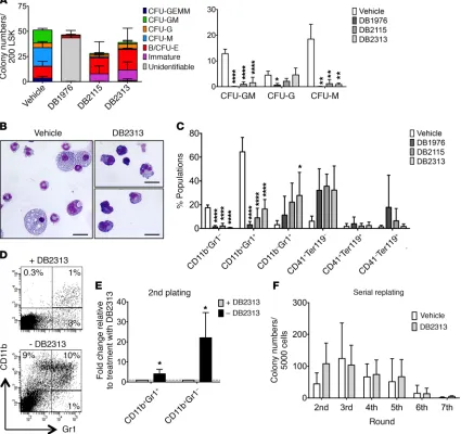
with the known important role of PU.1 in myelomonocytic differ-entiation. Of note, the initial compound DB1976 appeared to show toxicity on LSK cells, with a majority of colonies unidentifiable and a majority of dead cells, contrary to what we observed with the 2 newer compounds (DB2115 and DB2313), which we therefore pri-oritized for further investigation. Overall, the numbers of viable cells per colony were reduced upon treatment with PU.1 inhibitor (Supplemental Figure 6A), again consistent with an effect of PU.1 inhibition on more mature, differentiating cells. Consistently, cytomorphologic analysis revealed almost no macrophages, sig-nificantly fewer mature granulocytes, and an increase in immature cells, characterized by a higher nucleocytoplasmic ratio and baso-philic cytoplasm after treatment (Figure 6B). This was confirmed by flow cytometric analysis, which revealed a reduction in mature
monocytes (CD11b+Gr1–) and granulocytes (CD11b+Gr1+) and a
slight increase in immature granulocytes (CD11b–Gr1+) and CD41+
cells, but no significant difference in erythrocyte (Ter119+CD41–)
generation (Figure 6C and Supplemental Figure 6B). To further explore the granulocyte population and pinpoint the exact stage of maturation at which the small molecules impact differentiation, we used a combination of markers described by Guibal et al. (43) and further separated the cell populations into the myeloblast/ promyelocyte-metamyelocyte/mature stages (Supplemental Fig-ure 6C). After treatment, we observed a slight increase in the pro-portion of cells at the myeloblast/promyelocyte stage (stage 1), a strong increase of metamyelocytes (stage 2), and a decrease in more mature cells (stage 3) (Supplemental Figure 6D), indicat-ing that our compounds primarily inhibit later stages of granulo-cytic differentiation.
As we had observed a reduction in mature myelomonocytic colonies upon treatment with PU.1 inhibitors, but persistence or even slight increases in more immature cells (CFU-granulocyte/ erythrocyte/macrophage/megakaryocyte [GEMM] and immature colonies), we wanted to test whether these immature cells were still functional after drug removal. We focused on the compound with the lowest IC50 and the least effect on WT cells (DB2313). Inter-estingly, the production of mature granulocytes and monocytes increased significantly by 4-fold and 22-fold, respectively, after removing the treatment in the second plating, showing that the effects of PU.1 inhibition on granulocyte/monocyte (G/M) gener-ation are reversible (Figure 6, D and E). In addition, we performed serial replating assays with continuous treatment with PU.1 inhib-itors and found no significant differences in terms of serial replat-ing capacity, indicatreplat-ing that treatment with PU.1 inhibitors does not significantly affect the long-term colony-forming potential or in vitro self-renewal capacity of immature hematopoietic stem and progenitor cells (Figure 6F).
We also explored the effect of the inhibitors on lymphoid cells. We treated B cells in liquid culture for 3 days and found that the compounds had no effect on the apoptotic fraction (Sup-plemental Figure 6G). To assess T cells, we performed culturing on OP9-Delta1 cells and found that compound treatment led to slightly delayed, but not completely blocked, T cell differentia-tion (Supplemental Figure 6, E and F). Finally, we assessed PU.1 inhibitor–treated BM cells in congenic transplantation assays and found no changes in T cell numbers and only modestly decreased B cell numbers after 20 weeks (Supplemental Figure 6H). those from The Cancer Genome Atlas (TCGA), the MILE
(Microar-ray Innovations in LEukemia) study, and GEO GSE1159 to iden-tify the PU.1 regulon specifically in AML cells using the ARACNe algorithm (40–42), and then tested the activity of PU.1 using this gene set in the DB2313-induced transcriptional signature using GSEA. In these analyses, the PU.1 “regulon” generated by the ARACNe algorithm was partitioned in a positive and a negative component on the basis of a positive or negative Spearman cor-relation (P < 0.05) between expression levels of PU.1 and the tar-get gene across all samples of the data set used to generate the network. Strikingly, for each of these patient cohorts, we found that the PU.1 positive regulon (“PU.1 UP,” genes upregulated when PU.1 activity was high) was enriched under vehicle-treated conditions, consistent with a negative effect of DB2313 on PU.1 transcriptional activity in AML cells (Figure 5, G and H, and Sup-plemental Figure 4, F–I).
In addition, we performed a comparative analysis with publicly available PU.1 ChIP-sequencing (ChIP-seq) data (GEO GSE63317) (20) and found significant enrichment of PU.1 binding in the pro-moters of genes deregulated after treatment with PU.1 inhibitor (Figure 5I). Up- and downregulated genes were similarly affected, consistent with an interference of our drugs with PU.1-chromatin interaction and independent of downstream transactivating or repressive mechanisms.
We performed PU.1 ChIP-seq experiments and analyzed PU.1-bound sites in the vehicle-treated sample and PU.1 sites in the PU.1
inhibitor–treated (DB2313-treated) sample in PU.1 URE–/– cells.
Comparison of PU.1 ChIP-seq of vehicle versus DB2313 treatment as the background showed a strong enrichment of PU.1 motifs, while the opposite comparison showed enrichment of motifs that are not related to ETS transcription factors (data not shown). This is in line with better PU.1 binding under control conditions. The most common motif in the PU.1 ChIP was AAAGAGGAAGTG, and the respective position weight matrix (PWM) found by Homer was present in 41% of peaks in the vehicle sample versus only 16% of the peaks in the DB2313-treated sample, which is again in line with PU.1 inhibitor treatment causing decreased binding of PU.1 to its canonical target sites. When we looked specifically at the peaks with a PU.1 motif in the vehicle-treated samples and subtracted peaks with a PU.1 motif in the DB2313-treated samples, we arrived at 268 bona fide PU.1 sites that were “lost” upon drug treatment. A complete list of these peaks/sites is provided in Supplemental Table 2. Examples of peaks from this analysis are shown in Supple-mental Figure 5. Overall, the results from the ChIP-seq analyses are consistent with a genome-wide inhibitory effect of DB2313 on PU.1 binding to its canonical sites in AML cells.
PU.1 inhibitors have limited effects on normal hematopoiesis.
In order to determine the effect of our PU.1 inhibitors on normal
hematopoietic differentiation, we sorted immature Lin–Sca1+
c-Kit+ (LSK) cells from WT mice and treated them with either
The Journal of Clinical Investigation
R E S E A R C H A R T I C L EDiscussion
During the past decade, increasing evidence has shown the importance of PU.1 dosage in leukemia development. Our study provides proof of concept that targeting PU.1, a commonly dereg-ulated transcription factor in AML, has antileukemic effects.
Given the findings from Iwasaki et al. (15) showing that com-plete loss of PU.1 in HSCs leads to their exhaustion, we explored in this work inhibition of PU.1 activity as a therapeutic strategy in AML, with the hypothesis that leukemic cells with an already dysregulated PU.1 transcriptional network would be more suscep-tible to further PU.1 inhibition than normal cells. Our approach is similar in principle to what has been described recently for casein kinase 1A1, a tumor-suppressor gene located on chromosome 5q that is frequently heterozygously deleted in myelodysplastic syn-drome. Haploinsufficiency of CSNK1A leads to HSC expansion, whereas complete deletion leads to HSC failure. Cells with already reduced levels of CSNK1A were more sensitive to CSNK1A phar-macological inhibition than were cells with normal levels (44).
We used 2 methods to decrease PU.1 activity: RNA interfer-ence and new PU.1 inhibitors. Only a few drugs directly targeting dysregulated transcription factors have been described so far, with one recent example being an inhibitor targeting a protein-protein
interaction, RUNX1 binding to CBFβ-SMMHC, which is an
onco-genic transcription factor fusion found in inv(16) AML (45). The inhibitors reported here do not bind the PU.1 protein directly but instead prevent binding of PU.1 to cognate DNA sites, the requisite molecular event for gene regulation, via an allosteric mechanism. Specifically, occupancy by the compounds in the minor groove induces perturbations in DNA conformation that are transmitted to the orthosteric site of PU.1 in the major groove. Dissociation of DNA-bound PU.1 by DB2313 and distinctive DNA footprints of the compounds demonstrated that the bound confor-mations of PU.1 and the compounds with the same DNA sequence are not compatible. The compounds are derivatives of the hetero-cyclic diamidine family, some members of which (e.g., DB75 [fur-amidine] and derivatives for trypanosomiasis) have been estab-lished in the context of other clinical applications (46, 47).
Decreasing PU.1 activity and levels in human and murine
PU.1lo-induced leukemic cells led to decreased cell growth and
clonogenicity. We found that this phenotype was due to increased apoptosis after PU.1 inhibition. We also found inhibitory effects in primary human samples harboring various genetic alterations, suggesting that such a strategy could be applied to a larger frac-tion of patients with AML. Addifrac-tionally, treatment with DB2313 increased survival and reduced tumor burden in vivo. Even though these inhibitors are early tool compounds and need further opti-mization for improved bioavailability in vivo, the fact that com-pounds from the same family have reached the clinic for a differ-ent indication offers encouragemdiffer-ent for further developmdiffer-ent.
The reduction in PU.1 binding to chromatin, combined with changes in PU.1 target gene expression and the PU.1-dependent “reg-ulon” in AML cells, demonstrates the on-target effect of the com-pounds. Of note, our compounds have a preference for PU.1-binding sites, which are characterized by highly AT-rich flanking sequences, a feature that is not shared by most other ETS transcription factors. And while the effect of our compounds does not seem to be exclu-sively restricted to PU.1-binding sites, we found significant selec-Taken together, our results indicate that treatment with our
PU.1 inhibitors leads to effects on normal hematopoiesis that are consistent with fundamental roles of PU.1 function during hematopoiesis in myelomonocytic lineages, with limited effects on T and B cell lymphopoiesis. The effects on myeloid cells are reversible upon treatment discontinuation and seem to primarily affect more mature cells. However, in future studies, it will be of interest to perform longer-term in vivo treatment with further optimized compounds (once available) for a more comprehensive and definitive evaluation of the more subtle effects on normal hematopoiesis.
Treatment with PU.1 inhibitors decreases leukemia progression in vivo. To assess the effect of PU.1 inhibitors on the growth of
AML cells in vivo, we treated PU.1 URE–/– AML cells with DB2313
for 2 days and injected 2 × 10⁵ viable cells into sublethally
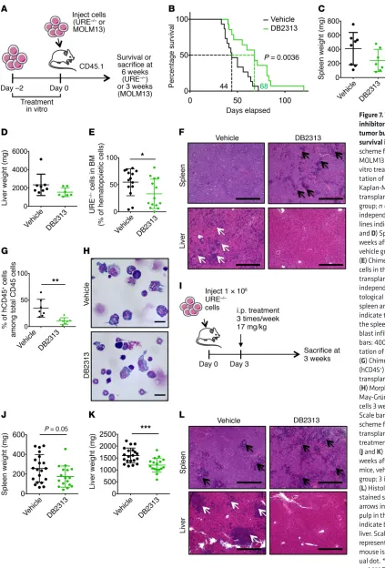
irradi-ated recipient mice (Figure 7A). Mice that received vehicle-treat-ed AML cells succumbvehicle-treat-ed to leukemia, with a mvehicle-treat-edian latency of 44 days, whereas mice that received DB2313-treated AML cells survived significantly longer (P = 0.0036), with a median latency of 68 days (Figure 7B). Assessment of tumor burden 6 weeks after transplantation showed a decrease in hepatoplenomegaly after treatment; mean splenic weight was 410 mg for vehicle-treated mice and 243 mg for DB2313-treated mice (Figure 7C); mean liver weight was 2,347 mg for vehicle-treated mice and 1,548 mg for DB2313-treated mice (Figure 7D). AML cell engraftment in the BM was significantly decreased after treatment with DB2313, with a mean chimerism of 55% for the vehicle-treated group and 33% for the DB2313-treated group (Figure 7E and Supple-mental Figure 7A). Histological analyses revealed severe blast infiltration, with disruption of the splenic architecture and com-plete loss of the red pulp in the vehicle-treated group, and sig-nificantly less pronounced effects in the DB2313-treated group. Likewise, infiltration of the liver by leukemic blasts was substan-tially reduced in the DB2313-treated group compared with that detected in the vehicle-treated group (Figure 7F). When we per-formed the same experiment with human MOLM13 AML cells, we observed a similarly significant decrease in leukemic burden in the BM after treatment, with a mean chimerism of 35% for the vehicle-treated group and 11% for the DB2313-treated group (Figure 7, A, G, and H).
Further, we performed in vivo treatment by i.p. injections of DB2313 following transplantation of PU.1 URE–/– AML cells (Figure
7I). The effect on AML cells in the BM was modest, with a mean chimerism of 47% for the vehicle-treated group versus 37% for the DB2313-treated group, which was likely due to the low concentra-tion of this prototype compound in the serum (Supplemental Fig-ure 7, B and C). However, we observed decreased splenomegaly (mean weight of 257 mg for vehicle-treated mice and 174 mg for DB2313-treated mice) and hepatomegaly (mean weight of 1,604 mg for vehicle-treated mice and 1,203 mg for DB2313-treated mice) (Figure 7, J and K), reduced leukemic infiltration in the spleen (Figure 7L and Supplemental Figure 7D), and no detectable leu-kemic infiltration in the liver after DB2313 treatment (Figure 7L).
The Journal of Clinical Investigation
R E S E A R C H A R T I C L Eimmunodeficiency IL-2 receptor γ–null (NSG) mice were housed in
the animal facility.
Patients’ characteristics. See the Supplemental Methods and Sup-plemental Table 1.
Lentiviral vector and transduction. shRNA sequences for knock-down of PU.1 have been previously described and validated (shPU.1_1 [ref. 50], and shPU.1_2 [ref. 51]); shPU.1_3 and shPU.1_4 were newly designed (shPU.1_3: 5′-AGATGCACGTCCTCGATAC-3′; shPU.1_4: 5′-TCCAGTTCTCGTCCAAGCACAA-3′). These sequences recognize both mouse and human sequences. Control (shCtrl) or shPU.1 sequences were cloned under an H1 promoter and inserted into the lentiviral vec-tor pRRLsin-PGK-eGFP-WPRE (provided by Hana Raslova, Institut Gustave Roussy, Villejuif, France). PU.1 overexpression was performed using the lentiviral vector PCAD-PU.1-IRES-GFP or GFP alone as pre-viously described (18). All viruses were produced using the 293T cell line. Cells were transduced by spin infection at 1,000 g for 60 minutes at 37°C in the presence of Polybrene. Seventy-two hours after
transduc-tion, cultures were sorted for GFP+ cells and used for further studies.
Molecular dynamics simulations. See the Supplemental Methods. Biosensor-SPR assays for compound binding affinity and PU.1-DNA complex inhibition by compound. See the Supplemental Methods.
DNA footprinting. See the Supplemental Methods.
Cell proliferation assays. To evaluate the effect of the small mol-ecules on cell viability, cells were cultured in the presence of vehicle (water) or different compounds. Cells were plated at a density between
0.5 × 10⁵ and 2 × 10⁵ in 100 μl or 500 μl of media and manually counted
every 24 hours for 4 days using a trypan blue exclusion assay. For IC50
determination, cell viability was first assessed using a resazurin cell viability assay (Cell Titer Blue; Promega) after 48 hours of culture and confirmed by manual counting using a trypan blue exclusion assay. If not further specified, drugs for the treatment of mouse cells were
used at the following concentrations: 25 μM for DB1976, 700 nM for
DB2115, and 330 nM for DB2313. For the treatment of human cells, the same concentrations were used, with the exception of DB2313, which was used at a concentration of 660 nM.
Colony formation assays and serial replating assays. For human cell line colony assays, 1,000 or 3,000 cells were plated per milliliter of
methylcellulose in HSC002SF or HSC003 methylcellulose (R&D
Sys-tems). For mouse cell colony assays, 1,000 cells per milliliter of
meth-ylcellulose were plated in HSC007 methmeth-ylcellulose (R&D Systems)
and scored after 7 days. For human primary AML cell colony assays, 150,000 mononuclear cells were plated per milliliter of methylcel-lulose in HSC003 media and the colonies scored after 12 to 14 days.
For LSK cells, 200 cells per 500 μl methylcellulose were plated in
HSC007. Vehicle or inhibitors were added to the methylcellulose at
the following concentrations: 25 μM for DB1976, 700 nM for DB2115,
and 330 nM for DB2313 for murine cells and 660 nM for DB2313 for human cells. For serial replating of cell lines, the same numbers of cells initially plated were replated. For serial replating of primary LSK
cells, 5,000 cells per 500 μl methylcellulose were replated.
Lymphoid cell culture. See the Supplemental Methods.
Flow cytometric analysis and cell sorting. See the Supplemental Methods.
Cytomorphology. See the Supplemental Methods. Western blot analysis. See the Supplemental Methods.
Inhibition of the PU.1-DNA complex in live cells (reporter assay). The functional inhibition of PU.1 transactivation by heterocyclic
diami-tivity for PU.1, which was sufficient to target AML cells and prevent unspecific toxicity. Consistent with known PU.1 function in normal hematopoiesis and myelomonocytic differentiation, we identified a transient decrease in the generation of terminally differentiated cells, especially from the myelomonocytic lineages, after treatment of immature stem and progenitor cells with PU.1 inhibitors. However, removal of the compounds led to restoration of normal hematopoi-etic differentiation, indicating that normal stem and progenitor cells are not significantly affected by treatment with PU.1 inhibitors.
In summary, our study provides proof of principle for inhibi-tion of the transcripinhibi-tion factor PU.1 as a therapeutic strategy in the treatment of AML. Furthermore, we present small molecules of the heterocyclic diamidine family as first-in-class PU.1 inhibitors that act via allosteric inhibition of PU.1-chromatin interaction and have anti-leukemic efficacy in vitro and in vivo. Our findings show that thera-peutic interference with transcription factor–chromatin binding can be achieved with small molecules through a minor groove–targeting strategy in principle and support the further development of PU.1 inhibitors as potential therapeutics.
Methods
Cells. THP1, MOLM13, BaF3, Kasumi-1, HEK293, and 293T cells were purchased from ATCC. MOLM13 and Kasumi-1 cells were cultured in RPMI 1640 medium supplemented with 20% heat-inactivated FBS and 1% penicillin/streptomycin (P/S). THP1, BaF3, and HEK293 cells were cultured in RPMI 1640 medium completed with 10% FBS and 1% P/S. BaF3 cells were supplemented with 10 ng/ml IL-3. 293T cells were maintained in DMEM supplemented with 10% FBS and 1% P/S.
PU.1 URE–/– and PU.1 URE+/–Msh2–/– leukemia cells (17, 18) were
cul-tured in M5300 medium (STEMCELL Technologies) supplemented with 10% FBS, 15% WEHI supernatant, 15% BHK supernatant, and
1% P/S. PU.1 URE–/– and PU.1 URE+/–Msh2–/– cell lines were established
from leukemic URE–/– and URE+/–Msh2–/– mice, respectively.
Mouse cells from peripheral blood (PB), BM, spleen, or liver were isolated, treated with ACK buffer (pH 7.4) to lyse red blood cells, and cultured or stained for FACS analysis. Cells were maintained in Iscove’s Modified Dulbecco’s Media (IMDM) (Gibco, Thermo Fisher Scientif-ic) supplemented with 20% FBS, 20 ng/ml recombinant mouse SCF (rmSCF) (Miltenyi Biotec), 10 ng/ml rmIL-3 (Gemini), 10 ng/ml rmIL-6 (Gemini), 20 ng/ml rmTPO (Miltenyi Biotec), and 1% P/S.
Human MNCs from BM or PB obtained from healthy or leuke-mic patients were isolated by density ultracentrifugation using Ficoll-Hypaque (GE Healthcare) and cultured in IMDM medium supplemen-ted with 2% FBS, cytokines (10 ng/ml rhIL-3 [Preprotech], 25 ng/ml recombinant human IL-6 [rhIL-6], 50 ng/ml rhSCF, 50 ng/ml rhFLT3
ligand, and 100 ng/ml rhTPO [all from Gemini]), 40 μg/ml hLDL, and
1% P/S. All cell lines and primary cells were maintained in an incubator
at 37°C and 5% CO2.
Small molecules. The syntheses of DB1976 (23), DB2115 (48), and DB2457 (49) have been previously reported. The synthesis of DB2313 and the experimental details, along with characterization data, are provided in the supplemental materials.
After synthesis, DB1976, DB2115, DB2313, and DB2457 were dis-solved in sterile water and stored at –20°C.
Mice. PU.1 URE–/–, PU.1 URE+/–Msh2–/–, and PU.1 heterozygous
Among the genes dysregulated in PU.1 URE–/– after DB2313 treatment,
we identified those with PU.1 occupancy in their promoters, defined as being within 1 kb upstream of the transcription start site (TSS). To deter-mine the significance of correlation of differential gene expression upon PU.1 activation with the presence of PU.1 binding in the gene promoter, we performed a Fisher’s exact test using UCSC Genes (mm9) as a refer-ence (https://genome.ucsc.edu/cgi-bin/hgGateway?db=mm9).
Interactome analyses (ARACNe Networks). See the Supplemental Methods.
ChIP-seq analysis. See the Supplemental Methods. BM transplantation. See the Supplemental Methods.
Determination of serum compound concentrations. See the Supple-mental Methods.
In vivo leukemia models. NSG mice were sublethally irradiated (2.0
Gy) and transplanted via retro-orbital vein injection with PU.1 URE–/–
AML cells or via tail vein injection with MOLM13 cells. For pretreat-ment experipretreat-ments, AML cells were treated in vitro with vehicle or DB2313, viable cells were counted after 2 days of treatment, and 2 ×
105 cells (PU.1 URE–/– AML) or 1 × 10⁶ cells (MOLM13) per mouse were
transplanted. Mice were monitored for signs of leukemia. For survival experiments, live BM aspiration was performed in mice 6 weeks after
transplantation of PU.1 URE–/– cells. Otherwise, mice were sacrificed
at 6 weeks (PU.1 URE–/– AML group) or 3 weeks (MOLM13 group). For
i.p. treatment with PU.1 URE–/– AML cells, mice were transplanted
with 1 × 10⁶ cells per mouse. Mice were divided into vehicle-treated
and DB2313-treated (17 mg/kg) groups. Treatment started 3 days after transplantation and continued for 3 weeks, with 3 injections per week. Mice were sacrificed 3–4 weeks after transplantation.
After sacrifice, BM, spleens, and livers were harvested from the mice, and engraftment was assessed by FACS using CD45.1, CD43,
and c-Kit antibodies for PU.1 URE–/– AML cells, and CD45.1 and
human CD45 antibodies for MOLM13 cells.
Statistics. Data are presented as the mean ± SD of independent experiments, unless otherwise specified. Statistical analyses were performed with GraphPad Prism 7.0 (GraphPad Software) using 1-way ANOVA with Tukey’s post-test correction for multigroup comparisons or a 2-tailed Student’s t test for 2-group comparisons, unless otherwise specified in the figure legends. A P value of less than 0.05 was con-sidered significant. For survival analyses, a log-rank test was used to determine statistical significance.
Study approval. Human samples were collected after informed consent was obtained from patients and upon approval of the IRB of Albert Einstein College of Medicine (protocol 2008-842). All mouse experiments were approved by the IACUC of Albert Einstein College of Medicine (protocol 2013-1202).
Author contributions
IAD and US designed the study and experiments. AP and WDW designed the biosensor-SPR experiments. GMKP designed the DNA footprinting and (with HMK) the reporter experiments. GMKP, KH, AP, and WDW designed and executed the molecular simulations. DWB designed the novel compound syntheses, which were performed by AK and AAF. IAD, JL, HMK, SRN, KM, AP, and GMKP performed experiments. BB analyzed Chip-seq data sets. IAD and JC analyzed microarray data sets. LAC, TIT, BW, IM, AV, EG, AAI, and AAF analyzed and interpreted data. IAD, JL, US, AP, WDW, DWB, and GMKP wrote the manuscript.
dines in live cells was measured using a fluorescent EGFP reporter. For this assay, a PU.1 expression plasmid was cloned by inserting a frag-ment encoding full-length PU.1 fused to an infrared RFP (iRFP) (52) reporter between the NheI/BamHI sites of pcDNA3.1(+). The fusion was linked by a sequence encoding a self-cleaving 2A peptide (53). Cultured HEK293 cells, which do not express PU.1, were transfected with the PU.1 expression plasmid for 24 hours. Cells were then retrans-fected, in the absence or presence of compounds, with an EGFP-based reporter under the control of a minimal promoter that was downstream
from a synthetic enhancer harboring 5 tandem repeats of the λB motif
(23). After an additional 24 hours, cells were trypsinized and counted by flow cytometry using an Accuri C6 instrument (BD) on 2 spectrally isolated channels for EGFP (488/511 nm) and iRFP (640/>670 nm).
Molecular modeling and docking. See the Supplemental Methods. ChIP. PU.1 URE+/–Msh2–/– cells (10 × 106) were treated with vehicle,
50 μM DB1976, 1.4 μM DB2115, or 660 nM DB2313 for 8 hours. ChIP
experiments were performed as previously described (54, 55), except that Dynabeads Protein A (Thermo Fisher Scientific) were used for the pulldown. Briefly, cells were cross-linked using a 1% formaldehyde solution for 15 minutes at room temperature. Samples were sonicated for 10 minutes to generate fragments below 1 kb. For
immunoprecipi-tation, 5 μg PU.1 antibody (Santa Cruz Biotechnology; sc-352) or
nor-mal rabbit IgG (Santa Cruz Biotechnology; sc-3888) was used. DNA fragments were purified using a QIAquick PCR Purification Kit (QIA-GEN) and subjected to qPCR amplification. The percentage of input was calculated using the comparative cycle threshold method. The primers used are listed in Supplemental Table 3.
qRT-PCR and microarray experiments. RNA was extracted using an RNeasy Mini or Micro Kit (QIAGEN) and, for qRT-PCR, reverse transcription of RNA was performed using an iScript cDNA Synthesis Kit (Bio-Rad). qPCR reactions were performed on a ViiA7 instrument using SYBR Green PCR Master Mix (both from Life Technologies, Thermo Fisher Scientific). Expression levels of genes were normal-ized to Gapdh. The primers used are listed in Supplemental Table 3.
For microarrays, RNA was isolated from PU.1 URE–/– AML cells after
24 hours of treatment with vehicle or DB2313 (330 nM). After check-ing RNA quality uscheck-ing a 2100 Bioanalyzer (Agilent Technologies), RNA was labeled with the Affymetrix GeneChip WT Terminal Label-ing Kit, hybridized to Affymetrix 2.0 Mouse GeneST microarrays, and scanned by an Affymetrix GeneChip Scanner 3000 7G system using standard protocols. Microarray data are available in the NCBI’s GEO database (GEO GSE77651). Raw data were normalized using Expres-sion Console software (Affymetrix), and differentially expressed genes were determined using a fold-change of 1.2 and a P value of 0.1 after analysis with Transcriptome Analysis Console software (Affyme-trix). Pathway analyses were performed using IPA (QIAGEN). A –log (P value) cutoff of 1.3 was used as the significance threshold. GSEA was performed with gene sets from the Molecular Signatures
Data-base (56, 57) and from the PU.1 URE–/– HSC signature database (12).
Integrative analysis with published data sets. For comparative analy-sis, we used the NCBI GEO data set GSE13125 (39), derived from a PU.1 overexpression system with tamoxifen-inducible “PUER” cells. Com-parative analysis between differentially expressed genes in this data set
and in PU.1 URE–/– AML cells after treatment was performed using IPA.
For GSEA, we used the NCBI GEO data set GSE5654, derived from PU.1
URE–/– HSC (12). To identify genes directly regulated by PU.1, we used
The Journal of Clinical Investigation
R E S E A R C H A R T I C L Ethe Leukemia & Lymphoma Society (to US); and a research grant
from the Taub Foundation for Myelodysplastic Syndromes (MDS) Research (to US). IAD was partially supported by a postdoctoral fellowship from the Association pour la Recherche sur le Cancer. JC was supported by the Einstein Training Program in Stem Cell Research from the Empire State Stem Cell Fund through New York State Department of Health contract C30292GG. US is the Diane and Arthur B. Belfer Scholar in Cancer Research of the Albert Ein-stein College of Medicine and is a Research Scholar of the Leukemia
& Lymphoma Society. AF is supported by NIH grant R35CA210065.
Address correspondence to: Ulrich Steidl, Albert Einstein College of Medicine/Montefiore Medical Center, Chanin Institute for Cancer Research, Rm. 601-605, 1300 Morris Park Avenue, New York, New York 10461, USA. Phone: 718.430.3437; Email: ulrich. steidl@einstein.yu.edu.
Acknowledgments
We thank A. Skoultchi, K. Gritsman, J.B. Micol, and all members of the Steidl laboratory for helpful suggestions and discussions, and M. Ferreira for technical assistance. We also thank D. Sun from the Einstein Stem Cell Isolation and Xenotransplantation Facility (funded through New York Stem Cell Science [NYSTEM] grant C029154); D. Reynolds and W. Tran from the Einstein Genomics Core Facility; P. Schultes from the Department of Cell Biology at Albert Einstein College of Medicine; and S. Wang from the Georgia State University Mass Spectrometry facility for their expert techni-cal support. We thank the Albert Einstein Cancer Center Shared Resources (NIH grant P30CA013330) and the Herbert Irving Can-cer Center Shared Resources (NIH grant P30CA013696). This work was supported by NIH grants R01CA166429 and R01CA217092 (to US), R01GM111749 (to WDW and DWB), and R21HL129063 (to GMKP); a Translational Research Program (TRP) grant from
1. Facts 2014-2015. Leukemia & Lymphoma Society. https://www.lls.org/sites/default/files/file_ assets/facts.pdf. Accessed September 28, 2017. 2. Cancer Genome Atlas Research Network, et al.
Genomic and epigenomic landscapes of adult de novo acute myeloid leukemia. N Engl J Med. 2013;368(22):2059–2074.
3. Mizuki M, et al. Suppression of myeloid tran-scription factors and induction of STAT response genes by AML-specific Flt3 mutations. Blood. 2003;101(8):3164–3173.
4. Vangala RK, et al. The myeloid master regula-tor transcription facregula-tor PU.1 is inactivated by AML1-ETO in t(8;21) myeloid leukemia. Blood. 2003;101(1):270–277.
5. Mueller BU, et al. ATRA resolves the dif-ferentiation block in t(15;17) acute myeloid leukemia by restoring PU.1 expression. Blood. 2006;107(8):3330–3338.
6. Forbes SA, et al. COSMIC: exploring the world’s knowledge of somatic mutations in human cancer. Nucleic Acids Res. 2015;43(Database issue):D805–D811.
7. Grimwade D, et al. Refinement of cytogenetic classification in acute myeloid leukemia: deter-mination of prognostic significance of rare recurring chromosomal abnormalities among 5876 younger adult patients treated in the United Kingdom Medical Research Council trials. Blood. 2010;116(3):354–365.
8. Bonadies N, Pabst T, Mueller BU. Heterozygous deletion of the PU.1 locus in human AML. Blood. 2010;115(2):331–334.
9. Lavallée VP, et al. The transcriptomic landscape and directed chemical interrogation of MLL- rearranged acute myeloid leukemias. Nat Genet. 2015;47(9):1030–1037.
10. Mueller BU, et al. Heterozygous PU.1 mutations are associated with acute myeloid leukemia. Blood. 2002;100(3):998–1007.
11. Steidl U, et al. A distal single nucleotide polymor-phism alters long-range regulation of the PU.1 gene in acute myeloid leukemia. J Clin Invest. 2007;117(9):2611–2620.
12. Steidl U, et al. Essential role of Jun family transcrip-tion factors in PU.1 knockdown-induced leukemic stem cells. Nat Genet. 2006;38(11):1269–1277.
13. McKercher SR, et al. Targeted disruption of the PU.1 gene results in multiple hematopoietic abnormalities. EMBO J. 1996;15(20):5647–5658. 14. Scott EW, Simon MC, Anastasi J, Singh H.
Requirement of transcription factor PU.1 in the development of multiple hematopoietic lineages. Science. 1994;265(5178):1573–1577.
15. Iwasaki H, et al. Distinctive and indispensable roles of PU.1 in maintenance of hematopoietic stem cells and their differentiation. Blood. 2005;106(5):1590–1600.
16. Staber PB, et al. Sustained PU.1 levels balance cell-cycle regulators to prevent exhaustion of adult hematopoietic stem cells. Mol Cell. 2013;49(5):934–946.
17. Rosenbauer F, et al. Acute myeloid leukemia induced by graded reduction of a lineage- specific transcription factor, PU.1. Nat Genet. 2004;36(6):624–630.
18. Will B, et al. Minimal PU.1 reduction induces a preleukemic state and promotes develop-ment of acute myeloid leukemia. Nat Med. 2015;21(10):1172–1181.
19. Staber PB, et al. The Runx-PU.1 pathway pre-serves normal and AML/ETO9a leukemic stem cells. Blood. 2014;124(15):2391–2399. 20. Sive JI, Basilico S, Hannah R, Kinston SJ, Calero-
Nieto FJ, Göttgens B. Genome-scale definition of the transcriptional programme associated with compromised PU.1 activity in acute myeloid leukaemia. Leukemia. 2016;30(1):14–23. 21. Durual S, et al. Lentiviral PU.1 overexpression
restores differentiation in myeloid leukemic blasts. Leukemia. 2007;21(5):1050–1059. 22. Munde M, Poon GM, Wilson WD. Probing the
electrostatics and pharmacological modulation of sequence-specific binding by the DNA-binding domain of the ETS family transcription factor PU.1: a binding affinity and kinetics investigation. J Mol Biol. 2013;425(10):1655–1669.
23. Munde M, et al. Structure-dependent inhibition of the ETS-family transcription factor PU.1 by novel heterocyclic diamidines. Nucleic Acids Res. 2014;42(2):1379–1390.
24. Pham TH, et al. Mechanisms of in vivo bind-ing site selection of the hematopoietic master transcription factor PU.1. Nucleic Acids Res.
2013;41(13):6391–6402.
25. Soeiro MN, Werbovetz K, Boykin DW, Wilson WD, Wang MZ, Hemphill A. Novel amidines and analogues as promising agents against intracel-lular parasites: a systematic review. Parasitology. 2013;140(8):929–951.
26. Wilson WD, Tanious FA, Mathis A, Tevis D, Hall JE, Boykin DW. Antiparasitic compounds that target DNA. Biochimie. 2008;90(7):999–1014. 27. Wei GH, et al. Genome-wide analysis of
ETS-family DNA-binding in vitro and in vivo. EMBO J. 2010;29(13):2147–2160.
28. Eisenbeis CF, Singh H, Storb U. PU.1 is a component of a multiprotein complex which binds an essential site in the murine immuno-globulin lambda 2-4 enhancer. Mol Cell Biol. 1993;13(10):6452–6461.
29. Nguyen B, Tanious FA, Wilson WD. Biosen-sor-surface plasmon resonance: quantitative analysis of small molecule-nucleic acid interac-tions. Methods. 2007;42(2):150–161.
30. Nanjunda RM, Munde M, Liu M, Wilson WD. Real-time monitoring of nucleic acid interactions with biosensor surface plasmon resonance. In: Wanunu Y, ed. Methods for Studying DNA/ Drug Interactions. Boca Raton, FL: CRC Press; 2011:92–122.
31. Stephens DC, Kim HM, Kumar A, Farahat AA, Boykin DW, Poon GM. Pharmacologic efficacy of PU.1 inhibition by heterocyclic dications: a mechanistic analysis. Nucleic Acids Res. 2016;44(9):4005–4013.
32. Wang S, Poon GMK, Wilson WD. Quantitative Investigation of Protein–Nucleic Acid Interac-tions by Biosensor Surface Plasmon Resonance. In: Leblanc BP, Rodrigue S, eds. DNA-Protein Interactions. New York, NY: Humana Press; 2015:313–332.
33. Wei D, Wilson WD, Neidle S. Small-molecule binding to the DNA minor groove is mediated by a conserved water cluster. J Am Chem Soc. 2013;135(4):1369–1377.
34. Kodandapani R, et al. A new pattern for helix-turn-helix recognition revealed by the PU.1 ETS-domain-DNA complex. Nature. 1996;380(6573):456–460.
specificity studies of four ETS proteins support an indirect read-out mechanism of protein-DNA rec-ognition. J Biol Chem. 2000;275(37):28363–28370. 36. Chen H, et al. PU.1 (Spi-1) autoregulates
its expression in myeloid cells. Oncogene. 1995;11(8):1549–1560.
37. Zhang DE, Hetherington CJ, Chen HM, Tenen DG. The macrophage transcription factor PU.1 directs tissue-specific expression of the macro-phage colony-stimulating factor receptor. Mol Cell Biol. 1994;14(1):373–381.
38. Back J, Allman D, Chan S, Kastner P. Visualizing PU.1 activity during hematopoiesis. Exp Hematol. 2005;33(4):395–402.
39. Weigelt K, Lichtinger M, Rehli M, Langmann T. Transcriptomic profiling identifies a PU.1 regu-latory network in macrophages. Biochem Biophys Res Commun. 2009;380(2):308–312.
40. Basso K, Margolin AA, Stolovitzky G, Klein U, Dalla-Favera R, Califano A. Reverse engineering of regulatory networks in human B cells. Nat Genet. 2005;37(4):382–390.
41. Margolin AA, et al. ARACNE: an algorithm for the reconstruction of gene regulatory networks in a mammalian cellular context. BMC Bioinformat-ics. 2006;7 Suppl 1:S7.
42. Margolin AA, Wang K, Lim WK, Kustagi M, Nemenman I, Califano A. Reverse engineering cellular networks. Nat Protoc. 2006;1(2):662–671. 43. Guibal FC, et al. Identification of a myeloid
committed progenitor as the cancer-initiating cell in acute promyelocytic leukemia. Blood. 2009;114(27):5415–5425.
44. Schneider RK, et al. Role of casein kinase 1A1 in the biology and targeted therapy of del(5q) MDS. Cancer Cell. 2014;26(4):509–520.
45. Illendula A, et al. Chemical biology. A small- molecule inhibitor of the aberrant transcription factor CBFbeta-SMMHC delays leukemia in mice. Science. 2015;347(6223):779–784. 46. Das BP, Boykin DW. Synthesis and antiprotozoal
activity of 2,5-bis(4-guanylphenyl)furans. J Med Chem. 1977;20(4):531–536.
47. Wenzler T, Boykin DW, Ismail MA, Hall JE, Tidwell RR, Brun R. New treatment option for second- stage African sleeping sickness: in vitro and in vivo efficacy of aza analogs of DB289. Antimicrob Agents Chemother. 2009;53(10):4185–4192.
48. Liu Y, Chai Y, Kumar A, Tidwell RR, Boykin DW, Wilson WD. Designed compounds for recogni-tion of 10 base pairs of DNA with two at binding sites. J Am Chem Soc. 2012;134(11):5290–5299. 49. Guo P, et al. The thiophene “sigma-hole” as a
concept for preorganized, specific recognition of G.C base pairs in the DNA minor groove.
Chemis-try. 2016;22(43):15404–15412.
50. Yoon H, Boss JM. PU.1 binds to a distal regulatory element that is necessary for B cell-specific expres-sion of CIITA. J Immunol. 2010;184(9):5018–5028. 51. Lausen J, Liu S, Fliegauf M, Lübbert M, Werner
MH. ELA2 is regulated by hematopoietic tran-scription factors, but not repressed by AML1-ETO. Oncogene. 2006;25(9):1349–1357. 52. Filonov GS, Piatkevich KD, Ting LM, Zhang J,
Kim K, Verkhusha VV. Bright and stable near- infrared fluorescent protein for in vivo imaging. Nat Biotechnol. 2011;29(8):757–761.
53. Kim JH, et al. High cleavage efficiency of a 2A peptide derived from porcine teschovirus-1 in human cell lines, zebrafish and mice. PLoS ONE. 2011;6(4):e18556.
54. Carvajal LA, Hamard PJ, Tonnessen C, Man-fredi JJ. E2F7, a novel target, is up-regulated by p53 and mediates DNA damage-depen-dent transcriptional repression. Genes Dev. 2012;26(14):1533–1545.
55. Espinosa JM, Verdun RE, Emerson BM. p53 functions through stress- and promoter-specific recruitment of transcription initiation compo-nents before and after DNA damage. Mol Cell. 2003;12(4):1015–1027.
56. Matthews CP, et al. Dominant-negative activator protein 1 (TAM67) targets cyclooxygenase-2 and osteopontin under conditions in which it specifically inhibits tumorigenesis. Cancer Res. 2007;67(6):2430–2438.