organic papers
o392
Vicki-Anne Tolhurstet al. C19H17NS2 DOI: 101107/S1600536801005293 Acta Cryst.(2001). E57, o392±o393 Acta Crystallographica Section EStructure Reports Online
ISSN 1600-5368
2,6-Bis(phenylthiomethyl)pyridine
Vicki-Anne Tolhurst,a* Rachel J.
Ballb and Anthony J. R. Gengeb
aSchool of Chemistry, The University of
Tasmania, GPO Box 252-75, Hobart 7001, Australia, andbDepartment of Chemistry,
University of Southampton, Highfield, Southampton SO17 1BJ, England
Correspondence e-mail: [email protected]
Key indicators Single-crystal X-ray study
T= 150 K
Mean(C±C) = 0.004 AÊ
Rfactor = 0.034
wRfactor = 0.041
Data-to-parameter ratio = 12.5
For details of how these key indicators were automatically derived from the article, see http://journals.iucr.org/e.
#2001 International Union of Crystallography Printed in Great Britain ± all rights reserved
The crystal structure of 2,6-bis(phenylthiomethyl)pyridine, 2,6-(C6H5SCH2)2C5H3N or C19H17NS2, shows the molecule to
havesyn-phenylthiol substituents with respect to the pyridyl ring.
Comment
Compounds that have both nitrogen and sulfur centres available for coordination to metals are becoming topical with respect to their role in enzyme mimicry, where the N-donor fragments somewhat resemble the imidazole fragment of histidine and the S-donor fragments model the amino acid cysteine. Interest in the structure of such ligands comes from the desire to understand how the preferred conformation of the uncoordinated compound affects the coordination mode of the ligand when coordinated to a metal centre or metal centres, and how the ligand may best be modi®ed to achieve a desired coordination mode. In the course of our work on transition metal halide complexes of mixed-donor N/S ligands (Ball et al., 2001), we isolated the title compound, (I), as colourless blocks from methanol. This ligand has previously been complexed with inorganic metal compounds (Teixidoret al., 1989, 1991; VinaÄset al., 1998). Until now, it has not been possible to compare the structure of the uncoordinated ligand with the ligand incorporated into such complexes.
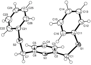
An ORTEP drawing of (I) is shown in Fig. 1. The bond distances and angles are comparable with those of 2,6-bis(p -nitrophenylthiomethyl)pyridine (SillanpaÈaÈet al., 1994), which posesses deactivating nitro groups at theparaposition of the phenyl rings. The difference in structure between the two compounds arises in the position of the thiolate groups with respect to the plane of the pyridyl ring. Compound (I) has both phenyl thiolate moietiessynwith respect to the pyridyl ring [SÐCÐCÐN torsion angles ÿ129.1 (2) and 77.4 (2)], whereas the nitro-substituted compound has one thiolate S-centre almost in the plane of the pyridyl ring [SÐCÐCÐN angle 6.8 (5)] with the other directed away from the pyridyl
ring plane [SÐCÐCÐNÿ112.0 (4)] (SillanpaÈaÈet al., 1994). The lone pairs of electrons on the two sulfur centres in (I) are directed away from each other [CÐCÐSÐCPh 71.1 (3) and
63.8 (2)]. We have observed, however, that the conformation of (I) can adjust so as to act as a bidentate or tridentate ligand toward metal centres (Ballet al., 2001). The phenyl substi-tuents are orientated in such a way that one is almost perpendicular to the plane of the pyridyl ring while the other tends towards the plane of the pyridyl ring.
Experimental
The title compound was prepared according to literature procedures (Teixidor et al., 1991). Crystals suitable for single-crystal X-ray diffraction studies were grown from a methanol solution at 269 K.
Crystal data
C19H17NS2
Mr= 323.47 Triclinic,P1 a= 10.165 (2) AÊ b= 10.365 (2) AÊ c= 9.053 (2) AÊ
= 112.93 (1)
= 100.12 (2)
= 102.37 (1) V= 821.6 (3) AÊ3
Z= 2
Dx= 1.307 Mg mÿ3 MoKradiation Cell parameters from 25
re¯ections
= 45.2±49.9
= 0.32 mmÿ1
T= 150 K Block, colourless 0.30.30.2 mm
Data collection
Rigaku AFC-7Sdiffractometer
!/2scans
Absorption correction: scan (Northet al., 1968) Tmin= 0.883,Tmax= 0.938 3073 measured re¯ections 2894 independent re¯ections 2487 re¯ections withF> 2(F)
Rint= 0.01
max= 25.0
h= 0!12 k=ÿ12!12 l=ÿ10!10 3 standard re¯ections
every 150 re¯ections intensity decay: 1.3%
Re®nement
Re®nement onF R= 0.034 wR= 0.041 S= 3.02 2487 re¯ections 199 parameters
H-atom parameters constrained w= 1/[2(F) + 0.008|F|2] (/)max< 0.001
max= 0.25 e AÊÿ3
min=ÿ0.31 e AÊÿ3
Table 1
Selected geometric parameters (AÊ,).
S1ÐC1 1.816 (2) S1ÐC11 1.770 (2) S2ÐC7 1.832 (2)
S2ÐC21 1.781 (2) N1ÐC2 1.344 (3) N1ÐC6 1.343 (3)
C1ÐS1ÐC11 104.13 (10) C7ÐS2ÐC21 100.90 (10) C2ÐN1ÐC6 118.5 (2) S1ÐC1ÐC2 115.1 (1)
N1ÐC2ÐC1 115.6 (2) N1ÐC2ÐC3 122.3 (2) S2ÐC7ÐC6 112.3 (1)
S1ÐC1ÐC2ÐN1 ÿ129.1 (2) S1ÐC1ÐC2ÐC3 52.3 (2) S1ÐC11ÐC12ÐC13 ÿ176.9 (2) S1ÐC11ÐC12ÐC13 ÿ176.9 (2) S2ÐC7ÐC6ÐN1 77.4 (2) S2ÐC7ÐC6ÐC5 ÿ101.9 (2) S2ÐC21ÐC22ÐC23 178.5 (2) S2ÐC21ÐC26ÐC25 ÿ176.9 (2) C1ÐS1ÐC11ÐC12 ÿ176.5 (2)
C1ÐS1ÐC11ÐC16 5.5 (4) C1ÐC2ÐN1ÐC6 ÿ177.5 (1) C1ÐC2ÐC3ÐC4 177.5 (1) C2ÐN1ÐC6ÐC7 ÿ179.6 (1) C4ÐC5ÐC6ÐC7 178.7 (2) C6ÐC7ÐS2ÐC21 63.8 (2) C7ÐS2ÐC21ÐC22 63.3 (2) C7ÐS2ÐC21ÐC26 ÿ119.5 (2)
No re¯ections had unacceptable values for (Fo±Fc)/(Fo)
although 140 re¯ections had values of(Fo±Fc)/(Fo) between 5 and
10, and 20 re¯ections had values of(Fo±Fc)/(Fo) greater than 10.
The large number of these re¯ections most probably accounts for the large goodness-of-®t value (3.02) for the re®ned structure.
Data collection: MSC/AFC Diffractometer Control Software
(Molecular Structure Corporation, 1988); cell re®nement:MSC/AFC Diffractometer Control Software; data reduction: TEXSAN (Mole-cular Structure Corporation, 1992); program(s) used to solve struc-ture: SHELXS86 (Sheldrick, 1990); program(s) used to re®ne structure:TEXSAN; software used to prepare material for publica-tion:TEXSAN.
We thank EPSRC for funding.
References
Ball, R. J., Radford, A. L., Skelton, B. W., Tolhurst, V.-A. & White, A. H. (2001).J. Chem. Soc. Dalton Trans.In preparation.
Johnson, C. K. (1976).ORTEPII. Report ORNL-5138. Oak Ridge National Laboratory, Tennessee, USA.
Molecular Structure Corporation (1988).MSC/AFC Diffractometer Control Software. MSC, 3200 Research Forest Drive, The Woodlands, TX 77381, USA.
Molecular Structure Corporation (1992). TEXSAN. MSC, 3200 Research Forest Drive, The Woodlands, TX 77381, USA.
North, A. C. T., Phillips, D. C. & Mathews, F. S. (1968).Acta Cryst.A24, 351± 359.
Sheldrick, G. M. (1990).Acta Cryst.A46, 467±473.
SillanpaÈaÈ, R., KivekaÈs, R., Escriche, L., SaÁnchez-CastelloÂ, G. & Texidor, F. (1994).Acta Cryst.C50, 1284±1286.
Teixidor, F., Escriche, L., Rodriguez, I., CasaboÂ, J., Rius, J., Molins, E., MartõÂnez, B. & Miravitlles, X. (1989).J. Chem. Soc. Dalton Trans.pp. 1381± 1384.
Teixidor, F., SaÁnchez-CastelloÂ, G., Lucena, N., Escriche, L., Kivekas, R. Sundberg, M. & CasaboÂ, J. (1991).Inorg. Chem.34, 4931±4935.
VinaÄs, C., AngleÂs, P., SaÁnchez, G., Lucena, N., Teixidor, F., Escriche, L., CasaboÂ, J., Piniella, J. F., Alvarez-Larena, A., KivekaÈs, R. & SillanpaÈaÈ, R. (1998). Inorg. Chem.37, 701±707.
Figure 1
supporting information
sup-1
Acta Cryst. (2001). E57, o392–o393
supporting information
Acta Cryst. (2001). E57, o392–o393 [doi:10.1107/S1600536801005293]
2,6-Bis(phenylthiomethyl)pyridine
Vicki-Anne Tolhurst, Rachel J. Ball and Anthony J. R. Genge
S1. Comment
Compounds that have both nitrogen and sulfur centres available for coordination to metals are becoming topical with
respect to their role in enzyme mimicry, where the N-donor fragments somewhat resemble the imidazole fragment of
histidine and the S-donor fragments model the amino acid cysteine. Interest in the structure of such ligands comes from
the desire to understand how the preferred conformation of the uncoordinated compound affects the coordination mode
of the ligand when coordinated to a metal centre or metal centres, and how the ligand may best be modified to achieve a
desired coordination mode. In the course of our work on transition metal halide complexes of mixed-donor N/S ligands
(Ball et al., 2001), we isolated the title compound, (I), as colourless blocks from methanol. This ligand has previously
been complexed with inorganic metal compounds (Teixidor et al., 1989, 1991; Vinãs et al., 1998). Until now, it has not
been possible to compare the structure of the uncoordinated ligand with the ligand incorporated into such complexes.
An ORTEP drawing of (I) is shown in Fig. 1. The bond distances and angles are comparable with those of 2,6-bis(p
-nitrophenylthiomethyl)pyridine (Sillanpää et al., 1994), which posesses deactivating nitro groups at the para position of
the phenyl rings. The difference in structure between the two compounds arises in the position of the thiolate groups with
respect to the plane of the pyridyl ring. Compound (I) has both phenyl thiolate moieties syn with respect to the pyridyl
ring [S—C—C—N torsion angles -129.1 (2) and 77.4 (2)°], whereas the nitro-substituted compound has one thiolate
S-centre almost in the plane of the pyridyl ring [S—C—C—N angle 6.8 (5)°] with the other directed away from the pyridyl
ring plane [S—C—C—N -112.0 (4)°] (Sillanpää et al., 1994). The lone pairs of electrons on the two sulfur centres in (I)
are directed away from each other [C—C—S—CPh 71.1 (3) and 63.8 (2)°]. We have observed, however, that the
conformation of (I) can adjust so as to act as a bidentate or tridentate ligand toward metal centres (Ball et al., 2001). The
phenyl substituents are orientated in such a way so that one is almost perpendicular to the plane of the pyridyl ring while
the other tends towards the plane of the pyridyl ring.
S2. Experimental
The title compound was prepared according to literature procedures (Teixidor et al., 1991). Crystals suitable for
single-crystal X-ray diffraction studies were grown from a methanol solution at 269 K.
S3. Refinement
No reflections had unacceptable values for Δ(Fo—Fc)/σ(Fo) although 140 reflections had values of Δ(Fo—Fc)/σ(Fo)
between 5 and 10, and 20 reflections had values Δ(fo—Fc)/σ(Fo) greater than 10. The large number of these reflections
Figure 1
A view of the title molecule showing the atom-labelling scheme. Ellipsoids are at the 50% probability level (Johnson,
1976).
(I)
Crystal data
C19H17NS2
Mr = 323.47
Triclinic, P1
a = 10.165 (2) Å
b = 10.365 (2) Å
c = 9.053 (2) Å
α = 112.93 (1)°
β = 100.12 (2)°
γ = 102.37 (1)°
V = 821.6 (3) Å3
Z = 2
F(000) = 340
Dx = 1.307 Mg m−3
Mo Kα radiation, λ = 0.7107 Å Cell parameters from 25 reflections
θ = 45.2–49.9°
µ = 0.32 mm−1
T = 150 K Block, colourless 0.3 × 0.3 × 0.2 mm
Data collection
Rigaku AFC-7S diffractometer
Radiation source: X-ray tube Graphite monochromator
ω/2θ scans
Absorption correction: ψ scan (North et al., 1968)
Tmin = 0.883, Tmax = 0.938 3073 measured reflections
2894 independent reflections 2487 reflections with F > 2σ(F)
Rint = 0.01
θmax = 25.0°, θmin = 2.1°
h = 0→12
k = −12→12
l = −10→10
supporting information
sup-3
Acta Cryst. (2001). E57, o392–o393
Refinement
Refinement on F
Least-squares matrix: full
R[F2 > 2σ(F2)] = 0.034
wR(F2) = 0.0407 2487 reflections 199 parameters 0 restraints
H-atom parameters constrained
Weighting scheme based on measured s.u.'s w = 1/[σ2(F) + 0.008|F|2]
(Δ/σ)max < 0.001 Δρmax = 0.25 e Å−3 Δρmin = −0.31 e Å−3
Fractional atomic coordinates and isotropic or equivalent isotropic displacement parameters (Å2)
x y z Uiso*/Ueq
S(1) 0.56351 (6) 0.65575 (6) 0.34517 (7) 0.0322 (2)
S(2) 0.14211 (6) 1.01495 (6) 0.64662 (8) 0.0377 (2)
N(1) 0.4444 (2) 0.9347 (2) 0.6760 (2) 0.0261 (4)
C(1) 0.5986 (2) 0.8310 (2) 0.5255 (3) 0.0292 (6)
C(2) 0.5072 (2) 0.8291 (2) 0.6384 (2) 0.0265 (5)
C(3) 0.4910 (2) 0.7260 (2) 0.7034 (3) 0.0326 (6)
C(4) 0.4093 (2) 0.7354 (2) 0.8115 (3) 0.0345 (6)
C(5) 0.3454 (2) 0.8454 (2) 0.8528 (3) 0.0324 (6)
C(6) 0.3654 (2) 0.9426 (2) 0.7816 (2) 0.0270 (5)
C(7) 0.2973 (2) 1.0617 (2) 0.8179 (3) 0.0327 (6)
C(11) 0.3996 (2) 0.6303 (2) 0.2137 (3) 0.0279 (6)
C(12) 0.3443 (2) 0.4962 (2) 0.0675 (3) 0.0331 (6)
C(13) 0.2199 (2) 0.4682 (3) −0.0478 (3) 0.0399 (7)
C(14) 0.1493 (2) 0.5715 (3) −0.0211 (3) 0.0410 (7)
C(15) 0.2020 (2) 0.7031 (3) 0.1256 (3) 0.0386 (7)
C(16) 0.3273 (2) 0.7333 (2) 0.2433 (3) 0.0323 (6)
C(21) 0.0296 (2) 0.8583 (2) 0.6482 (3) 0.0317 (6)
C(22) −0.0177 (3) 0.8703 (3) 0.7851 (3) 0.0556 (8)
C(23) −0.1078 (3) 0.7483 (3) 0.7833 (4) 0.0580 (9)
C(24) −0.1543 (2) 0.6143 (3) 0.6432 (3) 0.0443 (7)
C(25) −0.1095 (3) 0.6032 (3) 0.5060 (3) 0.0476 (7)
C(26) −0.0160 (2) 0.7243 (3) 0.5082 (3) 0.0402 (7)
H(1a) 0.5975 0.9140 0.4876 0.0598
H(1b) 0.6989 0.8442 0.5776 0.0598
H(3) 0.5480 0.6388 0.6717 0.0598
H(4) 0.3958 0.6620 0.8633 0.0598
H(5) 0.2788 0.8532 0.9248 0.0598
H(7a) 0.3671 1.1585 0.8297 0.0598
H(7b) 0.2732 1.0852 0.9335 0.0598
H(12) 0.3950 0.4161 0.0391 0.0598
H(13) 0.1886 0.3668 −0.1610 0.0598
H(14) 0.0506 0.5415 −0.1119 0.0598
H(15) 0.1511 0.7920 0.1619 0.0598
H(16) 0.3715 0.8334 0.3580 0.0598
H(22) 0.0000 0.9746 0.8789 0.0598
H(24) −0.2276 0.5214 0.6390 0.0598
H(25) −0.1395 0.4988 0.4056 0.0598
H(26) 0.0244 0.7157 0.4129 0.0598
Atomic displacement parameters (Å2)
U11 U22 U33 U12 U13 U23
S(1) 0.0385 (3) 0.0253 (3) 0.0326 (3) 0.0132 (2) 0.0067 (2) 0.0125 (2)
S(2) 0.0358 (3) 0.0361 (3) 0.0475 (4) 0.0099 (3) 0.0088 (3) 0.0268 (3)
N(1) 0.0284 (9) 0.0199 (9) 0.0248 (9) 0.0021 (7) 0.0019 (7) 0.0101 (7)
C(1) 0.032 (1) 0.022 (1) 0.030 (1) 0.0049 (9) 0.0065 (9) 0.0113 (9)
C(2) 0.027 (1) 0.021 (1) 0.025 (1) 0.0009 (9) 0.0006 (9) 0.0098 (9)
C(3) 0.031 (1) 0.028 (1) 0.040 (1) 0.0064 (9) 0.006 (1) 0.021 (1)
C(4) 0.033 (1) 0.034 (1) 0.042 (1) 0.0056 (10) 0.006 (1) 0.027 (1)
C(5) 0.031 (1) 0.033 (1) 0.032 (1) 0.0040 (10) 0.0058 (10) 0.018 (1)
C(6) 0.027 (1) 0.023 (1) 0.023 (1) 0.0012 (9) 0.0013 (9) 0.0091 (9)
C(7) 0.035 (1) 0.026 (1) 0.032 (1) 0.0046 (9) 0.0058 (10) 0.0118 (10)
C(11) 0.032 (1) 0.025 (1) 0.030 (1) 0.0052 (9) 0.0098 (9) 0.0174 (9)
C(12) 0.040 (1) 0.028 (1) 0.030 (1) 0.0050 (10) 0.013 (1) 0.0129 (10)
C(13) 0.040 (1) 0.040 (1) 0.031 (1) −0.001 (1) 0.009 (1) 0.015 (1)
C(14) 0.031 (1) 0.054 (2) 0.036 (1) 0.002 (1) 0.007 (1) 0.025 (1)
C(15) 0.035 (1) 0.044 (1) 0.046 (1) 0.012 (1) 0.013 (1) 0.028 (1)
C(16) 0.035 (1) 0.028 (1) 0.036 (1) 0.0086 (10) 0.009 (1) 0.0176 (10)
C(21) 0.028 (1) 0.032 (1) 0.039 (1) 0.0100 (9) 0.0076 (10) 0.020 (1)
C(22) 0.066 (2) 0.040 (2) 0.050 (2) 0.002 (1) 0.031 (1) 0.010 (1)
C(23) 0.063 (2) 0.060 (2) 0.055 (2) 0.008 (1) 0.032 (2) 0.029 (2)
C(24) 0.033 (1) 0.042 (1) 0.061 (2) 0.005 (1) 0.009 (1) 0.031 (1)
C(25) 0.047 (2) 0.036 (1) 0.047 (2) 0.002 (1) 0.002 (1) 0.016 (1)
C(26) 0.041 (1) 0.041 (1) 0.034 (1) 0.007 (1) 0.007 (1) 0.018 (1)
Geometric parameters (Å, º)
S(1)—C(1) 1.816 (2) C(11)—C(12) 1.403 (3)
S(1)—C(11) 1.770 (2) C(11)—C(16) 1.390 (3)
S(2)—C(7) 1.832 (2) C(12)—C(13) 1.384 (3)
S(2)—C(21) 1.781 (2) C(13)—C(14) 1.382 (3)
N(1)—C(2) 1.344 (3) C(14)—C(15) 1.391 (3)
N(1)—C(6) 1.343 (3) C(15)—C(16) 1.395 (3)
C(1)—C(2) 1.500 (3) C(21)—C(22) 1.378 (3)
C(2)—C(3) 1.401 (3) C(21)—C(26) 1.377 (3)
C(3)—C(4) 1.379 (3) C(22)—C(23) 1.385 (3)
C(4)—C(5) 1.390 (3) C(23)—C(24) 1.377 (4)
C(5)—C(6) 1.391 (3) C(24)—C(25) 1.369 (3)
C(6)—C(7) 1.497 (3) C(25)—C(26) 1.393 (3)
C(1)—S(1)—C(11) 104.13 (10) S(1)—C(11)—C(16) 125.0 (2)
supporting information
sup-5
Acta Cryst. (2001). E57, o392–o393
S(1)—C(1)—C(2) 115.1 (1) C(12)—C(13)—C(14) 120.6 (2)
N(1)—C(2)—C(1) 115.6 (2) C(13)—C(14)—C(15) 119.5 (2)
N(1)—C(2)—C(3) 122.3 (2) C(14)—C(15)—C(16) 120.7 (2)
C(1)—C(2)—C(3) 122.1 (2) C(11)—C(16)—C(15) 119.5 (2)
C(2)—C(3)—C(4) 118.5 (2) S(2)—C(21)—C(22) 121.0 (2)
C(3)—C(4)—C(5) 119.7 (2) S(2)—C(21)—C(26) 119.8 (2)
C(4)—C(5)—C(6) 118.3 (2) C(22)—C(21)—C(26) 119.2 (2)
N(1)—C(6)—C(5) 122.6 (2) C(21)—C(22)—C(23) 120.4 (2)
N(1)—C(6)—C(7) 116.2 (2) C(22)—C(23)—C(24) 120.6 (2)
C(5)—C(6)—C(7) 121.2 (2) C(23)—C(24)—C(25) 119.0 (2)
S(2)—C(7)—C(6) 112.3 (1) C(24)—C(25)—C(26) 120.9 (2)
S(1)—C(11)—C(12) 115.4 (2) C(21)—C(26)—C(25) 120.0 (2)
S(1)—C(1)—C(2)—N(1) −129.1 (2) C(3)—C(2)—N(1)—C(6) 1.1 (2)
S(1)—C(1)—C(2)—C(3) 52.3 (2) C(3)—C(4)—C(5)—C(6) 0.5 (2)
S(1)—C(11)—C(12)—C(13) −176.9 (2) C(4)—C(5)—C(6)—C(7) 178.7 (2)
S(1)—C(11)—C(12)—C(13) −176.9 (2) C(6)—C(7)—S(2)—C(21) 63.8 (2)
S(2)—C(7)—C(6)—N(1) 77.4 (2) C(7)—S(2)—C(21)—C(22) 63.3 (2)
S(2)—C(7)—C(6)—C(5) −101.9 (2) C(7)—S(2)—C(21)—C(26) −119.5 (2)
S(2)—C(21)—C(22)—C(23) 178.5 (2) C(11)—C(12)—C(13)—C(14) 0.3 (4)
S(2)—C(21)—C(26)—C(25) −176.9 (2) C(11)—C(16)—C(15)—C(14) −0.3 (5)
N(1)—C(2)—C(3)—C(4) −1.0 (2) C(12)—C(11)—C(16)—C(15) −1.2 (4)
N(1)—C(6)—C(5)—C(4) −0.4 (2) C(12)—C(13)—C(14)—C(15) −1.9 (4)
C(1)—S(1)—C(11)—C(12) −176.5 (2) C(13)—C(12)—C(11)—C(16) 1.2 (4)
C(1)—S(1)—C(11)—C(16) 5.5 (4) C(13)—C(14)—C(15)—C(16) 1.9 (5)
C(1)—C(2)—N(1)—C(6) −177.5 (1) C(21)—C(22)—C(23)—C(24) −1.7 (4)
C(1)—C(2)—C(3)—C(4) 177.5 (1) C(21)—C(26)—C(25)—C(24) −1.7 (5)
C(2)—N(1)—C(6)—C(5) −0.4 (2) C(22)—C(21)—C(26)—C(25) 0.4 (4)