TOXICOLOGICAL STUDIES OF ETHANOLIC LEAVES EXTRACT
OF
SARCOSTEMMA VIMINALE
Moussa Yahaya, *Angela Nnenna Ukwuani, Caleb Francis
Department of Biochemistry, Faculty of Science, Kebbi State University of Science and
Technology, Aliero, Kebbi State, Nigeria.
ABSTRACT
The effects of Sarcostemma viminale leaves extract on the
haematological, toxicological and histopathological indices were
evaluated in rats. The extracts at a single dose of 5000 mg/kg did not
produce any behavioural sign of toxicity or mortality in any of the
animals tested during 14 days observation period and as such, LD50 of
this plant was estimated to be greater than 5000 mg/kg. In the 28days
repeated dose oral sub-chronic toxicity study, administration of 250,
500 and 1000 mg/kg body weight Sarcostemma viminale leaves
extracts revealed no significant difference (p>0.05) in the
haematological parameters. Analysis of biochemical parameters
reveals no significant difference (p>0.05) in the extract treated groups
when compared to the control group. The histopathological
examination of the treated groups showed no visible lesions or signs of
the liver damage while the kidney revealed tubular congestion and necrosis, focal
haemorrhage and hyaline degeneration. These results suggest that the sub-chronic
administration of ethanolic extracts of Sarcostemma viminale leaves has no marked toxic
effect on the liver while unmasking the possible dangers of this plant to the kidney.
KEYWORDS: Toxicity, Sarcostemma viminale, leaves extracts, ethanolic.
INTRODUCTION
Herbal remedies have a therapeutic effect and are acceptable interventions for diseases and
symptoms. Interestingly, demand for medicinal plants is progressively rising in industrialized
nations as it is in developing countries.[1] The World Health Organisation (WHO) estimates
that about 80% of the developing world‘s population meets their primary healthcare needs
Volume 4, Issue 10, 311-319. Research Article ISSN 2277– 7105
Article Received on 8 Aug 2015,
Revised on 28 Aug 2015, Accepted on 17 Sep 2015
*Correspondence for
Author
Angela Nnenna Ukwuani
Department of
Biochemistry, Faculty of
Science, Kebbi State
University of Science and
Technology, Aliero,
through traditional medicine.[1,2,3,4] African indigenous herbal medicines are widely used
throughout the African continent, despite an apparent lack of scientific evidence for their
quality, safety and efficacy.[5] Most plants consumed locally in Nigeria have not been
thoroughly evaluated for their toxicity profiles [6] including the plant of this study.
Though, herbal medicines have recently attracted much attention as alternative medicines
useful for treating or preventing life-style related disorders.[7] However, relatively very little
knowledge is available about their mode of action and safety. The study of toxic or adverse
effect of crude drugs of plant origin is essential in order to prove a guide to their safe usage
and eventual standardization. This is especially important as traditional medicine
practitioners often administer such preparations without regards to their possible adverse
effects. The leaves of Sarcostemma viminale is known in Nigerian traditional medicine to
promote milk production in animals and for treating dysentery, indigestion, and haemorrhoid
in man. However, there is no scientific work on safety or toxicity of this plant. In this study,
ethanolic leaves extract of Sarcostemma viminale (ELSV) is assessed in vivo for its toxicity.
MATERIALS AND METHOD Identification of the plant
Identification of Sarcostemma viminale was done in the field by a botanist. The plant parts
(leaves, stems-bark and roots) were collected from the wilds of Aliero Local Government
Area of Kebbi State, Nigeria. Herbarium specimens (Voucher specimens No. 165) were
prepared and deposited in the Herbarium, Botany Unit, Biological Science Department,
Kebbi State University of Science and Technology, Aliero, Nigeria, where identity of the
plants was confirmed by comparison with available voucher specimens.
Extraction of plant materials
The plant materials were open air dried under the shade and chopped into smaller pieces. The
dried leaves were pulverized into moderately coarse powder. The powdered plant material
(100 g) was macerated in ethanol in an air tight aspirator bottle for 72 hours. This was then
filtered with the aid of sterile sieving cloth and evaporated using a Water bath at 45oC. The
dried extract collected was weighed, labelled and stored in an air tight bottle container.
Animals
White Wistar strain albino rats of both sexes, weighing averagely 100 - 210g were used for
Ahmadu Bello University, Zaria and were transported to the Department of Biochemistry
Laboratory, Kebbi State University of Science and Technology, Aliero, Kebbi State. They
were allowed to acclimatise to 2 weeks with free access to drinking water and standard diet
(Vital feeds, Jos, Nigeria).
Acute Oral Toxicity Study (LD50)
After acclimatization period, the acute oral toxicity study as described by Dixon.[8] was
performed as per the OECD-423 guidelines (acute toxicity class method). Five (5) rats of
either sex selected by random sampling technique were used for this study. The animals were
fasted over night providing only water, after which ELSV was administered orally at a dose
of 5000 mg/kg body weight to each rat at 48 hours interval respectively and subsequent
observed for 14 days. The behavioral changes (abdominal constriction, hyperactivity,
sedation, grooming), and body weight were observed for 14 days.
Sub-chronic Toxicity Study
Rats were divided into five (4) groups of five (5) rats each for sub-chronic toxicity study.
ELSV extract was orally administered daily for 28 days. Group 1 serve as control receiving
normal saline (5ml/kg) while Groups 2 to 4 served as ELSV treated Groups receiving (250,
500 and 1000mg/kg) bodyweight, respectively. The weights of the rats were recorded
weekly. The rats were fasted overnight on the 28th and on the 29th day, weights were taken
and blood samples were collected via cardiac puncture for further analysis.
Markers of toxicity
The following parameters were analyze from the blood samples collected at the end of the
sub-chronic toxicity studies; Aspartate aminotransferase (AST).[9] Alanine aminotransferase
(ALT).[9] Alkaline phosphatase (ALP).[10] Bilirubin.[11] Albumin.[12] Total protein.[13] and the
haematological parameters were analyse using the method described by Dacie and lewis.[14]
Histopathological Studies
The liver and kidney were harvested, weighed and preserved in 10% formalin for
histopathological analysis according to reported procedures of Aliyu et al.[15]
Statistics Analysis
Data collected in this study were expressed as Means ± Standard Deviation (SD) and
differences among experimental and control groups were determined using Turkey-Kramer
and Dunnett Multiply Comparism Tests respectively.
RESULTS AND DISCUSSION
In this study, extraction process yielded 27.5% of ELSV. The limit dose of 5000 mg/kg body
weight ELSV did not cause mortality nor show any clinical signs of acute toxicity in all of
the five rats tested in the short term (48 hours) and long term (14 days) observatory period.
The result showed the LD50 of ELSV is greater than 5000mg kg-1 suggesting that at the limit
dose tested ELSV is essentially non-toxic and safe for oral formulation. Sub-chronic toxicity
study after 28 days of daily administration of hydromethanolic extract of ELSV showed a non
significant decrease in body weights of the control and treated animals of this study (Figure
1).
0 100 200 300
WEEK O WEEK 1 WEEK 2 WEEK 3 WEEK 4
B
OD
Y
WE
IGH
T
(g)
TREATMENT PERIOD
CONTROL 250mg/kg ELSV
500mg/kg ELSV 1000mg/kg ELSV
Figure 1: Effect of sub-chronic administration of ELSV on Body weight of rats.
Reductions in body and internal organ weights have been considered sensitive indices of
toxicity after exposure to toxic substance.[16] In the present study, there was no significant
difference (p<0.05) in the organ weight of extract treated groups compared to the control
[image:4.595.109.479.315.481.2]groups (Table 1).
Table 1: Effect of sub-chronic administration of ELSV on rat organ weight Groups Left Kidney Right Kidney Liver Heart Control 0.65±0.08 0.54±0.05 4.87±0.65 0.73±0.11
250mg/Kg 0.57±0.08 0.52±0.08 4.73±0.83 0.62±0.07
500mg/Kg 0.63±0.02 0.57±0.03 5.83±0.37 0.60±0.03
The haematopoietic system is one of the most sensitive targets of toxic compounds and is an
important index of physiological and pathological status in man and animals.[17,18]
Assessment of the haematological parameters can be used to determine the extent of
deleterious effect of extracts on the blood of an animal or determine blood relating functions
of a plant extract or its product. Such analysis is relevant to risk evaluation as changes in the
haematological system have higher predictive value for human toxicity, when the data are
translated from animal studies.[19] In the present study, there were no significant differences
(p>0.05) in the hematocrit, hemoglobin, Total Leukocyte Count (TLC), lymphocytes,
neutrophils and platelets when compared to the control (Table 2). However, there was a non
significant dose-dependent decrease in lymphocytes of the ELSV treated groups.
Lymphocytes, the main effector cells of the immune system, usually show increase in activity
in response to toxic environment.[20] Leucocytosis observed in the present study indicates a
stimulation of the immune system which protects the rats against infection that might have
[image:5.595.63.539.386.510.2]been caused by chemical and secondary infections.[19]
Table 2: Effect of sub-chronic administration of ELSV on Hematological parameters.
Parameter Control ELSV
250 mg/kg
ELSV 500mg/kg
ELSV 1000mg/kg Hematocrit (%) 39.75 ± 5.05 36.50 ± 0.57 39.00 ± 5.56 37.50 ± 13.12
Hemoglobin(g/dl) 13.25 ± 1.68 12.16 ± 0.19 13.00 ± 1.8 12.50 ± 4.37
TLC(X109 cells /L) 2.45 ± 0.58 2.62 ± 0.45 2.86 ± 1.10 3.72 ± 1.28
Lymphocyte (%) 77.25 ±14.17 77.50 ± 1.91 74.00 ± 6.00 52.25 ± 19.50
Neutrophils(%) 22.00 ± 12.75 17.50 ± 3.78 20.66 ± 1.15 40.50 ± 22.17
Platelet(x109/l) 150.00 ± 35.59 173.75 ±23.59 181.66 ±20.20 144.50 ±49.80 Values are Mean ± Standard deviation (n = 4). *Significant different (p<0.05) compared to
control.
The result of hepatic biomarkers (AST, ALT and ALP, Bilirubin, albumin and protein) of
liver function is shown in Tables 3. Biochemical indices monitored in the serum such as
secretory substances of the liver can be used as ‘markers’ for assessing its functional
capacities.[21] Plasma proteins can be used to examine specific biochemical functions and the
general status of the body’s protein metabolism while increased billirubin level reflects the
depth of jaundice.[22] Increase in the level of AST, ALT and ALP reflects the structural and
functional dysfunction of hepatocellular membrane or cell rupture, and thereby indicates liver
damage. The results of this study revealed no significant difference (P>0.05) in all the
biochemical parameters of ELSV treated groups as compared to the control respectively.
suggests that sub-chronic administration of ELSV has no hepatotoxic effects in treated
[image:6.595.69.530.142.276.2]animals.
Table 3: Effect of sub-chronic administration of ELSV on Biochemical parameters.
Parameter Control ELSV
250 mg/kg
ELSV 500mg/kg
ELSV 1000mg/kg Total Bilirubin (mg/l) 0.65 ± 0.05 0.55 ± 0.12 0.67 ± 0.11 0.70 ± 0.12
Con. Bilirubin (mg/l) 0.15 ± 0.01 0.14 ± 0.02 0.17 ± 0.01 0.17 ± 0.01
Total protein(g/dl) 63.50 ± 3.41 53.25 ± 8.65 61.00 ± 2.64 60.20 ± 8.25
Albumin 29.50 ± 1.29 29.25 ± 7.36 30.00 ± 1.00 29.20 ± 3.63
ALP(m/l) 65.00 ± 8.16 59.25 ± 10.71 71.00 ± 2.65 73.20 ± 8.22
AST(U/l) 53.50 ± 8.06 50.25 ± 9.97 61.00 ± 2.00 65.00 ± 6.67
ALT(U/l) 59.00 ± 7.95 53.00 ± 6.97 67.00 ± 3.60 64.20 ± 8.07 Values are mean ± standard deviation (n =4). Significant different (p>0.05) compare with
control.
All groups treated with ELSV showed no significant damage of the liver while the kidney
showed various degrees of tubular necrosis and congestion, glomerular and hyaline
degeneration, and focal heamorrhage when compared with the control group (Figure 2 – 4).
The result from histological screening was in agreement with the serum biomarkers as no
apparent damage to the liver was observed in all the treated groups when compared with the
control group. This further confirms the plant extract to be non toxic to the liver within the
treatment durations. However, this finding also exposes the possible risk of kidney damage
upon sub-chronic administration of ELSV.
Figure 2: Photomicrograph of a group 2 (250mg/kg ELSV) rat’s kidney. Showing Tubular necrosis (arrow A), focal haemorrhage (Arrow B) and slight congestion (arrow C). H&E Stained, Magnification x 40.
A
B
[image:6.595.142.455.492.683.2]Figure 3: Photomicrograph of a group 3 (500mg/kg ELSV) Rat’s Kidney.
Showing: Glomerular cellular degeneration (arrow D) and Tubular necrosis (Arrow E). H&E
Stained, Magnification x 40.
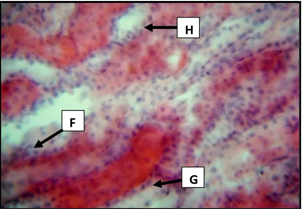
Figure 4: Photomicrograph of a group 4 (1000mg/kg ELSV) rat’s Kidney.
Showing: Hyaline degeneration (arrow F), tubular congestion (Arrow G) and Vacuum
(Arrow H). H&E Stained, Magnification x 40.
CONCLUSION
In conclusion, sub-chronic administration of ELSV showed no effect on the haematological
indices, biochemical function and hepatic secretions while eliciting nephrotoxic effects.
G
F
H
D
[image:7.595.145.453.389.602.2]However, further work is needed to confirm and justify the risk of kidney injury (via
biochemical markers of toxicity) as this was not covered within the scope of this study.
REFERENCE
1. Abere TA, Okoto PE, Agoreyo FO. Antidiarrhoea and toxicological evaluation of the leaf
extract of Dissotis rotundifolia Triana (Melastomataceae). BMC Complement Altern
Med, 2010; 10: 71.
2. Calixto JB. Efficacy, safety, quality control, marketing and regulatory guidelines for
herbal medicines (phytotherapeutic agents). Braz J Med Biol Res, 2000; 33: 179-189.
3. Green EC. The WHO forum on traditional medicine in health systems, Harare,
Zimbabwe, February 14-18, 2000. J Altern Complement Med, 2000;6(5): 379-382.
4. Johnson Q, Syce J, Nell H, Rudeen K, Folk WR. A randomized, double-blind,
placebo-controlled trial of Lessertia frutescens in healthy adults. PLoS Clin Trials, 2007; 2(4): 16.
5. Jadeja RN, Thounaojam MC, Jadav SV, Patel MD, Patel DK, Salunke SP, Padate GS,
Devkar RV, Ramachandran AV. (2011). Toxicological evaluation and hepatoprotective
potential of Clerodendron glandulosum Coleb leaf extract. Hum Exp Toxicol, 2011;
30(1): 63-70.
6. Musa TY, Adebayo OJ, Egwim EC, Owoyele VB. Increased liver alkaline phosphatise
and amino transferases activities following administration of ethanolic extract of Khaya
senegalensis stem bark to rats. Biochem, 2005; 17(1): 27–32.
7. Agyare C, Asase A, Lechtenberg M, Niehues M, Deters A, Hensel A. An
ethnopharmacological survey and in vitro confirmation of ethnopharmacological use of
medicinal plants used for wound healing in Bosomtwi-Atwima-Kwanwoma area, Ghana.
J Ethnopharmacol, 2009; 125(3): 393-403.
8. Dixon WJ. Staircase bioassay; the up and down method. Nuero Sci Biobehav Rev, 1991;
15: 47-50.
9. Reitman S, Frankel S. A colorimetric method for the determination of serum glutamic
oxaloacetic and glutamic pyruvic transaminases. Amer. J. Clin. Pathol, 1957; 28: 56.
10.Roy AV. Direct colorimetric determination of alkaline phosphatase in human serum. Clin
Chem, 1970; 16: 431.
11.Jendrassik G, Grof A. Quantitative in vitro determination of total and direct bilirubin in
serum or plasma. Clin Chem, 1938; 27: 102.
12.Rodkey FL. Quantitative determination of albumin in serum and plasma. Clin Chem,
13.Weichselbaum TE. Biuret method of serum total protein estimation. Am J Clin Pathol,
1946; 16: 40.
14.Dacie JV, Lewis SM (1994). Practical hematology, 8th Edition. Longman group Ltd
Hong Kong, 1994; 965–983.
15.Aliyu R, Adebayo AH, Gatsing D, Garba IH. The effects of ethanolic leaf extract of
Commiphora africana (Burseraceae) on rat liver and kidney functions. J Pharmacol
Toxicol, 2007; 2: 373-379.
16.Thanabhorn S, Jenjoy K, Thamaree S, Ingkaninan K, Panthong A. Acute and subacute
toxicity study of the ethanol extract from Lonicera japonica Thunb. J Ethnopharmacol,
2006; 107: 370-373.
17.Diallo A, Gbeassor M, Vovor A, Eklu-Gadegbeku K, Aklikokou K et al. Effect of
Tectonagrandis on phenylhydrazine-induced anaemia in rats. Fitoterapia, 2008; 79:
332-336.
18.Dickson AM, Fred OC, Eleojo O. Phytochemical, antibacterial and toxicity studies of
aqueous extract of Eucalyptus canaldulensis. Asian J Plant Sci Res, 2011; 1(3): 1.
19.Abiodun HA, Guang ZZ, Jun TF, Chang JJ, Wen JH, Jun JX, Yu MZ, Afolabi AA,
Roseline K, Ning HT. Biochemical, haematological and histopathological studies of
extract of Ageratum conyzoides L. in Sprague Dawley rats. J Med Plants Res, 2010;
4(21): 2264-2272.
20.Robins SL. Lymph nodes and spleen: Pathologic basis of disease. WB Saunders Co.,
Philadelphia, 1974; 1050.
21.Yakubu MT, Bilbis LS, Lawal M, Akanji MA. Evaluation of selected parameters of rat
liver and kidney function following repeated administration of yohimbine. Biochem,
2003; 15: 50-56.
22.Tedong L, Desire P, Dzeufiet D, Dimo T, Asongalem EA, Sokeng SN. Acute and
sub-chronic toxicity of Anacardium occidentale Linn (Anacardiaceae) leaves hexane extract