DOSE-RESPONSIVE SYSTEMIC TOXICITY OF CYPERMETHRIN IN
WISTAR RATS
Anurag Paramanik1, Tuhina Das1, Rini Ghosh1, Ananya Pradhan1, Subhabrata Das1,
Suman Mondal1, Prasanta Maiti2 and Sujata Maiti Choudhury1*
1
Department of Human Physiology with Community Health, Vidyasagar University,
Midnapore, West Bengal, India, Pin-721102. 2
Imgenex India Pvt. Ltd, Bhubaneswar, Odisha, India, Pin-751024.
ABSTRACT
Pesticides are used frequently and have various adverse effects on
human health in different ways. Cypermethrin (CYP), a synthetic
pyrethroid, is used extensively to control a wide variety of pests in
agriculture, forestry, horticulture, and public health. This study
designed to investigate the dose-dependent gonadal, immune and
systemic toxicity of CYP in mature male and female Wistar rats. Rats
were randomly divided into nine groups, different doses (1/11th, 1/10th, 1/9th, 1/7th, 1/6th, ¼.5th, 1/4th, 1/3rd incase of male rat; 1/11th, 1/10th, 1/9th, 1/7th, 1/6th, 1/5th, 1/4th incase of female rat) of CYP were administered for 14 consecutive days and different gonadal, immune
and systemic parameters were assessed. Sperm viability, testicular acid
phosphatase, WBC count, lymphocyte count, cerebellar and
cerebellum reduced glutathione (GSH) content were significantly diminished. Serum urea,
creatinine, cerebellar and cerebellum malondialdehyde (MDA) content were increased
following CYP treatment in male rats at 40 and 80 mg/kg body weight (1/9th and 1/4.5th LD50). Decreased SOD and GST levels were also observed in CYP-exposed female rats at a
dose level of 34.33 and 51.5 mg/kg body wt. (1/9th and 1/6th LD50). In conclusion, cypermethrin induced gonadal, immune and systemic toxicity in mature male rats with a body
weight of 40 mg/kg (1/9thLD50) and gonadal toxicity in mature female rats with a body weight of 34.33 mg/kg (1/9thLD50) and above.
Volume 7, Issue 16, 1690-1703. Research Article ISSN 2277– 7105
*Corresponding Author
Sujata Maiti Choudhury
Department of Human Physiology with Community Health, Vidyasagar University, Midnapore, West Bengal, India, Pin-721102. Article Received on 17 July 2018,
Revised on 07 August 2018, Accepted on 28 August 2018,
KEYWORDS: Cypermethrin; Gonadal; Immune; systemic; Testicular ACP; Cerebellar
MDA.
1. INTRODUCTION
Pesticides are the chemical formulation increasingly used in agriculture, animal husbandry
and public health operation to kill the insects, weeds and fungus and to get rid of insect
transmitted diseases. These pesticides are toxic not only to insects and pests but at different
levels to animals and human beings (Aktar et al., 2009). Improper use of these agrochemicals
may pose serious hazards to human and animal health. The use of pyrethroids has been
increasing during the past decade with the declining use of organophosphates, which are
more acutely toxic to birds and mammals than pyrethroids (Shafer and Meyer, 2004).
However, although pyrethroids are less acutely toxic but some studies have demonstrated that
synthetic pyrethroids possess hormone-mimicking action and have been classified as
endocrine-disrupting compounds (EDCs), which potentially pose a threat to human and
wildlife. Synthetic pyrethroids have been considered potentially toxic to male reproductive
system (Kilian et al., 2007).
Cypermethrin is a synthetic pyrethroid commonly used in agriculture, veterinary, and
household-insect management (Solati et al., 2010). Cypermethrin behaves as a fast-acting
neurotoxin in insects. It causes neurotoxicity insects by causing a long-lasting prolongation of
the normally transient increase in sodium permeability of nerve membrane channels during
excitation. These long-lasting trains can cause hundreds to thousands of repetitive nerve
impulses in the sense organs. This repetitive activity is induced by pyrethroid damage to the
voltage-dependent sodium channel, causing sodium channels to stay open much longer than
normal (Vijverberg and van den Bercken, 1990). Although considered to be safe for
household applications, some studies reported the adverse effects of cypermethrin on nervous
system of laboratory animals (Sayim et al., 2005).
Kidney plays an essential role in health, disease and overall development and growth. The
main function of kidney is to maintain total body fluid volume, its composition and acid base
balance. A number of environmental variables including certain xenobiotics (e.g. pesticides)
influence these functions (Rasoul et al., 2012). In fact, oxygen free radicals are involved in
toxicity of numerous chemicals including pesticides and in pathogenesis of many diseases
The exposure concentration is important for any type of pesticide, in analyzing the variation
of its toxicity in any system. So, in this present study, Wistar rats were exposed to different
concentrations of CYP to study the impact of CYP on gonadal, immune and systemic
parameters in rats.
2. MATERIALS AND METHODS
2.1. Chemicals and reagents
A commercial formulation of cypermethrin 10% emulsifiable concentrate (EC), named ‗‗Ustad‘‘ (United Phosphorus Limited) was used in the experiments. Zinc sulphate (ZnSO4), WBC dilution fluid, chloroform, ethylene-diamine-tetra-acetic acid, phosphate buffer (0.1 M,
pH 7.4), histopacque-1077, and all other chemicals used in this study were of analytical grade
and obtained from Merck Ltd., Himedia, Mumbai, India.
2.2. Animal maintenance
Healthy Wistar albino male rats (weighing 130-150 g) were selected for this experiment.
Standard laboratory feed and water were provided throughout the period of experimentation.
Experimental protocol and surgical methods were approved by the Institutional Animal
Ethical Committee, registered under CPCSEA.
2.3. Treatment protocol
After 10 days of acclimatization, the animals were randomly assigned to both the
experimental groups and the control group, each containing six rats. Mature male, female rats
were divided into nine groups where each group contains six animals. Group I was
considered as control and Group II to IX were cypermethrin treated groups. According to
FAO specifications (FAO specifications and evaluations for agricultural pesticides, 2006)
oral LD50 dose of cypermethrin formale and female rats were 360 and 309 mg/kg body
weight respectively (Dee An Jones, 1992). Commercial formulation of cypermethrin 10%
emulsifiable concentrate (EC) was administered at 1/11th, 1/10th, 1/9th, 1/7th, 1/5th, 1/4.5th, 1/4th and 1/3rd of LD50 concentration respectively. After treatments for14 consecutive days, all animals were anesthetized with pentobarbital sodium and sacrificed by cervical dislocation
on 15th day. Blood samples were drawn from animals from all the treatment groups and
allowed to fall drop by drop into a graduated centrifuge tubes containing anticoagulant
ethylene-diamine-tetra-acetic acid (EDTA) for the estimation of haematological parameters.
Tissue samples were collected and stored at -80ºC until analysis. Epididymis were collected
2.4. Sperm viability assay
The eosin-nigrosin staining was measured to determine the sperm viability (WHO, 1999).
One drop of sperm suspensions was added to two drops of 1% eosin Y. Then, after 30 s, three
drops of 10% nigrosin were added and mixed well. A drop of mixture was placed on a clean
glass slide, then air dried and was examined under the light microscope.
2.5. Assay of testicular acid phosphatase
The acid phosphatase activity was measured using p-nitrophenol phosphate (PNPP) as a
substrate (Vanha-Perttula and Nikkanen, 1973). Amount of liberated PNP was
spectrophotometrically measured at 420 nm.
2.6. Determination of ovarian superoxide dismutase (SOD)
The ovarian superoxide dismutase (SOD) was determined from its ability to inhibit the
auto-oxidation of pyrogallol according to Marklund and Marklund (1974).
2.7. Estimation of ovarian glutathione-S-transferase (GST)
Ovarian glutathione-S-transferase (GST) activity was measured spectrophotometrically by
the method of Habig et al. (1974) using 1-chloro-2, 4-dinitrobenzene and detected at 340 nm.
The activity of GST in testis was expressed in terms of μmol/min/mg protein.
2.8. Total leukocyte count (TLC)
Total leukocyte count (Wintrobe, 1967) was determined by diluting blood in 1:20 dilution
with white blood corpuscle (WBC) dilution fluid and then total leukocytes were counted in
Neubaur haemocytometer chamber.
2.9. Differential leukocyte count (DLC)
Thin blood smear was made by anticoagulant-added whole blood in a clean glass slide and
was stained with Leishman`s stain and then was observed under oil immersion objective of
the microscope. The percentage of granulocytes and agranulocytes were calculated
(Wintrobe, 1967).
2.10. Estimation of malondialdehyde (MDA)
Malondialdehyde was assessed by the mixing of 1 ml of sample with 0.2ml of 8.1% sodium
dodecyl sulfate, 1.5ml of acetate buffer (20%, pH-3.5) and 1.5 ml of aqueous solution of
thiobarbituric acid (0.8%) and the mixtures were boiled for 60 min at 95˚C. After heating
(15:1) and centrifuged at 5000 rpm for 10 min at room temperature. The optical density of
supernatants was measured at 535nm (Ohkawa et al., 1979).
2.11. Determination of reduced glutathione (GSH) content
At first100µl of sulfosalicylic acid was mixed with 200μl of sample and the mixture was
centrifuged at 3000 rpm for 10 min. With the supernatant, 1.8 ml of DTNB was mixed
(Griffith, 1981) and the final reading of the supernatant was noted at 412 nm.
2.12. Estimation of urea
Urea was determined by the modified method of Natelson (Natelson et al., 1951). To 0.1 ml
of serum, 3.3 ml of water, 0.3 ml of 10% sodium tungstate and 0.67 N sulphuric acid were
added. The suspensions were centrifuged at 2000 rpm and 1.0 ml of water, 0.4 ml of
diacetylmonoxime and 2.6 ml of 0.67 N sulphuric acid-phosphoric acid reagents were added
to the supernatant. Standard were prepared in a similar way and all the tubes were heated in a
boiling water bath for 30 min and cooled. The developed color was measured at 480 nm in
spectrophotometer (UV-Shimadzu-245, Japan).
2.13. Estimation of creatinine
Creatinine was measured according to the modified method of Brod and Sirota (Brod and
Sirota, 1948). At first protein free filtrate was prepared by mixing 1ml of serum with 8.0 ml
of water, 0.5 ml of 2/3 N sulphuric acid and 0.5 ml of 40% sodium tunsgstate. After that, 5.0
ml of filtrate was taken and 1.5 ml of saturated picric acid and 1.5 ml of 0.75 N sodium
hydroxide were added to it. Standard and blank were also prepared similarly. The color
intensity was measured at 530 nm in spectrophotometer.
Statistical analysis
The data was expressed as Mean±SEM, The differences between the means of each group
were tested using a one-way ANOVA test (using a statistical package, Origin 6.1,
Northampton, MA). P<0.05 was considered to indicate a statistically significant difference.
3. RESULTS
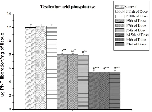
3.1. Effect of cypermethrin on sperm viability and testicular acid phosphatase
As shown in figure 1 the cypermethrin produced reproductive toxicity as reflected by the
reduced sperm viability (p<0.001). Acid phosphatase activities were lowered in the testis
Figure 1: Effect of cypermethrin on sperm viability. Results are expressed as Mean ±
SEM. Analysis is done by one way ANOVA. Superscript a Control group versus all other
[image:6.595.176.446.73.268.2]groups (** indicates p<0.01; *** indicates p<0.001).
Figure 2: Effect of cypermethrin on testicular acid phosphatase. Results are expressed as
Mean ± SEM. Analysis is done by one way ANOVA. Superscript a Control group versus all
other groups (** indicates p<0.01; *** indicates p<0.001).
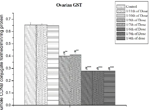
3.2. Effect on ovarian SOD and GST
[image:6.595.186.431.386.567.2]Figure 3: Effect of cypermethrin on ovarian SOD. Results are expressed as Mean ± SEM.
Analysis is done by one way ANOVA. Superscript a Control group versus all other groups
(** indicates p<0.01; *** indicates p<0.001).
Figure 4: Effect of cypermethrin on ovarian GST. Results are expressed as Mean ± SEM.
Analysis is done by one way ANOVA. Superscript a Control group versus all other groups
(** indicates p<0.01; *** indicates p<0.001).
3.3. Effect of cypermethrin on immunological parameters
From the study it was seen that total WBC count was decreased significantly (p<0.05) from
[image:7.595.186.441.393.577.2]significantly (p<0.01) from the dose level of 1/4.5th LD50 to to 1/3rd LD50 in male rat. No significant changes were observed below 1/9th LD50 dose. Maximum toxic effect of cypermethrin was seen in the 1/4.5th LD50 dose without showing any mortality. From the 1/4th LD50 dose, the rate of mortality was increased.
Table 1: Effect of different doses of cypermethrin on WBC count, leukocyte count.
WBC Count /µl Lymphocyte count (%)
Control 5591±58 54.33±0.557 1/11th Dose 5591±58 54.33±0.57 1/10th Dose 5591±58 54.33±0.881
1/9th Dose 7275±83 a*** 63.66±0.666 a*** 1/7th Dose 7275±83a*** 63.666±0.666 a*** 1/5th Dose 7275±83a*** 63.666±0.666 a*** ¼.5th Dose 6866±102 a*** 64.833±0.477 a*** 1/4th Dose 6866±102 a*** 64.833±0.477 a*** 1/3th Dose 6866±102 a*** 64.833±0.477 a***
Results are expressed as Mean±SEM. Analysis is done by ANOVA followed by multiple
comparison two-tail t-tests. Superscript a, Control versus all other experimental groups; (***
indicates p<0.001).
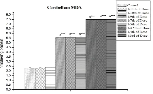
3.4. Effect of cypermethrin on cerebellar and cerebellum MDA and GSH content
A noticeable dose-dependent increase (p<0.001) in the MDA level and decline in GSH
content were seen in cypermethrin induced rat from 1/9th LD50 dose level onwards (Figure 5,6,7,8).
Figure 5: Effect of cypermethrin on cerebellum MDA. Results are expressed as Mean ±
SEM. Analysis is done by one way ANOVA. Superscript a Control group versus all other
Figure 6: Effect of cypermethrin on cerebellum GSH. Results are expressed as Mean ±
SEM. Analysis is done by one way ANOVA. Superscript a Control group versus all other
groups *** indicates p<0.001).
Figure 7: Effect of cypermethrin on cerebellar MDA. Results are expressed as Mean ±
SEM. Analysis is done by one way ANOVA. Superscript a Control group versus all other
groups *** indicates p<0.001).
Figure-8 Effect of cypermethrin on cerebellar GSH. Results are expressed as Mean ±
SEM. Analysis is done by one way ANOVA. Superscript a Control group versus all other
[image:9.595.203.421.76.230.2] [image:9.595.199.427.326.459.2] [image:9.595.211.408.557.696.2]3.5. Effect of cypermethrin on serum urea and creatinine content
Significant decrease in serum urea and creatinine were observed at 1/9th LD50 to 1/6th LD50 dose level which indicate cypermethrin induced nephro-toxicity (Table-2).
Table 2: Effect cypermethrin on serum creatinine and urea at different dose levels.
Creatinine(mg/dl) Urea(mg/dl)
Control 2.8±0.002 25±0.15
1/11th Dose 2.82±0.003 26±0.21
1/10th Dose 2.82±0.003 26±0.21
1/9th Dose 8±0.001a*** 55±0.85a***
1/7th Dose 8±0.001a*** 55±0.85a***
1/5th Dose 8±0.001a*** 55±0.85a***
¼.5th Dose 10.5±0.001a*** 65±0.92a***
1/4th Dose 10.5±0.001a*** 65±0.92a***
1/3rd Dose 10.5±0.001a*** 65±0.92a***
Results are expressed as Mean±SEM. Analysis is done by ANOVA followed by multiple
comparison two-tail t-tests. Superscript a, Control versus all other experimental groups; (***
indicates p<0.001).
4. DISCUSSION
The present study was conducted to explore the systemic toxic effect of cypermethrin in male
and female Wistar rats.
In this study, reduced sperm viability was observed and this may be due to reduced
spermatozoal mitochondrial enzyme activity, altered fructose synthesis and secretion by the
accessory glands.
Acid phosphatases are enzymes capable of hydrolyzing orthophosphoric acid esters in an acid
medium. The testicular acid phosphatase gene is up-regulated by androgens and is
down-regulated by estrogens (Yousef et al., 2001). Activities of phosphatase enzymes have been
shown to rise when testicular steroidogenesis is increased (Mathur and Chattopandhyay,
1982). From our result it is confirmed that after cypermethrin exposure testicular
steroidogenesis was decreased as testicular acid phosphatase was reduced.
Maintaining the balance between ROS and antioxidant enzymes, such as superoxide
dismutase (SOD) and glutathione-S-transferase (GST), is, therefore, crucial and could be an
(SOD) is involved in the clearance of superoxide. In the present investigation, decreased
SOD level was observed in cypermethrin induced male Wistar rats.
The increased WBC counts were noted in CYP-treated groups. These findings were also
supported by the findings of Yousef et al. in sheep (Yousef et al., 1998) and in rabbits
(Yousef et al., 1999). This increase in leukocyte count may indicate an activation of the animal‘s defence mechanism and immune system of the body (Nasuti et al., 2007). This may
also result in an increase in the release of WBC from the bone marrow storage pool into the
blood. The primary function of WBCs is to defend the body from foreign bodies, which are
generated by leukocytosis and antibody production. Pathological leukocytosis may occur due
to exposure to chemicals or acute hemorrhages and hemolysis. Leukocytosis may result due
to a resistance developed by the animal towards the localization of the inflammatory
response. Another possible cause of leukocytosis may be severe hemorrhages in the liver and
lungs (Latimer et al., 2004). This increase may be related to an increase in the lymphocyte
percentage.
In the present study, serum urea levels exhibited a significant increase in
cypermethrin-treated rats according to dose-dependent manner. Also, our results revealed that the
concentration of creatinine was significantly elevated in cypermethrin-treated group
compared to the control value. The creatinine excretion is dependent almost on the process of
glomerular filtration, though tubular secretion contributes slightly (Kassirer, 1971). The slight
and significant rise in the serum creatinine level of male rats may be due to the impairment of
the glomerular function and tubular damage of the kidneys. The increased levels of these end
products in blood especially serum creatinine and serum urea indicate poor clearance of these
substances by the kidneys, rather than excessive production.
Oxidative stress defines an imbalance between the production of reactive oxygen species
(ROS) and antioxidative defense mechanisms. During pyrethroid metabolism, ROS are
generated and cause oxidative stress in intoxicated animals. In oxidative stress, lipid
peroxidation occurs due to excessive free radical production and is considered a primary
mechanism of cell membrane destruction and cell damage. MDA is the end product of lipid
peroxidation. Increased MDA levels in brain suggested that CYP caused testicular damage
(Gupta et al., 1999). Reduction in GSH levels in brain following CYP treatment is indicative
of oxidative stress, whereas GSH was utilized for the detoxification of ROS. As one of the
Normal cellular function was executed based on a balance between ROS production and
antioxidant defense mechanisms existing in the cell (Ghosh and Maiti Choudhury, 2015).
5. CONCLUSION
The present study confirmed that cypermethrin exposure was responsible for gonadal,
immune and systemic toxicity at 40 mg/ kg body weight (1/9th LD50), and 80 mg/kg body weight (1/4.5th LD50) in male rats, and at 34.33 mg/kg body weight (1/9th LD50) and 51.5 mg/kg body weight (1/6th LD50) in female rats.
COMPETING INTERESTS
Authors have declared that no competing interests exist.
ACKNOWLEDGEMENTS
We are gratefully to acknowledge the authorities of Vidyasagar University in doing this
experimental work.
REFERENCES
1. Aktar, M.W., Sengupta, D., Chowdhury, A. Impact of pesticides use in agriculture: their
benefits and hazards. 2009; 2(1): 1-12.
2. Shafer, T.J., Meyer, D.A. Effects of pyrethroids on voltage-sensitive calcium channels: a
critical evaluation of strengths, weaknesses, data needs, and relationship to assessment of
cumulative neurotoxicity. Toxicol Appl Pharmacol, 2004; 196: 303–318.
3. Kilian Delport, E.R., Bornman, M.S., De Jager, C. Simultaneous exposure to low
concentrations of dichlorodiphenyltrichloroe-thane, deltamethrin, nonylphenol and
phytoestrogens has negative effects on the reproductive parameters in male Sprague
Dawley rats. Andrologia, 2007; 39: 128-35.
4. Solati, J., Hajikhani, R., Zaeim, R.T. Effects of Cypermethrin on Sexual Behaviour and
Plasma Concentrations of Pituitary-Gonadal Hormones. International Journal of Fertility
and Sterility, 2010; 4(1): 23-28.
5. Vijverberg, H.P., van den Bercken, J. Neurotoxicological effects and the mode of action
of pyrethroid insecticides. Crit Rev Toxicol,1990; 21: 105-126.
6. Sayim, F., Yavasoglu, N.U., Uyanikgil, Y., Aktug, H., Yavasoglu, A., Turgut, M.
Neurotoxic effect of cypermethrin in Wistar rats: A haematological, biochemical and
7. Rasoul, M.A., Marei, G.I., Abdelgalei, S.A. Evaluation of antibacterial properties
andbiochemical effects of monoterpenes on plant pathogenic bacteria. Afr J Microbiol
Res, 2012; 6(15): 3667-3672.
8. Kalender, Y., Kaya, S., Durak, D., Demi, F. Protective effects of catechin and quercetin
on antioxidant status, lipid peroxidation, and testis-histoarchitecture induced by
chloropyrifos in male rats. Environ Toxicol Pharmacol, 2012; 33: 141-148.
9. Mossa, A.T., Heikal, T.M., Omara, E.A. Physiological and histopathological changes in
the liver of male rats exposed to paracetamol and diazinon. Asian Pac J Trop Biomed,
2012; 2: S1683-S1690.
10.Dee An Jones. Environmental fate of cypermethrin, Environmental Monitoring & Pest
Management, Department of Pesticide Regulation, Sacramento, Data from EPA‘s
Pesticide Fact Sheet Database. 1992; CA 95814-3510.
11.WHO laboratory manual for the examination of human semen and sperm–cervical mucus
interaction. Fourth edition. New York. Cambridge University Press; 1999.
12.Vanha-Perttula, T., Nikkanen, V. Acid phosphatases of the rat testis in experimental
conditions. Acta Endocrinology, 1973; 72: 376– 390.
13.Marklund, S., Marklund, G. Involvement of superoxide anion radical in the auto
oxidation of pyrogallol and a convenient assay for superoxide dismutase. European
Journal of Biochemistry, 1974; 47: 469-474.
14.Habig, W.H., Pabst, M.J., Jakoby, W.B. Glutathione-S-Transferase: the first enzymatic
step in mercapturic acid formation. The Journal of Biological Chemistry, 1974; 249:
7130–7139.
15.Wintrobe, M.M. Clinical hematology. Lea and Febiger, Philadelphia, Library of
Congress, Print USA; 1967.
16.Ohkawa, H., Onishi, N., Yagi, K. Assay for lipid per-oxidation in animal tissue by
thiobarbituric acid reaction. Analytical Biochemistry, 1979; 95: 351-358.
17.Griffith OW. Glutathione turnover in human erythrocytes. The Journal of Biochemistry,
1981; 256: 4900-4904.
18.Natelson, S., Scott, M.L., Beffa, C. A rapid method for the estimation of urea in
biological fluids. American Journal of Clinical Pathology, 1951; 21: 275-81.
19.Brod, J., Sirota, J.H. The renal clearance of endogenous ―creatinine‖ in man. Journal of
20.Yousef, G.M., Diamandis, M., Jung, K., Diamandis, E.P. Molecular cloning of a novel
human acid phosphatase gene (ACPT) that is highly expressed in the testis. Genomics,
2001; 74: 385-395.
21.Mathur, P.P., Chattopadhyay, S. Involvement of lysosomal enzymes in flutamide-induced
stimulation of rat testis. Andrologia, 1982; 14: 171-176.
22.Yousef, M.I., Ibrahim, H.Z., Yacuot, M.H.M., Hassan, A.A. Effects of cypermethrin and
dimethoate on some physiological and biochemical parameters in Barki sheep. Egyptian
J. Nutr. Feeds, 1998; 1: 41–52.
23.Yousef, M.I., Abbassy M.S., Yacout, MH.M. Assessment of cypermethrin and
dimethoate toxicity in Barki sheep: Biochemical and histological changes and tissue
residues. Egyptian J. Anim. Prod, 1999; 36: 25–41.
24.Nasuti, C., Rosita, G., Maria, L.F., Antonio, D.S., Franco, C. Dopaminergic system
modulation, behavioral changes and oxidative stress after neonatal administration of
pyrethroids. Toxicol, 2007; 229: 194-205.
25.Latimer, K.S., Mahaffey, E.A., Prasse, K.W. Clinical Pathology: Veterinary Laboratory
Medicine. 4th ed. Iowa State University Press, Ames, Iowa, USA. 2004.
26.Kassirer, J.P. Clinical evaluation of kidney function—glomerular function. N Engl J Med,
1971; 285(7): 385-389.
27.Gupta, A., Agarwal, A.K., Shukla, G.S. Functional impairment of blood‑brain barrier
following pesticide exposure during early development in rats. Human Exp Toxicol, 1999;
18: 174‑9.
28.Ghosh, R., Maiti Choudhury, S. Alteration in estrous cycle, lipid peroxidation and
antioxidant status in female rat after exposure to lambda cyhalothrin and its attenuation