CLINICAL SIGNIFICANCE OF TROCHANTER SHAFT ANGLE AS
DETERMINANT OF ALIGNMENT OF FEMORAL CANAL AND
TROCHANTER IN MANAGEMENT OF FRACTURE SHAFT OF
FEMUR
1
Lakhwani O. P., *2Chetiwal Rajesh and 3Mittal P. S.
1
Associate Professor, *2Associate Professor, 3Assistant Professor, ESIC – PGIMSR, ESI
Hospital, Basaidarapur, Ring Road, New Delhi – 110015.
ABSTRACT
Introduction: Femoral nailing is the one of the common procedure used for the fixation of fracture shaft of femur. Since relation of the
medullary canal is not constant and collinear with the any specific
point over trochanter hence it is difficult to insert nail and various entry
points including pirriformis fossa, tip of greater trochanter has been
used but anatomical determinant are not clearly defined. Aims and Objectives: Study aimed at analyzing the relevant anatomy of proximal femur on dry femur bone and skigram to determine the
anatomical alignment of throchanter with medullary canal as angle
between and its significance. Materials and Methods: Adult dry
cadaveric femora and skigram were taken to measure angle between
the trochanter and shaft of femur for safe and anatomical femoral nail insertion confirmed by
retrograde insertion of remur and radiaogram. Results: Most favourable and least strainful path for uniplaner intramedullary nail lies in the line of medullary canal can be correlated
with angle between the Trochanter and Shaft which ranges between 5-17 degrees on dry
cadaveric femora and 4-14 degrees on skigram. Discussion: since trochanter is not collinear
with the medullary canal and variation in the proximal femoral anatomy, it is difficult to
define the any specific point as universal entry point for femoral nailing. Relation and angle
between trochanter and central medullary cavity line can guide for selection of appropriate
and safe entry point for intramedullary femoral nail insertion.
KEYWORDS: pirriformis, skigram, throchanter, uniplaner, remur and radiaogram.
Article Received on 19 Sep 2015,
Revised on 25 Sep 2015, Accepted on 30 Sep 2015
*Correspondence for Author
Dr. Chetiwal Rajesh
Associate Professor, ESIC – PGIMSR, ESI Hospital,
Basaidarapur, Ring Road, New Delhi – 110015.
INTRODUCTION
Entry site for antegrade femoral nail insertion is one of the most crucial step in overall
alignment, reduction and healing of fracture and avoiding complications in procedure.
Various entry sites including Piriformis fossa.[1],[2] Tip of greater trochanter (GT)[2],[3], lateral
tip of Greater Trochanter and other have been studied but, controversy still exists as
anatomical and radiological basis of selection of most appropriate and safe site is not
defined.
Most appropriate entry site for uniplanar femoral nail insertion should lie in the direction of
medullary canal. However, due to anatomical variations and the greater trochanter not being
strictly collinear with axis of the medullary canal it becomes difficult to ascertain a ideal
universal area for antegrade femoral nail insertion. Therefore, Present study was undertaken
to know the alignment relationship between Trochanter and shaft of femur using dry
cadaveric femur, radiogram and anatomical dissection in reference to entry sites to serve as
anatomical and radiological basis for femoral ante grade nail insertion site.
MATERIALS AND METHODS
Study involved fifty dry adult cadaveric non- fractured femora without any deformity.
Selected femora and their digital radiogram were taken for anthropometric and radiological
measurements in reference to nail insertion site.
Dry cadaveric femora were taken to know the most favorable anatomical entry site lying in
the direction of medullary canal. Since the greater trochanter is not collinear with long axis of
the femoral medullary canal hence, this relation can be expressed as Trochanter-shaft angle,
which is formed between a vertical line along axis of medullary canal and a line drawn from
a point on this vertical line at the level of base of lesser trochanter to the tip of Greater
Trochanter (GT) (which can be easily identified on bones and digital radiogram). Level of
base of lesser Trochanter signifies the transitional zone between cortical and cancellous bone
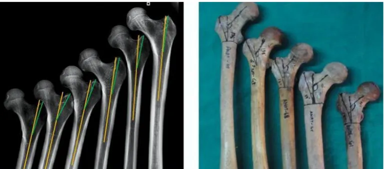
at junction of Trochanter and femoral shaft. (Figure-1) This angle was also measured and compared on computerized digital radiograms of the dry cadaveric femora after nullifying the
angle of torsion. (Figure - 2) Proximal femur according to Trochanter shaft angle (TSA) was
Four formalin fixed lower limbs of adult Indian cadavers having atleast 60 degrees of hip
flexion and no scars in hip region, thus excluding any previous hip surgery were selected in
study. Anatomical dissection of these formalin fixed adult cadaveric hip was carried out.
With an awl a small hole was created at entry sites of antegrade femoral nail and reamed.
Relation of entry sites with Gluteus maximus, Gluteus medius and Gluteus minimus muscles,
greater trochanter capsular attachment vascular supply network of femoral head and sciatic
and gluteal neuovasolar structure was explored.
OBSERVATIONS
In anthropometric study Trochanter shaft angle (TSA) was measured with maximum of 17
degree and minimum of 5 degree with average of 10.2 degrees. On Radiological
measurement the range was 4 to 14 degree with average of 11.4 degrees.
In 34 cases (68%) which had Trochanter-shaft angle range between 6-12 degrees in both
radiological and Anthropometric measurement, the alignment of the shaft or medullary canal
(confirmed by the digital radiogram) lied just medial to the tip of Greater Trochanter (GT). In
12 cases (24%) which had Trochanter shaft angle less than 6 degree corresponded with tip of
greater trochanter. In 4 (8%) cases of Trochanter-shaft angle 12-17 degrees alignment of
medullary canal falls in the direction of femoral neck. (Figure – 3 and 4).
The level of base of lesser trochanter mark the transition zone between cancellous to cortical
bones i.e. trochanter area and the shaft. This distance from tip of greater trochanter to the
base of lesser trochanter was measured in Morphological study it was average 6.77cms and in
Radiological study it was 6.73 cms.
In dissection of formalin fixed adult cadaveric hips anatomical relation was explored.
Piriformis fossa (Figure - 5) was identified as depression over the posterior-medial aspect of greater trochanter providing insertion of obturator externus muscle. Branches of medial
circumflex femoral vessels lied in close intricacy and partly encroached at this entry site in
one dissection. Attachment of capsule of the hip joint was encroached in two dissected
specimen in which entry site extended medial to the central axis of the femoral canal. Tip and
lateral aspect of greater trochanter provided attachment to the gluteus muscle and entry site in
this region in one dissection partially violated this insertion. Inferior most branches of the
and were liable to damage if incision, awl or reamer got inserted more medially and higher.
Area just medial to the tip of greater trochanter lying in the direction of the femoral shaft was
found relatively safer in order to avoid the important structures.
Longitudinal section of the femur bone (Figure - 1) revealed bony architecture with transitional zone lying between cancellous bone (Trochanter) and cortical bone (Shaft) at the
level of base of lesser trochanter. In Morphological study it was average 6.77cms and on
[image:4.595.195.407.242.425.2]skigram it was measured as 6.73 cms average.
Figure - 1 Measurement of Trochanter shaft angle (TSA) on skigram, B distance at the level of base of lesser trochanter, C distance from tip of Trochanter to base of lesser trochanter mark junction of cancellous bone (Trochanter) and cortical bone (shaft femur).
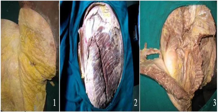
[image:4.595.108.494.529.698.2]Figure - 3 and 4 illustrating anatomical zones with Zone “A” corresponding to trochanter shaft angle 0-6 degree, Zone “B” corresponding to trochanter shaft angle 6-12 degrees, Zone “C” corresponding to trochanter shaft angle 6-12-17 degrees.
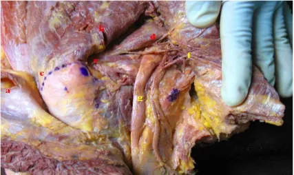
[image:5.595.80.518.348.572.2]Figure - 6 Illustation (a) and (b) incision in Gluteus maximus and medius approaching entry site. (c) Attachment of main abductor Gluteus medius over greater trochanter, (d) Piriformis muscle (e) Attachment of short external rotators obturators, Gemulli and Piriformis over pirriformis fossa and posterior medial aspect of Greater Trochanter, deep to this lies capsule of hip joint, (f) Branching of the Gluteal neurovascular bundle (g) Sciatic nerve.
DISCUSSION AND CONCLUSIONS
Conventional intramedullary nails being flexible enough provide the variation in entry point
selection hence Piriformis fossa, Greater trochanter or any nearby point can be used without
much inconvenience and complications. Rigid interlocking nails are less forgiving in
selection of entry point hence various complications.[4]-[7] like iatrogenic fracture of neck,
communition of the proximal part of femur, osteonecrosis of the femoral head have been
reported. Anatomical site for the safe insertion of uniplanar rigid interlocking nail should lie
in the direction of medullary canal of femur but medullary canal of femur is not collinear
with trochanter hence, to decide the safe anatomical entry point consideration needs to be
given to this alignment which can be better objectified by determining Trochanter – shaft
angle (TSA).
It was measured on dry femora and Computerized digital radiograms of the dry cadaveric
femora after nullifying the angle of torsion. Zoning of proximal femur on the basis
point in a particular patient. In patients having zone “A” (TSA 0-60
) tip of greater trochanter
practically lies in line with long axis of femoral medullary canal and thus can be taken as
asuitable entry site. For patients with zone B (TSA 6-120), which was found most common in 68 percent cases, most appropriate site lie just medial to graeter trochanter. zone C (TSA more than 12) warrants the use of biplanar bent nails to avoid complication and difficulty in nail insertion.
The level of base of lesser trochanter in longitudinal section of femur revealed a transitional
zone between cancellous to cortical bones i.e. trochanter area and the shaft. (Figure -1) It the distance between tip of GT and base of lesser trochater may serve as a guide for bending site
in biplanar nails. Wolfgang Grechenig etal.[8] studied the Anatomy of the greater trochanter
and its clinical importance for intramedullary femoral nailing in cadaver specimens. They
reported that trochanter may cover the actual entry site resulting in a much more medial entry
site and recommended resection of parts of the Trochanter and preoperative CT scan, but this
facility may not be available in all cases hence application of Trochanter shaft angle (TSA) is
useful during surgery under image intensifier guidance. In a clinical study by Robert F.
Ostrum[3] and cadaveric study by Wolfgang Grechenig[8] and Gausepohl T et al[9] it has been
found that anatomy of the Greater Trochanter (GT) is variable and sought its clinical
importance but how it can made to the clinical application was not clear. In our study we tried
to develop useful actual clinical application by measuring the Trochanter-Shaft angle (TSA)
and its significance in deciding selection of entry point.
Edward A. Perez.[10] (2007) Studied the Gluteus medius tendon injury in medial Trochanter
entry point. Study by Adam J. Starr in a prospective, randomized comparison of Trochanteric
Versus Piriformis Fossa Entry Portal found the ideal starting point for nail placement in
proximal femur fractures is controversial.
Georgiadis GM.[11] (1996) and T Gausepohla[9] (2002) had studied and found anatomical
determination of the correct entry point in 88% of the specimen. The ideal entry point for a
straight nail was found constantly at the medial border of the greater Trochanter, this
correlated with zone B in our study with Trochanter shaft angle 6-12 degrees. C. Dora etal.[12]
in a cadaveric study reported soft tissue damage in antegrade femoral nailing at entry point
and concluded that, entry point selection with ease of nail insertion must be weighed against
Exploring the entry sites by anatomical dissection showed entry point at piriformis fossa
(Figure -7) lie in close vicinity of external roatator muscles obturator externus, gemulli, pirriformis muscles and medial femoral circumflex vessels and likely to damage these
structures. entry site at tip of greatertrochanter lie at the tendinous insertion of gluteal medius
and may cause disruption of tendon. Entry site just medial to greater trochanter in region “B”
is relatively safer site for nail insertion.
Anatomical entry point lying in region “C” with trochanter shaft angle 12-17 degree lie at
junction of neck and trochanter and risks fracture and vascular damage, hence requires to
choose the bent biplane nail.
Anatomical and radiological evaluation revealed, the entry point for antegrade femoral nail
insertion is highly variable and it cannot be defined in terms of piriformis fossa or greater
trochanter. The anatomical entry site need to be correlated with the proximal femoral
anatomy, anatomy of the trochanter itself and trochanter-shaft angle (TSA).
However, this study dealt with relatively small number of femora and it will be worthwhile to
perform similar studies with a larger number of bones from different age groups and from
diverse population.
REFERENCES
1. Miller SD; Burkart B; Damson E; Shrive N; Bray RC. The effect of the entry hole for an
intramedullary nail on the strength of proximal femur, J Bone Joint Surg Br., 1993; 75:
202-6.
2. Ricci WM, Schwappach J, Tucker M, Coupe K, Brandt A, Sanders R, Leighto R.
Trochanteric versus piriformis entry portal for the treatment of femoral shaft fractures. J
Orthop Trauma., 2006; 20: 663–667.
3. Robert F. Ostrum, A Critical Analysis of the Eccentric Starting Point for Trochanteric
Intramedullary Femoral Nailing J Orthop Trauma, 2005; 19: 681–686.
4. Kempf I, Grosse A, Beck G; Closed locked intramedullary nailing. Its application to
comminuted fractures of the femur, J Bone Joint Surg. Am., 1985; 67: 709-20.
5. Winquist RA, Hansen ST Jr, Clawson DK; Closed intramedullary nailing on femoral
fractures A report of five hundered and twenty cases J. Bone Joint surg Am., 1984; 66:
6. Buford D Jr, Christensen K, Weathrall P; intramedullary nailing of femoral fractures in
adolescents Clin Orthop., 1998; 350: 85-89.
7. Mileski RA, Gravin KL, Crosby LA; Avascular necrosis of the femoral head in an
adolescent following intramedullary nailing of the femur, A case report J Bone Joint Surg
Am., 1994; 76: 1706-8.
8. Wolfgang Grechenig, Wolfgang 1 Pichler1, Hans Clement1, Norbert Peter Tesch and
Stephan Grechenig Anatomy of the greater femoral Trochanter: clinical importance for
intramedullary femoral nailing Anatomic study of 100 cadaver specimens, Acta
Orthopaedica, 2006; 77(6): 899–901 899.
9. Gausepohl, D. Pennig, J. Koebke and S. Harnoss, Antegrade femoral nailing: anatomical
determination of the correct entry point, Injury, 2002; 33(8): 701–705.
10.Edward A. Perez, Is There a Gluteus Medius Tendon Injury During Reaming Through a
Modified Medial Trochanteric Portal? A Cadaver Study., J Orthop Trauma, 2007; 21:
617–620.
11.Georgiadis GM, Olexa TA, Ebraheim NA, Entry sites for antegrade femoral nailing. Clin
Orthop., 1996; 330: 281–287.
12.C. Dora, M. Leunig, M. Beck, D. Rothenfluh and R. Ganz, Entry point soft tissue damage
in antegrade femoral nailing: a cadaver study, Journal of Orthopaedic Trauma, 2001;