FORMULATION AND EVALUATION OF TOPICAL CREAM OF
PIPERINE FOR VITILIGO
P. Satyendra, P. Arun, P. Shailendra, *D. Neelesh and K. Neeraj
Shri Ram Group of Institutions Faculty of Pharmacy.
ABSTRACT
The main interest of the investigations gathered here was in pursuit of
developing a topical dosage form of a suitable antivitiligo agent. In the
light of reported works on antivitiligo activity of piperine, this project
was aimed to bring a ray of hope to the “down casted” by a dosage
form incorporated with the bioactive compound piperine, as the API
(active pharmaceutical ingredient) for antivitiligo. The formulation
must be able to achieve the drug to penetrate the stratum corneum and
get lodged in the dermal layers without further trespassing into the
systemic circulation. Thus the tissue bioavailability of the drug in the
dermal region must be maximized where the melanocytes proliferency and formation of
melanocytic dendrites can be achieved. The formulation and development of conventional
topical preparations-cream and ointment incorporated with piperineis also carried out with a
NDDS known as phytosomes.
KEYWORDS: Piperine, Vitiligo, Ointment, Phytosme, Creams.
1. INTRODUCTION
Vitiligo also known as Leukoderma is a chronic skin disease. This is caused by the loss of
pigment, resulting in irregular pale patches of skin. It is reported that Vitiligo is a disorder in
which the body destroys its own pigment cells, melanocytes (autoimmune) in various parts of
the skin. In affected areas, the pigment gradually disappears. Vitiligo is classified according
to the distribution, pattern and extent of depigmentation. There are many reports on
classification. However, most investigators distinguished two large subtypes of Vitiligo,
segmental Vitiligo(S) and non-segmental Vitiligo(NS). According to another classification
proposed by Norlund and Lerner, three types are identified i.e., localized, generalized and
universal Vitiligo. Localized Vitiligo is further classified into focal and segmental:
Volume 7, Issue 3, 714-724. Research Article ISSN 2277– 7105
Article Received on 05 Dec. 2017,
Revised on 26 Dec. 2017, Accepted on 16 Jan. 2018
DOI: 10.20959/wjpr20183-10831
*Corresponding Author
D. Neelesh
Shri Ram Group of
Institutions Faculty of
generalized into acrofacial, vulgaris and mixed subtypes.
The researchers found that piperine as antivitiligo agent compared the effects of Piperine and
its analogues tetrahydro Piperine (THP), cyclohexyl analogue of Piperine (CHP) and reduced
CHP (rCHP) when applied to the skin of mice, either alone or followed by UVR.
2. MATERIAL AND METHODS
Pepper fruits (berries) were collected from various organic farming from locations in Karoor,
Chalakudy, Thrissur Dist. Kerala, South India and authenticated. Piperine was isolated and
standardized in SRGI Faculty of Pharmacy, Jabalpur and also procured from Sigma-aldrich
INC, USA. Phosphatidyl choline was a generous gift sample from Lipoid GMBH,
Ludwigshafen, Germany. Poloxamer 407 was procured from Spectrum chemicals Mfg Corp,
Gardina, CA, USA. Tri ethanol amine and 4-Hydroxyanisole were procured from Universal
lab and chemicals, Penang, Malasia. Glyceryl monooleate was obtained from Himedia Pvt.
Ltd. Sagar. The following chemicals were purchased from SD Fine-chem Ltd., Sagar:
Carbopol 934, Stearic acid, Hard paraffin, Cetostyryl alchohol, Glycerine, Beeswax, Lanolin,
Ethanol, KOH, NaOH, Acetone, Hexane, Toulene. Silica gel 60 F 254 and Ethyl acetate were
received from Merck specialties Pvt. Ltd. Mumbai. Tween 80 and Span 80 were procured
from Accord labs, Secunderabad. 4-Hydroxyanisole was provided by Hychem Laborotaries,
Sagar. All reagents used were of high quality analytical grade.
2.1. Extraction, isolation, standardization and preformulation studies on piperine
As represented in the figure 24a, extraction of piperine was carried out by using two separate
methods soxhlation and reflux method. In both extraction methods was different but the
isolation procedure was kept same. A 50gm, aliquot of powdered black pepper was taken and
extracted with 500ml of 95% ethanol in Soxhlet apparatus/reflux for 3 hrs. The solution was
filtered and concentrated under vacuum in a water bath at 60˚C by solvent recovery method
either by distillation or by using rotary evaporator. Alcoholic potassium hydroxide 50ml was
added to the concentrate and the solution was stirred continuously for 30min. The obtained
solution was heated and water was added drop wise until yellow precipitate was formed.
Water was added until no more precipitate appeared to form and this was allowed to settle
overnight. Needles of Piperine were observed to be separated out. The solid was collected
and washed with cold ether 2-3 times. It was recrystallized by using acetone. For this,
for 24hrs so that crystals of piperine are formed. Yellow coloured rod shaped crystals were
recrystallized after 24 hrs.
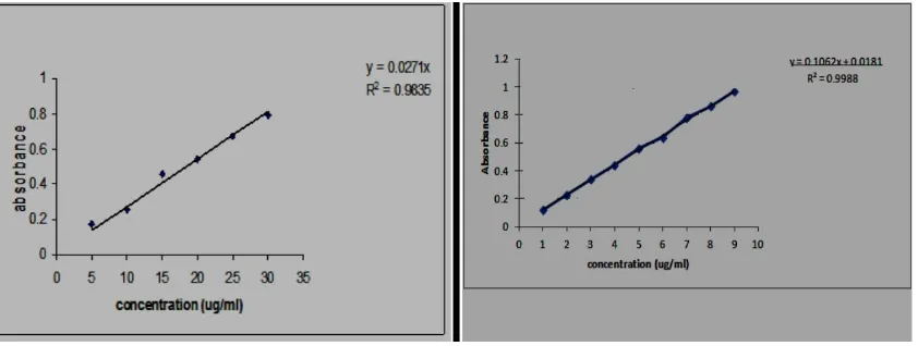
2.2. Construction of standard graph of piperine in ethanol and phosphate buffer(pH
6.6)
Piperine, 10ml was dissolved in 10ml of ethanol (1mg/ml). From this, 1ml was taken and
made upto 10ml with ethanol. From this 0.5, 1, 1.5, 2, 2.5, 3ml were taken and made up with
ethanol to 10ml separately to produce 5, 10, 15,20,25,30 µg/ml respectively. The solution
was suitably diluted and absorbance was taken at 344nm.
10mg of Piperine was dissolved in little of ethanol and then the volume was made upto 10ml
with buffer (1mg/ml). From this, 1ml was taken and made upto 10ml with buffer. From this
0.5, 1, 1.5, 2, 2.5, 3ml were taken and made up with buffer to 10ml separately to produce 5,
10, 15, 20, 25, 30 µg/ml respectively. The solutions were suitably diluted and the UV spectral
absorbance were taken at 344nm. The equation used was Y = mx + c; where Y= Absorbance,
[image:3.595.90.511.402.561.2]m = slope, x = Concentration, c = Intercept.
Figure 1: Standard calibration curves for Piperine at max 344nm in ethanol, (left) and
right (buffer pH 6.6) n=6.
2.3 Formulation of cream incorporated with 1% piperine- Formula to prepare 25g cream
incorporated with 1% piperine was calculated as follows. Beeswax (12.5 g), Lanolin (2 g)
and stearic acid (2.5 g) were taken in one beaker. In another beaker, Piperine (1%) was
dissolved in ethanol by sonication and introduced into glycerine (6.25 g), water (7.26 g),
Triethanolamine (0.44 g). Both the beakers were maintained at 60°C and all the ingredients
2.4Formulation of ointment incorporated with 1% Piperine
Ingredients were calculated for 15g ointment. Ointment was prepared by using fusion
method. Hard paraffin (50g) was taken in a china dish along with cetostearyl alcohol 50g and
was allowed to melt. Wool fat 50g and white soft paraffin 850g were also added after
sometime. After the excipients were melted 1% Piperine was incorporated. Stirred well at
reduced maintained temperature.
2.5 Characterization and evaluation of ointment and cream
(i) Drug content uniformity- Drug content uniformity of the formulation in ethanol as
solvent was performed. For 1g of cream, 9750μg of Piperine was found. So for 25 gms, the
amount of drug is 243.750mg which is almost equal to the total amount taken i.e., 250mg
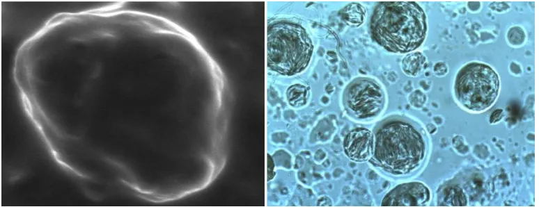
[image:4.595.105.492.326.475.2]establishing pass test for drug content uniformity.
Figure 2. SEM of cream (left) and phase contrast microphotograph (200X) of ointment
(right).
(ii) Organoleptic characters and consistency
Organoleptic characteristic studies of the cream were conducted. The cream was light yellow
colour and final preparation was found to be free from gritty nature with light yellow colour
(iii) Ex-vivo permeation and tissue bioavailability a. Preparation of the tissue
Figure 3. Drug diffusion profile from cream and ointment with and without urea into
buffer pH6.6 which was detected at 344 nm.
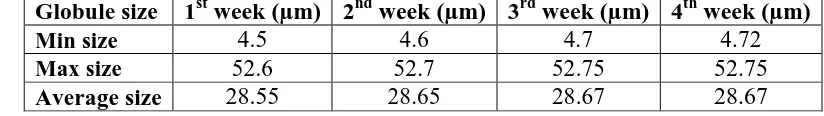
(iv) Globule size of cream under 10 x observed periodically as a part of stability
[image:5.595.117.473.83.267.2]assessment
Table no 1.
(v) Fabrication of phytosomes
The phytocomplex (phytosomes) was prepared by considering three drug-phosphatidyl
choline (PC) ratios of 1:1, 1:3, 1:5. The required amounts of piperine and phospholipids were
dissolved in anhydrous ethanol (30ml) and refluxed for 2 hrs while stirring with a magnetic
stirrer. Later the solution was transferred into a 100 ml round-bottom flask and ethanol was
evaporated under vacuum at 400C using rotary evaporator. The complex was washed with
n-hexane, dried residues were gathered and placed in desiccators overnight, then sieved through
a no. 100 mesh. The resultant extract–phospholipid complex was transferred into a glass
bottle and stored in the room temperature for further evaluations.
(vi) Composition and preparation of 1 % cream (25gm)-For a scope of comparative study
with the previously reported work, phytosomal complex was incorporated with a cream of
same composition. Emulsion system was prepared based on the procedure mentioned under
the formulation of piperine 1% cream. Briefly, beeswax, lanolin and stearic were taken in one
beaker. Glycerine, water, triethanolamine were taken in another beaker. Phytosome complex
Globule size 1st week (µm) 2nd week (µm) 3rd week (µm) 4th week (µm)
Min size 4.5 4.6 4.7 4.72
Max size 52.6 52.7 52.75 52.75
[image:5.595.86.498.392.450.2]was dissolved in ethanol by sonication and maintained at 400c. Both the beakers were
maintained at 60°C and all the ingredients were melted. Then oily phase is added to aqueous
phase and stirred continuously. The phytosome complex was added to the mixture when the
temperature dropped to 400C. Creams of three phytosomal complexes (1:1, 1:3, 1:5) and with
pure drug were coded as P1, P2,P3 and P0 respectively.
RESULT AND DISCUSSION
Evaluation of phytosomes and phytosomal cream
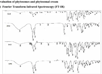
(i) Fourier Transform Infrared Spectroscopy (FT-IR)
Figure 4: FTIR reports: (a) piperine (b) phosphatidyl choline (c) physical mixture (d)
[image:6.595.80.507.210.519.2]complex.
Table no 2 FTIR Peaks and functional group.
Sr. No. Wave No. Functional group
1 2919.3 CH2
2 1738.2 C=O
3 1244.5 C – O
4 1092.3 C-N
5 3429 OH
6 3008.2cm-1 Un saturation
7 2937cm-1 CH2-CH2 –CH3
8 1634 cm-1 C=O stretching
9 1027cm-1 C-N stretching
(ii)Average particle size of phytosomes
was found to be 30.6 microns. From the drug content uniformity, the drug concentrations
found in P1, P2, P3 formulations are 247.36mg, 239.42 mg and 239.42mg/ml respectively.
Figure 6: Cumulative drug release profile of piperine from phytosomal cream
formulations (P1, P2, P3) diffused into buffer solution of pH6.6 detected at 344 nm.
Results are expressed as mean ± SD when= 6.
(iii) Curve fitting analysis
Table no 3. Drug release kinetic model fitting from phytosomes.
Sr.no. Phytosome code Zero First Higuchi Peppas n
1 P1 0.993752 0.878752 0.899746 0.9949 1.2994
2 P2 0.991302 0.863181 0.949023 0.9692 0.9582
3 P3 0.951157 0.894835 0.84127 0.9252 0.992
(iv) The pH- of the formulation F1 was found to be 6.2 which denotes the cream is slightly
acidic in nature.
(v) The globule size of the Formulation P1 has shown no significant change even after 4
months.
Table no 4. The globuler size of formulation.
Sr. no. Globule size
1stMonth (µm)
2ndMonth (µm)
3rdMonth (µm)
4thMonth (µm)
1 Min size 5.4 5.4 5.42 5.42
2 Max size 55.81 55.8 55.75 55.75
[image:7.595.145.455.148.327.2]Figure 7: Plots of log % drug concentration VS time for different temperatures.
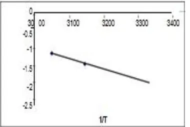
Figure 8: Plot of log k Vs 1 / T to determine k at T25°C.
1 / T values of 45°C and 55°C in Kelvin are 3.048 × 10⁻³ and 3.144 × 10 -3.
A graph is plotted between the 1 / T values and log k values and the k value obtained by
extrapolating the curve at 27 °C is 0.006 / months.
DISCUSSION
Thecrystals isolated from piperine was the long needle shaped with the average size of 59.8
µm was able to bring down to 13.77µm. This truly facilitated the drug incorporation in both
ointments and creams. When urea as a permeation enhancer was used, the drug diffusion into
the buffer was increased by 0.87% which ironically reflected less tissue drug bioavailability.
Drug tailoring to the dermis was augmented by 0.6% which was negligible. Minimum,
maximum and average globule size of cream was found to be 4.72µ 52.75µ and 28.75 µ
respectively while the average size of piperine was in the range of 50-60 µm. Crystal growth
should be more cautious than the sedimentation, since the former ruptures the lamellar
structure. Sedimentation can be overcome by simple physical agitation. Thus the optimised
formulation (TS1) was showing better tissue bioavailability (75.25%±1.72) than the previous
formulation (phytosomes having 72.07 ±0.01) which clearly indicates TS are persuasive
[image:8.595.205.395.239.366.2]Drug release kinetics are greatly influenced by crystallinity, solubility, particle size amount of
API (Active Pharmaceutical Ingredients) etc. A Water soluble API homogenously distributed
in a matrix system was mainly released by diffusion. At the same time, liphophilic drugs, the
mechanism was based on self erosion. In this case the API was liphophilic.
Comprehensive discussion on drug release kinetics of all formulations
Table 16: Drug release kinetics of cream and ointment.
Sr. no. Formulation Zero First Higuchi Peppas n
1 Cream 0.777322389 0.51691858 0.947135035 0.505880848 0.6286 2 Ointment 0.962658117 0.837180238 0.93231288 0.739151109 0.7695 3 Drug+urea 0.975245 0.800999799 0.960843795 0.721089635 0.7679
Table 17: Drug release kinetics of phytosomal formulation.
Sr no Phytosome code Zero First Higuchi Peppas n
1 P1 0.993752 0.878752 0.899746 0.9949 1.2994
2 P2 0.991302 0.863181 0.949023 0.9692 0.9582
3 P3 0.951157 0.894835 0.84127 0.9252 0.992
Formulation P1 best fit into Korsmeyer-Peppas model with R2 value 0.9949, then zero order
followed by higuchi equation. Formulations P2 and P3 best fit into zero order with R2 value >
0.9511, then Korsmeyer-Peppas followed by higuchi equation.
CONCLUSION
The main interest of the investigations gathered here was in pursuit of developing a topical
dosage form of a suitable antivitiligo agent. As many as 65 million world population suffers
from this skin disease. In Vitiligo, white blood cells attack and destroy pigment cells in
discrete areas of the skin. It is called an auto-immune disorder, because the pigment cells
become regarded by the body as "foreign" and are therefore rejected. Attempts in classifying
Vitiligo has two approaches. In one approach Vitiligo is classified as segmental Vitiligo and
non-segmental Vitiligo.
Phytosomes were considered because of its unique ability of bonding together with the phyto-
constituent as its integral part. Such a complex is obtained by reaction of stoichiometric
amounts of phospholipid and the substrate in an appropriate solvent. Phytosomal cream was
prepared and slight increase in the tissue retention of piperine in the dermis when compared
REFERENCES
1. Loyed Allen V, Nicolas Popovich G, Howard Ansel. Ansel’s pharmaceutical dosage
forms and drug delievery system. 8th ed., Lippincott Williams & Wilkins, Philadelphia.,
1-2: 93-94.
2. Michlae Altun, Kevin Tailor. Aulton’s pharmaceutics. The design and manufacture of
medicines. 3rd ed. Churchill Livingstone, Ney York., 1-2.
3. Gurib-Fakim A. Medicinal plants: Traditions of yesterday and drugs of tomorrow. Mol
Aspects Med., 2006; 27(1): 1-93.
4. Khan S, Balick MJ. Therapeutic plants of Ayurveda: a review of selected clinical and
other studies for 166 species. J Altern Complement Med., 2001; 7(5): 405-51.
5. Sasd B, Azaizeh H, Said O. Tradition and perspectives of arab herbal medicine: a
review, Evid Based Complement Alternat Med., 2005; 2(4): 475-479.
6. Zhou Y, Baker T. Theory of traditional Chinese medicine and therapeutic method of
diseases. Md Med., 2002; 3(1): 13-15.
7. Mahady GB. Global harmonization of herbal health claims, J Nutr, 2001; 131(3s):
1120S-3S.
8. World Health Organization. Traditional Medicine Strategy 2002-2005 WHO, Geneva, p,
19-26: 43-48.
9. Barnes J. Quality, efficacy and safety of complementary medicines: fashions, facts and
the future. Part I. Regulation and quality. Br J Clin Pharmacol, 2003; 55(3): 226-233.
10. Liang YZ, Xie P, Chan K. Quality control of herbal medicines. In: J. Chromatogr B
Analyt Technol Biomed Life Sci., 2004; 812(102): 53-70.
11. Gan F, Ye R. New approach on similarity analysis of chromatographic fingerprint of
herbal medicine. J Chromatogr A., 2006; 1104(1-2): 100-105.
12. Leung KS, Bian ZX, Moher D, Dagenais S, Li YP, Liu L, Wu TX, Miao JX. Improving
the quality of randomized controlled trials in Chinese herbal medicine, part III, In:
Zhong Xi Yi Jie He Xue Bao. Quality control of Chinese herbal medicine used in
randomized controlled trials., 2006; 4(3): 225-232.
13. Fong HH. Integration of herbal medicine into modern medical practices: issues and
prospects. Integr Cancer Ther., 2002; 1(3): 287-293.
14. CPMP Guideline 3AQ22a. Quality of herbal remedies, May, 1989; 195-243.
15. Boullata JI, Nace AM. Safety issues with herbal medicine. Pharmacotherapy, 2000;
20(3): 257-269.
Chinese herbal toxicology database. J Toxicol Clin Toxicol, 2002; 40(2): 159-167.
17. Woodward KN. The potential impact of the use of homeopathic and herbal remedies on
monitoring the safety of prescription products. Hum Exp Toxicol, 2005; 24(5): 219-233.
18. Ernst E. Are herbal medicines effective? Int J Clin Pharmacol Ther., 2004; 42(3):
157-159.
19. Le Poole JC, Das PK, Van Den Wijnguard RM, Bose JD, Westerh of W. A review on
vitiligo. Exp. Dermatol, 1993; 2: 146-147.
20. Available at: http://www.vithappens.com/wp-content/uploads/2011/07/vit_hands.jpg
21. Taieb A. Intrensic and extrinsic pathomechanisms of vitiligo. Pigment Cell Res., 2000;
8: 41.
22. Nordlund JJ, Lerner AB. Vitiligo-it is important. Arch Dermatol, 1982; 118: 5-8.
23. Kemp EH, Gawkrodger DJ, MacNeil S, Watson PF, Weetman AP. Detection of
tyrosinase in autoimmune mediated vitiligo patients using 35S labeled recombinant
human tyrosinase in radioimmuno assay. J Invest Dermatology, 1997; 109: 69.
24. Le Poole IC, Vanden Vigngaard RM, Westerhoff W, Das PK. Presence of T cells and
macrophages in inflammatory vitiligo skin parallels melanocytes disappearance. Ame J
Pathol, 1996; 148: 1219.
25. Shajil EM, Sreejata chaterjee, Deepali Agarwal, Bagchi T, Rasheedunissa Begum.
Vitiligo: pathomechanisms & genetic polymorphism of susceptible genes. Indian J Exp
Biology, 2006; 44: 526-539.
26. Caixia T, Hongwen F, Xiran L. Levels of soluble interleukin-2 receptor in the sera and
skin tissue fluids of patients with vitiligo. J Dermatol, 1999; 21: 59.
27. Cucchi ML, Frattini P, Santagostino G, Preda S, Orecchia G. Catecholamines increase in
the urine of non-segmental vitiligo especially during its active phase. Pigment cell Res.,
2000; 13: 28.
28. Prota G. Melanin and Melanogenesis, 1st ed. Academic Press, New York, 1992; 1–290.
29. Nicolaus RA, Piattelti M, Fattorusso E. Structure of melanin and melanogenesis-IV on
some natural melanin. Tetrahedron, 1964; 20: 1163.
30. Ito S, Homma K, Kiyota M, Fujitha K, Jimbow K Characterization of structural
properties for morphologic differentiation of melanosomes. III. Free and protein-bound
dopa and 5-S-cysteinyldopa in B16 and Harding-Passey melanomas. J Invest Dermatol,