Original Article
Quercetin improves rat brain function
after ischemia by regulating DJ-1/Nrf2
Cheng Xiao1*, Juan Wang2*, Xia Zhang2, Guodong Dai3
1Department of Neurology, Xianning Central Hospital, The First Affiliated Hospital of Hubei University of Science
and Technology, Xianning 437000, Hubei Province, China; 2School of Nursing, Hubei University of Science and
Technology, Xianning 437100, Hubei Province, China; 3Department of Neurosurgery, The First Affiliated Hospital
of Hubei University of Science and Technology, Xianning Central Hospital, Xianning 437000, Hubei Province, China. *Equal contributors.
Received October 11, 2018; Accepted April 8, 2019; Epub July 15, 2019; Published July 30, 2019
Abstract: Nuclear transcription factor NF-E2 related factor 2 (Nrf2) is an important nuclear transcription factor in anti-oxidative stress. DJ-1 is a multifunctional protein widely distributed in various tissues. It plays a role in
enhanc-ing Nrf2 stability and promotenhanc-ing Nrf2 protein expression. Quer is a flavonoid that provides antioxidant and neuro -protective effects. In this study, a rat brain I-R injury model was established to detect caspase-3 activity,
malondi-aldehyde (MDA) levels, and superoxide dismutase (SOD) levels. Modified nerve injury severity (mNSS) scores were
used to evaluate neurological function. DJ-1 and Nrf2 expression was compared. Rat Rn-c neurons were cultured
in vitro and divided into three groups, including the control group, IR group, and I-R+Quer group. Cell apoptosis and
reactive oxygen species (ROS) were detected using flow cytometry. MDA content and caspase-3 activity levels were significantly elevated, SOD activity was obviously reduced, and mNSS scores were increased in brain tissues of
the I-R model rats. DJ-1 and Nrf2 expression was apparently upregulated in the IR model. MDA content, caspase-3
activity, and mNSS scores were significantly decreased, while SOD activity, DJ-1, and Nrf2 expression levels were
obviously elevated in the I-R+Quer treatment group. IR-treatment induced Rn-c cell apoptosis and intracellular ROS production. It also upregulated DJ-1 and Nrf2 expression. Quer treatment further enhanced DJ-1 and Nrf2 levels, attenuated ROS production, and reduced apoptotic rates in H9C2 cells under IR treatment. Quer attenuates brain cell apoptosis and neuronal damage after I-R by increasing DJ-1 levels, enhancing DJ-1/Nrf2 pathways, and elevat-ing anti-oxidation levels.
Keywords: IR injury, DJ-1, Nrf2, oxidation, apoptosis, brain
Introduction
Ischemic cerebral infarction (ICI), also known as ischemic stroke, is a common cerebral vas-cular disease (CVD) [1-3]. ICI is characterized by a rapid onset, critical illness, death, and disabil-ity. Thus, it poses a serious threat to human life. Ischemia-reperfusion (I-R) injury is brain tissue damage caused by reperfusion of blood after ICI, occurring mainly in the treatment stage of ICI [4, 5].
Nuclear factor NF-E2-related factor (Nrf2)/anti-oxidant response element (ARE) is the most important endogenous anti-oxidative stress (OS) signaling pathway. Activation of Nrf2 plays a role in reducing OS response and alleviating
brain IR damage [6, 7]. DJ-1 is a regulatory pro-tein widely distributed in various tissues, play-ing an important role in various biological effects, such as anti-OS, reduction of apopto-sis, and promotion of cell growth and survival
[8-10]. Many studies have confirmed that DJ-1
plays a role in enhancing Nrf2 stability, as well as promoting Nrf2 protein expression and acti-vation. Anti-OS effects of DJ-1 depend on regu-lation of Nrf2 [10, 11].
whether Quer affects the activity of DJ-1/Nrf2 pathways and alleviates I-R injuries. The cur-rent study established a rat model of cerebral I-R injury, investigating the roles of Quer in regu-lating DJ-1/Nrf2 pathway activity and alleviat-ing rat brain I-R injuries.
Materials and methods
Main reagents and materials
Healthy adult male Sprague-Dawley rats (6 weeks old, weighing 200-220 g) were purch- ased from Beijing Weitong Lihua Experimental Animal Co., Ltd. Rat brain nerve RN-c cell line was purchased from Shanghai Zibo Bio. DMEM medium and fetal bovine Serum (FBS) were purchased from Gibco. Rabbit anti-rat Nrf2,
DJ-1, and β-actin were purchased from Abacm.
HRP-conjugated secondary antibody was pur-chased from Sangon. PrimeScriptTM RT reagent Kit was purchased from Takara. MDA, SOD, ROS, and Caspase-3 enzyme activity detection kits were purchased from Beyotime. Annexin V-FITC/PI apoptosis detection kit was purch- ased from Solarbio.
Rat brain I-R model establishment
Rats were anesthetized by intraperitoneal injec-tions of 10% chloral hydrate and placed on the operating table in the supine position. After skin disinfection, the skin of the neck was cut at the intersection of the internal carotid artery. The plug line was slowly inserted from the small mouth to the internal carotid artery, about 18 mm in depth, with obvious resistance. This indi-cated that the suture had caused middle cere-bral artery embolization. Next, the plug line was slowly pulled out after 2 hours, restoring the blood supply and establishing the rat brain IR injury model. Sham-operated group rats were only exposed to the right common carotid artery, internal carotid artery, and external ca- rotid artery, with no plug lines inserted. The rats
were sacrificed at 24 hours after surgery. Brain
tissues were collected to detect expression of DJ-1 and Nrf2. Brain homogenates were pre-pared to detect MDA content and SOD enzyme activity levels.
Quer treatment
I-R injury rats were divided into two groups. The I-R+Quer group was treated with 50 mg/Kg
Quer intraperitoneal injections once a day for 7 continuous days, while the IR group was
treat-ed with 50 μL/Kg normal saline intraperitoneal
injections once a day for 7 continuous days.
mNSS scoring
At 24 hours after model establishment,
modi-fied neurological severity scores (mNSS) were
used to evaluate motor, sensory, balance, and
reflex function. The total score was 18 and the
normal rat score was 0. Higher scores indicate more serious neurological damage.
Caspase-3 activity detection
According to kit instructions, the pNA standard product was diluted in a concentration gradient to prepare standard products with different concentrations. Absorbance was measured at 405 nm to make a standard curve. Tissues were smashed to prepare the homogenate. The homogenate was added with Caspase lysis buf-fer on ice for 20 minutes and centrifuged at 10,000 g and 4°C for 20 minutes. The superna-tant was taken to a new 1.5 mL centrifuge tube
and quantified using the BCA kit. Next, 65 μl assay buffer, 25 μl lysate supernatant, and 10 μl Ac-DEVD-pNA (2 mM) were added to a
96-well plate and incubated for 2 hours at 37°C. When the color change was obvious, the plate was measured at 405 nm on a microplate reader (Awareness, USA). Relative enzyme activity was calculated based on A405, with the experimental group/A405 and the control group × 100%.
MDA and SOD detection
Rat cerebral homogenate was prepared and
quantified using the BCA method. MDA and
SOD contents were tested in accordance with instructions, assessing oxidative stress condi-tions and antioxidant capacity.
RN-c cell I-R treatment
USA). The cells were changed to a routine medi-um after 12 hours and further cultured under normal conditions for 12 hours.
ROS detection
Cells were washed in PBS and incubated in 0.1% DCFH-DA probe at 37°C for 20 minutes. Afterward, they were washed by serum-free DM- EM medium. The cells were then tested using a
Beckman FC500MCL flow cytometer.
Cell apoptosis detection
The cells were washed twice in PBS and digest-ed by trypsin. Next, the cells were resuspenddigest-ed
in 500 μL binding solution and added with 5 μL of Annexin V-FITC and 5 μL of PI solution in
sequence. They were incubated, void of light, for 15 minutes. Finally, the cells were tested
using a flow cytometer.
qRT-PCR
Total RNA was extracted using TRIzol Reagent. PrimeScript™ RT reagent kit was used to reverse transcribe RNA to cDNA for PCR reac-tions. Moreover, the qPCR reaction system
con-tained 5.0 μL 2 × SYBR Green Mixture, 0.5 μL forward primer (5 μm/L), 0.5μL reverse primer (5 μm/L), 1.0 μL cDNA, and ddH2O. Reverse transcription conditions were 50°C for 15 min-utes and 85°C for 5 minmin-utes. Furthermore, qPCR reaction conditions were: pre-denatured at 95°C for 5 minutes, followed by 40 cycles of 95°C for 15 seconds and 60°C for 1 minute using the Bio-Rad CFX96 Real-Time PCR De- tection System.
Western blotting
Cells and tissues were lysed by RIPA. Next, 40
μg proteins were separated by 12% SDS-PAGE
gel and 4% concentrated gel. Proteins were then transferred to PVDF membranes at 300 mA for 90 minutes. Next, the membrane was blocked with 5% skim milk at room temperature
for 60 minutes and incubated in a primary
anti-body (DJ-1, Nrf2, and β-actin at 1:1000, 1:600,
1:8000, respectively) at 4°C overnight. After- ward, the membrane was incubated with HRP-conjugated goat anti-rabbit IgG (H+L) second-ary antibody (1:15000) at room temperature for 60 min. Finally, it was detected by ECL chemiluminescence.
Statistical analysis
Data analyses were performed using SPSS 18.0 software. Measurement data are ex- pressed as mean ± standard deviation and were compared by t-test or one-way ANOVA. Bonferroni’s method was adopted for post-hoc testing. P < 0.05 indicates statistical signi-
ficance.
Results
Brain function decreased in I-R model rats
Rats in the Sham group exhibited good mental
states, rapid activity, and no neurological defi -cits, with mNSS scores of 0 points. In contrast, the IR group showed hemiplegia, physical coor-dination dysfunction, unstable standing, and unresponsiveness at 24 hours after modeling, with abnormally elevated mNSS scores (Table 1).
Brain OS upregulated and caspase-3 activity enhanced in I-R model rats
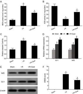
Results showed that MDA content significantly
increased (Figure 1A) and SOD enzyme activity was obviously decreased (Figure 1B) in brain tissues of I-R model rats. Caspase-3 activity in
the brain tissues of I-R model rats was signifi -cantly enhanced, compared with the Sham group, indicating an increase of apoptosis in brain tissues (Figure 1C).
Quer upregulated DJ-1 and Nrf2 expression and improved neurologic function in I-R rat brain tissues
Intraperitoneal injections of Quer significantly
reduced MDA content in brain tissues of I-R model rats (Figure 2A). Quer intraperitoneal injections obviously enhanced SOD activity in brain tissues of I-R model rats (Figure 2B). Caspase-3 enzyme activity in the Quer treat-ment group was markedly lower than that of the
Table 1. mNSS scores between the two groups
Group mNSS score
Sham 0
I-R 12.67±2.13*
I-R group (Figure 2C). Moreover, qRT-PCR re- vealed that DJ-1 and Nrf2 mRNA expression in brain tissues of the IR group was apparently higher than that of the Sham group. Compared with the I-R group, DJ-1 and Nrf2 mRNA levels in brain tissues of the Quer treatment group
[image:4.612.97.522.71.178.2]elevated D-1 and Nrf2 mRNA levels under IR treatment (Figure 3A, 3B). Western blotting demonstrated that, compared with the control group, D-1 and Nrf2 protein levels were obvi-ously upregulated in the IR treatment group. Quer intervention further enhanced D-1 and Figure 1. Brain OS upregulated and caspase-3 activity enhanced in I-R model rats. (A) MDA content in the rat brain tissue; (B) SOD enzyme activity in rat brain tissue; (C) Caspase-3 activity in rat brain tissue; *P < 0.05, compared with the Sham group.
Figure 2. Quer upregulated DJ-1 and Nrf2 expression and improved neuro-logic function in I-R rat brain tissues. (A) MDA content in the rat brain tissue; (B) SOD enzyme activity in rat brain tissue; (C) Caspase-3 activity in rat brain tissue; (D) qRT-PCR detection of mRNA expression; (E) Western blot detec-tion of protein expression; (F) mNSS scores of neuro funcdetec-tion. *P < 0.05, compared with the Sham group; #P < 0.05, compared with the I-R group.
were further upregulated (Fi- gure 2D). Western blotting de- monstrated that, compared wi- th the Sham group, DJ-1 and Nrf2 protein levels in brain tis-sues of the IR group were markedly increased. Compa- red with the I-R group, DJ-1 and Nrf2 protein expression in brain tissues of the Quer treat-ment group was further en- hanced (Figure 2E). Neuro- logical function assessment showed that, compared with the I-R group, neurological fun- ctions, such as hemiplegia, physical coordination dysfunc-tion, unstable standing, and unresponsiveness, were
sig-nificantly improved in the Quer
treatment group. Thus, mNSS scores were markedly reduced (Figure 2F).
Quer alleviated Rn-c cell apoptosis and ROS produc-tion induced by I-R by upregu-lating DJ-1 and Nrf2
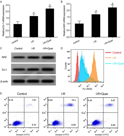
In this study, qRT-PCR showed that, compared with the con-trol group, D-1 and Nrf2 mRNA
expression was significantly
[image:4.612.91.373.238.556.2]Nrf2 protein levels under IR conditions (Figure 3C). Flow cytometry revealed that, compared with the control group, ROS content was mark-edly increased (Figure 3D), while apoptosis was apparently enhanced in the IR treatment group (Figure 3E). Rn-C-cell intracellular ROS
production and apoptosis were significantly
attenuated after treatment with Quer (Figure 3D, 3E).
Discussion
[image:5.612.92.519.74.542.2]activity. Obvious oxidative stress changes the structure of Keap. At this time, Nrf2 dissociates from Keap1, enters the nucleus, binds to ARE,
and upregulates HO-1, γ-GCS, SOD, glutathione
transferase (Glutathione), S-Transferase (GST), and nicotinamide adenine dinucleotide (phos-phoric acid) [NAD (P)H], achieving the goal of reducing OS levels and alleviating OS damage [17-20]. Activation of Nrf2 plays a role in reduc-ing OS response and alleviatreduc-ing brain I-R inju-ries [6, 7].
DJ-1/PARK7 (Parkinson gene 7) is located at chromosome 1p36.2-36.3, with a length of 24 kb. It encodes a protein with a molecular weight of 21 kD, consisting of 189 amino acids [21].
Numerous studies have confirmed that DJ-1
plays a role in enhancing Nrf2 stability and pro-moting Nrf2 protein expression and activation. Anti-OS effects of DJ-1 depend on regulation of Nrf2 [10, 11].
Quer is a natural plant-derived flavonoid with
the chemical name of
3,3’,4’,5,7-pentahydroxy-flavone and molecular formula of C15H10O7. Quer is widely found in a variety of fruits, vegetables, grains, and medicinal plants, including tea, apples, onions, grapes, quercetin, hyperoside, red dates, dodder, mulberry, and ginkgo leaves. In recent years, it was shown that Quer pro-vides various neuroprotective effects, such as delaying Alzheimer’s disease [12, 13], alleviat-ing cerebral ischemic injury [14], and improvalleviat-ing neurocognition and motor function [15]. How- ever, remains unclear whether Quer mitigates I-R damage by affecting DJ-1/Nrf2 pathways. The current study established a rat model of cerebral I-R injury and treated rats with Quer, investigating the role of Quer in regulating DJ-1/ Nrf2 pathway activity and alleviating rat brain IR injuries.
Compared with the Sham group, the I-R model group showed neurological impairment, such as limb coordination and movement disorder, unresponsiveness, and hemiplegia, indicating successful modeling. Compared with the Sham group, MDA content and caspase-3 activity
lev-els were significantly increased, while SOD
activity was obviously reduced in brain tissues of the IR model group. This indicated that neu-rological function impairment was related to OS elevation and increasing apoptosis. DJ-1 and
Nrf2 levels in the I-R model group were signifi -cantly higher than those in the Sham group.
Results indicated that the IR model initiated anti-OS emergency mechanisms by upregulat-ing DJ-1 and Nrf2. However, the actual anti-OS
ability in the body remained insufficient, lead -ing to high MDA contents and reduced SOD activities in the brain. Intraperitoneal injections
of Quer before I-R modeling significantly
re-duced MDA content and caspase-3 activity. They obviously elevated antioxidant enzyme SOD activity in rat brain tissues. Compared with the I-R group, DJ-1 and Nrf2 expression was apparently increased in brain tissues of the Quer treatment group. Intraperitoneal injec-tions of Quer improved cranial nerve function, alleviated I-R injuries after cerebral ischemia, and reduced mNSS scores by elevating DJ-1 and Nrf2 expression, thus enhancing anti-OS mechanisms and abilities. Pei et al. [22] report-ed that Quer can suppress the apoptosis of brain tissue induced by IR damage and OS by inhibiting ASK1/JNK3/caspase-3 pathways, im-
proving brain function. Yao et al. [23] revealed that Quer can reduce brain cell apoptosis, attenuate I-R injuries after brain tissue isch-emia, and improve rat brain function by activat-ing BDNF-TrkB-PI3K/Akt pathways. Ahmad et al. [24] found that Quer treatment can signifi -cantly reduce infarct size, improve neurological function, and decrease neuron loss and apop-tosis in rats after ischemia. The current study
found that Quer has a significant impact in the
reduction and treatment of I-R injuries after
brain ischemia, confirming the neuroprotective
effects of Quer. Present results are in accord
with those of Yao [23] and Ahmad [24].
Using in vitro studies, it was found that IR
treat-ment significantly induced Rn-c cell apoptosis
demonstrated that Quer can protect cells from high glucose-induced cell damage by activating Nrf2/ARE antioxidation, reducing ROS produc-tion, and alleviating apoptosis in ganglion cells. However, whether Quer exerts anti-oxidative damage and apoptosis through regulating Nrf2 by DJ-1 has not yet been reported. Present re- sults suggest that Quer plays a role in upregu-lating DJ-1 expression, activating DJ-1/Nrf2 pathways, enhancing neuronal antioxidant da- mage, and reducing neuronal apoptosis after I-R.
Conclusion
Quer attenuates brain cell apoptosis and neu-ronal damage after I-R by increasing DJ-1 lev-els, enhancing DJ-1/Nrf2 pathways, and elevat-ing anti-oxidation levels.
Disclosure of conflict of interest
None.
Address correspondence to: Dr. Guodong Dai, De- partment of Neurosurgery, Xianning Central Hos-
pital, The First Affiliated Hospital of Hubei University
of Science and Technology, No. 228, Jingui Road, Xian’an District, Xianning City 437000, Hubei Pro- vince, China. Tel: 0715-8896013; Fax: +86-0715-8896012; E-mail: [email protected] References
[1] Zhang X, Li Y, Liang Y, Sun P, Wu X, Song J, Sun
X, Hong M, Gao P, Deng D. Distinguishing intra-cerebral hemorrhage from acute intra-cerebral in-farction through metabolomics. Rev Invest Clin 2017; 69: 319-328.
[2] Deguchi I, Tanahashi N, Takao M. Clinical study of intravenous, low-dose recombinant tissue plasminogen activator for acute cerebral in-farction: comparison of treatment within 3 hours versus 3-4.5 hours. J Stroke Cerebrovasc Dis 2018; 27: 1033-1040.
[3] Nai Y, Liu H, Bi X, Gao H, Ren C. Protective ef -fect of astaxanthin on acute cerebral infarction in rats. Hum Exp Toxicol 2018; 37: 929-936. [4] Zuluaga M, Gueguen V, Letourneur D,
Pavon-Djavid G. Astaxanthin-antioxidant impact on excessive reactive oxygen species generation induced by ischemia and reperfusion injury. Chem Biol Interact 2018; 279: 145-158. [5] Yu W, Gao D, Jin W, Liu S, Qi S. Propofol pre
-vents oxidative stress by decreasing the isch-emic accumulation of succinate in focal cere-bral ischemia-reperfusion injury. Neurochem Res 2018; 43: 420-429.
[6] Lou J, Cao G, Li R, Liu J, Dong Z, Xu L. beta-caryophyllene attenuates focal cerebral isch-emia-reperfusion injury by Nrf2/HO-1 pathway in rats. Neurochem Res 2016; 41: 1291-304. [7] Takagi T, Kitashoji A, Iwawaki T, Tsuruma K,
Shimazawa M, Yoshimura S, Iwama T, Hara H.
Temporal activation of Nrf2 in the penumbra and Nrf2 activator-mediated neuroprotection in ischemia-reperfusion injury. Free Radic Biol Med 2014; 72: 124-33.
[8] Liu C, Chen Y, Kochevar IE, Jurkunas UV. De -creased DJ-1 leads to impaired Nrf2-regulated antioxidant defense and increased UV-A-in-duced apoptosis in corneal endothelial cells. Invest Ophthalmol Vis Sci 2014; 55: 5551-60. [9] Wu L, Xu H, Cao L, Li T, Li R, Feng Y, Chen J, Ma
J. Salidroside protects against MPP+-induced neuronal injury through DJ-1-Nrf2 antioxidant pathway. Evid Based Complement Alternat Med 2017; 2017: 5398542.
[10] Yan YF, Chen HP, Huang XS, Qiu LY, Liao ZP,
Huang QR. DJ-1 mediates the delayed cardio-protection of hypoxic preconditioning through activation of Nrf2 and subsequent upregula-tion of antioxidative enzymes. J Cardiovasc Pharmacol 2015; 66: 148-58.
[11] Sun Q, Shen ZY, Meng QT, Liu HZ, Duan WN, Xia ZY. The role of DJ-1/Nrf2 pathway in the
pathogenesis of diabetic nephropathy in rats. Ren Fail 2016; 38: 294-304.
[12] Nakagawa T, Itoh M, Ohta K, Hayashi Y, Hay
-akawa M, Yamada Y, Akanabe H, Chikaishi T, Nakagawa K, Itoh Y, Muro T, Yanagida D, Naka -bayashi R, Mori T, Saito K, Ohzawa K, Suzuki C, Li S, Ueda M, Wang MX, Nishida E, Islam S, Tana, Kobori M, Inuzuka T. Improvement of memory recall by quercetin in rodent contex-tual fear conditioning and human early-stage Alzheimer’s disease patients. Neuroreport 2016; 27: 671-6.
[13] Wang DM1, Li SQ, Wu WL, Zhu XY, Wang Y, Yuan HY. Effects of long-term treatment with
quercetin on cognition and mitochondrial func-tion in a mouse model of Alzheimer’s disease. Neurochem Res 2014; 39: 1533-43.
[14] Lei X, Chao H, Zhang Z, Lv J, Li S, Wei H, Xue R, Li F, Li Z. Neuroprotective effects of quercetin in a mouse model of brain ischemic/reperfu-sion injury via anti-apoptotic mechanisms based on the Akt pathway. Mol Med Rep 2015; 12: 3688-3696.
[15] Sabogal-Guáqueta AM, Muñoz-Manco JI, Ramírez-Pineda JR, Lamprea-Rodriguez M,
Osorio E, Cardona-Gómez GP. The flavonoid
[16] Yang EJ, Kim GS, Kim JA, Song KS. Protective
effects of onion-derived quercetin on gluta-mate-mediated hippocampal neuronal cell death. Pharmacogn Mag 2013; 9: 302-8. [17] Zhang JC, Yao W, Dong C, Han M, Shirayama Y,
Hashimoto K. Keap1-Nrf2 signaling pathway confers resilience versus susceptibility to ines-capable electric stress. Eur Arch Psychiatry Clin Neurosci 2018; 268: 865-870.
[18] David JA, Rifkin WJ, Rabbani PS, Ceradini DJ. The Nrf2/Keap1/ARE pathway and oxidative stress as a therapeutic target in type II diabe-tes mellitus. J Diabediabe-tes Res 2017; 2017: 4826724.
[19] Keleku-Lukwete N, Suzuki M, Yamamoto M. An
overview of the advantages of KEAP1-NRF2
system activation during inflammatory disease
treatment. Antioxid Redox Signal 2018; 29: 1746-1755.
[20] Zhu C, Dong Y, Liu H, Ren H, Cui Z. Hesperetin
protects against H2O2-triggered oxidative
dam-age via upregulation of the Keap1-Nrf2/HO-1 signal pathway in ARPE-19 cells. Biomed Pharmacother 2017; 88: 124-133.
[21] Kim RH, Peters M, Jang Y, Shi W, Pintilie M,
Fletcher GC, DeLuca C, Liepa J, Zhou L, Snow B, Binari RC, Manoukian AS, Bray MR, Liu FF, Tsao MS, Mak TW. DJ-1, a novel regulator of the tumor suppressor PTEN. Cancer Cell 2005; 7: 263-73.
[22] Pei B, Yang M, Qi X, Shen X, Chen X, Zhang F.
Quercetin ameliorates
ischemia/reperfusion-induced cognitive deficits by inhibiting ASK1/
JNK3/caspase-3 by enhancing the Akt signal-ing pathway. Biochem Biophys Res Commun 2016; 478: 199-205.
[23] Yao RQ, Qi DS, Yu HL, Liu J, Yang LH, Wu XX.
Quercetin attenuates cell apoptosis in focal ce-rebral ischemia rat brain via activation of BDNF-TrkB-PI3K/Akt signaling pathway. Neuro-chem Res 2012; 37: 2777-86.
[24] Ahmad A, Khan MM, Hoda MN, Raza SS, Khan MB, Javed H, Ishrat T, Ashafaq M, Ahmad ME, Safhi MM, Islam F. Quercetin protects against oxidative stress associated damages in a rat model of transient focal cerebral ischemia and reperfusion. Neurochem Res 2011; 36: 1360-71.
[25] Shi Y1, Liang XC, Zhang H, Wu QL, Qu L, Sun Q.
Quercetin protects rat dorsal root ganglion neurons against high glucose-induced injury in vitro through Nrf-2/HO-1 activation and NF-kappaB inhibition. Acta Pharmacol Sin 2013; 34: 1140-8.
[26] Shi Y, Liang XC, Zhang H, Sun Q, Wu QL, Qu L.