THE INFLUENCE OF PERIPHERAL CONNEXION ON THE
DIAMETER OF REGENERATING NERVE FIBRES
BY F. K. SANDERS* AND J. Z. YOUNG
Department of Zoology and Comparative Anatomy, Oxford
{Received 8 January 1946)
(With Plates 4-7 and Four Text-figures)
INTRODUCTION
Nerve fibres subserving different functions have different diameters, not only in a normal nerve but also in one which has been crushed and has under-gone degeneration and regeneration (Gutmann & Sanders, 1943). Therefore, there must be factors operating during the regeneration of any particular nerve which tend to restore the fibre size pattern typical of that nerve.
Simpson & Young (1945) have shown by cross union of visceral and somatic nerves containing fibres of different diameters that the central stump exerts an influence in determining the size of fibres regenerated. Thus after union of the central stump of a somatic nerve with the postganglionic trunk of the anterior mesenteric nerve, the latter comes to contain many medullated fibres, whereas normally this nerve corltains very few medullated fibres in the rabbit.
However, there is also evidence of a strong peri-pheral influence upon the size attained by regene-rating fibres. When a mixed nerve is severed and its ends united, there is such confusion of fibres at the line of suture that if central influences alone con-trolled the diameter of regenerated fibres, then all branches of the nerve below the suture would be expected to regenerate fibres of similar sizes. This in fact is not the case. Nageotte & Guyon (1918) and Sanders & Young (1944) have shown that below a suture in a mixed nerve the muscular branches come to contain larger fibres than the cutaneous. Mtef degeneration the cutaneous branches contain smaller Schwann tubes, and it has been suggested that it might be the restrictive influence of these which produced the difference (Gutmann & Sanders, 1943; Sanders & Young, 1944). Such a restrictive influence operates to some extent, but further experiments have shown that the most powerful influence lies not in the peripheral stump itself, but in connexion with the end-organ (Sanders & Young, 1945 ; Simpson & Young, 1945). Fibres which become connected with a muscle become larger than those which end • Leverhulme Research Scholar, Royal College of Surgeons of England.
blindly. Weiss & .Taylor (1944) found evidence of a similar effect in the course of experiments in which they completed the disproof of the theory of 'neurotropism'. More' recently, Weiss, Edds & Cavanaugh (1945) have extended this work and shown in various nerves of rats that a stretch of nerve regenerating without opportunity for its fibres to reach the periphery becomes filled with many and small nerve fibres.
The present work contains an investigation of the numbers and sizes of fibres found in nerves re-generating with and without normal peripheral connexions. Since fibres with no peripheral con-nexions are shorter than those regenerating from the same'level all the way to the periphery, a control investigation has been made of the effect of length of nerve on the diameter reached during regenera-tion. Finally, in order to discover whether the influence of peripheral connexion can override that of size of peripheral Schwann tubes, we have also made a study of regeneration through nerve grafts which originally contained fibres of different sizes. This serves also to answer a question of considerable practical importance to the surgeon who wishes to use pieces of cutaneous nerve for grafting into de-fects in motor nerve trunks (see Sanders & Young, 1944; Hammond & Hinsey, 1945).
METHODS
204 F. K. SANDERS AND J. Z. YOUNG The peroneal nerves of rabbits were crushed in
this way in the thigh, and then severed lower down, the ends being so treated as to provide different con-ditions for the reception of the regenerating fibres. The details of the various experiments are given below. All unions of the nerves were made by means of concentrated cockerel plasma, for the supply of which we are indebted to Mr P. B. Medawar.
For measurement of the regenerating fibres the nerves were fixed in Flemming's fluid,* sectioned transversely at 5^ after paraffin embedding, and stained with a modified Weigert method (Gutmann & Sanders, 1943). This did not always prove satis-factory, since the fixative in some cases failed to penetrate adequately to the centre of the nerve. The fibres were counted and measured on negative photographs made by direct projection on bromide paper at a magnification of 750 x . In some cases the whole nerve was photographed, while in others sample strips across the nerve were used. After photographing, a number of sample areas were selected for counting by marking out squares 50 x 50 mm. on the finished photograph, usually in a ring round the periphery of the nerve, where fixation, by ordinary histological criteria, was deemed to be adequate. The area of the whole nerve was also determined in each case by planimetry either of the photograph of the whole nerve or of a projection. The fibres in each sample square were then counted and marked off in size-groups with an interval of 2 p. The fibres of various sizes are not scattered quite at random throughout the nerve but sufficient squares were taken to provide an adequate total sample of the nerve. Except in special cases, mentioned elsewhere in the text, 25-30 squares were measured in the case of each nerve, an area equivalent to rather more than a quarter of the total area of the nerve. From the total number of fibres in the sample measured and the total nerve area obtained by planimetry an estimate of the total population could be obtained. The errors involved in such counts are discussed by Gutmann & Sanders (1943) and Simpson & Young (1945). In the present experi-ments nerves of the opposite sides of individual animals were compared, and since the technique was kept constant, errors of absolute estimation were unimportant.
Method of expressing results
In quantitative investigations of the present kind it is important to use a measure which shall express adequately the constitution of each individual nerve examined. The average fibre diameter is not very suitable for this purpose. Since the curve of distri-bution of fibre sizes.departs quite widely from the form of the 'normal curve', being in many cases • 1% chromic acid in distilled water, 15 ex., 2% osmic acid, 4 c.c, glacial acetic acid, 2 drops.
quite strongly bimodal, the average fibre ^ does not typify the diameter of the greatest n of fibres in the nerve. Moreover, there is such a numerical preponderance of small myelinated fibres in most nerves that nerves which differ strikingly in fibre constitution (e.g. muscular and cutaneous bundles) differ only slightly in average fibre dia-meter. Accordingly, we have in this paper used the root mean square of the whole population of dia-meters as our estimate of 'mean' fibre diameter. This figure (D) was calculated separately for each square of the sample counted, and the value of D given for each nerve was the mean of all the D'a found for separate squares within that nerve. This method of calculating D has the advantage that an estimate of error can be made in each case, and the values of D obtained for different nerves compared statistically. This estimate of the 'mean' diameter has an additional advantage in that it is directly related to. the total volume of nerve fibres present in the nerve. If the nerve were wholly composed of fibres of diameter D it would have a volume of fibres per unit length equal to that actually found in the nerve, but there spread over fibres of various diameters.
RESULTS
Effect of length on diameter of regenerated fibres
The object of the experiments described in this section was to discover whether the diameter reached by the fibres at any level in a regenerating nerve is affected by the length of fibre regenerating peri-pheral to diat level. Extension of a nerve beyond its normal length can be achieved either by the use of a graft or by joining it with another nerve. Both methods have considerable disadvantages. The dif-ficulty of obtaining suitable lengths of nerve for autografting and the inevitable variability in the resulting junctions suggested that the method of cross union might be more suitable. The peroneal nerves were therefore crushed as high as possible on both sides of each animal, cut lower down, and joined to the tibial nerves in such a way as to make on one side a nerve of normal length, and on the other one in which fibres have to grow much farther than is usual.
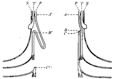
This was done by freeing the peroneal nerve along its whole length, cutting it peripherally, turning it back along itself and joining it to the tibial high in the thigh. The fibres regenerating from the crushed points thus have to (pass through a much shorter length of peroneal nerve on the one side before entering the tibial and passing to the end-organs of the latter.
J
\ endings such that stimulation of the nervees contraction of these muscles, but it is un-certain to what extent such abnormal connexions function during life. In any case, the final termina-tions are equally abnormal on both sides of the animal, and the only difference is therefore in the length of nerve to be regenerated.
Two experiments of this kind were made, rabbit 1011 being killed 107 days, and rabbit 1010, 200 days after operation.*
s r P P T S
A
Text-fig. 1. Diagrams of operations performed in order
to increase the length of fibres regenerated in the peroneal nerve. Peroneal nerve, P, is crushed high in the thigh
{A, A') and lower down severed and joined to the tibial
nerve, T, at B or B'; C or C, peripheral stumps of peroneaJ nerve; S, sural nerve.
regenerating axons on both sides cross the region of crush 5 days after the operation, <and advance through the peroneal nerve at 4-4 mm./day. Conditions in the two nerves are the same, except that on the one side the fibres have to travel an extra 55 mm. We may, therefore, expect that the fibres will arrive in the muscles supplied by the long nerve 55/4-4= 12-5 days later than on the short side. Out of a total regeneration time of 107 days, the fibres in the short nerve will have had 12-5 days longer for maturation after the arrival of fibres.
In order to discover what difference this increased length made to the process of maturation, sections were made of the peroneal nerves 10 mm. below the crushed point. Table 1 shows that the estimated total number of fibres is similar in the two nerves.
D, the ' mean' fibre diameter (see p. 204), is slightly
greater in the longer nerve, but since differences of this magnitude can be expected to occur in 60-70% of cases as a result of the operation of chance factors, the difference cannot be termed significant. There-fore, neither the fact that a greater length of nerve was regenerated, nor that contact with the muscle was made later, had operated to affect the diameter of the fibres immediately below the crushed point
(PI. 4. figs.. 1, 2).
[image:3.595.89.287.246.384.2]Sections were also made farther down both nerves, and showed plentiful medullated fibres below the suture points on both sides (PI. 4, figs. 3, 4; ionf,k). No large difference was evident in the fibre diameters but counts were not made. It seems that at this level
Table 1. The effect of length of nerve on fibre maturation
Animal ion g 1011 b IOII I ion d IOIO C 1010 m Days 107 107 107 107 200 20O Level Central stump Central stump Peripheral stump Peripheral stump Peripheral stump Peripheral stump Length of nerve below crush Long Short Long Short Long Short No. of fibres 9817 9412 6577 5539 7326 7066
D in fi
7-4 81 61 60 69 63 Comparison Degrees of freedom 63 41 34 t 1-691 0869 3-5I9 P 0-5-0-10 0-3-0-4 O-OOI-O-OI
107 days of regeneration. In i o n the distance on the side with a short nerve (AB; see Text-fig. 1) between crushed point and the union with the tibial nerve was 20 mm., whereas A'B', the distance from the crushed point to the union on the opposite sMe of the animal, was 75 mm. From the results of Gutmann, Guttmann, Medawar & Young (1942) we may assume that the tips of • the
• Every piece of nerve removed from an animal is indicated by a small letter, thus ion 6 means piece b of rabbit ion ; see tables.
206 F. £. SANDERS AND J. Z. YOUNG We were also interested to discover whether the
length of new nerve fibres to be formed affected the diameter of fibres central to the lesion. Table i shows that D was in fact smaller in the nerve which had the longer distance to regenerate, the difference between the two sides being of a magnitude that would only be found in 5-10% of cases as a result of random sampling alone. 100 days after injury is the time at which the fibres of the central stump show their greatest decrease in volume (Gutmann & Sanders, 1943), and if this decrease is in some way connected with the outpouring of material to make the new fibres, it might be expected to be greater in the nerve which has the greater length to regenerate. Sections of the points of union of the peroneal and tibial nerves showed good junctions on both sides (PI. 4, figs. 7, 8; i o n e,f) without any waist-like constriction. Although, unfortunately, there is no means of making a quantitative assessment of such junctions, we feel safe in concluding that these are similar on the two sides, and therefore that the absence of any difference in fibre diameter is significant.
200 days of regeneration. In rabbit 1010 the nerves were arranged so far as possible exactly as in 1011, and left to regenerate for 200 days. Using the same assumptions as before, we may conclude that the new fibres arrived at the gastrocnemius muscles 10 days later on the 'long' side, and therefore had that much longer for maturation in the short nerve.
Table 1 shows that 10 mm. below the point of crushing there were again approximately the same number of fibres on both sides. At this level, how-ever, tjiere were rather larger fibres on the side with the longer nerve. Moreo^Jer, this difference is greater than any to be expected as a result of random samp-ling. It will be noticed that the fibres are larger than after 100 days, though not greatly so (PI. 5, figs. 9,10).
Below the points of suture of the peroneal to the tibial nerves no actual counts were made, but the fibres appear smaller on the side with the longer nerve. On both sides there is a distinct difference between levels above and below the suture in that, whereas at the Former there is usually only one large fibre per tube, at the latter there are several smaller ones (PI. 5, figs. 9-12; 1010 c,m,g,q). This is a distinction regularly observed between nerves regenerated below crushed and sutured points. In this case the only injury of the axons was by crushing, and it is therefore demonstrated that the larger number of small fibres is due to interruption of the whole nerve and its tubes.
In the posterior tibial nerves at the heel the fibres were distinctly larger on the side with the shorter nerve (PI. 5, figs. 13, 14; IOIOJ, s). On both
sides, however, they were larger than after 107 days. Sections of the crushed points showed similar conditions on the two sides (PI. 5, figs. 15, 16; 1010 a, k). The unions between the peroneal and tibial nerves had been rather less successful than in
rabbit ion.(PI. 6, figs. 17, 18; 1010/, n). ticular, that on the s,ide with the lengthened showed a considerable lateral deviation of fibres. Though the stumps were closely'apposed, conditions had evidently not been favourable for the formation of a clot with good longitudinal orientation. The most probable explanation for this is that the union was made without longitudinal tension, and the case is indeed a warning of the bad results which may follow from a union made under apparently ideal surgical conditions (see Weiss, 1943). The difference between the two suture lines may explain the some-what smaller fibres found immediately below the crush in the shorter nerve.
Conclusions from the results of regeneration through artificially lengthened nerves. We may conclude from
these two experiments that the length of nerve to be regenerated below an injured point does not greatly affect the rate of increase of diameter of the fibres in the section of nerve immediately below the injury. Maturation does not proceed uniformly along the whole length of a regenerating fibre, but spreads centrifugally, so that at any given time more distant points show a smaller diameter (see also Gutmann & Sanders, 1943).
Effect of absence of peripheral connexion on fibre diameter
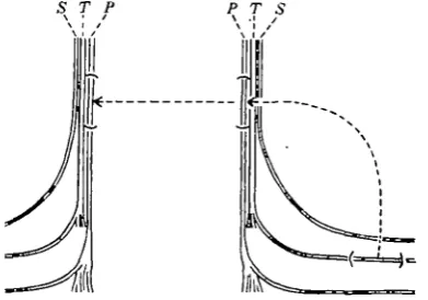
Two rabbits (1012 and 1013) were operated on in such a way as to reveal the effect of presence or absence of a connexion with the periphery on re-generation in the sensory and motor bundles of the peroneal nerves. On each side the nerve was crushed in the upper third of the thigh, above the point at which it divides into separate bundles destined to supply muscles and skin respectively. The former bundles of course contain somatic motor as well as sensory fibres, whereas the cutaneous branches (anterior .tibial nerve) contain sensory and sympa-thetic fibres only, the latter being almost entirely non-medullated. The nerve was also severed below the knee, where it passes under the lateral head of the gastrocnemius. On one side the cut lower ends were immediately reunited with plasma, whereas on the other side the severed end of the peroneal was turned aside and fixed in such a manner as to allow no possibility of reunion.
on the side with a peripheral union, and 5 that with none, but the fibres were distinctly smaller in the latter. This is in itself
i very interesting fact, suggesting that the recovery
in diameter of central fibres following their decrease luring regeneration is influenced in some way by Functional connexion with the periphery.
S T P
A' A
-B
Text-fig. 2. Diagram of experiment to show effect of presence and absence of peripheral connexion on re-generation. The peroneal nerve is crushed in the thigh at A or A'. It is cut at the knee and on one side the central end is turned forward so that no union is possible. On the other side the stumps are joined at the lower level B.
as in the central stump. This confirms the finding of Simpson & Young (1945) that when regenerating fibres fail to make contact with the periphery, far more of them are found in the peripheral than in the central stump. Weiss et al. (1945) have recently shown a similar effect in the nerves of rats. Further, the diameter of the regenerated fibres remains very small in this nerve, and D is not greater in the
motor than in the sensory bundles (PI. 6, figs. 2 1 ,
32). This last is a result of the utmost importance, since it strongly suggests that the factor which makes somatic motor fibres large is a peripheral, or ' func-tional' one. For since the upper lesion is a simple crush of the nerve, it is to be presumed that the large central fibres proceeded down large peripheral tubes and yet failed to reach a large size. Examination of the sections indeed shows the large tubes of the motor branches most clearly, each filled with numerous small fibres (PI. 6, figs. 21, 22; 1012 c, sensory and motor). The sensory and motor branches can still be distinguished by the large tubes of the latter, in spite of the fact that the fibre diameters are similar in both. Histograms reinforce the pictures, showing that not only is the fibre diameter similar in the two'nerves, but further that the large motor fibres have not given rise to any large fibres between the point of crushing and the neuroma, the maximum diameter being 12/1 in the motor and io'/x in the Table 2. The effect of peripheral connexion upon fibre maturation
Animal 1012 e 1012 a IOI2£ IOI2 C IOI2£ 1012 c 1013 a 1013/ 1013 c 1013 »' 1013 c 1013 1 Days 172 172 172 172 172 172 2OO 20O 2OO 20O 2OO 20O Type of nerve Mixed Mixed Motor Motor Sensory Sensory Mixed Mixed Motor Motor Sensory Sensory Periphery Connected Unconnected Connected Unconnected Connected Unconnected • Connected Unconnected Connected Unconnected Connected Unconnected Level Central Central Peripheral Peripheral Peripheral Peripheral Central Central Peripheral Peripheral Peripheral Peripheral No. of fibres 4986 5O97 2898 7757 2463 3456 5475 5H8 1883 6257 288s 2588
Din n
84 7-4 72 4-6 5-2 4-8 8-5 6 9 8-2 4-9 5-5 4 3
Total vol. of fibres cu.mm. 2764
2 1 9 2
7-67 5-80 IO2O 2-81 31-07 19-25 6-46 S-3I 13-37 1-69 Below the crushed point in the nerve connected
with t h e periphery, there were 5361 fibres in motor and sensory bundles together, that is to say, only a few more than in the central stump. T h e diameter was distinctly larger in the motor than in the sensory branches (PI. 6, figs. 19, 20). T h e regenerated stretch of nerve which had not been allowed to make connexion with the periphery contained a total of 11,213 medullated fibres, more than twice as many
sensory bundles. However, the motor bundles contain a larger number of fibres 6—10 ft in diameter than the sensory bundles, giving some indication of the operation of factors other than peripheral connexion.
208 F. K. SANDERS AND J. Z. YOUNG in the central stump (5475). In the nerve with no
peripheral connexion the number of regenerated fibres was again much greater (8845) than that in the central stump (5148). The central stump fibres were, as before, smaller in the nerve with no periphery. Their reduction in diameter without, apparently,
increase in diameter also in the sensory t^^lles which had made a peripheral connexion, ^ r the motor bundles with no peripheral connexion D was at most only slightly greater (4-9^1) than in the sensory bundles (4-3 /J.). The results from this ex-periment, therefore, fully confirm that of the
300
200
100
0
0 t
i
_
1
Motor nerve normal
1
•
k
.
(
1
)
1
j
12 18 Motor
with periphery
I"
() 6
1
1
•
12 18
1
Motor without periphery
1
3o
V
a
XI
o 400
300 200 100
0
1
1
<
Sensory normal
1
1 -
1
_
Sensor)1with periphery I
L
1
J
Sensory without penpher r12 18 0 12 18 0 12 18
Diameter in microns
Text-fig. 3. Histograms to show the number of fibres of different sizes in normal motor and sensory branches of the peroneal nerve, and in these same branches after 200 days of regeneration below a crushed point, with and without connexion with the periphery.
reduction in thickness of their myelin sheaths, leads to the production of the remarkable fibres seen in PI. 7. %. 24 (1013/) [cf. fig. 23 (1013 a)]. The fibres of the motor bundles which had been allowed to make functional connexions showed a typical increase in diameter, and there had been some
previous one in showing that all regenerated fibres remain small unless connected with the periphery, such connexion causing some increase of diameter in sensory and still more in motor fibres.
experi-if Simpson & Young (1945) it was shown that >f splanchnic nerve grafted to post-intercostal lerves came to contain quite large fibres. The present eries included two animals in which a test of the ame sort was made by grafting into the peroneal lerve in the thigh. On one side of each animal, the ;raft was a piece of the peroneal from the opposite ide. The defect made by taking this graft was then died by a piece of posterior tibial nerve from the ame animal.
S T P T S
Text-fig. 4. Grafting experiment to show the effect of izes of tubes on the diameter reached by regenerating lerve fibres. On one side a portion of the posterior tibial lerve (small fibres) is grafted to the peroneal nerve in the high, the piece removed from this nerve (large fibres) is hen grafted to the peroneal on the opposite side.
view of the uncertainties introduced, by the poor union no counts were made. Below the grafts both peroneal nerves contained many meduljated fibres, that with the peroneal nerve graft containing more than that with the posterior tibial graft. This case, therefore, shows that in spite of a poor union the many small-fibred bundles of the posterior tibial, when used as a graft, can allow a connexion to be made.
The second animal (1037) was allowed to survive for 200 days after an exactly similar operation. Here the union of the peroneal and posterior tibial graft had been well made (PI. 7, fig. 25; 1037 b), though the presence of many bundles had led to some complications. However, it may reasonably be assumed that the opportunities for connexion were similar to. those presented by the union of the peroneal with the peroneal graft (PI. 7, fig. 26; 1037 h) on the opposite side. The lower unions of both grafts with the peroneal nerves were also well made.
Table 3 shows that the fibres were somewhat larger in the graft of the peroneal than in that of the posterior tibial (PI. 7, figs. 27, 28). In the peroneal the difficulties of non-uniformity of various parts of the graft, owing to poor fixation in the centre, make the difference in fibre diameters observed of doubtful significance. Certainly the small tubes of the posterior tibial graft have not grossly restricted the growth of the fibres, but the result suggests that they have done so to some extent.
Animal
[image:7.595.91.287.250.387.2]1037 i 1037 c
Table 3
Days
200 200
. The effect of tube tize on fibre maturation
Source of graft
Peroneal Posterior tibial
No. of fibres
7300
9377
D in n
59 5'5
Probability
o-i
The posterior tibial contains smaller medullated ibres than the peroneal (see Sanders & Young, 944), and the experiment therefore tests the extent o which the large fibres present in the peroneal »erve in the thigh are able to dilate the small tubes >f the posterior tibial when they have passed through he latter and made connexion with the peroneal nuscles. The grafts were z cm. in length and were ixed in place with clotted cockerel plasma.
The first animal (1015) was allowed to survive for >8 days, but the experiment was reduced in value jy the fact that the upper union of peroneal with xmterior tibial was poorly made, the two stumps >eing separated by about 0-5 mm. of irregular con-lective tissue. Very many nerve fibres had never-heless penetrated this, and the grafted bundles of he posterior tibial contained many medullated ibres. Those in the corresponding piece of grafted >eroneal nerve on the opposite side were more lumerous, but perhaps not larger. However, in
No strict comparison is. possible between the diameters here recorded and those shown in Tables 1 and 2, which are of fibres regenerating below a point of crushing and not one of suture as in the present case. However, it is interesting that the diameter reached by the fibres is distinctly above that found in those nerves which made no connexion with the periphery. Evidently, after passing through the grafts, the fibres have been able to make connexion with their end-organs and to undergo a subsequent increase in diameter. Below the grafts fibres of large diameter were found on both sides (PI. 7, figs. 29, 30).
DISCUSSION
2 1 0 F. K. SANDERS AND J. Z. YOUNG contain far more and smaller fibres than others
com-parable in every way except that the fibres can make connexion with muscles and skin. A very important new result which emerges from these counts is that regenerating motor fibres which do not reach their end-organs become very little larger than sensory fibres. This shows the power of the peripheral effect very strikingly, and suggests that it is of paramount importance. It seems not unlikely that during normal development all fibres are of like diameter, and that the spectrum of fibre sizes develops as a result of the peripheral connexions, perhaps by the very act of functioning. Indeed, it may well be that the influence extends back from the fibres to the cell-bodies themselves, and that it is the peripheral connexion which determines the size of cells in the spinal cord (see Young, 1945). Such an effect of the periphery upon the centres recalls the theories of Weiss. He has always maintained that such an influence exists, though not quite ip the sense now revealed.
The figures of Table 2 show for the first time that sensory as well as motor fibres are affected by their connexions with the periphery. The anterior tibial nerve of the rabbit is a pure skin nerve (see Sanders & Young, 1944), its only motor fibres being sympa-thetic, presumably non-medullated and therefore not considered here. Yet during regeneration its fibres become larger if they are allowed to reach the periphery. It should perhaps be noted, however, that since the suture at S of Text-fig. 2 is of the whole peroneal nerve, many of these sensory fibres will enter the peroneal muscles. However, Gutmann (1945) has confirmed the previous evidence that sensory fibres cannot form motor end plates in muscle. Further evidence about the factors which control the maturation of sensory fibres is needed: these experiments show that the peripheral con-nexions exercise a large influence.
The present investigations still leave it quite uncertain how connexion with the periphery comes to exert its effect on maturation of the fibres. The experiments in which the nerves were artificially increased in length show that the diameter reached by fibres immediately below a crushed point is not greatly affected by the length of nerve to be re-generated more distally.
Hand in hand with the increase in size of the fibres which have become connected with the periphery goes the reduction and disappearance of those re-maining without connexion, but it is impossible at present to say whether the one process controls the other. The growth of one fibre in a tube may have the effect of constricting the others and hence leading to their degeneration. Until such degenera-tion has been actually observed it is impossible to say more. It may be that the material used to make the large fibres is in some way derived from the others arising from the same' parent fibre, thus producing their degeneration. Certainly the whole . increase
produced by peripheral connexion is not oj^ by draining material from other fibres. For tn^ volume occupied by the fibres in the peripheral stump of an unconnected nerve is much less thari that of the fewer, but larger, fibres which develop when a peripheral connexion is allowed. Weiss et al, (1945) also found this in the nerves of rats. The last column of Table 2 contains estimates of the volumes occupied by fibres in the central and peripheral stumps of the nerves. Assuming that the distance from the point of crushing to the neuroma was 45 mm., to the peroneal muscles 65 mm., and to the skin 195 mm., the total volume of the fibres in the peripheral Stump is seen to be very much greater ir the nerves connected with the periphery. The tota increase of some of the fibres cannot therefore have been only at the expense of others. Moreover, it must be remembered that the fibres central to the crush alsc remain small in an unconnected nerve. Assuming that the diameters found immediately above the lesion are maintained to the cord, the volume in the central stumps would be 28 and 31 cu.mm. in the nerves connected with the end-organ, but only 22 and 19 cu.mm. in those ending blindly as neuromas assuming that all crushes are made at a distance oi 100 mm. from the cord. Weiss et al. (1945) have already noted that the fibres remain small in the central stumps of a regenerating nerve without peripheral connexion. As they observe, the small size of such fibres cannot be due entirely to drainage of axoplasm from the central fibres. The volume oi the many small fibres in an unconnected regenerating nerve is smaller than in a similar nerve having con-nexion with the periphery (see Table 2), whereas the central stump of the latter contains the larger fibres Hammond & Hinsey (1945) believed that the smal decrease which they observed in the central stumps of sutured nerves might be due to the presence oi many small fibres turned back at the junction A similar explanation cannot apply in the present case, since the number of fibres in the central stump of the unconnected nerves was not significantlj greater than in those with a peripheral connexion However, it seems likely that some part of the large number of fibres seen above the neuromas was due to 'doubling back' towards the centre as suggested by Weiss et al. (1945).
2 1 1
SUMMARY
1. Stretches of regenerating rabbit's nerve in which no connexion with the periphery is allowed contain numerous and small nerve fibres.
2. Both cutaneous sensory and motor nerve fibres become large when allowed to reach their end-organs, but the motor fibres attain greater diameters than the cutaneous sensory fibres.
3. Motor fibres regenerating without oppor-tunity of reaching muscle fibres become little if any larger than sensory fibres when both are unable to reach their end-organs. It is, therefore, probable that the peripheral connexion is of great importance in determining fibre diameter.
4. The fibres of the central stump of a nerve regenerating unconnected with the periphery remain small and have relatively very thick myelin sheaths.
5. The diameter of nerve fibres regenerated in the region below a crushed point on a nerve is not in-fluenced by the length of nerve to be regenerated distally.
6. Increase of diameter and medullation proceeds centrifugally from a lesion, more distant points showing a lesser degree of maturation at any given time.
7. Fibres regenerated in a grafted piece of posterior tibial nerve were only slightly smaller than those in a grafted piece of peroneal nerve. Con-nexion with the periphery is, therefore, able to produce an increase in fibre diameter which over-rides any constriction of fibre growth produced by the slightly smaller Schwann tubes.
Our thanks are due to the Rockefeller Foundation for their support which made this work possible.
REFERENCES
GUTMANN, E. (1945). J. Anat., Lond., 79, 1.
GUTMANN, E. & SANDERS, F. K. (1943J. J. Physiol. 101,
489.
GUTMANN, E., GUTTMANN, L., MEDAWAR, P. B. & YOUNC, J. Z. (1942). J. Exp. Biol. 19, 14.
HAMMOND, W. S. & HINSEY, J. C. (1945). J. Comp.
Neurol. 83, 79.
NAGEOTTE, J. & GUYON, L. (1918). C.R. Soc. Biol.,
Paris, 81, 571.
SANDERS, F. K. & YOUNG, J. Z. (1944). J. Physiol. 103,
119.
SANDERS, F. K. & YOUNG, J. Z. (1945). Nature, Lond.,
155,
237-SIMPSON, S. A. & YOUNG, J. Z. (1945). J. /Lnat., Lond., 79. 48.
WEISS, P. (1943). Arch. surg. 46, 525.
WEISS, P. & TAYLOR, A. C. (1944). J. Exp.'Zool. 95, 233.
WEISS, P., EDDS, MACV. Jr. & CAVANAUCH, M. (1945). J. Comp. Neurol. 9a, 215.
YOUNG, J. Z. (1945). In Essays on Growth and Form. Oxford.
DESCRIPTION OF PLATES
The photographs of Weigert preparations of transverse sections are direct bromide prints at a magnification of 430 diameters.
The photographs of the junctions are of preparations stained by Bodian's protargol method at a magnification of 21 diameters.
PLATE 4
Fig. 1. Fibres below crushed point above lengthened peroneal nerve. 107 days ( i o n i).
Fig. 2. Fibres below crushed point on the opposite side where the nerve is not lengthened ( i o n d).
Fig. 3. Lower down in lengthened peroneal nerve
(1011 k). '
Fig. 4. Lower down in peroneal nerve, not lengthened
(1011/).
Fig. 5. Posterior tibial nerve supplied with fibres from peroneal which have run a long course ( i o n I). Fig. 6. Posterior tibial nerve on opposite side supplied with peroneal fibres without lengthening ( i o n m). Fig. 7. Junction of peroneal with tibial nerve on lengthened side. Peroneal to left (10117).
Fig. 8. Junction of peroneal with tibial nerve without lengthening.-. Peroneal to left ( i o n e).
PLATE 5
Fig. 9. Fibres immediately below crushed point of lengthened peroneal nerve. 200 days (1010 c).
Fig. 10. Fibres of opposite crushed peroneal nerves, not lengthened (1010 m).
Fig. 11. Lower down in lengthened peroneal nerve (ioiog).
Fig. 12. Fibres lower down in peroneal nerve, not lengthened (1010 q).
Fig. 13. Posterior tibial nerve supplied with fibres for 200 days from lengthened peroneal nerve (IOIOJ). Fig. 14. Fibres of posterior tibial nerve supplied from peroneal nerve without lengthening (1010 s).
Fig. 15. Crushed point, of lengthened peroneal nerve
(1010 a).
Fig. 16. Crushed point of peroneal nerve not lengthened
2 1 2 F. K. SANDERS AND J. Z. YOUNG
PLATE 6
Fig. 17. Union of peroneal with tibial nerve to give increased length (1010/). Peroneal to right.
Fig. 18. Union of peroneal with tibial nerve without increase of length (1010 «). Peroneal to right.
Fig. 19. Fibres in motor branch of peroneal nerve below crush which have been allowed to reach periphery.
172 days (1012 g).
Fig. 20. Fibres of sensory branch of peroneal below crush which have been allowed to reach the periphery
( # )
Fig. 2i. Fibres of motor branch of peroneal nerve below crush which have not been allowed to reach periphery
(1012 c).
Fig. 22. Fibres of sensory branch of peroneal nerve below crush which have not been allowed to reach periphery (1012 c).
PLATE 7
Fig. 23. Fibres of central stump of peroneal nerve which have been crushed and allowed to reach periphery. 200 days (1013 a).
Fig. 24. Central stump on opposite side above crush without union with periphery (1013/).
Fig. 25. Point of union of peroneal nerves with grafted posterior tibial nerve. 200 days (1037 b). The graft lies to the right.
Fig. 26. Point of union of peroneal nerve with grafted piece of peroneal (1037 h). The graft is to the right. Fig. 27. Fibres in piece of posterior tibial grafted into the peroneal (1037 c).
Fig. 28. Fibres in peroneal graft to peroneal (1037 j). Fig. 29. Fibres in peroneal nerve below posterior tibial graft (1037 e).
PLATE 5 JOURNAL OF EXPERIMENTAL BIOLOGY, 22, 3, 4
15
PLATE 7 JOURNAL OF EXPERIMENTAL BIOLOGY, 22, 3, 4