Original Article
Efficacy analysis of bronchoscopic triple-drug
delivery combined with chemotherapy for re-treatment
of smear-positive cavitary pulmonary tuberculosis
Changjun Liu, Ming Gao, Dongmei Fan
Department of Infectious Disease, Lanling County People’s Hospital, Linyi 277799, Shandong Province, China
Received May 6, 2019; Accepted July 11, 2019; Epub September 15, 2019; Published September 30, 2019
Abstract: Objective: The aim of the current study was to analyze the clinical effects of bronchoscopic triple-drug de-livery combined with chemotherapy for re-treatment of smear-positive cavitary pulmonary tuberculosis. Methods: A total of 144 patients, experiencing failed initial treatments for cavitary pulmonary tuberculosis, were enrolled in the study. All patients were smear-positive before receiving re-treatment. The patients were divided into the observation group and control group, according to a random number table, with 72 patients in each group. The control group received the routine chemotherapy regimen for 8 months. The observation group received bronchoscopic triple-drug delivery in combination with the routine chemotherapy regimen. Sputum conversion rates, cavity closure rates, and symptom improvement levels were compared between the groups. Immune function and pulmonary function were also compared between the two groups. Results: Sputum conversion rates and cavity closure rates in the observa-tion group were 94.44% and 84.72%, respectively, significantly higher than those of the control group (P < 0.05). After the consolidation phase, the observation group had lower scores concerning tuberculosis symptoms, including lower fevers, coughing and sputum production, chest pain, and loss of appetite (P < 0.05). CD3+, CD4+ T-cells, and
CD4+/CD8+ ratios in the observation group were significantly higher at the end of 8 months. However, CD8+ T-cells
were lower than those of the control group (all P < 0.05). With the conclusion of treatment, pulmonary function in the observation group was better than that in the control group (all P < 0.05). Conclusion: Bronchoscopic triple-drug delivery combined with chemotherapy for re-treatment of smear-positive cavitary pulmonary tuberculosis is more effective than chemotherapy alone. Therefore, this method is worthy of promotion in clinical practice.
Keywords: Bronchoscopy, chemotherapy, re-treatment of smear-positive cavitary pulmonary tuberculosis, pulmo-nary function, immune function
Introduction
Tuberculosis is one of the most serious public health problems, worldwide. Current data indi-cates that about 1.8 million people die each year from tuberculosis [1]. At present, China is experiencing a serious tuberculosis epidemic. According to statistics, incidence and mortality rates of tuberculosis in China rank in the top 3 of Class A and Class B infectious diseases, making it a serious potential threat to the phy- sical and mental health of the people [2]. Pul- monary tuberculosis is a lung disease caused by mycobacterium tuberculosis (MTB) infec-tions. It is clinically characterized by coughing, sputum production, dyspnea, hemoptysis, low fever, loss of appetite, and chest pain. With the wide application of chemotherapy drugs in Chi- na, more than 95% of patients can be cured
with timely diagnosis and standardized treat-ment [3, 4]. However, in recent years, due to irrational and irregular drug use, the number of patients requiring re-treatment for tuberculosis has increased significantly. This is especially true for patients with smear-positive cavitary pulmonary tuberculosis. As the disease pro-gresses and recurs, the cavity wall may appear thickened and fibrotic. Thus, it is difficult for orally-delivered chemotherapy drugs to pene-trate the thickened cavity wall, reaching an inhibitory concentration. This factor is respon-sible for unsatisfactory therapeutic efficacy ra- tes.
delivery. This method can directly deliver che-motherapy drugs into lesions, improving local concentrations. This helps to avoid inefficien-cies of oral deliveries, producing full therap- eutic effects of the anti-tuberculosis drugs. In addition, bronchoscopy procedures can elimi-nate bronchial secretions and caseous necro-sis, clearing the bronchial lumen. This is con- ducive to the formation of fresh granulation tis-sue and absorption of lesions [5, 6]. However, there are few reports concerning the method of bronchoscopic drug delivery for manage-ment of tuberculosis. The current study con-ducted a randomized controlled trial, analyzing the efficacy of bronchoscopic triple-drug deliv-ery. The aim of the current study was to find a better alternative for re-treatment of smear-positive cavitary pulmonary tuberculosis. Materials and methods
Patients
A total of 144 patients, experiencing failed ini-tial treatments for cavitary pulmonary tuber- culosis, admitted to Lanling County People’s Hospital. From June 2017 to July 2018, were enrolled in the study. All patients were smear-positive before re-treatment. The patients were divided into the observation group and control group, according to a random number table, with 72 patients in each group. The control gr- oup received routine chemotherapy for 8 mon- ths. The observation group received broncho-scopic triple-drug delivery in combination with routine chemotherapy. All patients provided informed consent. The study was approved by the Ethics Committee of Lanling County Peo- ple’s Hospital.
Inclusion criteria: Patients met the diagnostic criteria for cavitary tuberculosis according to the Guidelines for Diagnosis and Treatment of Tuberculosis developed by the Chinese Tu- berculosis Society of the China Medical As- sociation in 2013 [7]; Chest X-rays showed lesions confined in one lobe and there was on- ly one tuberculosis cavity; Sputum smear was positive; Aged between 18 and 65 years old. Exclusion criteria: Patients with multiple tuber-culosis cavities, autoimmune diseases, diabe-tes, extrapulmonary tuberculosis, or lung can-cer; Patients with respiratory failure or hemo- ptysis; Patients with major organ dysfunction; Under immunosuppressive treatment or diag-nosed with HIV infection; History of cognitive or
mental disorders; Allergic to any of the drugs used in this study.
Methods
The control group received the conventional standard chemotherapy regimen 2HRZES/6- HRE: H: Isoniazid (Guangdong Huanan Ph- armaceutical Group Co., Ltd., China) 0.30 g/ day orally once a day; R: Rifampicin (Hainan Pharmaceutical Factory Co., Ltd., China) 0.60 g/day orally once a day; Z: Pyrazinamide (Shenyang Hongqi Pharmaceutical Co., Ltd., China) 1.50 g/day orally once a day; E: Eth- ambutol (Guangzhou Baiyunshan Mingxing Ph- armaceutical Co., Ltd., China) 0.75 g/day orally once a day; S: Streptomycin (Huabei Phar- maceutical Co., Ltd., China) 0.75 g/day by intra-muscular injections, once a day. After 2 months of the intensive phase, pyrazinamide and stre- ptomycin were discontinued. The other drugs were continued for another 6 months, including the consolidation phase. The observation group was treated with bronchoscopic-delivered tri-ple-drugs, based on the above regimen. Pa- tients in the observation group underwent el- ectronic bronchoscopy procedures, aiming to determine the distribution of lesions in the bronchus. Purulent secretions, caseous necro-sis, and granulation tissue were then remov- ed, clearing the bronchial lumen. A catheter was inserted 8 mm distal to the tip of the br- onchoscope, delivering the triple anti-tubercu-losis drug mixture. This included 5 mL 0.90% sodium chloride solution, 0.10 g isoniazid (Sh- anghai Xinya Pharmaceutical Co., Ltd., China), 0.10 g levofloxacin injections (Shandong Qidu Pharmaceutical Co., Ltd., China), and 0.20 g amikacin injections (Shanghai Fuda Pharma- ceutical Co., Ltd., China). Dosages were adjust-ed basadjust-ed on patient conditions, ensuring that the volume was no more than 10 mL. The drug mixture was aspirated and sprayed several times, achieving uniform drug distribution. Af- ter the catheter and the bronchoscope were withdrawn, patients maintained the lateral de- cubitus position for no less than 30 minutes. Bronchoscopic treatment was administered once a week for 8 months.
Observational indices
Sputum negative conversion rates
as two consecutive negative smear tests, with the smear tests remaining negative until the end of treatment [8].
Cavity changes
Chest X-rays were obtained after the inten- sive phase and consolidation phase, observing changes of the cavities. Cavity changes were assessed as follows: 1) Closed: Cavity disap-peared; 2) Shrunken: Reduction of the cavity
noon; 3 points: Persistent low fever in the after-noon. Coughing and sputum production: 0 po- ints: No symptoms; 1 point: Occasional ing with no sputum; 2 points: Frequent cough-ing with small amounts of sputum that moder-ately affected patient daily life; 3 points: Fre- quent coughing with large amounts of sputum, severely affecting patient daily life. Chest pain: 0 points: No symptoms; 1 point: Mild pain and tolerable; 2 points: Moderate pain that affected patient sleep, barely tolerable; 3 points: Severe pain and intolerable. Loss of appetite: 0 points: No symptoms; 1 point: Decreased appetite; 2 points: No appetite; 3 points: Anorexia accom-panied by fat aversion, fatigue, nausea, and vomiting.
Immune function
Before and immediately after treatment, 5 mL of peripheral blood was collected from each patient. ZS-AD6 flow cytometry (Suzhou Zhong- sheng Medical Technology Co., Ltd., China) was used to measure CD3+, CD4+, and CD8+ T-cells
in peripheral blood via monoclonal antibody immunofluorescence. CD4+/CD8+ ratios were
[image:3.612.91.368.89.232.2]calculated based on obtained values. Table 2. Comparison of sputum conversion rates (n, %)
Group group (n = 72)Observation Control group (n = 72) χ2 P
At the end of 2nd month 30 (41.67) 26 (36.11) 0.468 0.494
At the end of 4th month 37 (51.39) 31 (43.06) 1.003 0.317
At the end of 5th month 54 (75.00) 42 (58.33) 4.500 0.034
At the end of 6th month 58 (80.56) 45 (62.50) 5.763 0.016
At the end of 7th month 63 (87.50) 49 (68.06) 7.875 0.005
At the end of 8th month 68 (94.44) 53 (73.61) 11.642 0.001 Table 1. Comparison of general conditions (_x ± sd, n, %)
Group group (n = 72)Observation Control group (n = 72) χ2/t P
Gender 0.117 0.732
Male 43 45
Female 29 27
Age (year) 38.60 ± 7.50 37.30 ± 8.10 1.057 0.293 Course of disease (year) 3.05 ± 0.54 3.10 ± 0.56 0.545 0.586 Cavity type 0.417 0.812 Fibrous cavity 43 42
Caseous cavity 27 29
[image:3.612.91.367.267.371.2]Others 2 1
Figure 1. Comparison of sputum negative con-version rates. *P < 0.05, **P < 0.01, compared
with the observation group.
diameter was more than half of the original cavity; 3) Unchang- ed: Reduction or expansion of the cavity was less than half the diameter of the original cavity; and 4) Expanded: Expansion of the cavity was more than half the diameter of the original cav-ity [9].
Symptom improvements
[image:3.612.88.294.274.531.2]after-Pulmonary function
All patients were subject to pulmonary functi- on tests, before and after treatment, using the BH9CMS-2 lung function machine (Beijing Zhongxi Yuanda Technology Co., Ltd., China). Pulmonary function parameters, including for- ced vital capacity (FVC), forced expiratory vol-ume in 1 s (FEV1), and inspiratory capacity (IC), were collected for comparisons.
Statistical analysis
Data were analyzed with SPSS 19.0 statistical package. Quantitative values are expressed as mean ± standard deviation (_x ± sd) and dif-ferences between groups were evaluated us- ing t-tests. Enumeration data are expressed as number/percentages (n/%) and were compar- ed by χ2 tests. Overall cavity changes between
the two groups were compared using Mann-Whitney U-tests. P < 0.05 indicates statistical significance.
Results
Comparison of general conditions
There were no significant difference in general conditions between the two groups (all P > 0.05), as shown in Table 1.
Comparison of sputum negative conversion rates
Sputum negative conversion rates of the obser-vation group at the 5th, 6th, 7th, and 8th month
after treatment were significantly higher than those of the control group (all P < 0.05). See Table 2 and Figure 1.
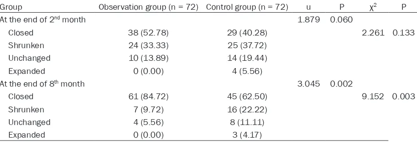
Comparison of cavity changes
When the intensive phase was completed at the end of 2nd month, although the cavity
clo-sure rate of the observation group was higher than that of the control group, differences were not significant (P > 0.05). When the consolida-tion phase was completed, at the end of 8th
month, the cavity closure rate was significant- ly higher in the observation group than in the control group. Overall cavity changes in the observation group were better than those of the control group, according to Mann-Whitney U-testing (P < 0.05). See Table 3 and Figure 2.
Comparison of improvement of symptoms
At the end of 8th month, the observation group
[image:4.612.93.520.85.232.2]showed lower scores concerning tuberculosis symptoms, including low fever, coughing and sputum production, chest pain, and loss of appetite (all P < 0.05). See Table 4 and Figure 3.
Table 3. Comparison of cavity changes (n, %)
Group Observation group (n = 72) Control group (n = 72) u P χ2 P
At the end of 2nd month 1.879 0.060
Closed 38 (52.78) 29 (40.28) 2.261 0.133 Shrunken 24 (33.33) 25 (37.72)
Unchanged 10 (13.89) 14 (19.44) Expanded 0 (0.00) 4 (5.56)
At the end of 8th month 3.045 0.002
Closed 61 (84.72) 45 (62.50) 9.152 0.003 Shrunken 7 (9.72) 16 (22.22)
[image:4.612.92.298.86.392.2]Unchanged 4 (5.56) 8 (11.11) Expanded 0 (0.00) 3 (4.17)
Figure 2. Comparison of cavity closure rates. **P <
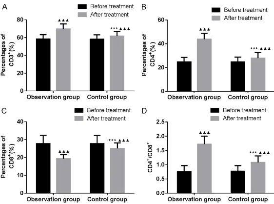
Comparison of immune function
At the end of 8th month, CD3+, CD4+ T-cells,
and CD4+/CD8+ ratios increased in both gr-
oups, compared with before treatment. How- ever, CD8+ T-cells decreased (all P < 0.05).
CD3+, CD4+ T-cells, and CD4+/CD8+ ratios in the
observation group were significantly higher than those in the control group. CD8+ T-cells
[image:5.612.147.505.81.318.2]were lower in the observation group than in the control group (all P < 0.05). See Table 5 and Figure 4.
Comparison of pulmonary function
When treatment was completed, pulmonary function indices, including FVC, FEV1, and IC, in both groups increased significantly, compar- ed with before treatment (all P < 0.05). Fur- thermore, increases in the observation group were more pronounced than those in the con-trol group (all P < 0.05). See Table 6 and Figure 5.
Discussion
Smear-positive re-treatment pulmonary tuber-culosis is a special type of tubertuber-culosis, charac-terized by a long course, severity, and difficulty in management [10, 11]. Statistics indicate that the incidence rate of chronic fibrous cavity in patients with smear-positive re-treatment pulmonary tuberculosis is 55%. The number of MTB in the cavity wall could reach 107-109 due
to vigorous proliferation of MTB in the caseous necrosis [12]. Additionally, thickening and fibro-sis of the cavity wall can hinder drug
penetra-ened cavity wall has poor blood circulation. Th- is makes drug penetration more difficult [15]. Therefore, the search for more effective treat-ment options has become a hot spot in clinical research.
[image:5.612.91.392.88.272.2]Bronchoscopic therapy is a new treatment method that combines lesion removal and dr- ug delivery. It can eliminate secretions and debris, clearing the bronchial lumen and cavi-ties. It can also deliver isoniazid, levofloxacin, amikacin, and other drugs directly into the le- sions, increasing local concentrations. This is a major advantage over oral administration. Increased local drug concentrations inhibit the proliferation of MTB in the cavity and improve sputum negative conversion rates. As a result, bronchoscopic therapy has been gradually rec-ognized and promoted. Satisfactory outcomes have been obtained [16, 17]. Yuan et al. found that the sputum negative conversion rates in- creased by 25% after 6 months of broncho-scopic therapy [18]. Fu et al. confirmed that cavity closure rates in the initial treatment for cavitary pulmonary tuberculosis reached 60% after 6 months of bronchoscopic lavage and drug delivery. This rate was significantly higher than that of 30% in the control group [19]. The above results indicate that bronchoscopic th- erapy can effectively improve the prognosis of patients with pulmonary tuberculosis. However, studies concerning bronchoscopic therapy for treatment of more complicated cases of tuber-culosis, including re-treatment smear-positive cavitary pulmonary tuberculosis, are scarce. In the current study, sputum negative conversion Table 4. Comparison of symptom improvement (_x ± sd)
Group group (n = 72)Observation Control group (n = 72) t P Low fever
Before treatment 2.36 ± 0.42 2.40 ± 0.40 0.585 0.559 At the end of treatment 0.58 ± 0.10 1.05 ± 0.15 22.122 < 0.001 Cough and sputum production
Before treatment 2.38 ± 0.43 2.36 ± 0.40 0.289 0.773 At the end of treatment 0.60 ± 0.12 1.09 ± 0.18 19.219 < 0.001 Chest pain
Before treatment 2.20 ± 0.35 2.18 ± 0.34 0.348 0.729 At the end of treatment 0.48 ± 0.08 0.76 ± 0.15 13.976 < 0.001 Loss of appetite
Before treatment 2.20 ± 0.37 2.19 ± 0.38 0.160 0.873 At the end of treatment 0.46 ± 0.07 0.75 ± 0.10 20.159 < 0.001
thick-Figure 3. Comparison of symptom improvement. A: Low fever; B: Cough and sputum production; C: Chest pain; D: Loss of appetite. ▲▲▲P < 0.001,
com-pared with before treatment. ***P < 0.001, compared with the observation
[image:6.612.89.375.71.296.2]group.
Table 5. Comparison of immune function (_x ± sd)
Group group (n = 72)Observation Control group (n = 72) χ2/t P
CD3+ (%)
Before treatment 58.45 ± 4.86 58.38 ± 4.80 0.087 0.931 At the end of treatment 69.62 ± 5.84 61.75 ± 5.27 8.489 < 0.001 CD4+ (%)
Before treatment 24.68 ± 3.87 24.73 ± 4.02 0.076 0.940 At the end of treatment 43.84 ± 5.05 27.86 ± 4.76 19.539 < 0.001 CD8+ (%)
Before treatment 27.80 ± 4.62 27.74 ± 4.57 0.078 0.938 At the end of treatment 19.30 ± 2.28 25.04 ± 3.12 12.604 < 0.001 CD4+/CD8+
Before treatment 0.76 ± 0.21 0.77 ± 0.20 0.293 0.770 At the end of treatment 1.72 ± 0.28 1.08 ± 0.23 14.987 < 0.001
symptoms, and improve pa- tient prognosis.
Under stimulation of MTB, the immune system is often com-promised. This is manifested as the suppression of T-cell-mediated cellular immunity. T-cell subsets usually show that CD3+ and CD4+ T-cells are
decreased and CD8+ T-cells
are increased. Additionally, CD4+/CD8+ ratios decrease,
indicating that the host’s im- mune system is impaired [20]. Previous studies have found that CD4+ and CD8+ T-cells
synergistically mediate immu- ne system to protect against MTB. CD4+ T-cells play a key
role in orchestrating adaptive immune response by activat-ing CD8+ T-cells and
stimulat-ing macrophage proliferation and phagocytosis. CD8+ T-ce-
lls are responsible for dissolv-ing macrophages that have phagocytized MTB [21]. Alter- ed CD4+/CD8+ ratios can lead
to activation of MTB prolifera-tion, even the emergence of drug-resistant and mutant str- ains. Results showed that CD3+, CD4+ T-cells, and CD4+/
CD8+ ratios in the observation
group were higher than those in the control group after the consolidation phase. More- over, CD8+ T-cells were lower
than those of the control gr- oup. Results further suggest that chemotherapy combined with bronchoscopic triple-drug and cavity closure rates of the observation
group after the consolidation phase were 94.44% and 84.72%, respectively, significant- ly higher than those in the control group. In addition, the observation group showed lower scores for tuberculosis symptoms, including low fever, coughing and sputum production, chest pain, and loss of appetite. Results sug-gest that the combination of routine chemo-therapy with bronchoscopic triple-drug delivery can improve therapeutic outcomes, alleviate
delivery can effectively enhance immune func-tion, compared to chemotherapy alone. Li et al. treated 63 patients with smear-positive re-treatment cavitary pulmonary tuberculosis us- ing bronchoscopic drug delivery. At the end of the 2nd and 6th month, CD3+, CD4+ T-cells, and
CD4+/CD8+ ratios increased and CD8+ T-cells
[image:6.612.90.373.387.569.2]Cavitary pulmonary tuberculosis can cause pr- ogressive fibrosis in lung tissues, resulting in mucociliary dysfunction and accumulation of bronchial secretion. Patients cannot effectively cough up sputum due to reduced ventilation and airway blockage. In addition, fibrosis can lead to poor local blood circulation, which has been associated with a continuous decline in lung function [23]. Hypoxia, in turn, aggravates fibrosis in the lung tissues and impedes healing of tuberculosis cavities. This often results in a rapid decline in the quality of life of patients. In the current study, after 8 months of treatment, FVC, FEV1, and IC levels in the observation group were higher than those in the control
One limitation of the current study was the re- latively small number of subjects from a single center. This factor may have impeded in-depth statistical analyses of the outcomes. Further research is necessary, investigating the under-lying mechanisms, aiming to find better alter- natives for treatment of cavitary pulmonary tuberculosis.
[image:7.612.91.372.70.279.2]In summary, bronchoscopic triple-drug delivery combined with chemotherapy for re-treatment of smear-positive cavitary pulmonary tubercu-losis is more effective than chemotherapy alone. Therefore, it is highly recommended for clinical practice.
Table 6. Comparison of pulmonary function (_x ± sd)
Group group (n = 72)Observation Control group (n = 72) χ2/t P
FVC (L)
Before treatment 3.40 ± 0.75 3.42 ± 0.74 0.161 0.872 At the end of treatment 4.32 ± 0.92 3.78 ± 0.85 3.658 < 0.001 FEV1 (L)
Before treatment 3.04 ± 0.68 3.05 ± 0.70 0.087 0.931 At the end of treatment 3.91 ± 0.75 3.31 ± 0.73 4.864 < 0.001 IC (L)
Before treatment 1.86 ± 0.38 1.88 ± 0.39 0.317 0.756 At the end of treatment 2.82 ± 0.45 2.34 ± 0.40 6.765 < 0.001
Note: FVC: Forced vital capacity; FEV1: Forced expiratory volume in 1s; IC: Inspira-tory capacity.
Figure 4. Comparison of immune function. A: CD3+; B: CD4+; C: CD8+; D:
CD4+/CD8+ ratios. ▲▲▲P < 0.001, compared with before treatment. ***P <
0.001, compared with the observation group.
[image:7.612.91.372.357.500.2]Disclosure of conflict of interest
None.
Address correspondence to: Dongmei Fan, Depart- ment of Infectious Disease, Lanling County People’s Hospital, No. 12 Tashan Road, Cangshan County, Linyi 277799, Shandong Province, China. Tel: +86-0539-5211790; E-mail: fandongmei5fg@163.com
References
[1] Houben RM and Dodd PJ. The global burden of latent tuberculosis infection: a re-estimation using mathematical modelling. PLoS Med 2016; 13: e1002152.
[2] Sun SH, Gao ZD, Zhao F, Zhang WY, Zhao X, Li YY, Li YM, Hong F, He XX and Zhan SY. Spatial-temporal analysis on pulmonary tuberculosis in Beijing during 2005-2015. Zhonghua Liu Xing Bing Xue Za Zhi 2018; 39: 816-820. [3] Nikitin MM, Puz’ko AS, Senchikhin PV and
Glo-tov AA. Modern methods of diagnosis and con-trol of the effectiveness of treatment for newly diagnosed fibro-cavernous pulmonary tubercu-losis caused by mycobacteria with multiple drug resistance. Vestn Rentgenol Radiol 2016; 97: 296-302.
[4] Pfannschmidt J and Schonfeld N. Interdisci-plinary treatment of patients with pulmonary tuberculosis. Zentralbl Chir 2017; 142: s53-s65.
[5] Hao XL, Lin JS, Cui LH, Wang SS, Feng T, Miao WW and Wang YY. Analysis of T-SPOT.TB test combined with bronchoscopy in the quick diag-nosis of smear-negative pulmonary tuberculo-sis. Journal of Practical Diagnosis and Therapy 2016; 30: 1039-1040.
drug-susceptible pulmonary tuberculosis. Me- dicine (Baltimore) 2016; 95: e4540.
[9] Lai HZ, Li SL, Cheng WS and Huang YP. Effect of bronchoscopic antituberculosis drug deliv-ery in the treatment of cavitary pulmonary tu-berculosis. Chinese Journal of Antituberculosis 2017; 39: 252-255.
[10] Sinha P, Srivastava GN, Gupta A and Anupurba S. Association of risk factors and drug resis-tance pattern in tuberculosis patients in North India. J Glob Infect Dis 2017; 9: 139-145. [11] Shenoy VP, Kumar A and Chawla K. Rapid
de-tection of multidrug resistant tuberculosis in respiratory specimens at a tertiary care centre in south coastal Karnataka using Genotype MTBDR plus assay. Iran J Microbiol 2018; 10: 275-280.
[12] Sharma S, Madan M, Agrawal C and Asthana AK. Genotype MTBDR plus assay for molecular detection of rifampicin and isoniazid resis-tance in mycobacterium tuberculosis. Indian J Pathol Microbiol 2014; 57: 423-426.
[13] Bouton TC, Phillips PPJ, Mitnick CD, Peloquin CA, Eisenach K, Patientia RF, Lecca L, Gotuzzo E, Gandhi NR, Butler D, Diacon AH, Martel B, Santillan J, Hunt KR, Vargas D, von Groote-Bidlingmaier F, Seas C, Dianis N, Moreno-Mar-tinez A and Horsburgh CR Jr. An optimized background regimen design to evaluate the contribution of levofloxacin to multidrug-resis-tant tuberculosis treatment regimens: study protocol for a randomized controlled trial. Tri-als 2017; 18: 563.
[14] Yew WW and Koh WJ. Emerging strategies for the treatment of pulmonary tuberculosis: promise and limitations? Korean J Intern Med 2016; 31: 15-29.
Figure 5. Comparison of pulmo-nary function. A: FVC; B: FEV1; C: IC. ▲▲▲P < 0.001, compared
with before treatment. ***P <
0.001, compared with the ob-servation group. FVC, forced vi-tal capacity; FEV1, forced expira-tory volume in 1 s; IC, inspiraexpira-tory capacity.
[6] Morales-Garcia C, Parra-Ruiz J, Sanchez-Martinez JA, Delgado-Martin AE, Am- zouz-Amzouz A and Her- nandez-Quero J. Concomi-tant tuberculosis and lung cancer diagnosed by bron-choscopy. Int J Tuberc Lung Dis 2015; 19: 1027-1032. [7] Zhang PY. Guidelines for
diagnosis and treatment of pulmonary tuberculosis. Chinese Journal of Tubercu-losis and Respiratory Dis-eases 2001; 24: 70-74. [8] Kang HK, Jeong BH, Lee H,
[15] Gillespie SH. The role of moxifloxacin in tuber-culosis therapy. Eur Respir Rev 2016; 25: 19-28.
[16] Mondoni M, Repossi A, Carlucci P, Centanni S and Sotgiu G. Bronchoscopic techniques in the management of patients with tuberculosis. Int J Infect Dis 2017; 64: 27-37.
[17] Ye W, Zhang R, Xu X, Liu Y and Ying K. Diagnos-tic efficacy and safety of endobronchial ultra-sound-guided transbronchial needle aspira-tion in intrathoracic tuberculosis: a meta-an- alysis. J Ultrasound Med 2015; 34: 1645-1650.
[18] Yuan ZA and Yu YM. Clinical efficacy of bron-choscopy in the treatment of pulmonary tuber-culosis and its effect on cellular immunity. Medical Journal of National Defending Forces in Southwest China 2017; 27: 1165-1167. [19] Fu HM, Xiao F, Liao JL and Fang Q. Efficacy
analysis of bronchoscopic drug delivery in the treatment of cavitary pulmonary tuberculosis. Guangdong Medicine 2008; 29: 863-864. [20] Liu YH, Gao WW, Li L, Du J, Ma Y, Shu W, Lyu XY,
Xie SH, Wang HH and Chen T. The effective-ness of individualized treatment regimen on smear-positive re-treatment pulmonary tuber-culosis with mono- and poly-drug resistance. Chinese Journal of Tuberculosis and Respira-tory Diseases 2018; 41: 25-31.
[21] Titarenko OT, D’Iakova M E, Esmedliaeva DS, Pavlova MV, El’kin AV, Alekseeva NP and Bond-arenko BB. Peculiarities of the circulating phagocytes functional activity in patients with different forms of drug-resistant pulmonary tu-berculosis. Biomed Khim 2012; 58: 467-474. [22] Li WB, Liu C, Sun Y, Guan WW, Guo Y, Li XH,
Zhao JC and Zang WB. Kangfuxing solution combined with bronchoscopic drug injection in the treatment of smear-positive re-treatment cavitary pulmonary tuberculosis and its effect on immune function and respiratory function. Modern Journal of Integrated Traditional Chi-nese and Western Medicine 2017; 26: 2972-2975.
[23] Krasnov DV, Skluev SV, Petrova YK, Skvortsov DA, Krasnov VA, Felker IG and Grischenko N. Modern collapse therapy for pulmonary tuber-culosis. Thorac Surg Clin 2019; 29: 47-58. [24] Zhao XG, Cheng HF, Cao WN, Hua SP, Zhu F,
Gao AX and Cheng L. Effect of bronchoscopic drug injection on smear-positive re-treatment cavitary pulmonary tuberculosis. Journal of Practical Medicine 2018; v34: 137-140. [25] Kumar R and Gupta N. Role of bronchoscopy in
evaluation of cases with sputum smear nega-tive pulmonary tuberculosis, interstitial lung disease and lung malignancy: a retrospective study of 712 cases. Indian J Tuberc 2015; 62: 36-42.
[26] Schnippel K, Shearer K, Evans D, Berhanu R, Dlamini S and Ndjeka N. Predictors of mortali-ty and treatment success during treatment for rifampicin-resistant tuberculosis within the South African National TB Programme, 2009 to 2011: a cohort analysis of the national case register. Int J Infect Dis 2015; 39: 89-94. [27] Guenaoui K, Harir N, Ouardi A, Zeggai S,