Original Article
Application value of MRI apparent diffusion coefficient
(ADC) before and after radical mastectomy
for patients with breast cancer
Liping Chen1*, Li Liu2*, Feng Zhao3, Xiaodong Li4, Baopeng Li5, Shujuan Lu6
1Magnetic Resonance Room, The Second People’s Hospital of Liaocheng, Liaocheng, Shandong Province, China; 2Department of Imaging, Maternity and Child Health Care of Zaozhuang, Zaozhuang, Shandong Province, China; 3Department of Imaging, Zaozhuang Hospital of Zaozhuang Mining Group, Zaozhuang, Shandong Province, China; 4CT Room, The People’s Hospital of Pingyi County, Linyi, Shandong Province, China; 5Department of Radiology,
The Affiliated Hospital of Shandong University of TCM, Jinan, Shandong Province, China; 6Department of Imaging,
The People’s Hospital of Shanting District, Zaozhuang, Shandong Province, China. *Equal contributors and co-first
authors.
Received June 5, 2019; Accepted August 9, 2019; Epub September 15, 2019; Published September 30, 2019
Abstract: Objective: To explore the correlation between Apparent Diffusion Coefficient (ADC) of preoperative Magnetic
Resonance Imaging (MRI) and the risk factors for prognosis among patients who have received radical mastectomy, and to study the role of ADC values measured in regions of interest (ROI) in the diagnosis of preoperative axillary lymph node metastasis and postoperative recurrence. Methods: Imaging data of diffusion weighted imaging (DWI) of 150 patients before and after radical mastectomy were reviewed. Multivariate linear regression was used to ana-lyze the correlation between preoperative ADC parameters of lesions and prognostic factors such as lymph node metastasis, human mammaglobin (hMAM) and low expression of Ki67. Receiver operating characteristic (ROC) curve was used to evaluate the value of ADC parameters obtained from the ROI in the diagnosis of preoperative axillary lymph node metastasis and postoperative recurrence. Results: Comparisons of ADC values for the
respec-tive preoperarespec-tive prognostic factor showed that there was a statistically significant difference between ADC values
for patients with lymph node metastasis, Ki67 overexpression, human mammaglobin positivity (hMAM+), estrogen receptor negativity (ER-), progesterone receptor negativity (PR-), human epidermal growth factor receptor 2 positivity (HER2+) and the ADC values for patients without these symptoms (all P<0.05). Further, multivariate linear
regres-sion showed that there was a significant correlation between the preoperative ADC value of tumors and lymph node
metastasis, Ki67 overexpression, hMAM (+), ER (-), PR (-) and HER2 (+) (all P<0.05). Pathological diagnosis of the 150 patients before surgery showed ipsilateral lymph node metastasis in 45 patients (30.00%). ROC curve analysis showed AUC=0.925 and IC 95% (0.791, 0.972). Youden index was 0.889, and the corresponding optimal threshold
value was 1.07. The sensitivity, specificity, positive predictive value, negative predictive value and diagnostic ac
-curacy were 88.89%, 92.38%, 83.38%, 95.10% and 91.33% respectively. Surgical pathology confirmed that there
were 33 cancer recurrences (22.00%) in the 150 patients 2 years after operation. The ROI ADC value for patients
with recurrent cancer was 1.32±0.19, which was significantly higher than that for patients without recurrent can
-cer (0.92±0.22), and the difference was statistically significant (t=10.302, P<0.001). ROC curve analysis showed
AUC=0.963 and IC 95% (0.839, 0.989). Youden index was 0.896, and the corresponding optimal threshold value
was 1.05. The sensitivity, specificity, positive predictive rate, negative predictive rate and diagnostic accuracy were 90.91%, 96.58%, 88.24%, 97.41% and 95.33%, respectively. Conclusion: There was a significant correlation be -tween the preoperative ADC values measured using DWI and lymph node metastasis, Ki67 overexpression, hMAM+, ER-, PR- and HER+ for patients who have received radical mastectomy. And ADC values played an important role in the diagnosis of preoperative axillary lymph node metastasis and evaluation of postoperative recurrence. It is worth popularizing and applying in clinic.
Keywords: Breast cancer, radical mastectomy, magnetic resonance imaging, apparent diffusion coefficient
Introduction
Breast cancer is a major public health problem facing the world. According to a report issued
With the continuous development in medical technology, many different treatment options for breast cancer are now available [3-5]. Ra- dical mastectomy is the optimal treatment of breast cancer as it can effectively remove the tumor, the surrounding cancer tissues, and the metastatic axillary lymph nodes. Research by Edward F et al. showed that the 5-year recur-rence rate after radical mastectomy was 21.78% and the local/regional recurrence rate was 10.4%, despite its effectiveness in treating breast cancer [6]. At present, the clinical diag-nosis of pre-operative axillary lymph node metastasis and post-operative recurrence of breast cancer mainly relies on invasive surgical biopsy, and assessment of prognosis mainly depends on the detection of tumor markers in the biopsy sample, which increases the pa- tients’ pain [7, 8]. Therefore, non-invasive pro-cedures to assess preoperative lymph node metastasis and postoperative prognosis (in- cluding recurrence) have been researched ex- tensively.
Magnetic resonance imaging (MRI) is an effec-tive means to examine soft tissue lesions, whi- ch works by placing the examined body parts in a static magnetic field and applying specific radio frequency pulses to stimulate the reso-nance of hydrogen protons. When the RF pulse is turned off, it can receive MR signals emitted by protons in relaxation process and recon-struct images of soft tissue lesions [10]. As a non-invasive diagnostic method, DWI has been widely used in the clinical diagnosis of breast cancer. With high-resolution, high-sensi-tivity and high-specificity imaging, it can help examiners effectively assess the size of tumors and the severity of disease. Research by Tho- mas E et al. showed that ADC values derived from measuring the average diffusion coeffi-cient in three orthogonal directions can effec-tively reflect the movement of water molecules in tissues; higher cell density of tumor tissues can lead to the decrease in water molecule movement, resulting in lower ADC values, which can be used for the diagnosis of benignity and malignancy of breast tumors [11]. However, there is no comprehensive report on the signifi-cance of ADC values in the diagnosis of preop-erative axillary lymph node metastasis and postoperative recurrence, and the correlation between ADC values and prognostic factors is not clear.
Therefore, the purpose of this study was to evaluate the correlation between ADC values and the level of pathological biomarkers in tumor tissues related to prognosis, and to ana-lyze the diagnostic value of preoperative axil-lary lymph node metastasis and postoperative recurrence in patients with breast cancer, so as to provide a reference for the selection of imag-ing examination among patients undergoimag-ing radical mastectomy. In order to ensure the reli-ability of the results, preoperative axillary lymph node metastasis and postoperative recurrence were confirmed by surgery.
Materials and methods
Patients
A total of 150 patients (all females) with breast cancer who underwent radical mastectomy in The People’s Hospital of Shanting District fr- om January 2014 to January 2017 were retro-spectively studied. All patients were between 18 and 67 years old, with an average age of 45.1±7.5. Inclusion criteria: patients met the diagnostic criteria of breast cancer set forth in the National Comprehensive Cancer Network (NCCN) Clinical Practice Guidelines for Breast Cancer Screening and Diagnosis (Version 3.2013) [12]; DWI was performed in patients before and after operation, and the imaging was clear and imaging data was complete; patients had complete perioperative clinical data. Exclusion criteria: patients who had se- vere concurrent hematological disease, im- mune system disease or other malignant tu- mors; patients who did not receive the speci-fied surgical treatment; patients with mental disorders who did not complete the follow-up. This study was approved by the Ethics Com- mittee of The People’s Hospital of Shanting District. All patients in this study received radi-cal mastectomy and signed informed consent.
MRI examination
3-4 mm, interslice gap 0-0.5 mm, matrix size 512×512. Chopper or mixed technique was used for chemical shift fat suppression with 2 excitations.
DWI: fast spin echo (FSE) plain scan, b=50 and 800 s/mm2, TR 2,000-3,000 ms, TE100-120 ms, FOV 320 mm×320 mm, slice thickness 4-5 mm, interslice gap 0.5-1.0 mm, FOV 320 mm× 320 mm, 2-4 excitation. DCE-MRI: Contrast agent Gd-DTPA (0.2 mg/kg) was injected into forearm vein after the plain scan, and 20 mL saline was then injected rapidly. Before and immediately after injection, patients were scan- ned using 64, 128, 191, 255, 318 s fast gradi-ent echo sequence. TR 4-5 ms, TE 2-3 ms, flip angle 12°, FOV 320 mm×320 mm, slice thick-ness 1-2 mm, interslice gap 0-0.5 mm, matrix size 336×336, total scan time 7 min and 7 s.
Calculation of ADC value
DWI images corresponding to different b va- lues were transmitted to the computer aided diagnosis platform FireVoxel (Center for Advan- ced Imaging Innovation and Research (CAI2R), New York University School of Medicine, New York, USA). The ADC values of ROI images were measured and calculated by two staff mem-bers. Three points were measured and the average was taken as the final ADC values. The two staff members alternated between calcu- lation and data checking.
Immunohistochemistry
Tumor tissues were resected during the sur-gery, which were immediately fixed with formal-dehyde and embedded in paraffin. Histological observation of the tissue section was then per-formed. MaxVisionTM rapid immunohistochem-ical kit (AMRESCO, Inc., USA) was used to de- tect the expressions of hMAM and Ki-67. Ki-67 staining ≤14% was low expression, otherwise it was high expression. ER and PR expression <10% was negative, otherwise it was positive. The expression of HER2 -/1+ was negative, oth-erwise it was positive.
Other examination
The tubular structure, pleomorphism and mitot-ic counts of the tumor cells were examined and WHO histological grading system was used as the reference index [13]. The axillary lymph node metastasis was confirmed by surgical pa- thology. The lymph node status was evaluated
by HE staining, and the results of biopsy were the gold standard.
Statistical analysis
The SPSS 24.0 statistical software was used. Enumeration data was expressed as n (%) and compared with χ2 test. The significance level was set at α=0.05 (bilateral). Measurement data was expressed as mean ± standard de- viation (_x ± sd). Independent t test was used for comparison between groups. The signifi-cance level was also set at α=0.05 (bilateral). ADC value was the independent variable, while lymph node metastasis, histological grade, low expression of Ki67, hMAM positivity, ER nega-tivity, PR negativity and HER2 positivity were dependent variables. Multiple linear regression was used to analyze the correlation between ADC parameters and prognostic factors. B stood for regression coefficient. SE stood for standard error. SC stood for standardization coefficient. ROC curve analysis was used to evaluate the significance of ADC values in the diagnosis of preoperative axillary lymph node metastasis and postoperative recurrence. Area under the curve (AUC) >0.5 meant the result was better than random guess, and there was a predictive value. The greater the AUC value, the greater the predictive value. P<0.05 meant the difference was statistically significant.
Results
Correlation between preoperative tumor ADC values and prognostic factors
Comparison of ADC values for preoperative prognostic factors showed that there was a statistically significant difference between the ADC values for patients with lymph node me- tastasis, Ki67 overexpression, hMAM (+), ER (-), PR (-) and HER2 (+) and those for patients with-out these symptoms (Table 1, all P<0.05). Further Multivariate linear regression showed that there was a significant correlation betwe- en the preoperative ADC value of tumors and lymph node metastasis, Ki67 overexpression, hMAM (+), ER (-), PR (-) and HER2 (+) (Table 2, all P<0.05).
Two cases of ADC value applied in lymphatic metastasis diagnosis
left axillary area. The larger one was about 1.7 cm*1.1 cm (Figure 1A), with an average ADC value of 1.32±0.17. No obvious enlarged echogenic lymph nodes were found in the ri- ght axillary area. The average ADC value was 0.91±0.10. The average ADC value of lymph nodes with tumor infiltration was significantly
Surgical pathology confirmed that there were 33 cancer recurrences (22.00%) in the 150 patients 2 years after operation. The ROI ADC value for patients with recurrent cancer was 1.32±0.19, which was significantly higher th- an that for patients without recurrent cancer (0.92±0.22), and the difference was statisti-Table 1. Comparison of preoperative prognostic factors related
to grouping ADC values
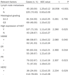
Relevant factors Cases (n, %) ADC value t P
Lymph node metastasis
Y 45 (30.00) 1.25±0.12 16.058 <0.001
N 105 (70.00) 0.93±0.09
Histological grading
G1-2 81 (54.00) 1.14±0.25 0.261 0.795
G3 69 (46.00) 1.15±0.22
High expression of Ki67
Y 107 (71.33) 1.11±0.26 2.280 0.025
N 43 (28.67) 1.22±0.27
hMAM
+ 88 (58.67) 1.19±0.22 2.080 0.040
- 62 (41.33) 1.11±0.24
ER
+ 95 (63.33) 1.13±0.21 2.390 0.018
- 55 (36.67) 1.21±0.19
PR
+ 79 (52.67) 1.11±0.19 2.307 0.023
- 71 (47.33) 1.19±0.23
HER2
+ 36 (24.00) 1.22±0.25 2.234 0.029
- 114 (76.00) 1.11±0.28
Note: ADC, apparent diffusion coefficient; hMAM, human mammaglobin; ER,
[image:4.612.90.357.97.384.2]estrogen receptor; PR, progesterone receptor; HER2, human epidermal growth factor receptor 2.
Table 2. Multiple linear regression analysis of factors related to preoperative tumor ADC value and patient prognosis
Relevant Factors B SE SC P
Intercept 1.597 0.192 0.000
Lymph node metastasis -0.132 0.045 -0.238 0.001
Histological grading (G3) -0.018 0.038 -0.149 0.512
High expression of Ki67 0.142 0.047 0.245 0.003
hMAM (+) 0.133 0.046 0.233 0.042
ER (+) 0.135 0.067 0.235 0.041
PR (+) -0.044 0.068 -0.074 0.048
HER2 (+) -0.089 0.037 -0.182 0.024
Adjusted R2 0.343
Note: ADC, apparent diffusion coefficient; hMAM, human mammaglobin; ER,
estrogen receptor; PR, progesterone receptor; HER2, human epidermal growth factor receptor 2.
higher than that of lymph nodes free of tumor infiltration (t=2.028, P<0.001).
Preoperative MRI findings (Patient B, 74 years old): Solid nodules in the right breast and multiple ly- mph nodes in the right axillary area. The lager one was about 1.3 cmx0.9 cm (Figure 1B), with an average ADC value of 1.29±0.14. There were no obvious enlarged lymph nodes in the left axillary area, and the average ADC value was 0.94±0.12. The average ADC value of lymph nodes with tumor infiltration was significantly hig- her than that of lymph nodes free of tumor infiltration (t=3.132, P< 0.001).
ROC curve analysis of preop-erative ROI ADC for diagnosis of preoperative axillary lymph node metastasis
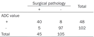
Pathological diagnosis of the 150 patients before operation show- ed ipsilateral lymph node me- tastasis in 45 patients (30.00%). ROC curve analysis showed AUC= 0.925 (Figure 2) and IC 95% (0.791, 0.972). Youden index was 0.889, and the corresponding op- timal threshold value was 1.07. Table 3 showed preoperative ROI ADC value for the diagnosis of pr- eoperative axillary lymph node metastasis. The sensitivity, speci-ficity, positive predictive value, ne- gative predictive value and diag-nostic accuracy were 88.89%, 92.38%, 83.38%, 95.10% and 91.33% respectively.
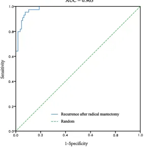
[image:4.612.92.359.468.597.2]cally significant (t=10.302, P<0.001). ROC cur- ve analysis showed AUC=0.963 (Figure 3) and IC 95% (0.839, 0.989). Youden index was 0.896, and the corresponding optimal thresh-old value was 1.05. Table 4 showed preope- rative ROI ADC value for the diagnosis of post-operative recurrence. The sensitivity, specifici-ty, positive predictive rate, negative predicti-
other fields before and after radical mastecto-my for breast cancer.
Application of ADC values in the diagnosis of preoperative lymphatic metastasis
[image:5.612.90.373.72.209.2]Lymphatic metastasis of breast cancer is one of the main factors affecting its prognosis. Figure 1. MRI examination image data of preoperative lymphatic
metas-tasis in 2 patients. A. The largest lymph node in the left axilla of patient A; B. The largest lymph node in the right axilla of patient B. MRI, Magnetic Resonance Imaging.
Figure 2. ROC curve analysis of preoperative ROI ADC value for diagnosis of preoperative axillary lymph node metastasis. ROC, receiver operating
char-acteristic; ADC, apparent diffusion coefficient; ROI, regions of interest.
ve rate and diagnostic accu- racy were 90.91%, 96.58%, 88.24%, 97.41% and 95.33% respectively.
Discussion
Theoretical basis for clinical diagnosis with ADC values
[image:5.612.90.371.280.576.2]Preoperative diagnosis of lymphatic metastas- is and complete dissection of metastatic lym- ph nodes during radical mastectomy are of great clinical significance. In the past, lymph node metastasis was mainly judged by the sta-tus of lymph nodes. Lymph node metastasis was confirmed by symptoms including enlarge-ment and increase of lymph nodes on physical examination as well as signs including matted lymph nodes, disappearance of lymph node hilum and blurred margin on imaging examina-tion [15, 21-23]. But this kind of diagnostic method is subjective and diagnostic errors may occur because of radiologists’ experience in taking and reading films. For metastatic lymph nodes of breast cancer, their cell growth pat-tern is similar to that of primary tumor cells because of the invasion of cancer cells, result-ing in the increase of cell density and the slow-down of water molecule diffusion [14]. The- refore, the theoretical basis for the application of ADC value in the clinical diagnosis of preop-erative lymphatic metastasis of breast can- cer is scientifically sound. In this study, 45 of the 150 patients were diagnosed with lym- phatic metastasis before operation. The ROC curve analysis showed that ADC value was highly valuable for clinical diagnosis (AUC= 0.925, Figure 2). Statistical analysis showed that the optimal threshold value was 1.07, and a 91.33% diagnostic accuracy was obtained after diagnosis with the optimal threshold va- lue was made. Xinghua et al. evaluated the diagnostic value of DWI in lymph node metas- tasis assessment, and studied whether ADC value could be used to differentiate metastatic and non-metastatic lymph nodes in breast can-cer patients [24]. A total of 13 studies were reviewed, including 676 cases of metastatic
lymph nodes and 811 cases of non-metastatic lymph nodes. It was clear that ADC value has a significant role to play in diagnosing lymphatic metastasis of breast cancer. The results of this study are consistent with the above findings.
Application of ADC values in postoperative prognosis evaluation
DWI can not only diagnose the change of cell density, but also effectively evaluate the str- ucture of cancer tissues. Tumor cell structure is a good index for assessing the invasiveness of cancer cells, which is closely related to the prognosis of patients. At present, the assess-ment of malignancy, benignity and prognosis of tumors is mainly based on pathological exami-nation, which comprehensively evaluate tumors by the study of cell morphology and the detec-tion of histopathological biomarkers [25]. Ki-67 is closely related to mitosis and recur-rence of tumor cells. Increased mitosis can lead to an increase in cell density. As discuss- ed above, cell density has an impact on the results of DWI imaging. HMAM was first found in tumor cells derived from patients with endo-metrial cancer and salivary gland cancer. Fol- low-up studies found the abnormal expression of hMAM in epithelial cells of tumor tissues. It has become one of the markers for the clinical evaluation of cell invasiveness and prolifera-tion activity. Steroid hormone receptor proteins ER and PR are good markers for prognosis. Some studies have shown that ER (a confound-ing factor affectconfound-ing PR) can affect the structure of tumor tissues by inhibiting vascular access and inducing tissue perfusion reduction, and the expression of ER and PR is related [26-28]. The expression of HER2 is mainly related to the growth and metastasis of tumor cells. It can increase angiogenesis of tumor tissues by in- ducing the expression of vascular endothelial growth factor (VEGF), improve the blood supply of tumor cells and promote cell metastasis. Studies have also shown a positive correlation between the ADC value provided by DWI and the blood supply and extracellular fluid volume of tumors, which serves as a proof of the theo-retical basis for the reflection of prognostic fac-tors by ADC values [29, 30].
[image:6.612.90.289.109.188.2]In this study, statistical analysis of the ADC values of lymph node metastasis, histological Table 3. Preoperative ROI ADC value for
diagnosis of preoperative axillary lymph node metastasis (n)
Surgical pathology Total
+
-ADC value
+ 40 8 48
- 5 97 102
Total 45 105
Note: ADC, apparent diffusion coefficient; ROI, regions of
grade, Ki67 overexpression, hMAM positivity, ER negativity, PR negativity and HER2 positivi- ty were performed, followed by multiple linear regression analysis. The results showed a sig-nificant correlation between preoperative ADC values of tumors and lymph node metastasis, Ki67 overexpression, hMAM (+), ER (-), PR (-) and HER2 (+), but no correlation with histologi-cal grade. This result was different from the resul- ts predicted. After literature review, we believe this discrepancy can be explained by the sub-jectivity involved in the histological grading of tumors.
between the results and the gold standard (results of biopsy) was small, which indicated that ADC value could be used for clinical diag-nosis of recurrence of breast cancer after radi-cal mastectomy.
Limitation of this study
[image:7.612.90.373.77.366.2]There are still some limitations in this study. First, subjects in this study are not representa-tive and the selection process may be biased as all patients were from a single medical insti-tution. Second, given the knowledge of results from previous imaging examinations, the pos-sibility of researchers’ impact on the results of this study during the follow-up period cannot be ruled out. Besides, the follow-up period of this study was 2 years, which only reflected the role of ADC value in diagnosis of recurrence shortly after surgery. Third, ADC values were not calculated with different b values when DWI was performed, so there may be some bi- as in perfusion or diffusion effects. Finally, the level of pathological tissue biomarkers used as a reference standard was obtained only th- Figure 3. ROC curve analysis of postoperative ROI ADC value for diagnosis
of postoperative recurrence. ROC, receiver operating characteristic; ADC,
[image:7.612.90.297.452.532.2]apparent diffusion coefficient; ROI, regions of interest.
Table 4. Preoperative ROI ADC value for diagno-sis of postoperative recurrence (n)
Surgical pathology
Total Recurrence No recurrence ADC value
Recurrence 30 4 34
No recurrence 3 113 116
Total 33 117
Note: ADC, apparent diffusion coefficient; ROI, regions of
interest.
Application of ADC value in the diagnosis of postoperative recurrence
rough a review of pathological reports; there could be changes in pathological measure-ments during the preservation and preparati- on of each tissue specimen.
Research conclusion
In conclusion, MRI is a valuable tool in the di- agnosis of preoperative axillary lymph node metastasis and the evaluation of postoperati- ve recurrence. There is a significant correlati- on between the preoperative ADC values mea-sured using DWI for patients who have received radical mastectomy and lymph node metasta-sis, Ki67 overexpression, hMAM positivity and HER2 positivity, which proves the role of ADC values in the assessment of prognosis for patients.
Disclosure of conflict of interest
None.
Address correspondence to: Shujuan Lu, Depart- ment of Imaging, The People’s Hospital of Shanting District, No.528 Xincheng Beijing Road, Shanting District, Zaozhuang 277200, Shangdong Province, China. Tel: 8833817; Fax: +86-0632-8833817; E-mail: [email protected]
References
[1] Craig AW. IARC Monographs on the evaluation of carcinogenic risk of chemicals to man. Vol. 12. Some Carbamates, Thiocarbamates and Carbazides (1976). Br J Cancer 1977; 36: 432. [2] Stewart BW and Wild CP. World Cancer Report
2014. Edited by Stewart BW and Wild CP. Lyon, International Agency for Research on Cancer 2014.
[3] Zembutsu H, Nakamura S, Akashi-Tanaka S, Kuwayama T, Watanabe C, Takamaru T, Takei H, Ishikawa T, Miyahara K, Matsumoto H, Hasegawa Y, Kutomi G, Shima H, Satomi F, Okazaki M, Zaha H, Onomura M, Matsukata A, Sagara Y, Baba S, Yamada A, Shimada K, Shi-mizu D, Tsugawa K, Shimo A, Tan EY, Hartman
M, Chan CW, Lee SC and Nakamura Y. Signifi -cant effect of polymorphisms in CYP2D6 on response to tamoxifen therapy for breast can-cer: a prospective multicenter study. Clin Can-cer Res 2017; 23: 2019-2026.
[4] Dubsky P, Curigliano G, Burstein HJ, Winer EP, Gnant M, Loibl S, Colleoni M, Regan MM, Pic-cart-Gebhart M, Senn HJ, Thurlimann B, Andre F, Baselga J, Bergh J, Bonnefoi H, Brucker SY, Cardoso F, Carey L, Ciruelos E, Cuzick J, Den-kert C, Di Leo A, Ejlertsen B, Francis P, Galim-berti V, Garber J, Gulluoglu B, Goodwin P,
Har-beck N, Hayes DF, Huang CS, Huober J, Khaled H, Jassem J, Jiang Z, Karlsson P, Morrow M, Orecchia R, Osborne KC, Pagani O, Partridge AH, Pritchard K, Ro J, Rutgers EJT, Sedlmayer F, Semiglazov V, Shao Z, Smith I, Toi M, Tutt A, Viale G, Watanabe T, Whelan TJ and Xu B. Re-ply to ‘The St Gallen international expert con-sensus on the primary therapy of early breast cancer 2017: the point of view of an interna-tional panel of experts in radiation oncology’ by Kirova et al. Ann Oncol 2018; 29: 281-282. [5] Kirova YM, Carroll S, Fourquet A, Offersen B,
Aristei C and Chen JY. The St Gallen interna-tional expert consensus on the primary thera-py of early breast cancer 2017: the point of view of an international panel of experts in ra-diation oncology. Ann Oncol 2018; 29: 280-281.
[6] Scanlon EF. Local recurrence in the pectoralis
muscles following modified radical mastecto -my for carcinoma. J Surg Oncol 1985; 30: 149-151.
[7] Heil J, Richter H, Golatta M and Sinn HP. Vacu-um-assisted biopsy to diagnose a pathological complete response in breast cancer patients after neoadjuvant systemic therapy. Ann Surg 2018; 268: e60-e61.
[8] Karahalli O, Acar T, Atahan MK, Acar N, Haci-yanli M and Kamer KE. Clinical and pathologi-cal factors affecting the sentinel lymph node metastasis in patients with breast cancer. In-dian J Surg 2017; 79: 418-422.
[9] Schwartzberg B, Lewin J, Abdelatif O, Bernard J, Bu-Ali H, Cawthorn S, Chen-Seetoo M, Feld-man S, Govindarajulu S, Jones L, Juette A, Ka-via S, Maganini R, Pain S, Shere M, Shriver C, Smith S, Valencia A, Whitacre E and Whitney R. Phase 2 open-label trial investigating percuta-neous laser ablation for treatment of early-stage breast cancer: MRI, pathology, and out-come correlations. Ann Surg Oncol 2018; 25: 2958-2964.
[10] Makela AV, Gaudet JM and Foster PJ. Quantify-ing tumor associated macrophages in breast
cancer: a comparison of iron and
fluorine-based MRI cell tracking. Sci Rep 2017; 7: 42109.
[11] Yankeelov TE, Lepage M, Chakravarthy A, Broome EE, Niermann KJ, Kelley MC, Meszoely I, Mayer IA, Herman CR, McManus K, Price RR and Gore JC. Integration of quantitative DCE-MRI and ADC mapping to monitor treatment response in human breast cancer: initial re-sults. Magn Reson Imaging 2007; 25: 1-13. [12] Theriault RL, Carlson RW, Allred C, Anderson
ML, Soliman H, Somlo G, Ward JH, Wolff AC, Zellars R, Shead DA and Kumar R. Breast can-cer, version 3.2013: featured updates to the NCCN guidelines. J Natl Compr Canc Netw 2013; 11: 753-760; quiz 761.
[13] Christgen M, Langer F and Kreipe H. Histologi-cal grading of breast cancer. Pathologe 2016; 37: 328-336.
[14] Lee SM, Lee KW, Kim MA, Song YS, Goo JM and Park CM. Serial texture analyses on ADC maps for evaluation of antiangiogenic therapy in rat breast cancer. Anticancer Res 2019; 39: 1875-1882.
[15] Kato F, Kudo K, Yamashita H, Baba M, Shimizu A, Oyama-Manabe N, Kinoshita R, Li R and Shi-rato H. Predicting metastasis in clinically nega-tive axillary lymph nodes with minimum
appar-ent diffusion coefficiappar-ent value in luminal A-like
breast cancer. Breast Cancer 2019.
[16] Cho GY, Gennaro L, Sutton EJ, Zabor EC, Zhang Z, Giri D, Moy L, Sodickson DK, Morris EA, Sig-mund EE and Thakur SB. Intravoxel incoherent motion (IVIM) histogram biomarkers for predic-tion of neoadjuvant treatment response in breast cancer patients. Eur J Radiol Open 2017; 4: 101-107.
[17] Kawashima H, Miyati T, Ohno N, Ohno M, Inokuchi M, Ikeda H and Gabata T. Differentia-tion between luminal-A and luminal-B breast cancer using intravoxel incoherent motion and dynamic contrast-enhanced magnetic reso-nance imaging. Acad Radiol 2017; 24: 1575-1581.
[18] Chen F, Chen P, Hamid Muhammed H and Zhang J. Intravoxel incoherent motion diffusion
for identification of breast malignant and be -nign tumors using chemometrics. Biomed Res Int 2017; 2017: 3845409.
[19] Hasanzadeh F, Faeghi F, Valizadeh A and Bay-ani L. Diagnostic value of diffusion weighted magnetic resonance imaging in evaluation of metastatic axillary lymph nodes in a sample of Iranian women with breast cancer. Asian Pac J Cancer Prev 2017; 18: 1265-1270.
[20] Baba S, Isoda T, Maruoka Y, Kitamura Y, Sasa-ki M, Yoshida T and Honda H. Diagnostic and prognostic value of pretreatment SUV in 18F-FDG/PET in breast cancer: comparison with
apparent diffusion coefficient from
diffusion-weighted MR imaging. J Nucl Med 2014; 55: 736-742.
[21] Li JT, Zhao HM, Guo XH, Tian PQ, Lu MH, Li LF, Liu ZZ, Cui SD and Zhang HW. Preoperative evaluation of sentinel lymph node biopsy using contrast-enhanced ultrasonography in early breast cancer patients and the involved dis-turbing factors. National Medical Journal of China 2019; 99: 1086-1089.
[22] Matsuda T, Iguchi E, Konishi E, Tokugawa T, Hamaoka A and Nakatsukasa K. A case of breast cancer with parenchymal and menin-geal central nervous system metastases treat-ed with multimodality therapy. Gan To Kagaku Ryoho 2019; 46: 463-465.
[23] Li J, Ma W, Jiang X, Cui C, Wang H, Chen J, Nie R, Wu Y and Li L. Development and validation of nomograms predictive of axillary nodal sta-tus to guide surgical decision-making in early-stage breast cancer. J Cancer 2019; 10: 1263-1274.
[24] Xing H, Song CL and Li WJ. Meta analysis of lymph node metastasis of breast cancer pa-tients: clinical value of DWI and ADC value. Eur J Radiol 2016; 85: 1132-1137.
[25] Horvat JV, Bernard-Davila B, Helbich TH, Zhang M, Morris EA, Thakur SB, Ochoa-Albiztegui RE, Leithner D, Marino MA, Baltzer PA, Clauser P, Kapetas P, Bago-Horvath Z and Pinker K. Diffu-sion-weighted imaging (DWI) with apparent
dif-fusion coefficient (ADC) mapping as a quanti -tative imaging biomarker for prediction of immunohistochemical receptor status, prolif-eration rate, and molecular subtypes of breast cancer. J Magn Reson Imaging 2019.
[26] Jia Y, Shi L, Yun F, Liu X, Chen Y, Wang M, Chen C, Ren Y, Bao Y and Wang L. Transcriptome
se-quencing profiles reveal lncRNAs may involve
in breast cancer (ER/PR positive type) by inter-action with RAS associated genes. Pathol Res Pract 2019.
[27] Abubakar M, Figueroa J, Ali HR, Blows F, Lis-sowska J, Caldas C, Easton DF, Sherman ME, Garcia-Closas M, Dowsett M and Pharoah PD. Combined quantitative measures of ER, PR, HER2, and KI67 provide more prognostic infor-mation than categorical combinations in lumi-nal breast cancer. Mod Pathol 2019.
[28] Arena V, Pennacchia I, Vecchio FM and Car-bone A. ER-/PR+/HER2- breast cancer type shows the highest proliferative activity among all other combined phenotypes and is more common in young patients: experience with 6643 breast cancer cases. Breast J 2019; 25: 381-385.
[29] Maiborodin IV, Kozyakov AE, Babayants EV and Krasil’nikov SE. Features of blood supply to ax-illary lymph nodes in breast cancer patients. Bull Exp Biol Med 2017; 163: 82-86.