Original Article
Protective effect of dexmedetomidine on the brain of
patients receiving heart valve replacement and
its influence on inflammatory response
Bifeng Zhong1*, Ming Gao2*, Hongbo Qian3, Wei Dong2, Hanli Lin1
Departments of 1Cardiology, 2General Surgery, Zhejiang Putuo Hospital, Zhoushan, Zhejiang, China; 3Department
of Cardiac Surgery, Yijishan Hospital of Wannan Medical College, Wuhu 241001, China. *Co-first authors. Received May 22, 2019; Accepted August 9, 2019; Epub October 15, 2019; Published October 30, 2019
Abstract:Objective: This study was to investigate the protective effect of dexmedetomidine (Dex) on brain tissues of patients receiving heart valve replacement and its influence on the inflammatory response in brain tissues. Meth-ods: A total of 70 patients with heart disease were selected and underwent the heart valve replacement in vitro, and they were divided equally into observation group and control group. Patients in control group were anesthetized via propofol, while those in observation group were anesthetized via propofol + dexmedetomidine. The cognitive func-tion of patients in both groups was evaluated before operafunc-tion and at 3 d after operafunc-tion, and the peripheral blood was collected at different time points (before CPB (T0), immediately after CPB (T1), immediately after operation (T2), at 6 h after operation (T3) and at 24 h after operation (T4)) to detect expressions of tumor necrosis factor-α (TNF-α), interleukin-6 (IL-6), plasma S-100β and neuron-specific enolase (NSE). Results: The levels of plasma TNF-α and IL-6 in patients of both groups were gradually increased at T1-T4, and they were significantly lower in observation group than those in control group (P<0.05). The S-100β protein levels in both groups were increased to different degrees at different time points after operation, displaying statistically significant differences compared with those before operation (P<0.05). The NSE concentration at T2-T4 was increased compared with that at T0 (P<0.05), and it was significantly higher in control group at T1-T4 compared with that at T0 (P<0.05), while it was obviously lower at T1 and T2 in observation group compared with that in control group (P<0.05). Conclusion: Dexmedetomidine can sig-nificantly reduce the cerebral oxygen metabolic rate, alleviate the brain tissue damage and relieve the inflammatory response during heart valve replacement.
Keywords: Dexmedetomidine, heart valve replacement, brain protection, inflammatory response
Introduction
Cardiopulmonary bypass (CPB) is a kind of auxiliary surgical method. Oxidative stress and inflammatory response possibly occur in patients in the process of heart valve replace-ment due to various uncertain factors, leading to the release of a large number of oxygen free radicals and inflammatory factors. It even causes damage to a variety of organs in the patient’s body, such as the heart, lung and brain [1, 2]. Brain tissues of patients are in an ischemic-hypoxic state during CPB, and the brain nerve of patients is easily prone to dam-age in case of insufficient blood supply to brain tissues, thus threatening the patient’s life safe-ty. Therefore, the necessary protection on
breath. This study aims to investigate the pro-tective effect of dexmedetomidine on brain tis-sues of patients receiving heart valve replace-ment and its influence on the inflammatory response in brain tissues.
Materials and methods
General data
A total of 70 patients with heart disease diag-nosed and treated in our hospital were select-ed, and they all underwent heart valve replace-ment in vitro. There were 35 males and 35 females aged 40-71 years old with the body weight of 42-78 kg, left ventricular ejection fraction (LVEF) ≥35% and mini-mental state examination (MMSE) score ≥24 points in the American Society of Anesthesiologists (ASA) grade II or III. All patients enrolled were ran-domly and equally divided into observation group (dexmedetomidine) and control group according to the random number table. Exclusion criteria: patients with atrioventricular block, sinus bradycardia, a history of chronic inflammation or central nervous system diseas -es or taking anti-inflammatory drugs; those with severe diseases affecting cognitive func-tion or mental state after operafunc-tion; those with other brain diseases or allergy to anesthetics; or those with LVEF <40% or MMSE score <23 points. All patients and their families had signed the informed consent, and this study was approved by the Ethics Committee of Zhejiang Putuo Hospital. There were no statisti-cally significant differences in general clinical data (gender, age, height, weight, LVEF and aor -tic occlusion time) among patients (p>0.05). Methods
After fasting for solids and liquids at 12 h and 6 h before heart valve replacement, respective-ly, patients in observation group were injec- ted with 0.4 μg/kg dexmedetomidine solution, while those in control group were injected with 0.01 mg/kg atropine. Vital signs of patients in both groups were monitored using the monitor, and the anesthesia was induced using 0.2 g/ kg cisatracurium, 0.2 μg/kg etomidate and 0.5 μg/kg sufentanil. During operation, patients in control group were injected with 2 mg/kg pro-pofol, while those in observation group were injected with 2 mg/kg propofol + 0.4 μg/kg dexmedetomidine for anesthesia till the end of
the operation. During CPB, normal saline was injected continuously into patients in control group, while 0.4 μg/kg dexmedetomidine was injected continuously into patients in observa-tion group.
Evaluation of therapeutic effects
The cognitive function of patients in observa-tion group and control group was evaluated using the MMSE score before operation and at 3 d after operation. The total MMSE score was 30 points and the MMSE score ≥24 points in patients educated for more than 6 years indi-cates the normal cognitive function.
After anesthesia, 2 mL jugular bulb blood and 2 mL radial artery blood were drawn from patients. The jugular venous oxygen saturation
(SjvO2), cerebral artery-jugular venous oxygen content difference (Ca-jvO2) [Ca-jvO2 = Hb × 1.34 × (SaO2-SjvO2) + 0.003 (PaO2-PjvO2)] and cerebral oxygen extraction rate (ERO2) [ERO2 = (SaO2-SjvO2)/SaO2 × 100%] were detected. Radial arterial oxygen saturation and partial pressure of oxygen were detected and repre-sented the internal carotid arterial oxygen saturation and partial pressure of oxygen, respectively.
Before CPB (T0), immediately after CPB (T1), immediately after operation (T2), at 6 h after operation (T3) and at 24 h after operation (T4), 5 mL peripheral venous blood was collected from patients in observation group and control group, and the supernatant was obtained. Then expression levels of plasma tumor necrosis factor-α (TNF-α), interleukin-6 (IL-6), S-100β and neuron-specific enolase (NSE) were detect -ed via enzyme-link-ed immunosorbent assay (ELISA).
Postoperative recovery: The postoperative recovery (cured, effective and ineffective) of patients in both groups was evaluated accord-ing to the symptom evaluation criteria for heart failure of the New York Heart Association [7]. Effective rate of treatment = (cured cases + effective cases)/total cases × 100%.
Statistical analysis
mean ± standard deviation (_x ± sd). t test was used for the significance analysis between groups. Chi-square test was adopted for the significance analysis of enumeration data, and Radit analysis was used for ranked data. P<0.05 was the statistical test level.
Results
Evaluation of cognitive function of patients in both groups
In control group, the MMSE score of patients at 3 d after operation was significantly lower than that before operation (t = 6.133, P<0.05). The MMSE score of patients in observation group was significantly higher than that in control group at 3 d after operation (t = 5.902, P<0.05). There was no statistically significant difference in the incidence rate of cognitive dysfunction between control group and observation group (34.29% vs. 20.00%) (χ2 = 1.895, P>0.05)
(Table 1).
Expressions of inflammatory factors in plasma
of patients in both groups
The levels of TNF-α in patients in control group and observation group at T1-T4 were signifi -cantly higher than those at T0 (P<0.05). At T1-T4, the expressions of plasma TNF-α in observation group were significantly lower than those in control group (P<0.05) (Table 2). The levels of IL-6 in patients of control group and observation group at T1-T4 were signifi -cantly higher than those at T0 (P<0.05). At T2-T4, the expressions of plasma IL-6 in obser-vation group were significantly lower than those in control group (P<0.05) (Table 3).
Brain protection indexes of patients in both groups
The levels of S-100β protein in patients of con -trol group and observation group at T1-T4 were obviously higher than those at T0 (P<0.05). At
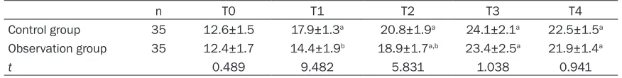
tistically higher than those at T0 (P<0.05). At T1-T2, the expressions of NSE in observation group were significantly lower than those in control group (P<0.05) (Table 5).
SjvO2 in observation group and control group at T1 was remarkably higher than that at T0 (P<0.05). SjvO2 in observation group at T1 was statistically higher than that in control group (P<0.05) (Table 6).
Ca-jvO2 in observation group and control group at T1 was statistically lower than that at T0 (P<0.05). Ca-jvO2 in observation group at T1 was statistically lower than that in control group (P<0.05) (Table 7).
PaO2 in observation group and control group at T1-T4 was remarkably higher than that at T0 (P<0.05). PaO2 in observation group at T1 was statistically higher than that in control group (P<0.05) (Table 8).
ERO2 in observation group and control group at T1-T2 was apparently lower than that at T0 (P<0.05). ERO2 in observation group at T1 was evidently lower than that in control group (P<0.05) (Table 9).
Clinical efficacy of patients in both groups
After treatment, the overall effective rate in observation group was 97.14% (34/35), which was significantly higher than that in control group (71.43%, 25/35) (P<0.05) (Table 10).
Discussion
At present, CPB has been widely applied in the operation of heart disease, but there are vari-ous uncertain factors affecting the operation results in the process of CPB. For example, ischemia-reperfusion injury of organs, immedi-ate contact between blood and non-physiologi-cal vessels and mechaninon-physiologi-cal shearing may cause damage to the lungs, thereby leading to inflammatory response in the body. Systemic
Table 1. MMSE score of patients in both groups before and after
operation (_x ± sd)
n operationBefore operation3 d after cognitive dysfunctionIncidence rate of Control group 35 27.98±1.22 25.99±1.64a 12 (34.29%) Observation group 35 28.04±1.31 28.25±1.63b 7 (20.00%) aP<0.05 vs. before operation, bP<0.05 vs. control group at 3 d after operation.
T3, the level of plasma S-100β protein in observation group were significantly lower than that in control group (P<0.05) (Table 4).
sta-Table 3. Plasma IL-6 levels in patients in both groups at different time points (_x ± sd)
n T0 T1 T2 T3 T4
Control group 35 34.1±4.3 42.9±4.5a 107.2±8.9a 155.8±15.7a 117.3±12.9a Observation group 35 33.6±3.5 40.2±4.1a 92.3±9.1a,b 140.6±16.3a,b 100.5±8.7a,b
t 2.301 2.016 6.795 4.572 6.356
[image:4.612.90.524.88.144.2]aP<0.05 vs. T0, bP<0.05 vs. control group at each time point.
Table 2. Plasma TNF-α levels in patients in both groups at different time points (_x ± sd)
n T0 T1 T2 T3 T4
Control group 35 350±246 439±55a 852±124a 1025±112a 859±37a
Observation group 35 347±214 394±40a,b 729±68a,b 934±97a,b 794±66a,b
t 0.019 4.482 5.317 3.958 6.274
[image:4.612.91.523.197.250.2]aP<0.05 vs. T0, bP<0.05 vs. control group at each time point.
Table 4. S-100β protein expression levels in patients in both groups at different time points (_x ± sd,
pg/mL)
n T0 T1 T2 T3 T4
Control group 35 103.7±9.2 122.4±7.5a 335±27a 427±22a 389±40a Observation group 35 102.8±7.4 120.7±3.3a 328±23a 370±44a,b 391±36a
t 0.359 1.148 1.167 6.352 0.137
aP<0.05 vs. T0, bP<0.05 vs. control group at T3.
Table 5. NSE expression levels in patients in both groups at different time points (_x ± sd, ng/mL)
n T0 T1 T2 T3 T4
Control group 35 12.6±1.5 17.9±1.3a 20.8±1.9a 24.1±2.1a 22.5±1.5a Observation group 35 12.4±1.7 14.4±1.9b 18.9±1.7a,b 23.4±2.5a 21.9±1.4a
t 0.489 9.482 5.831 1.038 0.941
aP<0.05 vs. T0, bP<0.05 vs. control group at T1-T2.
ably decrease the mortality rate of rats with sepsis [11]. It has been found that dexmedeto-midine can activate the complement system in the body, leading to the excessive synthesis and release of cytokines. It further results in endotoxemia and stimulates a variety of cells to synthesize and release such inflammatory fac -tors as TNF-α, IL-6 and IL-10 during heart valve replacement [12]. In this study, it was indicated that the levels of plasma TNF-α and IL-6 in patients of observation group and control group were significantly increased after CPB and reached the peak at T3, but they were signifi -cantly lower in observation group than those in control group, indicating that dexmedetomi-dine can effectively alleviate the inflammatory response in patients after operation. However, little is known concerning the related mecha-inflammatory response syndrome (SIRS) may
occur in severe cases, resulting in death of patients [8]. It has been reported that the selective α2 adrenergic receptor agonist has a favorable inhibitory effect on TNF-α produced during inflammatory response and attenuates inflammatory response [9]. According to animal experiments, dexmedetomidine can relieve the myocardial ischemia in rats, and thus signifi -cantly reduce the incidence rate of nerve injury after transient cerebral ischemia [10]. However, there is little research on whether dexmedeto-midine has a similar anti-inflammatory and pro -tective effect on the brain during heart valve replacement under CPB.
[image:4.612.89.524.312.366.2] [image:4.612.87.525.415.470.2]-nism. Previous study illustrated that the inci-dence rate of cognitive dysfunction was 15% at 1 week after joint replacement, but it was increased to 45% after coronary artery bypass grafting [13]. Results of this study revealed that there was no significant difference in the MMSE score between the two groups before operation, but the MMSE score of patients in
[image:5.612.88.388.96.151.2]T1-T4 than that at T0, while it was significantly lower at T1-T2 in observation group than that in control group, indicating that dexmedetomidine can relieve the NSE release after CPB during administration, but the plasma NSE level is not affected after drug withdrawal. The clinical detection of SjvO2, Ca-jvO2, PaO2 and ERO2 can reflect the metabolic status of brain tissues, in
Table 6. Comparison of SjvO2 between the two groups of patents at
different time points (_x ± sd, %)
n T0 T1 T2 T3 T4
Control group 35 65±5 72±6a 66±7 65±6 64±7
Observation group 35 66±6 81±8a,b 67±6 66±7 65±6
t 0.642 3.952 1.947 0.431 0.585
[image:5.612.89.387.209.262.2]aP<0.05 vs. T0, bP<0.05 vs. control group at T1.
Table 7. Comparison of Ca-jvO2 between the two groups of patents at
different time points (_x ± sd, mmol/L)
n T0 T1 T2 T3 T4
Control group 35 49±8 31±7a 47±9 57±8 56±7
Observation group 35 50±7 23±8a,b 46±8 58±7 57±8
t 0.503 4.286 1.382 0.741 0.575
[image:5.612.88.390.319.377.2]aP<0.05 vs. T0, bP<0.05 vs. control group at T1.
Table 8. Comparison of PaO2 between the two groups of patents at
different time points (_x ± sd, mmHg)
n T0 T1 T2 T3 T4
Control group 35 134±53 209±36a 178±38a 199±56a 203±51a Observation group 35 135±65 230±45a,b 189±36a 201±54a 203±50a
t 0.140 2.265 1.284 0.083 0
aP<0.05 vs. T0, bP<0.05 vs. control group at T1.
Table 9. Comparison of ERO2 between the two groups of patents at
different time points (_x ± sd, %)
n T0 T1 T2 T3 T4
Control group 35 38±7 26±8a 32±7a 41±8 40±7 Observation group 35 36±8 17±10a,b 29±8a 39±9 39±8
t 0.915 2.895 1.718 1.375 1.052
aP<0.05 vs. T0, bP<0.05 vs. control group at T1.
Table 10. Comparison of clinical efficacy between the two groups of
patients [n (%)]
n Cured Effective Ineffective Effective rate Control group 35 19 (54.29) 6 (17.14) 10 (28.57) 25 (71.43) Observation group 35 29 (82.86) 5 (14.29) 1 (2.9) 34 (97.14)a aP<0.05 vs. control group.
observation group was sig-nificantly higher than that in control group at 3 d af- ter operation, suggesting that dexmedetomidine can improve the cognitive fu- nction of patients after operation.
[image:5.612.89.388.433.488.2] [image:5.612.89.389.547.592.2]which the decline in SjvO2 indicates increased oxygen consumption or decreased oxygen sup-ply in the brain [19]. It was found in this study that SjvO2 and PaO2 in observation group at T1 were obviously higher than those in control group, while Ca-jvO2 and ERO2 in observation group were obviously lower than those in con-trol group. PaO2 was remarkably increased and ERO2 was extraordinarily decreased in both groups, suggesting that the brain metabolic rate and oxygen consumption of patients in observation group were decreased, the toler-ance ability of brain tissues to hypoxia was increased, and the brain injury was alleviated. At the same time, SjvO2 was higher than 50% in both groups of patients at each time point, indi-cating that no significant imbalance of cerebral oxygen supply and demand occurred in both groups during anesthesia [20]. After treatment, the overall effective rate in observation group (97.14%) was significantly higher than that in control group (71.43%), suggesting that dexme -detomidine presents good protective effect on the brain of patients and improves the patient’s quality of life.
Conclusion
In conclusion, dexmedetomidine can signifi -cantly reduce the cerebral oxygen metabolic rate, alleviate the brain tissue damage and relieve the inflammatory response during heart valve replacement, which finding provides new insights into an auxiliary mean for the heart valve replacement.
Disclosure of conflict of interest
None.
Address correspondence to: Ming Gao, Department of General Surgery, Zhejiang Putuo Hospital, No. 19 Wenkang Street, Donggang Street, Putuo Dis- trict, Zhoushan 316000, Zhejiang, China. Tel: +86-18058029488; Fax: +86-580-3031234; E-mail: Tielidong7sm@163.com
References
[1] Pagni S, Ganzel BL, Singh R, Austin EH, Mascio C, Williams ML, Akella PV and Trivedi JR. Clini-cal outcome after triple-valve operations in the modern era: are elderly patients at increased surgical risk? Ann Thorac Surg 2014; 97: 569-576.
[2] Chen TT, Jiandong-Liu, Wang G, Jiang SL, Li LB and Gao CQ. Combined treatment of
ulina-statin and tranexamic acid provides beneficial effects by inhibiting inflammatory and fibrino-lytic response in patients undergoing heart valve replacement surgery. Heart Surg Forum 2013; 16: E38-47.
[3] Liu Z, Wang Y, Wang Y, Ning Q, Zhang Y, Gong C, Zhao W, Jing G and Wang Q. Dexmedetomi-dine attenuates inflammatory reaction in the lung tissues of septic mice by activating cholin-ergic anti-inflammatory pathway. Int Immuno-pharmacol 2016; 35: 210-216.
[4] Fontes MT, McDonagh DL, Phillips-Bute B, Welsby IJ, Podgoreanu MV, Fontes ML, Staf-ford-Smith M, Newman MF, Mathew JP; Neuro-logic Outcome Research Group (NORG) of the Duke Heart Center. Arterial hyperoxia during cardiopulmonary bypass and postoperative cognitive dysfunction. J Cardiothorac Vasc Anesth 2014; 28: 462-466.
[5] Shaikh SI and Mahesh SB. The efficacy and safety of epidural dexmedetomidine and cloni-dine with bupivacaine in patients undergoing lower limb orthopedic surgeries. J Anaesthesi-ol Clin PharmacAnaesthesi-ol 2016; 32: 203-209.
[6] Engels M, Bilgic E, Pinto A, Vasquez E, Wollschläger L, Steinbrenner H, Kellermann K, Akhyari P, Lichtenberg A and Boeken U. A car-diopulmonary bypass with deep hypothermic circulatory arrest rat model for the investiga-tion of the systemic inflammainvestiga-tion response and induced organ damage. J Inflamm (Lond) 2014; 11: 26.
[7] Zhang J, Wang Z, Wang Y, Zhou G and Li H. The effect of dexmedetomidine on inflammatory response of septic rats. BMC Anesthesiol 2015; 15: 68.
[8] Wei Y, Hu J, Liang Y, Zhong Y, He D, Qin Y, Li L, Chen J, Xiao Q and Xie Y. Dexmedetomi- dine pretreatment attenuates propofol-in-duced neurotoxicity in neuronal cultures from the rat hippocampus. Mol Med Rep 2016; 14: 3413-3420.
[9] Pang XY, Fang CC, Chen YY, Liu K and Song GM. Effects of ulinastatin on perioperative in-flammatory response and pulmonary function in cardiopulmonary bypass patients. Am J Ther 2016; 23: e1680-e1689.
[10] Bulow NM, Colpo E, Pereira RP, Correa EF, Wac-zuk EP, Duarte MF and Rocha JB. Dexmedeto-midine decreases the inflammatory response to myocardial surgery under mini-cardiopulmo-nary bypass. Braz J Med Biol Res 2016; 49: e4646.
[11] Xiong B, Shi QQ and Miao CH. Dexmedetomi-dine renders a brain protection on hippocam-pal formation through inhibition of nNOS-NO signalling in endotoxin-induced shock rats. Brain Inj 2014; 28: 1003-1008.
ver-sus isoflurane anesthesia on brain injury after cardiac valve replacement surgery. J Cardio-thorac Vasc Anesth 2018; 32: 1581-1586. [13] Ueki M, Kawasaki T, Habe K, Hamada K,
Kawa-saki C and Sata T. The effects of dexmedetomi-dine on inflammatory mediators after cardio-pulmonary bypass. Anaesthesia 2014; 69: 693-700.
[14] Xiang H, Hu B, Li Z and Li J. Dexmedetomidine controls systemic cytokine levels through the cholinergic anti-inflammatory pathway. Inflam-mation 2014; 37: 1763-1770.
[15] Shou-Shi W, Ting-Ting S, Ji-Shun N and Hai-Chen C. Preclinical efficacy of Dexmedetomi-dine on spinal cord injury provoked oxidative renal damage. Ren Fail 2015; 37: 1190-1197. [16] Wang Z, Chen Q, Guo H, Li Z, Zhang J, Lv L and
Guo Y. Effects of dexmedetomidine on H-FABP, CK-MB, cTnI levels, neurological function and near-term prognosis in patients undergoing heart valve replacement. Exp Ther Med 2017; 14: 5851-5856.
[17] Al Nimer F, Thelin E, Nyström H, Dring AM, Svenningsson A, Piehl F, Nelson DW and Bel-lander BM. Comparative assessment of the prognostic value of biomarkers in traumatic brain injury reveals an independent role for se-rum levels of neurofilament light. PLoS One 2015; 10: e0132177.
[18] Zhang Q, Wu D, Yang Y, Liu T and Liu H. Effects of dexmedetomidine on the protection of hy-peroxia-induced lung injury in newborn rats. Int J Clin Exp Pathol 2015; 8: 6466-6473. [19] Steinbrenner H, Bilgic E, Pinto A, Engels M,
Wollschläger L, Döhrn L, Kellermann K, Boek-en U, Akhyari P and LichtBoek-enberg A. SelBoek-enium pretreatment for mitigation of ischemia/reper-fusion injury in cardiovascular surgery: influ-ence on acute organ damage and inflammato-ry response. Inflammation 2016; 39: 1363-1376.