Case Report
Comprehensive endovascular treatments of traumatic
pseudoaneurysm of the posterior communicating
artery combined with cavernous sinus fistula:
a case report and literature review
Wei Li1,2, Na Li2, Ming Lv1*, Youxiang Li1*
1Beijing Neurosurgical Institute, Capital Medical University, Beijing, China; 2Department of Neurosurgery, The
Second Affiliated Hospital of Xingtai Medical College, Xingtai, China. *Equal contributors.
Received January 27, 2018; Accepted October 8, 2018; Epub April 15, 2019; Published April 30, 2019
Abstract: A 23-year-old woman experienced a serious accident, resulting in multiple fractures of the skull base. Forty-five days after the injury, the patient presented with symptoms such as conjunctival congestion, ptosis of the eyelids, and blurred vision. Head CTA (CT angiography); revealed an arterial aneurysm in the cavernous sinus region. Angiography revealed a giant posterior communicating artery aneurysm combined with a cavernous sinus fistula. Subsequently, the patient was treated using a covered stent, coils, and Onyx- 18. After treatment, both the aneurysm and cavernous sinus fistula were embolized, and the clinical symptoms improved significantly. The combination of a covered stent, coils, and Onyx- 18 is a safe and effective treatment method for post-traumatic pseudoaneurysm of the posterior communicating artery combined with cavernous sinus fistula.
Keywords: Traumatic aneurysm, carotid-cavernous fistula, posterior communicating artery, endovascular, coil embolization, covered stent
Introduction
Traumatic cavernous sinus fistulas refer to an
abnormal vascular shunt between the cavern-ous sinus and surrounding vessels. The distri-bution of vessels around the cavernous sinus is
complex, and complicated cavernous sinus fis -tulas are easily formed after trauma, which pro-duces a variety of symptoms. The treatment for this condition is also complex. We report the case of a patient with a rare post-traumatic pseudoaneurysm of the posterior
communicat-ing artery combined with a cavernous sinus fis -tula, who was admitted to our center and was successfully treated using integrated approaches.
Case report
The 23-year-old woman sustained a head injury in an accident on October 6, 2016, and received treatment at another hospital. The right eye showed cyanosis and swelling, while the left
eye was asymptomatic. Head CT revealed sub-arachnoid hemorrhage, cerebral contusion and laceration, intracranial pneumatocele, fracture in the right frontal temporal bone, and multiple fractures of the skull base (Figure 1A and 1B). The following clinical diagnoses were made: multiple injury, cerebral contusion and lacera-tion, subarachnoid hemorrhage, intracranial pneumatocele, fracture of the right frontal tem-poral bone, multiple fractures of the skull base, and forehead scalp laceration. The forehead scalp laceration was treated by debridement and stitching. Meanwhile, drug therapy was administered, after which, the patient’s condi-tion gradually improved. On November 20, 2016, the patient was found to have ptosis of the left eyelid, and was readmitted to the other hospital for treatment. A physical examination revealed such symptoms as the enlargement of the left pupil by about 5 mm, the absence of
direct and indirect light reflexes, limited eyeball
eyeball, and dysdipsia. Head CT and CTA per-formed at the other hospital revealed a giant aneurysm in the left cavernous sinus region (Figure 1C). For further diagnosis and treat-ment, the patient was transferred to our hospital.
Endovascular treatment was performed under general anesthesia on December 22, 2016. Both femoral arteries were punctured. A 5F Navien catheter (EV3, Plymouth, MN, USA) was placed in the left internal carotid artery, and a 5F Guiding catheter (Godman USA) was placed in the right vertebral artery. A 4 mm × 7 mm Hyperform balloon (EV3, Plymouth, MN, USA)
was placed at the fistula of the posterior com -municating segment in the left internal carotid artery. Angiography of the left internal carotid artery revealed no development of aneurysm
and cavernous sinus fistula in the proximal to the balloon after balloon inflation, thus illus -trating that no rupture existed in the cavernous segment of the internal carotid artery. An- giography of the right vertebral artery revealed that the posterior communicating artery pseu-doaneurysm developed and the cavernous
sinus was inflated. The balloon was emptied,
and an Echelon-10 microcatheter (EV3, Ply- mouth, MN, USA) was placed at the rupture of
elon-10 microcatheter to completely block the
aneurysm fistula. The contrast injection via the vertebral artery confirmed that the aneurysm and cavernous sinus fistula had not developed.
The left internal carotid artery balloon was with-drawn, and left internal carotid angiography revealed the development of the aneurysm. A 4 mm × 10 mm WILLIS covered stents (Mi- croPort, Medical Company Shanghai, China) covered stent was placed at the posterior com-municating artery segment of the internal carotid artery (Figure 2A-G). Postoperative angiography revealed that the left internal carotid artery had been repaired, the distal branch had developed well, and the
pseudoan-eurysm and cavernous sinus fistula had not
developed (Figure 2H).
Two hours before the surgery, the patient was given 200 mg of aspirin and 300 mg of
clopido-grel at a draught. Moreover, 8 ml of tirofiban
(dosage: 5 mg, 100 ml) was intravenously injected and micropumped at the rate of 3 ml/h until 12:00 noon the following day. Bayaspirin
(100 mg qd) and clopidogrel (75 mg qd) were
given since 8:00 am the following day.
On the 3rd day post operation, the patient’s left
pupil diameter was about 5 mm, direct and indi-Figure 1. Preoperative CT images.
A. CT image reveals subarachnoid hemorrhage in the ambient cistern and intracranial pneumatocele after trauma. B. CT bone window sagittal image shows the fracture of the slope of the dorsum sella. C. CTA image shows the intracranial aneurysm after the development of eyelid ptosis.
the left posterior communicat-ing artery via the right verte-bral artery. Micro angiography revealed the location of the rupture. The coil could not be installed as planned because
of the fast blood flow.
Oc-clusion of the posterior seg-ment of the left internal carot-id artery with a balloon was performed again to reduce the
blood flow. Thereafter, three
ev3 coils (3D 4 mm × 12 cm; 3 mm × 8 cm; and Helix 2 mm × 8 cm) (EV3, Plymouth, MN,
USA) were placed quickly and
successively from the far end to the near end via the Echelon-10 microcatheter to block the posterior
communi-cating artery aneurysm
fistu-la. Angiography revealed that
the fistula still developed.
[image:2.612.89.376.73.358.2]rect light reflexes
disappe-ared, limitation of eye adduc-tion was better than the pre-operative level, conjunctival congestion was absent, and ptosis of the eyelid was better than before treatment. The patient’s blurred vision and hoarse voice both improved compared to preoperative lev-els. The dysdipsia symptom was absent. Review of cranial MRI showed thrombosis in the aneurysm and its shrinkage. Angiograms at 3 months after the operation. There revealed that the pseudoaneurysm and
cavernous sinus fistula disap -pearanced (Figure 3A and
3B). The patient’s left and right pupils diameter was ab- out 3mm with normal, direct
and indirect light reflexes.
Such symptoms as limitation of eye adduction, conjunctival congestion and eyelid ptosis were absent. The patient’s blurred vision and hoarse voice both improved com-pared to preoperative levels.
Discussion
Barrow [1] classified carotid cavernous fistulas into two types: high flow and low flow.
Traumatic carotid cavernous
fistula is often a high-flow fis -Figure 2. Angiograms in the operative. A. Lateral image of the left internal
ca-rotid artery showing the balloon placed at the suspicious rupture of the pos-terior communicating artery segment of the internal carotid artery. B. Lateral angiography of the right vertebral artery shows the rupture of the posterior communicating artery and the formation of the pseudoaneurysm and fis -tula. C. Lateral image of the right vertebral artery showing the Echelon-10 microcatheter placed at the rupture of the posterior communicating artery.
[image:3.612.90.375.71.644.2]tula. The general treatment for this condition is balloon occlusion.
The combination of a pseudoaneurysm with a
cavernous sinus fistula induced by the posteri -or communicating artery after trauma is rare. Only seven cases of this condition have been reported to date [2-8]. The anatomic relation-ship around the cavernous sinus determines the pathogenesis of the disease. The possible mechanism underlying the formation of an internal carotid artery aneurysm in the dura after trauma is as follows: 1) the fracture of the skull base directly injures the internal carotid artery; 2) the head injury excessively stretches and twists the siphon segment of the internal carotid artery; and 3) the internal carotid artery is impacted by the surrounding bony structures [9, 10]. In this case, the CT scan performed after injury did not reveal an aneurysm, but a review of the head CT scan performed 30 days after the injury revealed a parasellar aneurysm. We considered that the aneurysm was caused by trauma without any congenital cause. It is well-known showed that the posterior commu-nicating artery originates from subdural [11,
12]. Cavernous sinus fistulas do not form natu -rally after a posterior communicating artery injury. In this case, the patient had a clear trau-ma history and extensive bone fractures in the slope and dorsum sella. These structures are adjacent to the area of the posterior communi-cating artery from the internal carotid artery. Therefore, we considered that both excessive stretch-induced posterior communicating ar- tery injury and cavernous sinus dural rip caused
region, and the induced pressure differences between the posterior communicating artery and the cavernous sinus. If the cavernous sinus
fistula had not formed, the pseudoaneurysm
would have bursted as the pressure increased, and this would have been very dangerous for the patient.
The clinical symptoms of these diseases are generally as follows: cavernous sinus and ocu-lomotor nerve compression symptoms, con-junctival congestion, proptosis, ptosis of the eyelids, eyeball movement disorder, blurred vision, and murmur in the orbital region. The case reported by Chen [6] presented polyuria and hyponatremia. They argued that the poste-rior communicating artery provided pituitary blood supply, and the injury to this artery dis-rupted the transportation of antidiuretic hor-mone produced by the pituitary stalk, thereby resulting in diabetes insipidus and electrolyte disorder. In the present case, the cavernous sinus symptoms were less severe and chemo-sis was mild, with no proptochemo-sis and murmur in the orbital region, which could be attributed to the shorter time of onset from injury. These symptoms may also occur over time. Our patient also presented supervened dysdipsia, which was absent in the patients reported in the other cases. This may be related to the posterior cir-culation ischemia resulting from blood steal induced by the posterior communicating artery rupture into the cavernous sinus. After treat-ment, however, these symptoms also improved
[image:4.612.90.376.74.214.2]significantly.
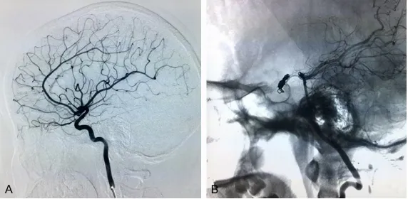
Figure 3. Angiograms at 3 months after the operative. A. Left internal carotid artery angiography reveals the disappearance of the pseudoaneurysm and cavernous sinus fistula. B. Angiography of the right vertebral artery reveals the disappearance of the pseudoaneurysm and cavernous sinus fistula.
by the fractures led to the pseudoaneurysm and
cavern-ous sinus fistula. We analyzed
the phenomenon by which the pseudoaneurysm and
cavern-ous sinus fistula were not
formed in the seven previously reported cases and in our patient immediately after trau-ma. We considered that the pseudoaneurysm and
cavern-ous sinus fistula might have
At present, the treatment of this rare disease is complicated. Tytle [2] and Fu [3] performed cra-niotomy and clipped the pseudoaneurysm.
Weaver [4] performed coil embolization at the
posterior communicating artery via the verte-bral artery. Oran [5] performed coil emboliza -tion via the internal carotid artery and vertebral artery. Chen [6] and Du [8] performed coil
embolization via the internal carotid artery. Jin bo [7] performed coil embolization at the pos -terior communicating artery via the vertebral
artery with simultaneous sealant embolization.
The purpose of the treatment is to eliminate
the cavernous sinus fistula and pseudoaneu -rysm. Moreover, these treatments differ from the common balloon occlusion of the carotid
cavernous fistula. The risk of craniotomy is high
and pseudoaneurysms lack a vessel wall; this may lead to massive hemorrhage during sur-gery. Therefore, endovascular treatment is the best choice. We used the following procedure for endovascular treatment: coil and Onyx- 18
embolization was used to eliminate the poste -rior cerebral artery pole of the poste-rior com-municating artery via the vertebral artery.
Although we encountered difficulties when placing the first coil because of the high blood flow, we could still place the coil successfully
with the assistance of a balloon. The place-ment of a covered stent in the internal carotid artery blocked the internal carotid artery pole of the posterior communicating artery, so that
the cavernous sinus fistula and pseudoaneu -rysm were trapped. The placement of a cov-ered stent to place the covcov-ered stents was rela-tively simple and fast, and helped maintain the normal anatomy of the internal carotid artery [13-16]. If we have used many coils to pack the pseudoaneurysm instead of a covered stent, the coils would have led to severe occupation
effect. To our knowledge, this is the first report
on the use of such a variety of treatments in a case of post-traumatic pseudoaneurysm of the posterior communicating artery combined with
cavernous sinus fistula. Moreover, the treat -ments proved safe and effective in treating this rare condition.
Acknowledgements
This research received no specific grant from
any funding agency in the public, commercial,
or not-for-profit sectors.
Disclosure of conflict of interest
None.
Address correspondence to: Ming Lv, Beijing Neurosurgical Institute, Capital Medical University, Beijing 100050, China. E-mail: dragontiger@163. com; Youxiang Li, Department of Interventional Neuroradiology, Beijing Tiantan Hospital, Capital Medical University, Beijing 100050, China. E-mail: liyouxiang@263.net
References
[1] Barrow DL, Spector RH, Braun IF, Landman JA, Tindall SC and Tindall GT. Classification and treatment of spontaneous carotid-cavernous sinus fistulas. J Neurosurg 1985; 62: 248-256. [2] Tytle TL, Loeffler CL and Steinberg TA. Fistula
between a posterior communicating artery an-eurysm and the cavernous sinus. AJNR Am J Neuroradiol 1995; 16: 1808-1810.
[3] Fu Y, Ohata K, Tsuyuguchi N and Hara M. Direct surgery for posttraumatic carotid-cavernous fistula as a result of an intradural pseudoaneu -rysm: case report. Neurosurgery 2002; 51: 1071-1073.
[4] Weaver KD, Ewend MG and Solander S. Suc-cessful transarterial guglielmi detachable coil embolization of posttraumatic posterior com -municating artery-cavernous sinus fistula: technical note. Neurosurgery 2003; 52: 458-460; discussion 460-451.
[5] Oran I, Parildar M, Memis A and Dalbasti T. Posttraumatic intradural internal carotid ar-tery-cavernous sinus fistula associated with ipsilateral carotid dissection. Transarterial em-bolization with detachable coils. Interv Neuro -radiol 2004; 10: 63-68.
[6] Chen Y, Jiang DY, Tan HQ, Wang LH, Chen XY and Sun JH. Successful transarterial guglielmi detachable coil embolization of post-traumatic fistula between a posterior communicating ar -tery aneurysm and the cavernous sinus. A case report. Interv Neuroradiol 2009; 15: 435-441.
[7] Jinbo Y, Jun L, Kejie M and Zheng Z. Successful transarterial embolization of a posttraumatic fistula between a posterior communicating ar -tery aneurysm and the cavernous sinus: a case report. Turk Neurosurg 2015; 25: 776-778.
[9] Enomoto H, Shibata T, Ito A and Harada T. Trau-matic aneurysm of the supraclinoid internal carotid artery: report of a case. Neurosurgery 1984; 15: 700-702.
[10] Masana Y and Taneda M. Direct approach to a traumatic giant internal carotid artery aneu-rysm associated with a carotid-cavernous fis -tula. Case report. J Neurosurg 1992; 76: 524-527.
[11] Pedroza A, Dujovny M, Artero JC, Umansky F, Berman SK, Diaz FG, Ausman JI and Mirchan -dani HG. Microanatomy of the posterior com-municating artery. Neurosurgery 1987; 20: 228-235.
[12] Rhoton JA. The supratentorial arteries. Neuro-surgery 2002; 51: S1-53-S51-120.
[13] Li MH, Li YD, Gao BL, Fang C, Luo QY, Cheng YS, Xie ZY, Wang YL, Zhao JG, Li Y, Wang W, Zhang BL and Li M. A new covered stent de-signed for intracranial vasculature: application in the management of pseudoaneurysms of the cranial internal carotid artery. AJNR Am J Neuroradiol 2007; 28: 1579-1585.
[14] Wang YL, Ma J, Ding PX, Li YD, Han XW and Wu G. Treatment of post-traumatic carotid-cavern-ous fistulas with the willis covered stent. A pre -liminary prospective study. Interv Neuroradiol 2012; 18: 172-177.
[15] Wang W, Li MH, Li YD, Gu BX and Lu HT. Recon-struction of the internal carotid artery after treatment of complex traumatic direct carotid-cavernous fistulas with the willis covered stent: a retrospective study with long-term follow-up. Neurosurgery 2016; 79: 794-805.