ARTIGO ORIGINAL
AMP
STUDENT
RESUMO
Introdução: A vitamina D tem vindo a ganhar importância como um modificador do risco de diabetes. O objetivo deste estudo foi avaliar a associação entre os níveis séricos de vitamina D e a prevalência de retinopatia diabética em pacientes com diabetes tipo 1. Material e Métodos: Estudo retrospetivo de uma população de doentes com diabetes tipo 1, seguidos num centro hospitalar terciário português. Os pacientes foram incluídos se tivessem uma avaliação oftalmológica e um doseamento dos níveis de 25-hidroxivitamina D no mesmo ano. Os ajustes para eventuais variáveis confundidoras foi realizado recorrendo a uma análise de regressão logística. Resultados: Foram incluídos 182 doentes (47% sexo masculino), dos quais 57% (n = 103) demonstravam sinais de retinopatia diabética. Foi encontrada uma associação significativa entre níveis inferiores de 25-hidroxivitamina D circulante sérica e uma maior prevalência de retinopatia diabética, depois do ajuste para os confundidores incluídos (duração da diabetes, taxa de filtração glomerular estimada, idade, sexo, controlo metabólico, estação do ano, dislipidemia e hipertensão) (OR = 0,94; 95% IC 0,90 - 0,99, p = 0,023). Uma maior duração da diabetes, assim como um pior controlo metabólico, mantiveram também uma associação significativa com uma maior prevalência de retinopatia diabética na análise multivariada (OR = 1,20; 95% IC 1,13 - 1,27, p < 0,001; OR = 4,13; 95% IC 1,34 - 12,7, p = 0,013, respetivamente).
Conclusão: Níveis inferiores de vitamina D séricos demonstraram-se associados a uma prevalência superior de retinopatia diabética em pacientes com diabetes tipo 1, após o ajuste para eventuais variáveis confundidoras. Futuramente, estudos experimentais poderão estabelecer as vias moleculares implicadas nesta associação, assim como um papel concreto da suplementação na prevenção das complicações microvasculares da diabetes.
Palavras-chave: Deficiência de Vitamina D; Diabetes Mellitus Tipo 1; Portugal; Retinopatia Diabética; Vitamina D
Association between Serum Vitamin D and Diabetic
Retinopathy in Portuguese Patients with Type 1 Diabetes
Associação entre a Vitamina D Sérica e a Retinopatia
Diabética numa População Portuguesa com Diabetes Tipo 1
1. Faculty of Medicine. Porto University. Porto. Portugal.
2. Department of Ophthalmology. Centro Hospitalar de Entre o Douro e Vouga. Santa Maria da Feira. Portugal. 3. Department of Ophthalmology. Centro Hospitalar de São João. Porto. Portugal.
4. Department of Endocrinology, Diabetes and Metabolism. Centro Hospitalar de São João. Porto. Portugal. 5. Department of Surgery and Physiology. Faculty of Medicine. University of Porto. Porto. Portugal. 6. Department of Anesthesiology. Centro Hospitalar de São João. Porto. Portugal.
7. I3S Instituto de Investigação e Inovação em Saúde. University of Porto. Porto. Portugal.
Autor correspondente: Manuel Falcão. [email protected]
Recebido: 30 de novembro de 2019 - Aceite: 06 de fevereiro de 2020 | Copyright © Ordem dos Médicos 2020
Miguel LOPES1, Rita LAIGINHAS1,2, Carolina MADEIRA3, João Sérgio NEVES4,5, Margarida BARBOSA1,6,7,
Vitor ROSAS3, Davide CARVALHO1,4, Fernando FALCÃO-REIS3,5, Manuel FALCÃO3,5
Acta Med Port 2020 Jul-Aug;33(7-8):459-465 ▪ https://doi.org/10.20344/amp.12890 ABSTRACT
Introduction: Recently, vitamin D has gained importance as a diabetes risk modifier. Our aim was to assess the association between serum vitamin D levels and the prevalence of diabetic retinopathy in patients with type 1 diabetes.
Material and Methods: Retrospective review of a population of patients with type 1 diabetes followed in a Portuguese tertiary center. Patients were included if they had an ophthalmological evaluation and a serum 25-hydroxyvitamin D level determination within the same year. Logistic regression analysis was used to adjust for possible confounders.
Results: We included 182 patients (47% male), and 57% (n = 103) had signs of diabetic retinopathy. We found a significant association between lower circulating levels of 25-hydroxyvitamin D levels and a greater prevalence of diabetic retinopathy after adjusting for confounders (duration of diabetes, estimated glomerular filtration rate, age, sex, metabolic control, season, dyslipidemia and hypertension) (OR = 0.94; 95% CI 0.90 - 0.99, p = 0.023). Longer duration of diabetes and worse metabolic control also remained associated with diabetic retinopathy in the multivariate analysis (OR = 1.20; 95% CI 1.13 - 1.27, p < 0.001 and OR = 4.13; 95% CI 1.34 - 12.7, p = 0.013, respectively).
Conclusion: Lower levels of vitamin D were associated with an increased prevalence of diabetic retinopathy in patients with type 1 diabetes, after adjusting for possible confounders. Future controlled studies may elucidate the molecular routes for this association as well as the role of supplementation in the prevention of diabetes microvascular complications.
Keywords: Diabetes Mellitus, Type 1; Diabetic Retinopathy; Portugal; Vitamin D; Vitamin D Deficiency
INTRODUCTION
Diabetic retinopathy (DR) is the most sight-threatening complication of type 1 diabetes (T1D), and it remains one of the most worrisome emerging causes of blindness,
par-ticularly among working-age individuals.1,2 It is known that
metabolic control and duration of diabetes are
independ-ent risk factors for DR.3 However, other pathophysiological
mechanisms involved in this condition are not fully clarified.
Vitamin D has been known to regulate bone and
min-eral homeostasis.4 However, aside from this role, several
recent studies have linked vitamin D to the pathogenesis
of diabetes5 and there is growing evidence that vitamin D
can interfere with the mechanisms involved in diabetes and
its complications.6 In in vitro studies, vitamin D showed a
ARTIGO ORIGINAL
AMP
STUDENT
protective effect on cytokine-suppressed insulin release by
1,25(OH)2D3, both in human and mouse models.8 Some
studies have pointed out vitamin D deficiency as a risk fac-tor for T1D as the genetic background of vitamin D
deficien-cy has demonstrated strong associations with T1D.9
Vitamin D has been shown to play an important part in the regulation of angiogenesis, being an inhibitor of the
HIF-1α/VEGF pathway in cancer cells.10 Other more recent
studies highlighted the molecular basis of a protective effect of vitamin D in the specific context of DR, as it decreased diabetes-induced reactive oxygen species (ROS) and in-hibited the ROS/TXNIP/NLRP3 inflammasome pathway, protecting against retinal vascular damage and cell
apopto-sis.11 One study has highlighted both the anti-inflammatory
and anti-angiogenic properties of vitamin D in the retinal tis-sue in vivo, indicating that it prevents the development and progression of DR by decreasing inflammation and
neo-vascularization.12 Genetics have also been emphasized in
this field, showing that a polymorphism, which increases the transcription of the vitamin D receptor and thus its biological
activity, results in less severe DR in T1D patients.13
There is increasing evidence suggesting an associa-tion between vitamin D deficiency and diabetic retinopathy
in type 2 diabetes patients.14–17 However, literature remains
scarce regarding this association in T1D patients, and some
contradictory findings have been reported.18–21
The present study aims to evaluate the association be-tween serum vitamin D levels and the prevalence of DR in T1D patients.
MATERIAL AND METHODS
This was an observational retrospective study of a pop-ulation of T1D patients followed in a tertiary eye care center (Centro Hospitalar Universitário de S. João - CHUSJ, Porto, Portugal). All procedures were performed in accordance with the ethical standards of the Ethics Committee of Centro Hospitalar de São João/Faculty of Medicine of Porto Uni-versity and with the 1964 Helsinki Declaration and its later amendments or comparable ethical standards, having the present study been approved by this institution without the need of written informed consent given the nature of the study.
Sampling
Our target population consisted of all patients diag-nosed with T1D from the catchment area of CHUSJ (ap-proximately 300 000 people), aged over 18 years, and alive at the time of the study (2018).
According to the last available census and epidemiologi-cal records from 2011, the hospital referral region of this ter-tiary center has a population of around 340 000, 200 000 of
which are aged above 18 years.22 A Portuguese study has
estimated the prevalence of T1DM in Northern Portugal to
be 1.66 cases per 1000.23 Assuming these estimations, a
population of about 332 T1DM patients is expected in our referral region.
By merging medical registries, we found 469 subjects
diagnosed with T1D between 1950 and 2017 and we re-viewed their medical records. From those, we excluded the cases in which the diagnosis of T1D was doubtful by ana-lyzing medical records (uncertainty about type 1 or type 2 diabetes), as well as those without an ophthalmological ob-servation and a serum vitamin D level determination within the same year.
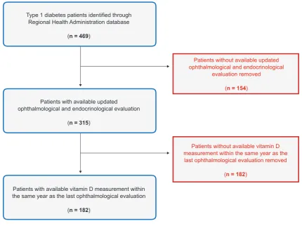
After applying the exclusion criteria, a total of 182 pa-tients were included. The sample size met our initial esti-mates, and we were able to consider the sample as an ad-equate representation of our target population. The patient selection process is further explained in Fig. 1.
Clinical and laboratorial data
We reviewed clinical and laboratory data from the se-lected sample. Patients were excluded if they had incom-plete medical records, namely regarding date of T1D diag-nosis, complete ophthalmological evaluation mentioning diabetic retinopathy stage, and serum 25-hydroxyvitamin D level determination within the same year of the last ophthal-mological evaluation.
DR status was classified as present/absent based on fundoscopy findings in medical records from the last oph-thalmological evaluation. We considered absent DR if there was no evidence in medical records of any DR manifesta-tion (microaneurysms, cotton wool spots, intra-retinal hem-orrhages, or macular edema).
We also analyzed subjects based on the presence and
type of DR according to the following stratification24: mild
non-proliferative DR; moderate to severe non-proliferative DR and proliferative retinopathy (PDR). People with panret-inal photocoagulation were also included in the PDR group. If there was asymmetric retinopathy, the eye in worse condi-tion was used for classificacondi-tion.
Total 25-hydroxyvitamin D levels (ng/mL) were obtained from the patient’s clinical records. The month when the measurement was assessed was also recorded to verify if there was any circannual pattern of variation. Vitamin D deficiency was defined at a 25-hydroxyvitamin D level ≤ 20 ng/mL, insufficiency at 21 - 29 ng/mL and sufficiency at 30 – 100 ng/mL, according to international clinical practice
guidelines.25
Data regarding sex, age, disease duration and age at T1D diagnosis were also collected. We extracted all avail-able glycated hemoglobin (HbA1c) levels in the medical re-cords for each patient. We used the final averaged value as an estimate of the patient’s metabolic control.
Further data extracted for analysis included history of hypertension, dyslipidemia, peripheral neuropathy, and ne-phropathy (classified as present/absent).
ARTIGO ORIGINAL
AMP
STUDENT
Kidney Disease Epidemiology Collaboration) equation.26
Potential confounders
In this analysis, we included potential confounders in the relationship between 25-hydroxyvitamin D levels and DR. We opted to evaluate the potential effect of age and sex in our model.
Duration of the disease (years) and metabolic control (dichotomized as good/bad) were included, as they are known to the be the main determinants for DR in patients
with T1D.3 Good metabolic control was defined as an
aver-age HbA1c < 7%, according to the latest recommendations
of the American Diabetes Association.27
History of hypertension and dyslipidemia were also in-cluded as potential confounders as they have been
associ-ated with vitamin D status.28,29 It is known that vitamin D
deficiency (< 20 ng/mL) and insufficiency (20 - 29 ng/mL)
are common among patients with chronic kidney disease,30
and we included an adjustment for glomerular filtration rate to account for this potential confounder effect.
Vitamin D is a secosteroid that is generated in the skin
under the influence of sunlight.31 As human sun exposure
is related to the seasonal position of the sun, vitamin D is generated at different rates according to the season. To en-compass this potential variation, we included an adjustment for the season by dividing the year according to common seasonal sun positions in Portugal.
All things considered, we performed a multiple logistic
regression model for predicting the presence of DR, and included 25-hydroxyvitamin D levels as well as the adjust-ments for the following variables: duration of T1D, age, sex, estimated glomerular filtration rate, hypertension, dyslipi-demia, metabolic control and seasonal variation.
Statistical analysis
Descriptive statistics have been used to document com-parability of baseline characteristics. Normal distribution was checked by histogram visual inspection. Categorical variables are presented as frequencies and percentages, and continuous variables are represented as mean ± stand-ard deviation or median (interquartile range), according to their normal distribution status. Comparisons of vitamin D levels in patients with and without DR and among different types of DR were performed using the Mann-Whitney and Kruskal-Wallis tests.
We analyzed the association between vitamin D levels and any DR, including potential adjustments, using multiple logistic regression. We also analyzed this relationship in the
different DR stages. A two-sided value of p < 0.05 was
con-sidered statistically significant. The statistical analysis was performed using IBM SPSS Statistics 25 software.
RESULTS
From our initial population of 469 patients, a total of 182 patients presented a serum vitamin D level determination within the same year as an ophthalmological evaluation.
Figure 1 – Patient selection flow chart
Type 1 diabetes patients identified through Regional Health Administration database
(n = 469)
Patients with available updated ophthalmological and endocrinological evaluation
(n = 315)
Patients without available updated ophthalmological and endocrinological
evaluation removed
(n = 154)
Patients with available vitamin D measurement within the same year as the last ophthalmological evaluation
(n = 182)
Patients without available vitamin D measurement within the same year as the last ophthalmological evaluation removed
ARTIGO ORIGINAL
AMP
STUDENT
Of this sample, 86 (47%) were male. The mean age at the evaluation was 43 ± 14 years and the mean disease dura-tion was 20 ± 12 years.
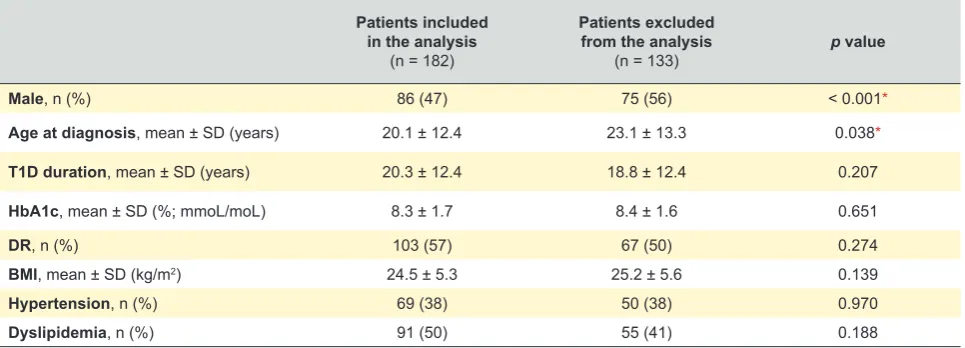
In the evaluation, 103 patients (57%) had fundus mani-festation of DR, and the mean 25-hydroxyvitamin D level was 21.3 ± 10.9 ng/mL. Table 1 details the clinical and bio-chemical characteristics of our sample, according to DR status. Further details regarding the characteristics of the excluded patients can be found in Table 2.
According to the adopted criteria, 53% (n = 96) of pa-tients had vitamin D deficiency (25-hydroxyvitamin D level ≤
20 ng/mL), 26% (n= 48) had vitamin D insufficiency (21 - 29
ng/mL) and 21% (n= 38) had adequate levels (30 – 100 ng/
mL).
Seasonal variation of the 25-hydroxyvitamin D levels ac-cording to the month in which the samples were taken is demonstrated in Fig. 1. The levels followed the expected circannual pattern of variation according to the sun expo-sure in Portugal: the highest levels were recorded during
the summer, as the lowest occurred during the winter. We stratified vitamin D levels in different duration of dia-betes intervals, and we found that mean 25-hydroxyvitamin D levels were constantly higher in patients without DR. For patients with a duration of diabetes < 25 years, the mean 25-hydroxyvitamin D serum level was 19.1 ng/mL if DR was
present (n= 46) and 21.7 ng/mL if DR was absent (n= 71),
p = 0.42. For patients with a duration of diabetes ≥ 25 years,
mean 25-hydroxyvitamin D serum level was 21.3 ng/mL if
DR was present (n= 57) and 31.6 ng/mL if DR was absent
(n= 8) (p = 0.02).
We also stratified vitamin D levels by type of DR: no DR
(n= 81), mild DR (n= 35), moderate to severe DR (n= 30)
and PDR (n= 36). We did not find any statistically
signifi-cant association between the mean vitamin D levels and the severity of the DR (mean vitamin D in patients without DR = 22.6 ± 11.2 ng/mL, with mild DR = 21 ± 9.5 ng/mL; with moderate to severe DR = 20.3 ± 11.8 ng/mL; with PDR =
19.8 ± 10.8 ng/mL; p = 0.56).
Table 1 – Clinical and biochemical characteristics of type 1 diabetes patients by diabetic retinopathy status
No Retinopathy
(n = 79) Retinopathy(n = 103) p value
Male, n (%) 35 (44) 51 (50) 0.55
Age, mean ± SD (years) 37.1 ± 13.2 46.7 ± 13.9 < 0.001*
T1D duration, mean ± SD (years) 11.6 ± 8.2 27.0 ± 10.9 < 0.001*
HbA1c, mean ± SD (%; mmoL/moL) 8.0 ± 1.7 (64 ± 19) 8.5 ± 1.7 (69 ± 19) 0.04*
BMI, mean ± SD (kg/m2) 24.5 ± 5.0 24.5 ± 5.5 0.70
25(OH)D, mean ± SD (ng/mL) 22.7 ± 11.1 20.3 ± 10.7 0.30
eGFR, mean ± SD (mL/min/1.73m2) 114.6 ± 20.3 95.7 ± 31.4 < 0.001*
Albuminuria, median (IQR) (mg/24h) 6 (7.6) 11.5 (45.8) < 0.001*
Hypertension, n (%) 16 (21) 53 (51) < 0.001*
Dyslipidemia, n (%) 24 (32) 67 (66) < 0.001*
Data are presented as mean ± standard deviation (SD), median (interquartile range) (IQR) or as n (%). The values represent means at the last evaluation. T1D: type 1 diabetes mellitus; HbA1c: glycated hemoglobin; BMI: body mass index; 25(OH)D: 25-hydroxyvitamin D; eGFR: estimated glomerular filtration rate *: Values were significantly different between groups in the unadjusted analysis, assuming a two-sided value of p < 0.05 as statistically significant.
Table 2 – Clinical and biochemical characteristics of the patients included in the initial sample (n= 315)
Patients included in the analysis
(n = 182)
Patients excluded from the analysis
(n = 133) p value
Male, n (%) 86 (47) 75 (56) < 0.001*
Age at diagnosis, mean ± SD (years) 20.1 ± 12.4 23.1 ± 13.3 0.038*
T1D duration, mean ± SD (years) 20.3 ± 12.4 18.8 ± 12.4 0.207
HbA1c, mean ± SD (%; mmoL/moL) 8.3 ± 1.7 8.4 ± 1.6 0.651
DR, n (%) 103 (57) 67 (50) 0.274
BMI, mean ± SD (kg/m2) 24.5 ± 5.3 25.2 ± 5.6 0.139
Hypertension, n (%) 69 (38) 50 (38) 0.970
Dyslipidemia, n (%) 91 (50) 55 (41) 0.188
ARTIGO ORIGINAL
AMP
STUDENT
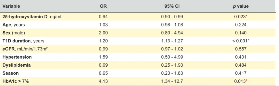
We performed a multiple logistic regression model for predicting the presence of DR, and included 25-hydroxyvi-tamin D levels as well as the adjustments for the duration of T1D, age, sex, estimated glomerular filtration rate, hyperten-sion, dyslipidemia, metabolic control and seasonal variation (Table 3). After the adjustments, duration of T1D, metabolic control and lower 25-hydroxyvitamin D levels remained as-sociated with a greater prevalence of diabetic retinopathy.
The final model was able to explain 61% (Nagelkerke R2) of
the outcome.
DISCUSSION
In the present study, we found that lower serum 25-hy-droxyvitamin D levels were associated with an increased prevalence of DR in a sample of Portuguese T1D patients after adjusting for possible confounders. It is possible that higher serum levels of vitamin D may have a protective ef-fect on the development of DR.
Recently, vitamin D has gained importance as a type 2 diabetes risk modifier. This was initially suggested by the observation of a seasonal variation in the glycemic con-trol where it was perceived to be worse during the winter
season.32 Additional evidence of a role played by vitamin D
in type 2 diabetes comes from a large number of observa-tional and cross-secobserva-tional studies that showed an inverse relationship between prevalence of type 2 diabetes and its complications and the vitamin D levels. Although there are several epidemiological studies reporting the association
between vitamin D and DR in patients with diabetes14–21,
to our knowledge, only four evaluated the association
be-tween DR and vitamin D in T1D.18–21 Kaur et al18 performed
a cross-sectional study of 517 T1D adolescents aged 8 – 20 years and found lower vitamin D levels in DR cases. Shimo et al19 reached the same conclusions when analyzing 75
young T1D Japanese patients. However, the previous stud-ies are limited in the fact that they only analyzed younger individuals, which means that most of them would not have had enough time to have developed retinal disease.
Con-trasting with these results, Joergensen et al20 reviewed 227
T1D patients and concluded that vitamin D deficiency was a predictor of all-cause mortality but not development of
microvascular complications in the eye and kidney. How-ever, this study only analyzed cases with severe vitamin D deficiency [defined as a plasma 25(OH)D3 value equal to or below the 10% percentile - 6.2 ng/mL], and less severe deficits are underrepresented. In addition, the authors high-light, as a limitation, that the results could be due to the lack of power based on a low number of events, as the hazard ratio for both retinopathy and albuminuria was above 1 in patients with severe vitamin D deficiency. Finally, a study
from the EURODIAB Prospective Complications Study21
concluded that vitamin D levels were not independently as-sociated with non-proliferative or proliferative retinopathy. In this study, the adjustments did not include the duration of disease, the strongest predictor for DR development. As seen in our sample, it is possible that confounders can con-ceal eventual associations.
Although the role of vitamin D in the pathophysiology of diabetes, is a subject of debate in the scientific community, there are several mechanisms that support a protective ef-fect. Vitamin D can affect the key processes that are in-volved in diabetes: insulin resistance, insulin secretion and
inflammation.28 Vitamin D can affect insulin resistance by
stimulating the insulin receptor expression, as it enhances the transcriptional activation of the insulin receptor gene. This has been shown in an in-vitro study exposing human promonocytic cells to active vitamin D, leading to an in-creased expression of mRNA encoding for insulin receptors and a 1.3 fold increase in glucose transport when compared
to untreated cells.33 The effect of vitamin D on insulin
secre-tion and beta cell occurs through the regulasecre-tion of extracel-lular calcium concentration and flux through the beta cell, as
insulin secretion is a calcium dependent process.28 In
addi-tion, glucose and sulfonylurea-stimulated insulin secretion was shown to be lower in the islets of vitamin D-deficient rats, when compared to the islets of vitamin D-sufficient rats
or vitamin D deficient rats with vitamin D replacement.34
Vi-tamin D also interferes in the downregulation of NF-kB, a major transcription factor for TNF-α and other
pro-inflam-matory molecules.35 Moreover, vitamin D can have other
im-portant effects in inflammatory cytokines by interfering with a number of other genes or transcription factors involved in
Table 3 – Multivariate regression analysis of retinopathy in type 1 diabetes patients
Variable OR 95% CI p value
25-hydroxyvitamin D, ng/mL 0.94 0.90 - 0.99 0.023*
Age, years 1.03 0.98 - 1.08 0.224
Sex (male) 2.00 0.80 - 4.94 0.140
T1D duration, years 1.20 1.13 - 1.27 < 0.001*
eGFR, mL/min/1.73m2 0.99 0.97 - 1.02 0.557
Hypertension 1.59 0.50 - 4.99 0.431
Dyslipidemia 0.69 0.25 - 1.93 0.484
Season 0.65 0.23 - 1.83 0.417
HbA1c > 7% 4.13 1.34 - 12.7 0.013*
ARTIGO ORIGINAL
AMP
STUDENT
cytokine generation.36
Regarding DR, the literature remains scarce in detailing the mechanisms which can explain the protective properties of vitamin D. Research has indicated that vitamin D pre-vents the development and progression of DR by inhibiting inflammation and neovascularization in retinal tissues. In
an experimental study using rats with DR,12 the
histopatho-logical examination showed the appearance of edema and disordered arrangement of retinal tissues in a non-vitamin D supplemented group, but milder pathological changes in a supplemented group. Vitamin D has also been shown to reduce the level of diabetes-induced proinflammatory cy-tokines, playing a protective role against retinal vascular
damage and cell apoptosis.11
However, despite having pointed out an association be-tween lower serum vitamin D levels and the presence of DR, our study failed to show an association with the sever-ity of the disease. This could be due to the small sample size for this comparison and its necessary adjustments, or because vitamin D does not play such an important part in the pathophysiology of the later stages of the disease. An important aspect to point out is the overall low mean level of serum vitamin D in our sample. The main perspec-tive for this problem is the specific context of vitamin D defi-ciency epidemiology in Portugal. A recent study carried out by the Dr. Ricardo Jorge National Institute of Health con-cluded that, in comparison to other European populations, the mean 25-hydroxyvitamin D levels observed in
Portu-guese populations were relatively lower.37 Another major
epidemiological study carried out in 2017, the PROMETS study, which, among other things, evaluated the vitamin D levels of a nation-wide sample, showed that only 14.4% of
the patients had vitamin D levels greater than 20 ng/mL.38
Two other studies carried out in older patients yielded
simi-lar results,39,40 which suggests that the lower overall vitamin
D levels we found may be related to a populational trend. From this data, we can conclude that there is still some con-troversy regarding the cut-offs established for the vitamin D levels, and that definitions should consider the geographical patterns.
The major limitation of our study is its retrospective de-sign. From the study, it is not possible to evaluate vitamin D levels in the past and how eventual changes in its levels interfered with the development of DR. Also, the fact that we only analyzed a single hospital narrows the generalizability
of the results. Another limitation of our study is that we were not able to determine quantitative sun exposure and dietary supplementation, as well as some systemic medication, which are important factors that can influence vitamin D
lev-els.41 Vitamin D measurements are not routinely performed
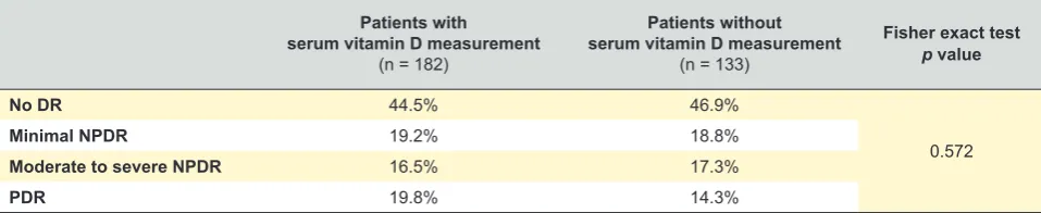
in all patients and this could lead to a selection bias by the attending physician. However, we did not find significant dif-ferences between the sample with the vitamin D measure-ments and the initial sample in terms of disease severity (Table 4).
Nevertheless, our study presents as a major strength the fact that it evaluates both adolescents and adults with T1D. This stands out from previous studies, as most of these only included young individuals and therefore, not many years of systemic disease. By including older patients, we present a wider and more comprehensive evaluation of this compari-son throughout the whole age spectrum.
It is possible that vitamin D is implicated in the patho-physiology of DR, and further studies are needed so that we can establish the biological pathways for this phenomenon, as well as evaluate future roles for supplementation.
CONCLUSION
This study supports an association between lower vi-tamin D levels and a greater prevalence of DR in T1D pa-tients. Unfortunately, despite improvements in glucose con-trol, diabetic complications are still common, leading most researchers to look for other preventive measures.
PROTECTION OF HUMAN SUBJECTS
The authors declare that the procedures were followed according to the regulations established by the Clinical Re-search and Ethics Committee and to the Helsinki Declara-tion issued by World Medical AssociaDeclara-tion.
CONFIDENTIALITY OF DATA
The authors declare having followed the protocols in use at their working center regarding patients’ data publica-tion.
CONFLICTS OF INTEREST
All authors declare no conflicts of interest.
FINANCIAL SOURCES
No funding was received for this research.
Table 4 – Prevalence of different DR status according to request of serum vitamin D measurement by the attending physician
Patients with serum vitamin D measurement
(n = 182)
Patients without serum vitamin D measurement
(n = 133)
Fisher exact test p value
No DR 44.5% 46.9%
0.572
Minimal NPDR 19.2% 18.8%
Moderate to severe NPDR 16.5% 17.3%
PDR 19.8% 14.3%
ARTIGO ORIGINAL
AMP
STUDENT
REFERENCES
1. Prokofyeva E, Zrenner E. Epidemiology of major eye diseases leading to blindness in Europe: a literature review. Ophthalmic Res. 2012;47:171-88.
2. Congdon N, Zheng Y, He M. The worldwide epidemic of diabetic retinopathy. Indian J Ophthalmol. 2012;60:428.
3. Yau JW, Rogers SL, Kawasaki R, Lamoureux EL, Kowalski JW, Bek T, et al. Global prevalence and major risk factors of diabetic retinopathy. Diabetes Care. 2012;35:556-64.
4. Thomas MK, Demay MB. Vitamin D deficiency and disorders of vitamin D metabolism. Endocrinol Metab Clin North Am. 2000;29:611-27. 5. Muscogiuri G, Sorice GP, Ajjan R, Mezza T, Pilz S, Prioletta A, et al.
Can vitamin D deficiency cause diabetes and cardiovascular diseases? Present evidence and future perspectives. Nutr Metab Cardiovasc Dis. 2012;22:81-7.
6. Chakhtoura M, Azar ST. The role of vitamin d deficiency in the incidence, progression, and complications of type 1 diabetes mellitus. Int J Endocrinol. 2013;2013:148673.
7. Riachy R, Vandewalle B, Belaich S, Kerr-Conte J, Gmyr V, Zerimech F, et al. Beneficial effect of 1,25 dihydroxyvitamin D3 on cytokine-treated human pancreatic islets. J Endocrinol. 2001;169:161-8.
8. Mathieu C. Vitamin D and diabetes: where do we stand? Diabetes Res Clin Pract. 2015;108:201-9.
9. Penna-Martinez M, Badenhoop K. Inherited variation in Vitamin d genes and type 1 diabetes predisposition. Genes. 2017;8:2-9.
10. Ben-Shoshan M, Amir S, Dang DT, Dang LH, Weisman Y, Mabjeesh NJ. 1alpha,25-dihydroxyvitamin D3 (Calcitriol) inhibits hypoxia-inducible factor-1/vascular endothelial growth factor pathway in human cancer cells.. Mol Cancer Ther. 2007;6:1433-9.
11. Lu L, Lu Q, Chen W, Li J, Li C, Zheng Z. Vitamin D 3 protects against diabetic retinopathy by inhibiting high-glucose-induced activation of the ROS/TXNIP/NLRP3 inflammasome pathway. J Diabetes Res. 2018;2018:1-11.
12. Ren Z, Li W, Zhao Q, Ma L, Zhu J. The impact of 1,25-dihydroxy vitamin D3 on the expressions of vascular endothelial growth factor and transforming growth factor-β1in the retinas of rats with diabetes. Diabetes Res Clin Pract. 2012;98:474-80.
13. Taverna MJ, Selam JL, Slama G. Association between a protein polymorphism in the start codon of the vitamin D receptor gene and severe diabetic retinopathy in C-peptide-negative type 1 diabetes. J Clin Endocrinol Metab. 2005;90:4803-8.
14. Alcubierre N, Valls J, Rubinat E, Cao G, Esquerda A, Traveset A, et al. Vitamin D deficiency is associated with the presence and severity of diabetic retinopathy in type 2 diabetes mellitus. J Diabetes Res. 2015;2015.
15. Luo BA, Gao F, Qin LL. The association between vitamin D deficiency and diabetic retinopathy in type 2 diabetes: a meta-analysis of observational studies. Nutrients. 2017;9:307.
16. Dow C, Mancini F, Rajaobelina K, Boutron-Ruault MC, Balkau B, Bonnet F, et al. Diet and risk of diabetic retinopathy: a systematic review. Eur J Epidemiol. 2018;33:141-56.
17. Zhang J, Upala S, Sanguankeo A. Relationship between vitamin D deficiency and diabetic retinopathy: a meta-analysis. Can J Ophthalmol / J Can d’Ophtalmologie. 2017;52:219-24.
18. Kaur H, Donaghue KC, Chan AK, Benitez-Aguirre P, Hing S, Lloyd M, et al. Vitamin D deficiency is associated with retinopathy in children and adolescents with type 1 diabetes. Diabetes Care. 2011;34:1400-2. 19. Shimo N, Yasuda T, Kaneto H, Katakami N, Kuroda A, Sakamoto F,
et al. Vitamin D deficiency is significantly associated with retinopathy in young Japanese type 1 diabetic patients. Diabetes Res Clin Pract. 2014;106:e41-3.
20. Joergensen C, Hovind P, Schmedes A, Parving HH, Rossing P. Vitamin D levels, microvascular complications, and mortality in type 1 diabetes. Diabetes Care. 2011;34:1081-5.
21. Engelen L, Schalkwijk CG, Eussen SJ, Scheijen JL, Soedamah-Muthu SS, Chaturvedi N, et al. Low 25-hydroxyvitamin D2 and
25-hydroxyvitamin D3 levels are independently associated with macroalbuminuria, but not with retinopathy and macrovascular disease in type 1 diabetes: the EURODIAB prospective complications study. Cardiovasc Diabetol. 2015;14:67.
22. Instituto Nacional de Estatística. Census 2011. [accessed 2018 Aug 20]. Available from: https://censos.ine.pt/xportal/ x m a i n ? x p i d = C E N S O S & x p g i d = i n e _ c e n s o s _ p u b l i c a c a o _ det&contexto=pu&PUBLICACOESpub_boui=156638623&PUBLICACO ESmodo=2&selTab=tab1&pcensos=61969554.
23. Jorge Z, Lacerda Nobre E, Macedo A, Jácome de Castro J. Prevalência da diabetes mellitus tipo 1 em Portugal, 1995-1999: coorte de jovens do sexo masculino. Acta Med Port. 2003;16:251–3.
24. Wilkinson CP, Ferris FL 3rd, Klein RE, Lee PP, Agardh CD, Davis M, et al. Proposed international clinical diabetic retinopathy and diabetic macular edema disease severity scales. Ophthalmology. 2003;110:1677-82. 25. Holick MF, Binkley NC, Bischoff-Ferrari HA, Gordon CM, Hanley DA,
Heaney RP, et al. Evaluation, treatment, and prevention of vitamin D deficiency: an endocrine society clinical practice guideline. J Clin Endocrinol Metab. 2011;96:1911-30.
26. Levey AS, Stevens LA, Schmid CH, Zhang Y, Castro AF, Feldman HI, et al. A new equation to estimate glomerular filtration rate. Ann Intern Med. 2009;150:604-12.
27. American Diabetes Association. 6. Glycemic targets: Standards of medical care in diabetes-2019. Diabetes Care. 2019;42:S61-70. 28. Rashidbeygi E, Rahimi MH, Mollahosseini M, Yekaninejad MS, Imani
H, Maghbooli Z, et al. Associations of vitamin D status and metabolic dyslipidemia and hypertriglyceridemic waist phenotype in apparently healthy adults. Diabetes Metab Syndr Clin Res Rev. 2018;12:985-90. 29. Chen S, Sun Y, Agrawal DK. Vitamin D deficiency and essential
hypertension. J Am Soc Hypertens. 2015;9:885-901.
30. Jean G, Souberbielle JC, Chazot C. Vitamin D in chronic kidney disease and dialysis patients. Nutrients. 2017;9:1-15.
31. Issa CM. Vitamin D and type 2 diabetes mellitus. Adv Exp Med Biol. 2017;996:193-205.
32. Campbell IT, Jarrett RJ, Keen H. Diurnal and seasonal variation in oral glucose tolerance: studies in the Antarctic. Diabetologia. 1975;11:139-45.
33. Maestro B, Campion J, Davila N, Calle C. Stimulation expression in U-937 by and human insulin responsiveness cells promonocytic D3 of insulin for glucose receptor transport. Endocr J. 2000;47:383-91. 34. Norman AW, Frankel JB, Heldt AM, Grodsky GM. Deficiency inhibits
pancreatic secretion of insulin. Science. 1980;209:823-5.
35. Cohen-Lahav M, Douvdevani A, Chaimovitz C, Shany S. The anti-inflammatory activity of 1,25-dihydroxyvitamin D3 in macrophages. J Steroid Biochem Mol Biol. 2007;103:558-62.
36. Gysemans CA, Cardozo AK, Callewaert H, Giulietti A, Hulshagen L, Bouillon R, et al. 1,25-dihydroxyvitamin D3 modulates expression of chemokines and cytokines in pancreatic islets: implications for prevention of diabetes in nonobese diabetic mice. Endocrinology. 2005;146:1956-64.
37. Bettencourt A, Marinho A, Martins A, Silva D, Martins Da Silva B, Pinho E, et al. Vitamina D e autoimunidade na população portuguesa. Inst Nac Saúde Doutor Ricardo Jorge. 2018:9-11.
38. Raposo L, Martins S, Ferreira D, Guimarães JT, Santos AC. Vitamin D, parathyroid hormone and metabolic syndrome - the PORMETS study. BMC Endocr Disord. 2017;17:71.
39. Santos MJ, Fernandes V, Garcia FM. Carência de vitamina D numa população hospitalar: uma fotografia pela perspetiva laboratorial. Acta Med Port. 2015;28:726-34.
40. Santos A, Amaral TF, Guerra RS, Sousa AS, Álvares L, Moreira P, et al. Vitamin D status and associated factors among Portuguese older adults: results from the Nutrition UP 65 cross-sectional study. BMJ Open. 2017;7:e016123.