225
THE ENHANCED EPITHELIALIZATION POTENTIAL OF THE AQUEOUS EXTRACT
OF PRUNELLA VULGARIS IN THERMALLY INJURED RATS
Cheng-Cun Li1,*, Qing-Guo Meng2, Xiu-Qin Liang3 and Ji-Zhou Xiao4
1 Burns Department, Affiliated Hospital of Taishan Medical College, Taian, 271000, Shandong, China 2 Department of General Surgery, The People’s Hospital of Dongping, Dongping, 271500, Shandong, China 3 Department of Rehabilitation, Tai’an Rongjun Hospital, Taian, 271000, Shandong, China
4 Burns Department, The Second People’s Hospital of Liaocheng, Linqing, 252600, Shandong, China
*Corresponding author: chengcunli@gmail.com
Received: May 12, 2015; Revised: May 26, 2015; Accepted: May 27, 2015; Published online: December 29, 2015
Abstract:The aim of the present study was to assess the wound-healing efficiency of the Prunella vulgaris extract (PVE) on thermally injured rats. Sixty adult, female rats were divided into four groups, with group I serving as a sham control (with-out a burn injury). A burn was inflicted on the dorsum of all the rats in the second, third and fourth groups. Group II rats served as the burn-injury control group without any treatment. The burn-injured areas of the rats in the third and fourth groups were treated topically by 1% silver sulfadiazine (SSD) cream or 10% PVE-based cream, respectively. Biochemical (antioxidant, lipid peroxidation, hydroxyproline, mucopolysaccharide), morphological and histopathological evaluations showed the burn-healing efficiency of PVE to be better than that of the SSD group. In conclusion, topical application of PVE enhanced collagen synthesis and antioxidant levels by suppressing lipid peroxidation as well as increasing proliferation of keratinocytes, and thus positively influenced wound-healing.
Key words: Prunella vulgaris; burn injury; silver sulfadiazine; histopathology; collagen
INTRODUCTION
Thermal injury is an acute wound result from expo-sure to heat and chemicals. Burn injuries represent a significant health burden all over the world. Every year, there are 18.1 burn injuries per 100,000 individu-als resulting in hospitalization (Kindividu-alson et al., 2012). Burn wounds are associated with a significant risk of mortality, as well as significant physical, functional and psychiatric sequela in survivors. Therefore, burn wounds represent a significant effect to patients, health care professionals, and the health care system (Brewster et al., 2013). Wound healing is a complex process during which multiple epithelial and stromal tissue elements interact to reconstitute an injured or-gan. At the onset of epithelial injury, the activation of multiple signaling pathways initiates a cascade of events that reestablish tissue architecture and barrier
function. The process of tissue regeneration and re-pair after epithelial injury can be divided into three sequential phases: inflammation, proliferation (epi-thelialization) and remodeling (Guo and DiPietro, 2010; Gurtner et al., 2008). The common treatment regime for burn wounds would be a topical applica-tion of a medicament to enhance wound healing by minimizing free radical generation and inflammatory reaction as well as to prevent opportunistic infections by microbes.
phenolic acids, flavonoids, triterpenes, alkaloids and tannins, might positively influence the healing pro-cess owing to their strong antioxidant activities by scavenging the highly reactive free radicals, which are involved in delaying the wound-healing process (Manca et al., 2015). In addition, those phytocompo-nents can also act as anti-inflammatory and antimi-crobial agents.
Prunella vulgaris L. (Labiatae) is a perennial herb that has been used in traditional medicine in China and western countries for treating sore throat, fever, for accelerating wound healing as well as for derma-tosis and skin allergies. In China, P. vulgaris is called ‘Xia Ku Cao’ and has a long history in therapeutic use as an antipyretic, and more recently for antikeratitis purposes (Kim et al., 2007). It was likewise employed as a major additive component herb in the Chinese traditional functional beverage Guangdong cool tea (Cheung and Zhang, 2008). In South Korea, P. vulgaris is applied to patients with goiter, dermatitis and skin allergy (Sun et al., 2005). In addition, it also exhibits antioxidative, anti-allergic, anti-inflammatory and an-timicrobial activities (Ryu et al., 2000; Psotova et al., 2003). Some studies also indicated its usefulness in the treatment of atopic dermatitis (Lee et al., 2008) and in providing photoprotection (Vostálová et al., 2010).
Phytochemical studies indicate that P. vulgaris is rich in phenolics such as rosmarinic, ellagic, ursolic and caffeic acids and flavonoids (quercetin), saponins and triterpenes (Vostálová et al., 2010). Rosmarinic acid (RA), the main phenolic acid constituent, is known for its wide-ranging biological activities, i.e. antioxidative, anti-inflammatory, antimutagenic, an-timicrobial (Skottova et al., 2004; Petersen and Sim-monds, 2003). Ursolic acid, a triterpene constituent of PVE, has been described as an effective inhibitor of UVA-modulated expression and activation of me-talloproteinase 2, whose increased level is a visible marker of destruction during skin photo-aging (Lee et al., 2003). Hence, the present study was designed to verify the wound-healing activity of PVE attributed to its various phenolic compounds with antioxidant, antimicrobial and anti-inflammatory activity in a thermally injured rat model.
MATERIALS AND METHODS Plant material
P. vulgaris were collected from Shandong, PR China, in August 2014 and authenticated by Professor Xiao-Ming Xin of the Taishan Medical University in China. A voucher specimen (no. PV23407) was deposited in the Herbarium of the Department of Pharmacology, Taishan Medical University, China.
Aqueous P. vulgaris extract
Two hundred g of dried whole plant material was added to 1000 mL of deionized water and incubated at 100oC for 50 min and the resulting aqueous extract
was filtered using Whatman filter paper. The residue from the filtration was extracted again by the same procedure. The filtrates obtained were combined and then lyophilized by freeze dryer and stored at -20oC
before the study. The final yield of aqueous extract was 28%.
Experimental animals
Sixty female albino Sprague-Dawley (SD) rats pro-cured from the Central Animal House Block, China, weighing 250±20 g were used in the study. The ani-mals were housed at 22±3°C, 55±5% humidity, 12 h light/dark cycles with ad libitum access to food and water. Rats were allowed to adapt to their environ-ment conditions for at least 10 days before initiation of the experiment. Animal experiments were conducted according to Institutional Animal Ethics Committee (Reg. No.14/2014/CPCSEA) guidelines and in com-pliance with the Animal Welfare Act, implementing Animal Welfare Regulations and the principles of the Guide for the Care and Use of Laboratory Animals.
Induction of burn injury
burn-ing with a square steel plate (1.5 cm2) heated at 100˚C,
for 10 s to create (Haghdoost et al., 2013). Postopera-tive pain was treated with acetaminophen for three days.
Experimental design
The 60 adult, female SD rats were divided into four groups. Group I served as a negative control group (n=6), without any burn injury. Group II served as a burn-injury control group (n=18) and did not re-ceive any treatment (positive control). The thermally injured areas of the rats in groups III and IV were treated topically with 0.5 g of 1% silver sulfadiazine (SS) cream (n=18) and 0.5 g of 10% (w/w) PVE-based cream (n=18), respectively. Six rats each of groups II, III and IV were killed by intraperitoneal administra-tion of sodium pentobarbitone (100 mg/kg bwt) at three different stages (3rd, 7th and 14th day) to assess the
progress of wound healing of each treatment group by comparing with group II. The harvested wound was bisected into half, one portion was embedded in par-affin for histological analysis and the other half stored at -80°C for biochemical analyses. The stored wound tissue was homogenized (10%) in 0.02 M potassium-phosphate buffer, pH 7.6 (1:10 w/v) and centrifuged at 3000xg at 4°C. The clear supernatant was used for further analyses.
Wound contraction rate and re-epithelialization
Wound contraction was calculated as a percentage of the original wound size by using wound image. Briefly, at time intervals ranging from 0 through 14 days after wounding, wounds were photographed using a digi-tal camera (20 MP). Digidigi-tal ImageJ software (NIH, Bethesda, MD) was used to measure the wound size and contraction rate with using the following formula:
[(Area of the original wound minus the area of the actual wound (on day n; n = 3, 7 and 14 days) divided by the area of the original wound] ×100. The re-epithelialization area of the wound was the area between the wound edge and the newly formed epithelial edge on days 3, 7 and 14.
Tensile strength
The tensile strength of a wound represents the degree of wound-healing processes. A samples of healing skin (from groups II, III and IV), along with normal skin (group I) were excised for tensile strength measure-ment using a computerized tensiometer (EZTEST I, Shimadzu Corp., Japan). Tensile strength was calcu-lated using the following formula:
Tensile strength (N/cm2)
= breaking force (N)/area (cm2);
where area (cm2) = thickness (cm) × width (cm) (Baie
and Sheikh, 2000).
Biochemical analysis
Dermal antioxidants and lipid peroxidation
The dermal antioxidant enzymes, i.e. superoxide dis-mutase (SOD) (Marklund and Marklund, 1974), cata-lase (CAT) (Sinha et al., 1972), glutathione peroxidase (GPx) (Rotruck et al., 1973) and reduced glutathione content (GSH) (Moron et al., 1979), were estimated according to previously reported methods. The pro-tein content of dermal tissue was determined based on the Biuret reaction of the BCA kit (Thermo, Rockford, IL, USA). The level of lipid peroxides in the dermal tissue was determined by the method of Devasagayam and Tarachand (1987).
Estimation of hydroxyproline
for 20 min and measured at 557 nm using a spectro-photometer. The hydroxyproline value was extrapo-lated from the standard curve.
Estimation of mucopolysaccharide
One hundred mg of delipidated residue of the tissues (skin) were hydrolyzed with 2 mL of 3N HCl at 80°C for 4 h. The hydrolyzed material was neutralized and used for estimation of hexosamine and hexuronic acid. Hexosamine was estimated by the method of Wagner (1979) and hexuronic acid by the method of Bitter and Muir (1962).
Enzyme-linked immunosorbent assay
Nitric oxide (NO) levels were quantified by the ELISA method using the manufactured protocol (Caymen Chemical, Ann Arbor, MI) in the granulation tissue of experimental rats. The tissue (skin) lysates were prepared by homogenizing frozen wound samples in dounces containing 500 μL of 0.1 M phosphate buffer (pH 7.4), 1 mM EDTA and 10 μM indomethacin. The samples were normalized to 1 mg/1 mL.
Histopathological studies
Wounded tissue was stained by the hematoxylin and eosin (H&E) method. Portions of granulation tissues were fixed in 10% formalin. The washed tissues were dehydrated in descending grades of isopropanol and cleared in xylene. The tissues were then embedded in molten paraffin wax. Sections were cut at 5-µm thick-nesses and stained with H&E. The sections were then viewed under a light microscope (Nikon microscope ECLIPSE E400, model 115, Japan) for histopathologi-cal changes.
Statistical analysis
The values were expressed as a mean±±standard de-viation (SD). Differences between each group were assessed by one-way analysis of variance (ANOVA) using SPSS (Statistical Package for Social Sciences) version 21. Post hoc testing was performed for
inter-group comparisons using the least significance dif-ference (LSD) test. P-values <0.05 were considered as statistically significant.
RESULTS
Effects of SSD and PVE on wound contraction, epithelialization and tensile strength
Wound contraction and re-epithelialization are the significant characteristics of the healing process. The progression of wound contraction was evaluated by microscopic and macroscopic observations. The ef-fects of SSD and PVE on wound contraction rate in experimental rats are shown in Table 1. On the 3rd and
7th day after wound creation, the wound contraction
rate of the SSD- and PVE-treated groups was 27.34, 60.82% and 32.45 and 71.58%, respectively. On the 14th day, the wound contraction rate of the SSD- and
PVE-treated groups was 85.76 and 99.74%, respec-tively. However, the burn-injured group showed the maximum contraction (65.23%) on day 14. The per-centage of wound contraction in the SSD- and PVE-treated groups was significantly higher (P<0.05 and P<0.01) than that of the burn-injured rats.
Fig. 1 depicts the effects of SSD and PVE on the wound re-epithelialization area in experimental rats. During the 3rd and 7th days, the re-epithelialization
area concomitantly increased on both treatment groups in comparison with the burn-injured group. The re-epithelialization area on the 14th day was also
higher in both treatment groups, but in the PVE-treat-ed group the wound area completely closPVE-treat-ed without leaving a raw wound behind, which indicates its
effec-Table 1. Effects of SSD and PVE on wound contraction in ex-perimental rats.
Groups 3 Days7 14
Burn-injured 21.24±2.29 45.23±5.21 65.23±7.24 SSD-treated 27.34±1.78 a* 60.82±7.23 a* 85.76±9.76 a*
PVE-treated 32.45±2.24 a** 71.58±6.38 a** 99.74±9.96 a**
tive wound-healing ability when compared with the SSD group, which still showed some wound area had yet to heal. The order of the re-epithelialization rate is PVE>SSD>burn-injured group.
Fig. 2 shows the effect of SSD and PVE on tensile strength in the dermal tissue of experimental rats. The tensile strengths of the burn-injured rats were substantially lower (P<0.05) than those of the nega-tive control group. A significant increase in tensile strength was observed in the SSD- and PVE-treated group (P<0.05 and P<0.01, respectively) compared with the burn-injured group. Our previous results of wound contraction, re-epithelialization rate and ten-sile strength demonstrated similar positive results of the enhanced wound-healing ability of PVE.
Effect of SSD and PVE on dermal antioxidants
The effects of SSD and PVE on dermal antioxidants in experimental rats are shown in Table 2. Increasing trends in the levels of antioxidants were observed in the SSD- and PVE-treated group on the 3rd and 7th
days, but the slight reduction on 14th day especially
in PVE group, was also observed. Thermal wounds treated with SSD (P<0.05) and PVE (P<0.01) revealed a maximum antioxidant activity on the 7th day
com-pared to control and burn-injured rats.
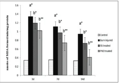
Effects of SSD and PVE on lipid peroxidation
The effects of SSD and PVE on lipid peroxidation (LPO) in the dermal tissue of experimental rats are shown in Fig. 3. LPO levels were substantially elevated on days 3 and 7 in the burn-injured rats, but started to decline after treatment with SS and PVE. The lev-els of malondialdehyde (MDA) were restored to nor-mal (P<0.01) in the PVE-treated rats on the 14th day,
whereas they were significantly reduced (P<0.05) in the SSD-treated rats on the 14th day. In the case of the
burn-injured group, the levels of MDA did not revert back to near normal till the 14th day due to non-medication. Fig. 1. Effects of SSD and PVE on re-epithelialization area of
dermal tissue in experimental rats. Values were expressed as mean±SD for six rats in each group. Statistical significance (P val-ue): **P<0.01, *P<0.05 compared with the burn-injured group (b).
Fig. 2. Effects of SSD and PVE on tensile strength (14th day) in
dermal tissue of experimental rats. Values were expressed as mean±SD for six rats in each group. Statistical significance (P
value): **P<0.01, *P<0.05 compared with the control (a) and burn-injured group (b).
Table 2. Effects of SSD and PVE on dermal antioxidant in ex-perimental rats.
Groups Parameters Day 3 Day 7 Day 14 Control
(No burn) Burn-injured SSD-treated PVE-treated
SOD (U/mg of protein)
1.35±0.13 1.65±0.18 a*
1.98±0.19 2.05±0.22 b*
1.41±0.22 1.48±0.16 2.82±0.29 b** 3.57±0.41 b**
1.37±0.14 1.12±0.15 a* 1.84±0.13 b* 1.63±0.19 b* Control
(No burn) Burn-injured SSD-treated PVE-treated
CAT (μm H2O2/min/ mg protein)
3.47±0.35 3.76±0.44 3.92±0.22 4.01±0.38 b*
3.43±0.39 2.95±0.28 a* 4.35±0.45 b* 5.07±0.68 b**
3.45±0.29 2.89±0.22 a* 3.69±0.36 b* 3.54±0.41 b* Control
(No burn) Burn-injured SSD-treated PVE-treated
GPx (nmole GSH utilized/ min/mg/ protein) 3.56±0.43 3.93±0.39 a*
4.05±0.42 4.17±0.34
3.52±0.26 3.01±0.39 a* 4.62±0.48 b* 4.85±0.51 b**
3.56±0.29 2.34±0.35 a** 3.91±0.36 b* 3.69±0.43 b* Control
(No burn) Burn-injured SSD-treated PVE-treated
Reduced glutathione (µg/mg protein)
3.55±0.22 3.97±0.45 a* 4.39±0.28 b* 4.28±0.33
3.51±0.34 3.31±0.37 4.48±0.45 b* 5.13±0.49 b**
3.56±0.30 2.72±0.28 a* 3.42±0.35 b* 4.07±0.41 b**
Effects of SSD and PVE on dermal
hydroxyproline, hexosamine and hexuronic acid
The collagen in granulation tissue was estimated by the breakdown of collagen into hydroxyproline, which is an index of collagen turnover. Table 3 demonstrates the effects of SSD and PVE on dermal hydroxyproline, hexosamine and hexuronic acid in experimental rats. The levels of dermal hydroxyproline, hexosamine and hexuronic acid were significantly elevated in SSD- and
PVE-treated animals (P<0.05 on 7th day and P<0.01
on 3rd and 7th day, respectively), but declined slightly
on the 14th day, when compared with the burn-injured
and negative control groups. No significant changes were noted between the negative control groups.
Effects of SSD and PVE on nitric oxide (NO)
Fig. 4 shows the effects of SSD and PVE on nitric oxide (NO) in the dermal tissue of experimental rats. Thermally injured rats showed a substantial reduction (P<0.01) in the levels of NO when compared with negative control rats on the 3rd, 7th and 14th days.
Treat-ment with SSD (P<0.05) and PVE (P<0.01) displaying a marked elevation in the levels of NO were noted on the 3rd, 7th and 14th days when compared with
burn-injured rats.
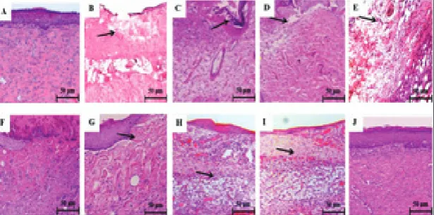
Effects of SSD and PVE on histological changes
The effects of SSD and PVE on histological changes in the dermal tissue of experimental rats are shown in Fig. 5. Transection of granulated tissue of negative control rats showed a normal architecture of the der-mis and epidermal layer with proper distribution of collagen fibers without any infiltration (H&E, 100X) (5A). Transection of burn-injured (untreated) rats on days 3 and 7 showed an irregular pattern of collagen fiber distribution with ulceration and infiltration of inflammatory cells (5B and C), whereas on the 14th
day the presence of immature collagen fibers and less inflammatory cells (5D) were noted. Transection of the SSD-treated group showed an irregular pattern of collagen fibers with ulceration and infiltration of inflammatory cells on the 3rd day (5E), whereas less
ulceration and infiltration with moderate collagen fiber were noted on the 7th and 14th days (5F and G).
Transection of the PVE-treated group showed mild ulceration with inflammatory cells on the 3rd day (5H),
whereas few infiltrations were noted on the 7th day
(5I). On the 14th day, a uniform accumulation of
col-lagen fibers with prominent bundles consisting of a moderate number of fibroblast without any inflam-matory cells (5J) was seen, similar to that of the nega-tive control group. A close examination of granulation
Fig. 3. Effects of SSD and PVE on lipid peroxidation (LPO) in dermal tissue of experimental rats. Values were expressed as mean±SD for six rats in each group. Statistical significance (P
value): **P<0.01, *P<0.05 compared with the control (a) and burn-injured group (b).
Table 3. Effects of SSD and PVE on dermal hydroxyproline, hex-osamine and hexuronic acid in experimental rats
Groups Param-eters Day 3 Day 7 Day 14 Control
(No burn) Burn-injured SS-treated PVE-treated
Hydroxy-proline (mg/100mg tissue)
3.13±0.36 3.19±0.32 3.41±0.27 b* 3.67±0.43 b*
3.15±0.23 3.28±0.32 a* 3.57±0.33 b* 3.98±0.39 b**
3.12±0.43 3.37±0.44 a* 3.45±0.36 b* 3.51±0.52 b* Control (No
burn) Burn-injured SS-treated PVE-treated
Hexos-amine (mg/g wet tissue)
6.57±0.54 6.67±0.45 6.79 ±0.48 6.83±0.62
6.52 ±0.58 6.75 ±0.43 a* 6.92 ±0.75 b* 7.03±0.73 b**
6.49 ±0.66 6.45 ±0.96 6.82 ±0.71
b* 6.76±0.80 b* Control
(No burn) Burn-injured SS-treated PVE-treated
Hexuronic acid (mg/g wet tissue)
8.27±0.86 8.24±0.53 8.26±0.48 8.24±0.74
8.28±0.53 8.21±0.61 8.40±0.72 8.48±0.56 b*
tissue sections revealed that tissue regeneration was much faster in the treated groups, especially the PVE-treated group, in comparison to unPVE-treated rats.
DISCUSSION
The healing of a skin burn is a multistep process in-volving several complex phases such as hemorrhage, inflammation, granulation tissue formation, re-epi-thelialization and the late remodeling phase of repair
Fig. 5. Effects of SSD and PVE on histological changes in dermal tissue of experimental rats. Transection of granulated tissue of control rats showed normal architecture of dermis and epidermal layer with proper distribution of collagen fibers without any infiltration (H&E, 100X) (5A). Transection of burn-injured (untreated) rats on 3rd and 7th days showed irregular pattern of collagen fibers with ulceration
and infiltration of inflammatory cells (5B and C), whereas on day 14, the presence of immature collagen fibers and less inflammatory cells (5D) was seen. Transection of SSD group showed irregular pattern of collagen fibers with ulceration and infiltration of inflammatory cells on the 3rd day (5E), whereas less ulceration and infiltration with moderate collagen fiber were noted on the 7th and 14th days (5F and
G). Transection of PVE group showed mild ulceration with inflammatory cells on the 3rd day (5H), whereas few infiltrations were noted
on the 7th day (5I). On the 14th day, a uniform accumulation of collagen fibers with prominent bundles consisting of moderate number
of fibroblast without any inflammatory cells (5J) was noted, similar to that of control group (H&E, 100X) (Scale bar-50µm).
Fig. 4. Effects of SSD and PVE on nitric oxide (NO) in dermal tissue of experimental rats. Values were expressed as mean±SD for six rats in each group. Statistical significance (P value): **P<0.01, *P<0.05 compared with the control (a) and burn-injured group (b).
(Kim et al., 2009). Management of skin burns often represents a difficult therapeutic challenge for phy-sicians: a successful treatment should lead to physi-ological function recovery, relief from symptoms and an aesthetic improvement of skin lesions. Silver sulfa-diazine (SSD) is the topical agent of choice in severe burns and is used almost universally today in prefer-ence to compounds such as silver nitrate and mafenide acetate and hence we used SSD as a standard for com-paring with PVE. Preliminary phytochemical analyses were done, which revealed the presence of phenolic (10.34±0.81 mg GAE/bwt), flavonoid (6.12±0.35 mg QE/bwt), saponin (0.41±0.02 mg/bwt) and alkaloid (3.45±0.24 mg/wt) contents.
The wound contraction and re-epithelialization rate of the PVE-treated group was significantly higher than that of the standard SSD in that it completely closed the wound area without leaving a raw wound behind, displaying its efficacious epithelialization rate on the 14th day. PVE treatment increased the rate of
re-epithelialization in these groups was not so signifi-cant when compared to the PVE-treated group. The enhanced rate of wound contraction and reduction in healing time may be due to enhanced epithelialization (Priya et al., 2002). Skin tensile strength is determined by the amount and quality of synthesized collagen, as well as degradation of preformed collagen (Chen et al., 2012). The tensile strengths of the SSD and PVE groups were substantially higher than that of the burn-injured group due to increased wound contraction rate, increased re-epithelialization area as well as regulation of the collagen disposition. The results of wound con-traction, re-epithelialization rate and tensile strengths demonstrated similar positive outcomes, which prob-ably influence the wound-healing ability of PVE.
Free radicals are the major contributor to poor healing ability, and to enhance the wound-healing process antioxidants play a vital role. It is well estab-lished that wounds initiate inflammation that in turn induces the production of free radicals by phagocytic cells such as neutrophils and macrophages. Overpro-duction of these reactive oxygen species (ROS) results in oxidative stress, thereby causing cytotoxicity and delayed wound healing. Free radicals will contribute to the cellular damage in the zone of metastasis, which leads to necrosis and conversion of superficial wounds into deeper wounds by triggering inflammation and promoting premature apoptosis in the extracellular matrix (Rai et al., 2005). Therefore, the elimination of ROS could be an important strategy in the healing of chronic wounds.
Flavonoids and polyphenols are known to lower the excessive production of protease and reactive oxygen species, often formed in the injured site to protect cells and extracellular matrix from oxidative damage by preventing or slowing down the progress of cell necrosis and its neoplastic transformation (Surh, 2003; Amin et al., 2009). The damage inflicted on the skin results in the production of ROS and also in a reduction in various enzymatic and non-enzymatic free radical scavengers, thereby hindering the healing process (Serarslan et al., 2007). Antioxidant enzymes like superoxide dismutase (SOD), glutathione peroxi-dase (GSH-Px) and catalase (CAT) break down free
radicals to accelerate wound healing. Treatment with SSD and PVE showed an increased antioxidant activ-ity compared with negative control and burn-injured rats. Previously it was reported that rosmarinic acid (RA), the main phenolic acid constituent, owing to its hydroxyl group, can effectively scavenge free radicals (Skottova et al., 2004). PVEs are rich in flavonoids (quercetin) and saponins, which might also act as good antioxidant agents.
Lipid peroxidation is an oxidative deterioration of polyunsaturated fatty acids in cell membranes by excessive production of free radicals, which contribute to cellular damage. Lipid peroxidation levels were el-evated in the burn-injured group due to an increased generation of free radicals. Similarly, several studies have reported increased levels of lipid peroxidation following burn injury (Sener et al., 2005; Toklu et al., 2006). In this study, PVE-treated rats exhibited showed decreased lipid peroxidation levels in com-parison to burn-injured rats. Thus, the above results indicate that due to the antioxidant activity of PVE, lipid peroxidation product levels were significantly decreased, which probably enhanced the healing ac-tivity. It is well documented that during burn injury the levels of free radicals are elevated with reduced antioxidant activity, thus delaying the wound-healing process. PVE is rich in phytocomponents (RA, UA, quercetin) and able to quench the free radical genera-tion and therefore endorse the wound-healing process by improving wound contraction and re-epitheliali-zation rates (Khaled-Khodja et al., 2014).
(collagen) levels at various stages of wound-healing processes. Based on the findings of this study, PVE can act as a chemoattractant for fibroblasts and stimu-lates fibroblast proliferation, which in turn increases the synthesis of collagen in thermally injured skin. In addition, it can effectively suppress lipid peroxidation and thereby protect the cells from damage and thus increase the viability of collagen fibrils. Collagen is the major protein component of connective tissue and is responsible for the tissue strength, and increased tensile strengths were observed as a characteristic of the healing activity of PVE.
Mucopolysaccharides (MPS) are a major compo-nent of the extra cellular matrix of the skin, joints, eyes and many other tissues and organs. MPS are the first components of the extracellular matrix to be synthe-sized during wound healing, and they form the scaffold for collagen and elastin deposition. MPS can protect granulating tissue from oxygen free radical damage and thereby stimulate wound healing (Trowbridge and Gallo, 2002). Uronic acid is one of the main glycosami-noglycan present in wounds, attracting fibroblast and stimulating collagen synthesis by providing more fluid, which facilitates greater cell mobility, early remodeling and helps the wound to heal without scar formation (Hu et al., 2003). Hexosamine and hexuronic acid con-tent increases in the early stages of wound healing and indicates that the fibroblasts have actively synthesized with mucopolysaccharides on which the collagen can be laid (Karthikeyan and Rani, 2003).
Both hexosamine and hexuronic acid were el-evated in the PVE-treated rats, but minimal changes were observed in the SSD group on days 3 and 7 when compared with negative control and burn-injured rats. A similar study by Murthy et al. (2013) reported in-creased hexosamine and hexuronic acid content in Bacopa monniera-treated rats, which may contrib-ute to rapid wound healing. The observed increase in hexosamine and hexuronic acid levels in the PVE group may accelerate the wound-healing process. A slight decline in the levels of hydroxyproline, hexos-amine and hexuronic acid were noted on day 14, when compared with the burn-injured and negative con-trol groups, and is an indication of the wound-healing
process reaching the final phase (re-epithelialization is almost completed). Suriyamoorthy et al (2014), using Acacia caesia in wound treatment, corroborate our findings with increased levels of these components during the early stages of wound healing followed by restoring to near normal.
Nitric oxide (NO) is an intercellular signaling mol-ecule, and an efficient, balanced production of it plays an important role in burn healing. The highly valuable effect of bioavailable NO is ascribed to the scavenging of superoxide, as the major component of oxidative stress. NO also has a beneficial effect on angiogen-esis, inflammation, matrix deposition, and remodeling (Soneja et al., 2005). Furthermore, NO has a regula-tory role in vascular endothelial growth factor (VEGF) through the wound-healing process. The levels of NO in burn-injured rats were substantially reduced as time passed owing to increased ROS generation. With PVE treatment, the levels of NO were markedly elevated on days 3, 7 and 14 when compared with burn-injured rats and the SSD group. Flavonoids and polyphenols present in PVE could favor endothelial cell and fibro-blast proliferation and migration. Therefore, they are capable of leading to the formation of new blood ves-sels and capillaries, regenerating new dermal tissue and remodeling the newly formed tissue by stimulating collagen fiber production (Paolino et al., 2012).
Our biochemical results correlated well with the histological findings. Transection of granulated tissue of negative control rats showed a normal architecture of the dermis and epidermal layer with proper dis-tribution of collagen fibers without any infiltration (H&E, 100X) (5A). Transection of burn-injured (un-treated) rats on days 3 and 7 showed an irregular pat-tern of collagen fibers with ulceration and infiltration of inflammatory cells (5B and C), whereas on the 14th
day the presence of immature collagen fibers and less inflammatory cells (5D) were noted. Transection of the SSD group showed an irregular pattern of collagen fibers with ulceration and infiltration of inflammatory cells on the 3rd day (5E), whereas less ulceration and
infiltration with moderate collagen fiber were noted on the 7th and 14th days (5F and G). Transection of the
cells on the 3rd day (5H), whereas few infiltrations were
noted on the 7th day (5I). On the 14th day, a uniform
ac-cumulation of collagen fibers with prominent bundles consisting of moderate number of fibroblast without any inflammatory cells (5J) was observed, similar to that of the negative control group. Our results are in correlation with other studies reporting that treatment with plant extracts led to an increase in collagen fibers and increased proliferation fibroblast and keratino-cytes (Manjunatha et al., 2007; Somboonwong et al., 2012). No pathological changes were noted in any of the negative control groups throughout the study.
PVE might promote fibroblast proliferation by activation of Akt /mTOR signaling. Activated mTOR stimulates the protein synthesis required for cell growth and metabolism via phosphorylation of 4EBP1 and p70S6K. Moreover, activation of the PI3K/AKT and mTOR/p70S6K signaling pathways regulates thrombin-induced retinal pigment epithelium (RPE) cell prolif-eration. Therefore, activation of AKT/mTOR signaling promotes cyclin D1 protein translation, leading to cell proliferation, which could be the major mechanism in-volved in acemannan-promoted wound repair (Xing et al., 2015), as well as in our model of burn injury.
CONCLUSION
Our results indicate that the topical application of PVE, owing to its high content of various phytocomponents, might accelerate the various phases of wound healing by reducing lipid peroxidation or enhancing antioxi-dants to halt or slowdown the onset of cell necrosis and apoptosis, thereby increasing the viability of collagen fibrils by increasing the strength of collagen fibers (ten-sile strength) as well as upregulating proliferation of keratinocytes, fibroblast (re-epithelialization), extracel-lular matrix formation (MPS) and lowering inflam-matory response on the final stage of healing. Further studies are required to reveal the molecular mechanism behind PVEs wound-healing activities.
Authors’ contributions: All the authors contributed equally.
Conflict of interest disclosure: The authors declare that there is no conflict of interest to disclose.
REFERENCE
Amin, A.R., Kucuk, O., Khuri, F.R. and D.M. Shin (2009). Perspec-tives for cancer prevention with natural compounds. J Clin.
Oncol.27(16), 2712-2725.
Baie, S.H. and K.A. Sheikh (2000). The wound healing
proper-ties of Channa striatus-cetrimide cream − tensile strength measurement. J Ethnopharmacol.71(1), 93-100.
Bitter, T. and H.M.Mui, (1962). A modified uronic acid carbazole reaction. Anal. Biochem. 4(4), 330-334.
Brewster, C.T., Coyle, B. and S.Varma (2013). Trends in hospital admissions for burns in England, 1991-2010: A descriptive population-based study. Burns39(8), 1526-1534.
Chen, X., Peng, L.H., Li, N., Li, Q.M., Li, P., Fung, K.P. and J.Q. Gao
(2012). The healing and anti-scar effects of astragaloside IV on the wound repair in vitro and in vivo. J Ethnopharmacol.
139(3), 721-727.
Cheung, H.Y. and Q.F. Zhang (2008). Enhanced analysis of
trit-erpenes, flavonoids and phenolic compounds in Prunella vulgaris L. by capillary zone electrophoresis with the addi-tion of running buffer modifiers. J Chromatogr. A, 1213(2), 231-238.
Devasagayam, T.P. and U.Tarachand (1987). Decreased lipid
peroxidation in the rat kidney during gestation. Biochem.
Biophys. Res. Com.145(1), 134-138.
Guo, S. and L.A. DiPietro (2010). Factors affecting wound healing.
J Dent. Res.89(3), 219-229.
Gurtner, G.C., Werner, S., Barrandon, Y. and M.T.Longaker
(2008). Wound repair and regeneration. Nature.453(7193), 314-321.
Haghdoost, F., Baradaran Mahdavi, M.M., Zandifar, A., Sanei, M.H., Zolfaghari, B. and S.H.Javanmard (2013). Pistacia atlantica resin has a dose-dependent effect on angiogenesis and skin burn wound healing in rat. J Evid Based Compl.
Altern Med.2013, 1-7.
Hu, M., Sabelman, E.E., Cao, Y., Chang, J. and V.R. Hentz (2003). Three‐dimensional hyaluronic acid grafts promote heal-ing and reduce scar formation in skin incision wounds. J
Biomed. Mat. Res. Part B: App. Biomaterials.67(1),
586-592.
Kalson, N.S., Jenks, T., Woodford, M., Lecky, F.E. and K.W.Dunn
(2012). Burns represent a significant proportion of the total serious trauma workload in England and Wales. Burns.
38(3), 330-339.
Karthikeyan, J. and P. Rani (2003). Enzymatic and non-enzymatic
antioxidants in selected Piper species. Ind. j exp. Biol.41(2), 135-140.
Khaled-Khodja, N., Boulekbache-Makhlouf, L. and K.Madani
(2014). Phytochemical screening of antioxidant and anti-bacterial activities of methanolic extracts of some Lamia-ceae. Ind. Crops Prod.61, 41-48.
Kim, H.S., Noh, S.U., Han, Y.W., Kim, K.M., Kang, H., Kim, H.O.
Kim, S.Y., Kim, S.H., Shin, H.Y., Lim, J.P., Chae, B.S., Park, J.S., T.Y.Shin (2007). Effects of Prunella vulgaris on mast cell-mediated allergic reaction and inflammatory cytokine pro-duction. Exp. Biol. Med.232(7), 921-926.
Lee, J., Jung, E., Koh, J., Kim, Y.S. and D.Park (2008). Effect of rosmarinic acid on atopic dermatitis. J Dermatol.35(12), 768-771.
Lee, Y.S., Jin, D.Q., Beak, S.M., Lee, E.S. and J.A. Kim (2003). Inhi-bition of ultraviolet-A-modulated signaling pathways by asiatic acid and ursolic acid in HaCaT human keratino-cytes. Eur J Pharmacol.476(3), 173-178.
Manca, M.L., Zaru, M., Bacchetta, G., Biggio, T., Cappai, N., Cabras,
A. and A.M. Fadda (2015). A new technological approach to
improve the efficacy of a traditional herbal medicinal prod-uct in wound healing. Ind. Crops Prod.63, 71-78.
Manjunatha, B.K., Vidya, S.M., Krishna, V., Mankani, K.L., Singh,
S.D. and Y.N. Manohara (2007). Comparative evaluation
of wound healing potency of Vitex trifolia L. and Vitex altissima L. Phytother, Res.21(5), 457-461.
Marklund, S. and G. Marklund (1974). Involvement of the
super-oxide anion radical in the autoxidation of pyrogallol and a convenient assay for superoxide dismutase. Eur.J Biochem.
47(3), 469-474.
Milovanović, I., Stajić, M., Ćilerdžić, J., Stanojković, T., Knežević,
A. and J. Vukojević (2014). Antioxidant, antifungal and
anticancer activities of se-enriched Pleurotus spp. mycelium extracts. Arch. Biol. Sci. 66(4), 1379-1388.
Moron, M.S., Depierre, J.W. and B.Mannervik (1979). Levels of
glutathione, glutathione reductase and glutathione S-trans-ferase activities in rat lung and liver. Biochim. Biophys. Acta (BBA)-General Subjects. 582(1), 67-78.
Murthy, S., Gautam, M.K., Goel, S., Purohit, V., Sharma, H. and
R.K. Goel (2013). Evaluation of in vivo wound healing
activity of Bacopa monniera on different wound model in rats. BioMed Res. Int.2013, 1-7.
Paolino, D., Cosco, D., Cilurzo, F., Trapasso, E., Morittu, V.M., Celia, C. and M. Fresta (2012). Improved in vitro and in vivo collagen biosynthesis by asiaticoside-loaded ultrade-formable vesicles. J Control. Rel.162(1), 143-151.
Petersen, M. and M.S.Simmonds (2003). Rosmarinic acid.
Phyto-chem. 62(2), 121-125.
Priya, K.S., Gnanamani, A., Radhakrishnan, N. and M.Babu
(2002). Healing potential of Datura alba on burn wounds in albino rats. J Ethnopharmacol.83(3), 193-199.
Psotova, J., Kolář, M., Soušek, J., Švagera, Z., Vičar, J. and J.
Ulrichová (2003). Biological activities of Prunella vulgaris
extract. Phytother Res.17(9), 1082-1087.
Rai, N.K., Tripathi, K., Sharma, D. and V.K.Shukla (2005). Apop-tosis: a basic physiologic process in wound healing. Int J
Low Extrem. Wounds4(3), 138-144.
Rotruck, J.T., Pope, A.L., Ganther, H.E., Swanson, A.B.,
Hafe-man, D.G. and W. Hoekstra (1973). Selenium:
biochemi-cal role as a component of glutathione peroxidase. Science.
179(4073), 588-590.
Ryu, S.Y., Oak, M.H., Yoon, S.K., Cho, D.I., Yoo, G.S., Kim, T.S. and
K.M.Kim (2000). Anti-allergic and anti-inflammatory trit-erpenes from the herb of Prunella vulgaris. Planta Medica.
66(4), 358-360.
Şener, G., Kabasakal, L., Çetinel, Ş., Contuk, G., Gedik, N. and B.
Ç. Yeğen (2005). Leukotriene receptor blocker montelukast
protects against burn-induced oxidative injury of the skin and remote organs. Burns.31(5), 587-596.
Serarslan, G., Altuğ, E., Kontas, T., Atik, E., G.Avci (2007). Caffeic acid phenethyl ester accelerates cutaneous wound healing in a rat model and decreases oxidative stress. Clin. Exp.
Dermatol.32(6), 709-715.
Sinha, A.K. (1972). Colorimetric assay of catalase. Anal. Biochem.
47, 389-94.
Škottová, N., Kazdová, L., Oliyarnyk, O., Večeřa, R., Sobolová,
L. and J. Ulrichová (2004). Phenolics-rich extracts from
Silybum marianum and Prunella vulgaris reduce a
high-sucrose diet induced oxidative stress in hereditary hyper-triglyceridemic rats. Pharmacol Res.50(2), 123-130.
Somboonwong, J., Kankaisre, M., Tantisira, B. and M.H.
Tanti-sira (2012). Wound healing activities of different extracts
of Centella asiatica in incision and burn wound models:
an experimental animal study. J Evid Based Compl. Altern Med.12(1), 103.
Soneja, A., Drews, M. and T.Malinski (2005). Role of nitric oxide, nitroxidative and oxidative stress in wound healing. Phar-macol Res.57, 108.
Sun, H.X., Qin, F. and Y.J. Pan (2005). In vitro and in vivo immu-nosuppressive activity of Spica Prunellae ethanol extract on the immune responses in mice. J Ethnopharmacol.101(1), 31-36.
Surh, Y.J. (2003). Cancer chemoprevention with dietary
phyto-chemicals. Nat. Rev. Cancer.3(10), 768-780.
Suriyamoorthy, S., Subramaniam, K., Durai, S.J.R., Wahaab, F.
and R.P.E. Chitraselv, (2014). Evaluation of wound healing
activity of Acacia caesia in rats. Wound Med.7, 1-7.
Toklu, H.Z., Şener, G., Jahovic, N., Uslu, B., Arbak, S. and B.Ç.
Yeğen (2006). β-glucan protects against burn-induced
oxi-dative organ damage in rats. Int. Immunopharmacol.6(2), 156-169.
Trowbridge, J.M. and R.LGallo (2002). Dermatan sulphate-new
functions from an old glycosaminoglycan. Glycobiol. 12, 117-125R.
Wagner, W.D. (1979). A more sensitive assay discriminating
galactosamine and glucosamine in mixtures. Anal. Bio-chem. 94(2), 394-396.
Woessner, J.F. (1961). The determination of hydroxyproline in
tissue and protein samples containing small proportions of this imino acid. Arch Biochem Biophys.93(2), 440-447.
Xing, W., Guo, W., Zou, C. H., Fu, T.T., Li, X.Y., Zhu, M. and X. Xu