IN VITRO CYTOTOXICITY AND GENOTOXICITY
EVALUATION OF NEWLY SYNTHESIZED STEROIDAL
THIAZOLES
Shamsuzzaman
[a]*, Ayaz Mahmood Dar
[a]#and Manzoor Ahmad Gatoo
[b]§
Keywords: Thiazole; 5α-iodocholestan-6-one; MTT assay; pBR322; Comet assay.
A preparation of new series of 2'-hydrazinocholest-6-eno[4,5-d]thiazoles 4-6 from 5α-cholestan-6-ones 1-3 are herein reported. After characterization by IR, 1H NMR, 13C NMR, MS and analytical data, the synthesized compounds 4-6 were tested for anticancer activity in
vitro against the human cancer cell lines A549, HepG2, HeLa, SW480 and HL-60 by MTT assay during which compounds 4-6 showed significant anticancer behaviour. The gel electrophoresis pattern demonstrated that the compound 4 alone or in presence of Cu(II) causes the nicking of super coiled pBR322. Further the compound 4 is also able to generate reactive oxygen species (hydroxyl radical) in a dose dependent manner, which correlates its ability to cause DNA breakage in cancer cells. The genotoxicity of the compounds was studied by comet assay involving potential apoptotic degradation of DNA and was analyzed by agarose gel electrophoresis and visualized by ethidium bromide staining.
Corresponding Authors
E-Mail: [email protected]*,
[email protected],# [email protected] §
[a] Department of Chemistry Aligarh Muslim University Aligarh 202002, India
[b] Department of Biochemistry Jawaharlal Nehru Medical College Aligarh Muslim University, Aligarh 202002, India
Introduction
Steroids have always attracted considerable attention because of being a fundamental class of biologically signalling molecules. In addition to their profound physiological and clinical importance1, they have the
potential to be developed as drugs for the treatment of a large number of diseases including cardiovascular, autoimmune, brain tumours, breast cancer, prostate cancer and osteoarthritis.2-4 Most of the steroid based
pharmaceuticals are semi-synthetic compounds prepared by connecting a special functionality to the core structure of a steroid.5 Most important of such functionalities are the
heterocyclic systems because of their potent receptor binding properties. The advantage of employing hydrophobic steroid units is their ability to interact with cell membranes and thus pave the way for biological activity of such hybrid molecules.4
Thiazoles and their derivatives have attracted continuing interest over the years because of their varied biological activities. They have been used for the treatment of allergies,6 hypertension,7 inflammation,8 schizophrenia,9
bacterial infections,10 HIV infections,11 hypnotics12 and
more recently for the treatment of pain,13 as fibrinogen
receptor antagonists with antithrombotic activity14 and as
new inhibitors of bacterial DNA gyrase B.15 The substituted
thiazoles have number of other characteristic pharmacological features such as relative stability and ease of starting materials built in biocidal unit, enhanced lipid solubility with hydrophilicity and easy metabolism of compounds.16
DNA cleaving agents have attracted extensive attention in the field of molecular biology due to their potential applications.17 Under uncatalyzed physiological conditions,
the phosphodiester bonds of DNA are extremely stable and the half life of DNA hydrolysis is estimated to be around 200 million years.18 Some of the metal complexes have been
widely investigated as efficient cleaving agents of nucleic acids19 but the serious issues over their lability and toxicity
restricted the practical usage of these compounds in pharmacy.20 To overcome these limitations, Gobel and
co-workers21 put forward the concept of ‘metal free cleaving
agents’ which are being applied to active phosphodiesters like ‘nucleic acid mimic’ and RNA. In view of the pharmacological importance of thiazoles, our aim here is to synthesize the new steroid derivatives with a substituted thiazole ring attached at ring B of tertracyclic core and to study the in vitro anticancer activity.
Experimental
Materials and instruments
All the reagents and solvents were obtained from best known commercial sources and were freshly distilled. Melting points were determined on a Kofler apparatus and are uncorrected. The IR spectra were recorded on KBr pellets with Pye Unicam SP3-100 spectrophotometer and values are given in cm-1. 1H and 13C NMR spectra were run
in CDCl3 on a JEOL Eclipse (400 MHz) instrument with
General procedure for the synthesis of steroidal thiazoles (4-6)
To a solution of steroidal ketones 1-3 (1 mmol) in absolute ethanol (15 mL) was added thiosemicarbazide (1 mmol) and iodine (2 mmol) in the same solvent (25 mL) and the reaction mixture was refluxed for about 13-17 h. The progress of the reaction was monitored by TLC. After completion of reaction, the excess solvent was removed to three fourths of the original volume under reduced pressure. The reaction mixture was cooled to room temperature, diluted with Na2S2O7 solution and subsequently with water.
The mixture was taken in ether, washed with water and dried over anhydrous Na2SO4. Evaporation of solvents and
recrystallization from methanol afforded respective products
4-6.
Scheme 1. Showing the synthesis of fused steroidal thiazoles
3β-Acetoxy-2'-hydrazinocholest-6-eno[4, 5-d]thiazole (4)
Yield 82 %, m.p.163-164 °C, IR (KBr, cm-1): 3395, 3310
(NH, NH2), 1730 (OAc), 1625 (C=C), 1555 (C=N), 1320
(C-N), 645 (C-S). 1H NMR (400 MHz, CDCl
3): δ 6.8 (brs,
2H, NH2,exchangeable with D2O), 4.7 (m, 1H, C3α-H, W½
=15 Hz), 4.4 (s, 1H, NH, exchangeable with D2O), 2.7 (dd,
1H, C5α-H, J =15 Hz, 5 Hz), 2.03 (s, 3H, OCOCH3), 1.18 (s,
3H, C10-CH3), 0.70 (s, 3H, C13-CH3), 0.97 & 0.83 (other
methyl protons). 13C NMR (100 MHz, CDCl
3): δ 171.2
(OCOCH3), 163 (C=N), 132 (C6), 120 (C7), 70.2 (C3), 46
(C14), 44 (C13), 42 (C4), 39 (C10), 35 (C5), 26 (C19), 24 (C11),
22 (C18), 20 (C15), 17 (C16). Anal. Calcd for C30H49N3O2S: C,
69.84, H, 9.39, N, 8.11 % found: C, 69.90, H, 9.51, N, 8.15 %. ESI MS: m/z 515 [M+.].
3β-Chloro-2'-hydrazinocholest-6-eno [4, 5-d] thiazole (5)
Yield 76 %, m.p.143-144 °C, IR (KBr, cm-1): 3370, 3320
(NH, NH2), 1622 (C=C), 1560 (C=N), 1323 N), 745
(C-Cl), 635 (C-S). 1H NMR (400 MHz, CDCl
3): δ 6.63 (brs, 2H,
NH2, exchangeable with D2O), 4.45 (s, 1H, NH,
exchangeable with D2O), 3.9 (m, 1H, C3α-H, W½ = 17 Hz),
2.8 (dd, 1H, C5α-H, J =17.05 Hz, 5.3 Hz), 1.18 (s, 3H, C10
-CH3), 0.70 (s, 3H, C13-CH3), 0.97 & 0.83 (other methyl
protons). 13C NMR (100 MHz, CDCl
3): δ 162 (C=N), 134
(C6), 120 (C7), 57.7 (C3), 46 (C14), 45 (C13), 42.6 (C4), 39
(C10), 35 (C5), 26 (C19), 24 (C11), 22 (C18), 20 (C15), 17 (C16).
Anal. Calcd for C28H46ClN3S: C, 68.37, H, 9.29, N, 8.49 %
found: C, 68.43, H, 9.36, N, 8.54%. ESI MS: m/z 491/489 [M+.].
2'-Hydrazinocholest-6-eno[4, 5-d]thiazole (6)
Yield 73 %, m.p.129-130 °C, IR (KBr, cm-1): 3376, 3328
(NH, NH2), 1617 (C=C), 1557 (C=N), 1328 N), 634
(C-S). 1H NMR (400 MHz, CDCl
3): δ 6.2 (brs, 2H, NH2,
exchangeable with D2O), 3.8 (s, 1H, NH, exchangeable with
D2O), 2.74 (dd, 1H, C5α-H, J=16.9 Hz, 5.5 Hz), 1.18 (s, 3H,
C10-CH3), 0.70 (s, 3H, C13-CH3), 0.97 & 0.83 (other methyl
protons). 13C NMR (100 MHz, CDCl
3): δ 163 (C=N), 130
(C6), 120 (C7), 46 (C14), 42.2 (C4), 39 (C10), 35 (C5), 26
(C19), 24 (C11), 22 (C18), 20 (C15), 17 (C16). Anal. Calcd for
C28H47N3S: C, 73.47, H, 10.19, N, 9.13 % found: C, 73.52,
H, 10.28, N, 9.19%. ESI MS: m/z 457 [M+.].
In vitro anticancer activity (MTT assay)
Cell culture and conditions: Human cancer cell lines SW480 (colon adenocarcinoma cells)/ATCC (CCL-228), HeLa (cervical cancer cells)/ATCC (CCL-2), A549 (lung carcinoma cells)/ATCC (CCL-185), HepG2 (hepatic carcinoma cells)/ATCC (CRL-8065) and HL-60 (Leukaemia cells)/ATCC (CCL-240) were taken for the study. SW480, A549, HL-60 and HepG2 cells were grown in RPMI 1640 supplemented with 10 % foetal bovine serum (FBS), 10U penicillin and 100 µg mL-1 streptomycin at 37 °C with 5 %
CO2 in a humidified atmosphere. HeLa cells were grown in
Dulbecco’s modified Eagle’s medium (DMEM) supplanted with FCS and antibiotics as described above for RPMI 1640. Fresh medium was given every second day and on the day before the experiments were done. Cells were passaged at preconfluent densities, using a solution containing 0.05 % trypsin and 0.5 mM EDTA.
Cell viability assay (MTT): The anticancer activity in vitro was measured using the MTT assay. The assay was carried out according to known protocol.22,23 Exponentially
growing cells were harvested and plated in 96-well plates at a concentration of 1×10⁴ cells/well. After 24 h incubation at
37 °C under a humidified 5 % CO2 to allow cell attachment,
the cells in the wells were respectively treated with target compounds and Cisplatin at various concentrations for 48 h. The concentration of DMSO was always kept below 1.25 %, which was found to be non-toxic to the cells. A solution of 3-(4,5-dimethylthiazo1-2-y1)-2,5-diphenyltetrazolium bro-mide (MTT), was prepared at 5 mg mL-1 in phosphate
buffered saline (PBS; 1.5 mM KH2PO4, 6.5 mM Na2HPO4,
137 mM NaCl, 2.7 mM KCl; pH 7.4). 20 µl of this solution was added to each well. After incubation for 4 h at 37 °C in a humidified incubator with 5 % CO2, the medium/MTT
mixtures were removed, and the formazan crystals formed by the mitochondrial dehydrogenase activity of vital cells were dissolved in 100 µl of DMSO per well. The absorbance of the wells was read with a microplate reader at 570 nm. Effects of the drug cell viability were calculated using cell treated with DMSO as control.
Data analysis: Cell survival was calculated using the formula: Survival (%) = [(absorbance of treated cells -absorbance of culture medium)/(-absorbance of untreated cells - absorbance of culture medium)]×100.24,25 The
experiment was done in triplicate and the inhibitory concentration (IC) values were calculated from a dose response curve.
O
N S
NH NH2
X X
H2N-CS-NH-NH2
Ethanol, Reflux
X
OAc (1) Cl (2) H (3)
OAc (4) Cl (5) H (6)
X
I2,
H
IC50 is the concentration in ‘µM’ required for 50 %
inhibition of cell growth as compared to that of cisplatin as the values is shown in Table 1. IC50 values were determined
from the linear portion of the curve by calculating the concentration of agent that reduced absorbance in treated cells, compared to control cells, by 50 %. Evaluation is based on mean values from three independent experiments, each comprising at least six microcultures per concentration level.
Treatment of supercoiled plasmid pBR322 DNA with compound 4
To investigate the mechanism of anticancer activity by studying the effect of compound 4 on supercoiled plasmid pBR322 DNA, an experiment was done in which the reaction mixture containing 10 mM Tris HCl (pH 7.5), 0.5 μg of pBR322 plasmid DNA, 100 µM copper, varying with concentrations of compound 4 was taken. Incubation at room temperature was performed for specified time periods. After incubation, 10 μL of a solution containing 40 mM EDTA, 0.05 % Bromophenol blue tracking dye and 50 % glycerol was added and the solution was subjected to electrophoresis in submarine 1 % agarose gel. The gel was stained with ethidium bromide (0.5 mg mL-1), viewed and
photographed on a transilluminator.
Detection of hydroxyl radicals (͘ OH)
The detection of hydroxyl radicals was investigated by the method studied by Quinlan and Gutteridge.26 The reaction
mixture (0.5 mL) containing Tris HCl (10 mM, pH 7.5), Calf thymus DNA (200 µg), increasing concentrations of compound 4 (12.5 µM, 25 µM, 50 µM, 75 µM, 100 µM, 200 µM, 400 µM, 600 µM), Cu(II) (100 µM) and volume is made up to 1mL by distilled water and incubated for 60 minutes at 37 ºC. Reaction is stopped using 0.5 ml of TCA (28 %) and 0.5 mL of 1% TBA is added and boiled for 15 minutes and cooled to room temperature. The intensity was read at 532 nm.
Molecular docking
The rigid molecular docking studies were performed using HEX 6.1 software.27 The initial structure of the steroidal
thiazoles was generated by Discovery Studio 3.5. The molecules of compound were optimized for use in the following docking study. The crystal structure of the B-DNA dodecamer d(CGCAAATTTCGC)2 (PDB ID: 1BNA) were downloaded from the protein data bank. All calculations were carried out on an Intel CORE i5, 2.6 GHz based machine running MS Windows 7 as the operating system. Visualization of the docked pose have been done using PyMol molecular graphics program.28
Comet assay
To assess the genotoxic effect of the steroidal thiazoles ( 4-6), comet assay29 was performed in SW480 cells. SW480
(1×10⁶) cells were treated with three different concentrations,
20 μg mL-1 of steroidal thiazoles (4-6) and cisplatin (20 μg
mL-1) for 24 h. The cells were then washed and 200 μL of
cell suspension in low melting agarose (LMA) was layered on to the labelled slides precoated with Agarose (1.5 %). The slides were placed on ice for 10 min and submerged in lysis buffer (2.5 % NaCl, 100 mM EDTA, 10 mM Tris, 10 % DMSO and 1 % Triton X-100) at pH 10 at 4 °C for more than 1 h. The slides were then equilibrated in alkaline buffer (30 mM NaOH, 1 mM EDTA) at pH 13 at 4 °C, electrophoresed at 0.86 V cm-1 at 4 °C, neutralized, washed
and dried. At the time of image capturing, the slides were stained with ethidium bromide (ETBr, 150 μL 1X) and cover slips were placed over them. For visualization of DNA-damage, ETBr stained slides were observed under 209 objectives of a fluorescent microscope (Olympus BX-51, Japan). The images of 50-100 randomly selected cells were captured per slide using a CCD camera.
Results and discussion
Chemistry
3β-Acetoxy-5α-cholestan-6-one 1, 3β-chloro-5α -choles-tan-6-one 2 and 5α-cholestan-6-one 3 were prepared according to the literature procedure.30-32 Steroidal thiazoles
4-6 were conventionally prepared in one pot synthesis by reacting steroidal ketones 1-3 with iodine and thiosemicarbazide in absolute ethanol (Scheme 1). The key intermediates, α-haloketones are important precursors for the synthesis of a variety of heterocyclic compounds. Literature reveals about the synthesis of thiazoles via a Hantzsch protocol which also makes the reaction of α-haloketones with thiosemicarbazide mechanistically analogous.33,34 The important feature of this reaction is the
formation of α-haloketone intermediate which may be obtained separately by the treatment of ketones with halogens. The advantage of this synthesis is to evade the α-haloketones as a starting material. In spite of this modification, the method still remains cumbersome (13-17 h reflux).
Scheme 2. Allylic displacement of iodine by the attack of sulphur atom of reagent
The formation of products 4-6 can be explained by considering that during the reaction the α-iodoketone 1a formed in situ undergoes allylic displacement of iodine via enolization and the subsequent attack of sulphur
X
H O
X
H I O
X H IHO
H2N C S
NH NH2
X
H O
HN C S
NH NH2
H
H
X H
HN C S
NH NH2 O
X
H N
C S
NH NH2 O
H
X
H N
CS
NH NH2
HO
H
X
H N
C S
NH NH2 H2O
H
N C S
NH NH2
X H
1a 1b
H
H
H
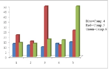
Table 1. Showing the IC50 values of compounds 4-6 against human cancer cell lines
Compound IC50, µmol L−1
SW480 A549 HepG2 HeLa HL-60
4 13.04±0.6 11.32±0.2 9.71±1.1 13.17±0.4 14.71±0.3
5 21.66±0.4 15.44±1.3 >50 11.74±0.7 26.27±0.5
6 14.03±0.2 13.22±0.7 17.37±1.5 16.62±0.4 >50
Cisplatin 3.52±0.3 10.51 ±0.2 9.8±0.9 9.43±0.5 7.8±1.5
of thiosemicarbazide followed by cyclization leads to the formation of products 4-6 as shown in Scheme 2. An enol tautomeric form 1b might be the driving force to accelerate the reaction towards product formation.35
In vitro anticancer activity
The growth inhibitory effect of compounds 4-6 towards the human cancer cells was measured by MTT [3-(4,5-Dimethylthiazol-2-yl)-2,5-diphenyltetrazolium bromide] assay. The conversion of the soluble yellowish MTT to the insoluble purple formazan by active mitochondrial lactate dehydrogenase of living cells has been used to develop an assay system for measurement of cell proliferation.22,23 The
results are expressed as IC50 values (Table 1) which indicate
that compounds 4-6 showed different levels of anticancer activities. The compound 4 showed minimum IC50=
9.71±1.1 (HepG2), 11.32±0.2 (A549), 13.04±0.6 (SW480) and 13.17±0.4 µmol L−1(HeLa). While compound 5 showed
minimum IC50=11.74±0.7 (HeLa) and 15.44±1.3 µmol L−1
(A549). The minimum inhibitions shown by compound 6
were 13.22±0.7 (A549), 14.03±0.2 (SW480) and 16.62 ±0.4 µmol L−1 (HeLa).
From these results it is clear that the IC50 for compound 4
against A549 cell line is 11.32 ± 0.2 which is very close to the IC50 of cisplatin (10.51 ± 0.2) against the same cell line.
IC50 for compound 5 against HeLa cell line is 11.74 ± 0.7
which is also close to the IC50 of cisplatin (9.43 ± 0.5)
against the same cell line. Similarly IC50 for compound 4
against HepG2 is 9.71 ± 1.1 which is also near to the IC50 of
cisplatin (9.6 ± 0.3) against the same cell line. It can be concluded that Compound 4 and 5 are showing potential anticancer activity against A549, HepG2, HeLa cell lines by showing IC50 close to that of standard drug, Cisplatin thus
can be considered as potential cytotoxic agents. The graphical representation of IC50 values in MTT assay is
shown in Fig. 1.
Figure 1. Graphical representation of IC50 values shown by compound 4-6 against SW480, A549, HepG2, HeLa and HL-60 by MTT assay
Treatment of supercoiled plasmid pBR322 DNA with compound 4 and detection of hydroxyl radicals ( OH)
Anticancer activity mechanism was also confirmed by studying the treatment of supercoiled plasmid pBR322 DNA with different concentrations of compound 4 and 100 µM copper. Our nucleolytic experiments suggest that cell death may be due to cleavage or fragmentation of DNA of these cancer cells and that the active species responsible for this are ROS (hydroxyl radical) which resulted from the in vitro
reaction of different concentrations of compound 4 with copper in presence of thiobarbituric acid. We observe from gel electrophoresis that after adding copper (100 µM) the concentration of radicals increase which in presence of different concentrations of compound 4 show the nicking of plasmid pBR322 DNA from its supercoiled form (form I) to open circular form (form II). Fig. 2 reveals that in lane 6, 7 and 8, the nicking is quite obvious by the disappearance of form I and appearance of form II and with the increase in concentration of compound 4 (lane 8) the band intensity (form II) became maximum, depicting the more pronounced cleavage at high concentration.
Figure 2. Fragmentation pattern of supercoiled plasmid pBR322, Lane 1 contains DNA only, lane 2 contains DNA and copper, lane 3, 4 and 5 contain DNA and compound 4 (100, 200 and 300 µM respectively) and lane 7, 8 and 9 contain DNA and compound 4 (100, 200, 300 µM respectively) plus 100 µM copper added to it.
In the DNA cleavage reactions mediated by various antioxidants in the presence of Cu(II), it has been established that Cu(II) is reduced to Cu(I) by the antioxidants and that Cu(I) is an essential intermediate in the DNA cleavage reactions.36,37 It is also generally understood
that DNA cleavage by various antioxidants and Cu(II) is the result of the generation of hydroxyl radicals. As mentioned in literature also, Cu(II) is reduced to Cu(I) and the re-oxidation of Cu(I) to Cu(II) by molecular oxygen gives rise to superoxide anion which in turn leads to the formation of H2O2.38 Presumably Cu(I) is oxidized to Cu(II) by H2O2 in a
Fenton type reaction giving rise to hydroxyl radicals. To determine the hydroxyl radical production and the role of copper ions in DNA cleavage, an experiment was performed where progressively increasing concentrations of compound
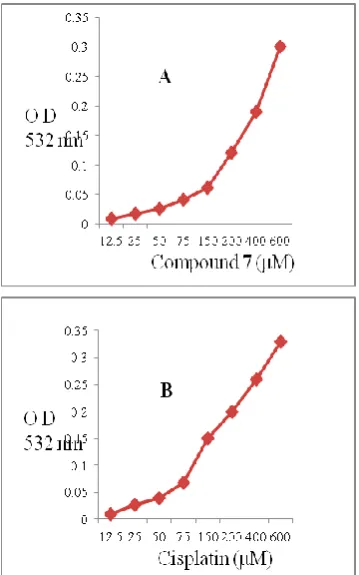
Figure 3. Showing comparative determination of hydroxyl radical production by compund 4 (A) and cisplatin (B) by the assay of thiobarbituric acid
The compound 4–Cu(II) (Fig. 3A) and cisplatin–Cu(II)
(Fig. 3B) are shown to generate the hydroxyl radicals that react with CT DNA, result in strand breaks. The assay is based on the fact that degredation of DNA by hydroxyl radical results in the release of TBA reactive material, which forms a coloured adduct readable at 532 nm.39 Increasing
concentrations of compound 4 or Cisplatin in presence of Cu(II) showed a corresponding increase in the generation of hydroxyl radicals. However, the generation of hydroxyl radical being more in case of cisplatin as shown in Fig. 3B. The results in Fig. 3 confirmed the relatively higher rate of formation of hydroxyl radicals and correlated with the rate of DNA degredation by the compound 4 as well as cisplatin.
Comet assay
In the comet assay, the images of SW480 cells treated with compounds (4-6) showed the formation of comets. Compound 4 presented maximum apoptotic DNA damage followed by compound 6 and 5, which is in accordance with its maximum cytotoxicity as seen in MTT assay. None of the steroidal thiazoles exhibited apoptotic DNA damage to the extent of Cisplatin. The quantified increase in DNA damage suggested that all three thiazole derivatives induced dose dependent fragmentation of chromosomal DNA leading to apoptosis. The images of comet assay for control, cells treated with Cisplatin (20 μg mL-1), 4 (20 μg mL-1), 5
(20 μg mL-1), and 6 (20 μg mL-1) are shown in Fig. 4. Slides
were analyzed for parameter like tail length (TL), using image analyzer CASP software version 1.2.2. The results of the assay for tail length are shown in graph given in Fig. 4.
Compound 4 Compound 5 Compound 6
Cisplatin
Figure 4. Detection of DNA damage in SW480 cells. Treated cells (24 h) were layered over agarose gel, lysed, electrophoresed in alkaline buffer and stained with propidium iodide. Control cells were treated with DMSO alone. The DNA fragmentation resulting in a comet-like appearance in cells treated with cisplatin and compounds 4-6.
Molecular docking studies with DNA
In our experiment, molecular docking studies of steroidal thiazoles with DNA duplex of sequence d(CGCGAATTCGCG)2 dodecamer (PDB ID: 1BNA) were
performed in order to predict the chosen binding site along with preferred orientation of the molecules inside the DNA groove. The resulted docked model (Fig. 5) depicted that all the three compounds recognized minor groove interaction leading to van der Waals and hydrophobic interaction with DNA functional groups which stabilizes the groove and leads to the stablity of the complex. The compound 4
showed electrostatic interaction in the form of hydrogen bonding with NH of 7thThiamine at a distance of 2.88 Å by
the acetate group at 3β-position of steroidal molecule. The compound 5 showed the groove fit behaviour and arranged in a perpendicular manner with respect to the minor groove walls of the DNA helix while as compound 6 showed electrostatic interaction in the form of hydrogen bonding with NH of 11thThiamine at a distance of 3.21 Å. The
resulting relative binding energies of docked steroidal thiazole (4-6)–DNA complexes were found to be -308, -319 and -314 kJ mol-1, respectively depicting the decrease in the
energy after forming complexes with DNA.
Fig. a Fig. b Fig. c
Conclusion
In summary, we have developed a facile and convenient approach for the preparation of new steroidal thiazole derivatives in one-pot synthesis. All the newly synthesized compounds were evaluated for the anticancer activity in vitro against five cancer cell lines. The preliminary results showed that compounds 4-6 were found active during anticancer as well as genotoxic screening but compounds 4
and 6 were found to be potential anticancer agents. These compounds were also found to catalyze the oxidative degradation of isolated DNA either alone or in the presence of transition metal ions such as copper. However, in presence of copper the oxidative cleavage was enhanced. As mentioned earlier cancer cells being rich in transition metal ion like copper40 we conclude that compound 4 in presence
of endogenous copper may give rise to hydroxyl radical this may lead to the oxidative DNA cleavage in cancerous cells. Hence this protocol provides a convenient strategy to annelate steroid nucleus with widespread bioactive thiazoles there by extending the categories of heterosteroids. This strategy may also provide valuable information for the further design and development of more active anticancer agents through various modifications and derivatizations.
Acknowledgement
Authors thank the Chairman, Department of Chemistry, AMU Aligarh, for providing basic research facilities and Department of Biochemistry, JNMC, AMU for biological study of the compounds.
References
1Dauvious, S., Parker, M. G., Steroid hormone action, IRL Press Oxford, 1993, 161-185
2Dubey, R. K., Oparil, S., Imthurn B., Jackson, E. K., Cardiovasc.
Res., 2002, 53, 688-708
3Latham, K. A., Zamora, A., Drought, H., Subramanian, S., Matejuk, A., Offner H., Rosloniec, E. F., J. Immunol.,2003,
171, 5820-5827
4Moudgil, V. K., Steroid receptors in health and disease, New York/London: Plenum Press, 1987.
5Gower D. B., Makin, H. L. J., “Biochemistry of steroid
hormones,” Oxford/ London/Edinburgh: Blackwell Scientific Publications. 1984, 122
6Hargrave, K. D., Hess F. K., Oliver, J. T., J. Med. Chem.,1983,
26, 1158-1163
7Bondock, S., Khalifa W., Fadda, A. A., Eur. J. Med. Chem., 2007,
42, 948-954
8Sharma, P. K., Sawnhney, S. N., Gupta, A., Singh G. B., Bani, S.
Indian J. Chem., 1998, 37B, 376-381
9Jaen, J. C., Wise, L. D., Caprathe, B. W., Tecle, H., Bergmeier, S., Humblet, C. C., Heffner, T. G., Meltzner L. T., Pugsley, T. A., J. Med. Chem., 1990, 33, 311-317
10Tsuji, K., Ishikawa, H., Bioorg. Med. Chem. Lett.,1994, 4, 1601-1606
11Bell, F. W., Cantrell, A. S., Hogberg, M., Jaskunas, S. R., Johansson, N. G., Jordon, C. L., Kinnick, M. D., Zhou, X. X.,
J. Med. Chem., 1995, 38, 4929-4936
12Ergenc, N., Capan, G., Günay, N. S., Ozkirimli, S., Güngör, M., Ozbey S., Kendi, E., Arch. Pharm. Pharm. Med. Chem., 1999,
332, 343-347
13Carter, J. S., Kramer, S., Talley, J. J., Penning, T., Collins, P., Graneto, M. J., Seibert, K., Koboldt, C., Masferrer J., Zweifel, B., Bioorg.Med. Chem. Lett., 1999, 9, 1171-1174
14Badorc, A., Bordes, M. F., De Cointet, P., Savi, P., Bernat, A., Lale, A., Petitou, M., Maffrand, J. P., Herbert, J. M., J. Med. Chem., 1997,40, 3393-3401
15Rudolph, J., Theis, H., Hanke, R., Endermann, R., Johannsen L., Geschke, F. U., J. Med. Chem., 2001, 44, 619-626.
16Pattan, S. R., Dighe1, N. S., Nirmal, S. A., Merekar, A. N., Laware, R. B., Shinde, H. V., Musmade, D. S., Asian J. Research Chem., 2009, 2, 196-201.
17Burger, R. M., Chem. Rev., 1998, 98, 1153-1169. 18Westheimer, F. H., Science, 1987, 235, 1173-1178
19Jin, Y., Cowan, J. A., J. Am. Chem. Soc., 2005, 127, 8408–8415. 20Smith, J., Ariga, K., Anslyn, E. V., J. Am. Chem. Soc., 1993, 115,
362-364
21Scheffer, U., Strick, A., Ludwig, V., Peter, S., Kalden, E., Gobel, M. W., J. Am. Chem. Soc., 2005, 127, 2211-2217
22Slater, T. F., Sawyer, B. Strauli, U., Biochim. Biophys. Acta., 1963, 77, 383-393,
23Mosmann, T., J. Immunol. Methods,1983, 65, 55-63
24Saxena, H. O., Faridi, U., Kumar, J. K., Luqman, S., Darokar, M. P., Shanker, K., Chanotiya, C. S., Gupta M. M., Negi, A. S.,
Steroids, 2007, 72, 892-900.
25Woerdenbag, H. J., Moskal, T. A., Pras, N., Malingre, T. M., El-Feraly F. S., Kampinga, H. H., J. Nat. Prod.,1993, 56, 849-856
26Quinlan, G. J., Gutteridge, Biochem. Pharmacol., 1987, 36, 3629-3633.
27Mustard, D., Ritchie, D. W., PROTEINS: Struct. Funct. Bioinf., 2005, 60, 269-274.
28Delano, W. L., The PyMOL Molecular Graphics System, DeLano Scientific, San Carlos, CA, USA, 2002.
29Singh, N. P., Mut. Res., 2000, 455, 111-127.
30Constantine, E., Anagnostopoulos, Fieser, L. F., J. Am. Chem.
Soc., 1954, 76, 532-536
31Backer, R. H., Squire, E. N., J. Am. Chem. Soc., 1948, 70, 1487-1490
32Rajnikant, Gupta, V. K., Firoz, J., Shafiullah, Gupta, R., Cryst.
Reports, 2000, 45, 785-788
33Hantzsch, A., Weber, J. H., Ber. Deutsch. Chem. Ges., 1887, 20, 3118-3132
34Rudolph, J., Tetrahedron, 2000, 56, 3161-3165
35Yang-i Lin, C. M., Seifert S. M., Kang J. P., Lang, S. A., J.
Heterocycl. Chem., 1979, 16, 1377-1383
36Shamsi, F. A., Husain S., Hadi, S. M., J. Biochemical Toxicol., 1996, 11, 67-71
37Ahmad, M. S., Fazal, F., Rahman, A., Hadi S. M., Parish, J. H.,
Carcinogenesis,1992, 13, 605-608
38Badwey J. A., Kaenovsky, M. L., Annual Rev. Biochem., 1980,
49, 695-726,
39Gupte A. Mumper, R. J., Cancer Treat. Rev., 2009, 35, 32-46, 40Halliwell B., Gutteridge, J. M. C., FEBS Lett., 1992, 307,
108-112