Edinburgh Research Explorer
Derivation of the human embryonic stem cell line RCe006-A
(RC-2)
Citation for published version:
De Sousa, P, Tye, B, Bruce, K, Dand, P, Russell, G, Gardner, J, Downie, JM, Bateman, M & Courtney, A
2016, 'Derivation of the human embryonic stem cell line RCe006-A (RC-2)', Stem cell research, vol. 16, no.
2, pp. 452-455. https://doi.org/10.1016/j.scr.2016.02.029
Digital Object Identifier (DOI):
10.1016/j.scr.2016.02.029
Link:
Link to publication record in Edinburgh Research Explorer
Document Version:
Publisher's PDF, also known as Version of record
Published In:
Stem cell research
Publisher Rights Statement:
© 2016 The Authors. Published by Elsevier B.V. This is an open access article under the CC BY license
General rights
Copyright for the publications made accessible via the Edinburgh Research Explorer is retained by the author(s)
and / or other copyright owners and it is a condition of accessing these publications that users recognise and
abide by the legal requirements associated with these rights.
Take down policy
The University of Edinburgh has made every reasonable effort to ensure that Edinburgh Research Explorer
content complies with UK legislation. If you believe that the public display of this file breaches copyright please
contact [email protected] providing details, and we will remove access to the work immediately and
investigate your claim.
Lab Resource: Stem Cell Line
Derivation of the human embryonic stem cell line RCe006-A (RC-2)
P.A. De Sousa
a,b,c,⁎
, B. Tye
a, K. Bruce
a, P. Dand
a, G. Russell
a, J. Gardner
a, J.M. Downie
a,
M. Bateman
a, A. Courtney
aa
Roslin Cells Limited, Nine Edinburgh Bio-Quarter, 9 Little France Road, Edinburgh EH16 4UX, UK b
Centre for Clinical Brain Sciences, University of Edinburgh, UK c
MRC Centre for Regenerative Medicine, University of Edinburgh, UK
a b s t r a c t
a r t i c l e i n f o
Article history: Received 9 February 2016 Accepted 11 February 2016 Available online 16 February 2016
The human embryonic stem cell line RCe006-A (RC-2) was derived from a frozen and thawed blastocyst volun-tarily donated as surplus to fertility requirements following ethics committee approved informed consent under licence from the UK Human Fertilisation and Embryology Authority. The cell line exhibits expression of expected pluripotency markers andin vitrodifferentiation potential to three germinal lineage representative cell popula-tions. It has a male trisomy 12 karyotype (47XY, +12). Microsatellite DNA marker identity and HLA and blood group typing data are available.
© 2016 The Authors. Published by Elsevier B.V. This is an open access article under the CC BY license (http://creativecommons.org/licenses/by/4.0/).
Resource table
Name of stem cell construct RCe006-A Alternative name RC-2, RC2 Institution Roslin Cells Ltd.
Person who created resource B. Tye, K. Bruce, P. Dand, G. Russell, J. Gardner. Contact person and email [email protected];
[email protected] [email protected] [email protected] [email protected] Date archived/stock date 21 December 2007 (pre-bank at
passage 12 on feeders)
29 November 2010 (banked at passage 31) Type of resource Biological reagent: cell line
Sub-type hESC, research grade
Origin Blastocyst with ICM and Trophoblast Key transcription factors Oct4 (confirmed byflow cytometry and
immunocytochemistry)
Authentication See Quality Control test summary,Table 1
Link to related literature (direct URL links and full references)
N/A Information in public databases http://hpscreg.eu/cell-line/RCe006-A http://www.nibsc.org/science_and_research/ advanced_therapies/uk_stem_cell_bank/ cell_lines.aspx
Ethics Informed consent obtained. Scotland A Research Ethics committee approval obtained (07/MRE00/56). Conducted under the UK Human Fertilisation and Embryology Authority licence no R0136 to centre 0202.
⁎ Corresponding author at: University of Edinburgh, Roslin Cells Limited, Nine Edinburgh Bio-Quarter, 9 Little France Road, Edinburgh EH16 4UX, UK.
E-mail addresses:[email protected],[email protected](P.A. De Sousa).
Resource details
RCe006-A (RC-2) was derived from a frozen and thawed, surplus to requirement, blastocyst. The cell line was derived by whole em-bryo outgrowth on mitotically inactivated human dermalfibroblast (HDF) feeder cells using HDF conditioned medium and expanded under feeder free conditions.
RCe006-A (RC-2) was shown to be pluripotent by expression of the pluripotency markers Oct4, Nanog Tra-1-60 and Tra-1-81, but not the differentiation marker SSEA-1 using immunocyto-chemistry (Table 1,Fig. 1). Byflow cytometric analysis, expres-sion of the pluripotency makers SSEA-4, Tra-1-60 and Tra-1-81 was 81.8%, 55.0% and 43.6%, respectively, whereas low expression of the differentiation marker SSEA-1 (2.3%), was observed at passage 6 (Fig. 2). Differentiation to the three germ layers, endo-derm, ectoderm and mesoendo-derm, was demonstrated using embry-oid body formation with all three germ layers present as shown by expression of α-fetoprotein, β-tubulin and muscle actin (Fig. 3).
A microsatellite PCR profile has been obtained for the cell line, and HLA Class I and II typing is available (Table 2). Blood group genotyping gave the blood group BO1(Table 2).
Verification and authentication
The cell line was analysed for genome stability by G-banding (Fig. 4) and showed an abnormal 47XY, + 12 male genotype in all 20 cells analysed. The cell line is free from mycoplasma contamina-tion as determined by RC-qPCR. Microsatellite PCR DNA profiling for cell identity is available.
http://dx.doi.org/10.1016/j.scr.2016.02.029
1873-5061/© 2016 The Authors. Published by Elsevier B.V. This is an open access article under the CC BY license (http://creativecommons.org/licenses/by/4.0/).
Contents lists available atScienceDirect
Stem Cell Research
Materials and methods
Ethics
Derivation of hESC from surplus to requirement and failed to fertilise/develop embryos was approved by The Scotland A Research Ethics Committee and local ethics board at participating fertility clinics and conducted under licence no R0136 from the UK HFEA with in-formed donor consent.
Cell culture
Frozen embryos were thawed using Embryo Thawing Pack (Origio (Medicult), Denmark) using standard techniques and were cultured EmbryoAssist (Origio) until Day 3 or BlastAssist (Origio) after Day 3 of development. Embryos were cultured at 36.5–37.5 °C, 5 ± 0.5% CO2, 5 ± 0.5% O2in drops under paraffin oil (Origio) and transferred to fresh medium at least every 2–3 days.
By Day 8 of development, or when spontaneous hatching occurred, embryos were placed in derivation conditions consisting of mitotically inactivated neonatal human dermalfibroblasts (HDFs) (ThermoFisher Scientific (Cascade Biologics), Paisley, UK) on tissue culture plastic pre-coated with 2μg/cm2human laminin (Sigma-Aldrich, Dorset, UK) as per manufacturer's recommendation. If required, assisted hatching was per-formed by removing the zona pellucidae mechanically using Swemed cutting tools (Vitrolife, Göteborg, Sweden).
HDF cells were cultured in DMEM (Lonza, Slough, UK), 10% FCS (GE Healthcare (PAA), Buckinghamshire, UK) and 2 mML -gluta-mine (ThermoFisher Scientific). HDFs were mitotically inactivated using gamma irradiation at 50 Gy using a Gammacell Elite 1000 machine. For use as a feeder layer, irradiated HDFs were plated at 2–50,000 cells/cm2in HDF conditioned medium (80% Knockout-DMEM, 20% Knockout serum replacement (KOSR), 1 mM glutamine, 0.1 mMβ-mercaptoethanol, 1% nonessential amino acids, and 4 ng/ml human bFGF (all ThermoFisher Scientific) over 24 h intervals over 7 days) supplemented with an additional 24 ng/ml human bFGF. Cells Table 1
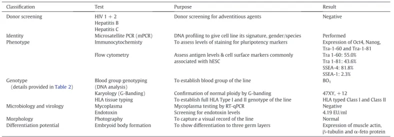
Summary of quality control testing and results for RCe006-A (RC-2).
Classification Test Purpose Result
Donor screening HIV 1 + 2 Hepatitis B Hepatitis C
Donor screening for adventitious agents Negative
Identity Microsatellite PCR (mPCR) DNA profiling to give cell line its signature, gender/species Performed
Phenotype Immunocytochemisty To assess levels of staining for pluripotency markers Expression of Oct4, Nanog, Tra-1-60 and Tra-1-81 Flow cytometry Assess antigen levels & cell surface markers commonly
associated with hESC
Tra 1-60: 55.0% Tra 1-81: 43.6% SSEA-4: 81.8% SSEA-1: 2.3% Genotype
(details provided inTable 2)
Blood group genotyping (DNA analysis)
To establish blood group of the line BO1
Karyology (G-Banding) Confirmation of normal ploidy by G-banding 47XY, +12
HLA tissue typing To establish full HLA Type I and II genotype of the line HLA typed Class I and Class II Microbiology and virology Mycoplasma Mycoplasma testing by RT-qPCR Negative
Endotoxin Screening for endotoxin levels 4.19 EU/ml Morphology Photography To capture a visual record of the line Normal
Differentiation potential Embryoid body formation To show differentiation to three germ layers Expression of muscle actin, β-tubulin andα-feto protein
Fig. 1.Immunostaining of RCe006-A (RC-2) show expression of the pluripotency markers Oct-4 (red), Nanog, Tra-1-60, Tra-1-81 (green), but not the differentiation marker SSEA-1 (green). Cell nuclei are counterstained with DAPI (blue).
Fig. 2.RCe006-A (RC-2) (passage 6) was subjected toflow cytometry analysis for markers of pluripotency with specific antibody or isotype control as indicated above the histograms. Percentage staining is indicated inTable 1.
Fig. 3.In vitro embryoid body differentiation of RCe006-A (RC-2). Specific staining shown in green, from left to right: control, ectoderm (β-tubulin III), mesoderm (muscle actin), and endoderm (α-fetoprotein). Cell nuclei are counterstained with DAPI (blue).
Table 2
Microsatellite PCR, blood group and HLA tissue typing results for RCe006-A (RC-2). Microsatellite PCR results
D3S1358 1 D3S1358 2 vWA 1 vWA 2 D16S539 1 D16S539 2 D2S1338 1 D2S1338 2
15 15 17 18 9 11 20 22
Amelogenin 1 Amelogenin 2 D8S1179 1 D8S1179 2 D21S11 1 D21S11 2 D18S51 1 D18S51 2
X Y 12 14 29 30 12 15
D19S433 1 D19S433 2 THO1 1 THO1 2 FGA 1 FGA 2 CSF1PO 1 CSF1PO 2
12 14 7 9 Iw* Iw* 11 12
D5S818 1 D5S818 2 D7S820 1 D7S820 2 D13S317 1 D13S317 2 TPOX 1 TPOX 2
11 12 7 8.2 12 13 0** 0**
*Peak falls below threshold to confidently score. **No peak detected.
Blood group genotyping
RhD RhC Rhc RhE Rhe Fy a Fy b Fy GATA
pos pos neg neg pos pos pos neg
Jka Jkb K k M N S S
pos pos neg pos neg pos neg pos
Kp a Kp b Do a Do b ABO
neg pos pos? pos? BO1
HLA tissue typing
HLA Class I Type HLA-A*01, A*26; B*08, B*37; C*06, C*07
HLA Class II Type HLA-DRB1*03, DRB1*10; DRB3*02; DQB1*02, DQB1*05 Comment DRB1*03 is expressed serologically as a DR17
were cultured at 36.5–37.5 °C, 5 ± 0.5% CO2, 5 ± 0.5% O2and 50% me-dium exchanged 6 days a week.
The established cell line was expanded and banked using CellStart matrix and Stempro hESC Serum Free Medium (ThermoFisher Scientif-ic). Passaging was performed mechanically using an EZ passage tool (ThermoFisher Scientific). hESC lines were expanded to 25–30 wells of a 6-well plate and cryopreserved in 0.5–1 ml KOSR based cryopreserva-tion solucryopreserva-tion (75% KO-DMEM, 15% Xeno-free KOSR (ThermoFisher Sci-entific) and 10% DMSO (Origen Biomedical, Texas, USA)) or Cryostor CS10 (Biolife Solution, Washington, USA).
Mycoplasma
Mycoplasma detection was performed using Applied Biosystems PrepSEQ™Mycoplasma Nucleic Acid Extraction Kit and MicroSEQ™
Mycoplasma Real-Time PCR Detection Kit (ThermoFisher Scientific (Ap-plied Biosystems, ThermoFisher Scientific) according to the manufacturer's instruction.
Endotoxin
Endotoxin levels were determined using the Kinetic-QCL assay (Lonza) and an incubating plate reader (BioTek ELx808) according to the manufacturer's instructions. Briefly, an unknown sample was com-pared with a standard curve of known levels of control endotoxin. An assay was deemed valid if the coefficient of correlation, r≥0.980 and the CV (%) for the standard curve was≤10%.
Flow cytometry
Human embryonic stem cells were dissociated using Trypsin (ThermoFisher Scientific). Non-specific staining was blocked using 5% goat serum (Sigma) in PBS (Lonza) containing 0.01% Tween-20 (Sigma). Cells were stained with antibodies against SSEA-4, SSEA-1, Tra-1-60 and Tra-1-81 (all BD, Oxford, UK), at 250 ng per reaction followed by Goat F(ab)2 anti-mouse IgM-PE Goat F(ab)2 anti-mouse IgG3-FITC (1:200; Santa Cruz Biotechnology, Texas, USA). Cells were analysed using a FACS Ariaflow cytometer (BD).
Immunocytochemistry
hESCs werefixed in 4% paraformaldehyde (ThermoFisher Scientific (Alfa Aesar)), permeabilised using 100% ethanol (ThermoFisher Scientific) and stained with AFP (1:500; Sigma), β-tubulin III (1:1000; Sigma), muscle-specific actin (1:50; DAKO, Glostrup, Denmark), Oct-4 (1:200; Santa Cruz Biotechnology), Nanog (1:20;
R&D Systems, Abingdon, UK), Tra-1-60, Tra-1-81, SSEA-1 (all 1:50; BD) and secondary antibodies anti-mouse IgG-FITC (1:200; Sigma), mouse IgG-AlexaFluor 488, goat IgG-AlexaFluor 488, anti-goat IgG-AlexaFluor-594, anti-donkey polyclonal AlexaFluor-594 (all 1:200; ThermoFisher Scientific). Images were acquired using a Zeiss S100 Axiovertfluorescence microscope or Nikon eC1 confocal microscope.
In vitro differentiation
hESC cells were pre-treated for 1 h with 10μM ROCK inhibitor in Stempro hESC SFM (ThermoFisher Scientific) and embryoid bodies EBs generated in ultra low attachment plates (Corning) for 7 days before being transferred into EB medium (20% FBS (GE Healthcare (PAA)), 80% KO-DMEM 1 mML-glutamine, 0.1 mMβ-mercaptoethanol, 1% nones-sential amino acids (all ThermoFisher Scientific)), on glass slide tissue culture chambers (Nunc, ThermoFisher Scientific) coated with 0.5% gel-atin (Sigma) at 0.1 ml/cm2for 14 days.
Genomic analysis
All outsourced assays were carried out under a Quality and Technical Agreement. DNA was extracted using the QIAamp DNA Mini kit (Qiagen, Manchester, UK) according to the manufacturer's recommen-dations and provided in recommended quantities to the service providers.
Microsatellite PCR, or Short Tandem Repeat analysis, was used to de-termine cell line identity and was carried out by Public Health England. A profile was obtained for the following core alleles: vWA, D16S539, Amelogenin, THO1, CSF1PO, D5S818, D75820, D135317 and TPOX.
Human Leukocyte Antigen (HLA) tissue typing was carried out by the Scottish National Blood Transfusion Service.
Blood group genotyping was carried out by the Molecular Diagnos-tics Laboratory at NHSBT.
Karyotype analysis was carried out by The Doctors Laboratory (London, UK) or the Western General Cytogenetics Laboratory (Edinburgh, UK). Live cells at 60–70% confluency were shipped over-night in warm containers,fixed and analysed by standard G-banding analysis. For research grade lines, 20 spreads were analysed.
Acknowledgements
Research culminating in the derivation of this line was funded by a grant from Scottish Enterprise Economic Development Agency (PM07321) to PDS, MB, and AC.