199 Address for correspondence
Dr. Uzma Sarwar
Assistant professor of Dermatology
Shalamar Medical & Dental College/ Shalamar Teaching Hospital, Lahore
E-mail: [email protected]
Original Article
Frequency and association of dermatological
manifestations of type 2 diabetes mellitus with body
mass index and HbA1c levels
Introduction
Diabetes mellitus (DM) is the common
endocrine disorder, which affects the people of all ages and socioeconomic groups. It is a syndrome in which there is impaired secretion or function of insulin resulting in hyperglycemia.1 More than 285 million people around the world are affected by DM and the prevalence will rise to 438 million by the year 2030.2 According to another study, it would affect 5.4% of the world Uzma Sarwar*, Habib Ur Rehman*, Sumara Rashid**, Muhammad Azam Bokhari*
* Department of Dermatology, Shalamar Medical & Dental College/ Shalamar Teaching Hospital, Lahore
** Department of Dermatology, Fatima Memorial Hospital & Medical College, Lahore
Abstract Objective To determine the frequency of cutaneous manifestations in patients suffering from type 2
diabetes mellitus and to observe the association of these manifestations with body mass index (BMI) and glycosylated hemoglobin (HbA1c) level.
Methods This cross-sectional study was conducted in Shalamar Teaching Hospital / Shalamar Medical and Dental College, Lahore from October 2016 to March 2017. A total of 200 adult patients belonging to both genders having type 2 diabetes mellitus attending the outpatient department were studied. Detailed history and cutaneous examination was performed after taking the informed consent. Glycemic profiles including HbA1c were done. Height and weight were recorded too. Normal range of HbA1c was taken as 5 - 6.5 %. Value of ≥ 6.5 % was taken as high. Normal range of BMI as taken as 18.5- 24.9 kg/m2. Patients having BMI 25-29.9 kg/m2 were taken as overweight and ≥ 30 kg/m2
as obese.
Results Of 200 patients, 174 (87%) patients were found to have cutaneous abnormalities. There were 53 (26.5%) males and 147 (73.5%) females. Mean age was 52.4±9.96 years. Mean HbA1c was 9.07±2.23%. The mean BMI was 29.46 kg/m2 with 20% having normal BMI, 38% were overweight and 41% were obese. Most frequently observed skin disease was cutaneous infections including bacterial and fungal infections, followed by generalized xerosis, diabetic dermopathy, acanthosis nigricans, pruritus and acrochordons. Statistically significant correlation was observed between HbA1c and diabetic foot and between BMI and acanthosis nigricans and generalized xerosis.
Conclusion About 87% patients with type 2 diabetes have cutaneous lesions, the most common of which was cutaneous infections. Hyperglycemia manifested with high HbA1c and obesity manifested with high BMI is associated with many cutaneous lesions.
Key words
200 population by the year 2025.3 South Asian
population is more predisposed to type 2 DM.4 There were approximately 6.6 million adult people in Pakistan having DM in 20125 making it the tenth largest nation with this problem worldwide.6 It can affect cardiovascular, renal, nervous system, eyes and the skin.7 Cutaneous manifestations in DM are attributed to the abnormal carbohydrate metabolism, atherosclerosis, microangiopathy, neuron degeneration, impaired host responses and some other yet undetermined mechanism. Almost two third of the diabetic patients both of type 1 and 2 are affected by cutaneous manifestations during the course of their disease. Many skin diseases are associated with DM with incidence rate ranging from 11.4%8 to 71%.9
Skin lesions of DM can be classified in four categories: 1) skin manifestations not specific for diabetes mellitus but with weak or strong association with diabetes including necrobiosis lipoidica, acanthosis nigricans, diabetic bulla, 2) cutaneous manifestations due to diabetic complications such as microangiopathy, macroangiopathy and neuropathy like diabetic foot ulcers, diabetic gangrene, diabetic dermopathy, 3) treatment-related cutaneous lesion like insulin allergic reaction, lipodystrophy due to insulin or oral hypoglycemic agents related skin lesions and finally, and 4) infections (bacterial, fungal).10 Skin infections in diabetics are associated with poor metabolic control and higher HbA1c and BMI. There are limited local data about the association of the skin lesions in diabetes in connection with levels of HbA1c and BMI.
The current study was conducted to determine the frequency of cutaneous manifestations in patients suffering from type 2 DM. The association of those skin manifestations with body mass index and glycemic control was the further aim of this study.
Methods
This cross-sectional study was carried out in the Department of Dermatology and Department of Endocrinology-SIDER (Sakina Institute of Diabetes & Endocrine Research), Shalamar Teaching Hospital/ Shalamar Medical & Dental College, Lahore from October 2016 to March 2017. All patients with type 2 DM (according to American Diabetes Association criteria: fasting blood sugar (FBS) ≥126 for 2 times) were included in the study. Patients with skin lesions secondary to other cutaneous disorders and pregnant patients with diabetes were excluded. After taking the informed consent from the patient, detailed history was taken. Dermatological and systemic examination was performed. Photographs were taken where needed. All the patients were assessed for height and weight to calculate body mass index (BMI). Normal range of BMI was taken as 18.5-24.9 kg/m2. Patients having BMI 25-29.9 kg/m2 were taken as overweight and ≥30 kg/m2
as obese. Fasting blood sugar, blood sugar random (BSR) and HbA1c were performed. HbA1c value of ≥ 6.5% was considered as having unsatisfactory glycemic control according to American Diabetes Association criteria.
Where needed other relevant laboratory investigations like complete blood count, lipid profile, renal and liver function tests, urine examination were advised. Wood’s lamp examination, fungal scraping, nail clipping and skin biopsy were performed when needed for diagnosis. The cutaneous lesions with duration of less than three months were studied to find their association with HbA1c levels.
201 descriptive variables like various skin changes
and their correlation with HbA1c and BMI. T-test was used for statistical analysis. p value <0.05 was considered statistically significant.
The study was approved by ethical committee of our institute research board.
Results
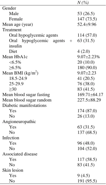
Among 200 patients, 53 (26.5%) were male and 147 (73.5%) were females. Age of the patients ranged from 30 to 83 year, mean being 52.4±9.96 year. One hundred and four patients were having disease of less than 5 year duration, 47 patients had disease duration of 5-10 years and 49 patients have disease for > 10 years.
Mean BSF was 169.7±64.17 g/dl and mean BSR was 227.5±88.29 g/dl. The mean HbA1c was 9.07±2.23 % with 180 (90%) patients having unsatisfactory glycemic control (≥ 6.5%). Mean BMI was 29.46±5.71 kg/m2 with 76 (38%) patients falling into overweight group (BMI 25-29.9 kg/m2) and 83 (41.5%) patients were obese (BMI ≥30 kg/m2
), (Table 1).
Oral hypoglycemic agents were used by 114 patients, insulin by 19 patients, 63 patients were using both oral hypoglycemic agents and insulin while only 4 patients had been advised diet for control of diabetes.
There were 174 (87%) patients who had cutaneous lesion. The most frequently observed individual lesion was generalized xerosis with 51 (29.3%) patients, followed by diabetic dermopathy in 49 (28.2%) and acanthosis nigricans in 48 (27.5%) patients. Different types of skin infections were seen in 96 (55.2%) of the patients, making it the most common dermatosis.
Other commonly observed dermatoses were
Table 1 Demographic and clinical characteristics of study population (n=200).
N (%) Gender
Male 53 (26.5)
Female 147 (73.5)
Mean age (year) 52.4±9.96
Treatment
Oral hypoglycemic agents 114 (57.0) Oral hypoglycemic agents +
insulin
63 (31.5)
Diet 4 (2.0)
Mean HbA1c 9.07±2.23%
<6.5% 20 (10.0)
≥6.5% 180 (90.0)
Mean BMI (kg/m2) 9.07±2.23
18.5-24.9 41 (20.5)
25-29.9 76 (38.0)
≥30 83 (41.5)
Mean blood sugar fasting 169.71±64.17 Mean blood sugar random 227.5±88.29 Diabetic manifestations
Yes 174 (87.0)
No 26 (13.0)
Angioneuropathic
Yes 63 (31.5)
No 137 (68.5)
Infection
Yes 96 (48.0)
No 104 (52.0)
Associated disease
Yes 117 (58.5)
No 83 (41.5)
Skin lesion
Yes 9 (4.5)
No 191 (95.5)
generalized pruritus in absence of any obvious skin lesion in 33 (19.0%) patients, acrochordons in 20 (11.5%) patients and diabetic foot in 16 (9.1%) patients. Other dermatoses had prevalence of less than 9 %.
202 Table 2 Frequency of cutaneous manifestations and their association with HbA1c and BMI (n=200)
Glycemic control P value Body mass index P value Satisfactory
(n=20) N (%)
Unsatisfactory (n=180) N (%)
Normal (n=41) N (%)
Overweight (n=76) N (%)
Obese (n=83) N (%)
Xerosis (n=51) 5 (9.8) 46 (90.2) 0.957 18 (35.3) 15 (29.4) 18 (35.3) 0.010* Diabetic dermopathy
(n=49) 2 (4.1) 47 (95.9) 0.112 14 (28.6) 18 (36.7) 17 (34.7) 0.245 Acanthosis nigricans
(n=48) 2 (4.2) 46 (95.8) 0.122 4 (8.3) 16 (33.3) 28 (58.3) 0.010* Dermatophytosis
(n=41) 7 (17.1) 34 (82.9) 0.090 6 (14.6) 19 (46.3) 16 (39.0) 0.389 Pruritus (n=33) 1 (3.0) 32 (97.0) 0.144 8 (24.2) 9 (27.3) 16 (48.5) 0.381 Onychomycosis
(n=32) 3 (9.4) 29 (90.6) 0.898 4 (12.5) 16 (50.0) 12 (37.5) 0.249 Furunculosis (n=23) 2 (8.7) 21 (91.3) 0.825 4 (17.4) 5 (21.7) 14 (60.9) 0.118 Acrochordons (n=20) 1 (5.0) 19 (95.0) 0.432 1 (5.0%) 11 (55.0) 8 (40.0%) 0.116 Diabetic foot (n=16) 5 (31.3) 11 (68.8) 0.003* 5 (31.3) 6 (37.5) 5 (31.3) 0.491 Intertrigo (n=15) 0 (0.0) 15 (100.0) 0.179 2 (13.3) 6 (40.0) 7 (46.7) 0.768 Lichen planus (n=6) 0 (0.0) 6 (100.0) 0.407 3 (50.0%) 1 (16.7) 2 (33.3%) 0.177 Vitiligo (n=6) 1 (16.7) 5 (83.3) 0.580 2 (33.3) 3 (50.0) 1 (16.7) 0.438 Carbuncle (n=5) 0 (0.0) 5 (100.0) 0.450 0 (0.0) 3 (60.0) 2 (40.0) 0.426 Scleredema
diabeticorum (n=5) 0 (0.0) 5 (100.0) 0.450 1 (20.0%) 1 (20.0) 3 (60.0%) 0.650 Insulin reaction (n=5) 0 (0.0) 5 (100.0) 0.450 0 (0.0%) 1 (20.0) 4 (80.0%) 0.190 Yellow skin (n=4) 1 (25.0) 3 (75.0) 0.312 0 (0.0) 3 (75.0) 1 (25.0) 0.276 Oral leukoplakia (n=4) 0 (0.0) 4 (100.0) 0.501 2 (50.0%) 1 (25.0) 1 (25.0%) 0.336 Candidiasis (n=4) 0 (0.0) 4 (100.0) 0.501 0 (0.0) 1 (25.0) 3 (75.0) 0.346 Stiff joints and waxy
skin (n=3) 0 (0.0) 3 (100.0) 0.561 0 (0.0) 2 (66.7) 1 (33.3) 0.541 Diabetic rubeosis
(n=3) 0 (0.0) 3 (100.0) 0.561 2 (66.7) 0 (0.0) 1 (33.3) 0.112
Diabetic bullae (n=2) 0 (0.0) 2 (100.0) 0.636 1 (50.0) 1 (50.0) 0 (0.0) 0.412 Lipodystrophy (n=2) 0 (0.0) 2 (100.0) 0.636 1 (50.0%) 0 (0.0) 1 (50.0%) 0.436 OHA-related skin
lesions (n=2) 0 (0.0) 2 (100.0) 0.636 0 (0.0%) 2 (100.0) 0 (0.0%) 0.192 Necrobiosis lipoidica
(n=1) 0 (0.0) 1 (100.0) 0.738 1 (100.0) 0 (0.0) 0 (0.0) 0.142
Eruptive xanthomas
(n=1) 0 (0.0) 1 (100.0) 0.738 0 (0.0) 0 (0.0) 1 (100.0) 0.492
Acquired perforating
dermatosis (n=1) 0 (0.0%) 1 (100.0) 0.738 0 (0.0) 0 (0.0) 1 (100.0) 0.492 *P value < 0.05 is significant, OHA: oral hypoglycemic agents.
faciei, 32 (18.3%) patients had onychomycosis of finger- or toenails and 15 (8.6%) patients had intertrigo and 4 (2.2%) patients had candidiasis. Majority of the patients 57% in our study had more than one type of dermatosis.
With reference to the glycemic control (Table 2), out of 180 patients who had unsatisfactory glycemic control (HbA1c ≥6.5%), 158 (87.7%) patients had diabetic manifestations. Out of 20
patients who have satisfactory glycemic control 16 (80%) patients had diabetic manifestations.
Out of 180 patients with unsatisfactory glycemic control (HbA1c ≥6.5%), 85 (47.2%) presented with infections and out of 20 patients with satisfactory glycemic control (HbA1c ≤6.5%), 11 (55%) presented with infections.
203 statistically significant correlation (p=0.025).
Similarly, the relationship between HbA1c and treatment modalities also came out to be statistically significant (p=0.001). Out of 20 patients who have HbA1c <6.5%, all were taking oral hypoglycemic agents. Out of 180 patients who had HbA1c ≥6.5%, 94 were on oral hypoglycemic drugs, 63 were on combination of oral hypoglycemic and insulin, 19 were on insulin alone and 4 patients were on management by diet control (Table 1).
Out of 180 patients with unsatisfactory glycemic control having (HbA1c ≥6.5%), 11 (6.1%) patients had diabetic foot and out of 20 with satisfactory glycemic control having HbA1c <6.5%, 5 (25%) patients presented with diabetic foot (p=0.003).
When BMI was studied (Table 2), it was found that out of 41 patients who had normal BMI (18.5-24.9 kg/m2), 35 (85.5%) patients had skin disease. Out of 76 patients who were overweight with BMI between (25-29.9 kg/m2), 67 (88.1%) patients had cutaneous manifestations and out of 83 patients who were obese with BMI ≥30 kg/m2, 72 (86.7%) patients had cutaneous manifestations.
When we studied the correlation between BMI and gender it came out to be statistically significant (p=0.034). Out of 83 obese patients who had BMI ≥30 kg/m2, there were 66 (79.5%) females while 17 (20.4%) were men. Similarly out of 76 overweight patients (BMI 25-29.9 kg/m2), there were 48 (63.1%) females while 28 (36.8%) were males. Thirty-three (80.5%) patients out of total 41 with normal BMI (18.5-24.9 kg/m2) were females and 8 (19.5%) were males.
The correlation between BMI and acanthosis nigricans was statistically significant (p=0.010). Out of total 48 patients with acanthosis
nigricans, 28 were obese, 16 were overweight and 4 had normal BMI. Similarly the correlation between BMI and xerosis was also statistically significant (p=0.010). Out of total 51 patients having xerosis, 18 were obese 15 were overweight and 18 had normal BMI.
Discussion
Diabetes mellitus is an endocrine disorder, which can involve the multiple systems of our body. It almost invariably affects the skin. Cutaneous involvement usually occurs after the primary disease but at times it appears with the onset of disease or even precedes the disease by many years.11 These clinical cutaneous signs are valuable for the clinicians. The proposed underlying mechanisms for many diabetic associated dermatoses are abnormal carbohydrate metabolism, impaired host responses, neuron degeneration, atherosclerosis and microangiopathy.
Our study shows 87% prevalence of skin lesions in type 2 DM. When compared with the other studies, frequency of skin lesions in type 2 DM has been reported to be in range of 49-84% which is almost comparable with our study.12,13 Deepika et al.14and Chatterjee et al.15 reported the prevalence of 76.8% and 73.9%, respectively. The bit higher values in our study may be due to the reason that most of the patients i.e. 90% in our study had unsatisfactory glycemic control shown by high values of HbA1c i.e. ≥6.5%.
204 The most frequent dermatosis was cutaneous
infections, which showed the prevalence of 55.2%. Fungal infections of the skin and nails were the most common and many patients have more than one type of infection. Almost similar results of 51.7% and 61% were reported by Gosh et al.16 and Naheed et al.17 However, Timshina (41.95%),18 Hosseini (41.5%),19 Chatterjee (40.9%),15 Vahora (39.75%)20 and Majeed (23%)21 reported variable lower prevalence rates. This difference can be explained by the different blood sugar levels and differences in the socioeconomic and hygienic status of the studied population.
The risk of infections seems to be more in patients with poor glycemic control; however we did not find any significant relationship between the glycemic control and the prevalence of skin infections. It is proposed that the poor metabolic control effect the host responses, as delayed phagocytosis, delayed chemotaxis and decreased white blood cell adherence.
Most commonly observed individual lesion was generalized xerosis in our study which was present in 51 (29.3%) of patients. Xerosis was the most skin manifestation 44% in a study conducted by Goyal et al.11and it was the second most common manifestation 34.45% in a study conducted by Chatterjee et al.15Moreover in our study the correlation between xerosis and BMI is significant (p=0.010)
Diabetic dermopathy was observed in 49 (28.2%) patients in the present study. Although not pathognomonic of diabetes, it is considered as one of the most common dermatosis seen in diabetes affecting 7-70% of the patient.14 It was the most frequent manifestation 32.3% in an Iranian study by Hosseini et al.19 and 36% in a study conducted by Goyal et al.11, although lower frequencies were noted by Chatterjee et al.15(12.75%) and Majeed et al.21 (6%).
Acanthosis nigricans is usually seen due to insulin resistance. It was observed in 48 (27.5%) of the patients in our study, which is comparable with a study conducted by Hosseini et al.19 who reported it to be the third most prevalent skin lesion 26.4% in his study. Almost similar results were seen by Niaz et al.22 as 22%; however, lower prevalence of 10.9%, 2.5% and 1.5% were reported by Deepika et al.14, Ahmad et al.23 and Majeed et al.21, respectively. The high prevalence in our study may be associated with high insulin resistance in our patients. In the above mentioned three studies both type 1 and type 2 diabetic patients were included and we know that insulin resistance and consequent acanthosis nigricans are more common in type 2 diabetes mellitus. Moreover, the high prevalence of acanthosis nigricans in our study correlated significantly with BMI (p=0.010).
Generalized pruritus in the absence of any skin lesion was observed in 33 (19.0%) patients. Chatterjee et al.15 noted a higher value of 50%. Other studies reported variable prevalence of 21%, 9.4%, 8%, 7.1% and 3%.17,24,22,23,21 The findings in different studies can be variable depending upon the setting, study design and the population of the study.
Acrochordons have been associated with diabetes mellitus, insulin resistance and increased BMI. 11.5% of our study population was suffering from acrochordons. Similar results were reported by Niaz et al.22 in a local study, however, higher prevalence of 19%, 21% and 32% are reported by various local and international studies.25,17,11
205 Higher prevalence of 18.1% have been reported
in a study conducted in Sudan27and 16% in a study conducted locally in Pakistan by Niaz et al.22 The lower prevalence rates ranging from 0.8%-3% are reported by different studies from India and Pakistan.14,21,24 Neuropathy and angiopathy along with impaired immunity, venous insufficiency and lymphedema are the different mechanisms contributing in development of diabetic foot. Apart from these, infection, advanced age, presence of comorbidities that can affect the healing of wounds or renal replacement therapy are the factors that determine the outcome of the diabetic foot ulcers. Moreover, in our study the correlation between the diabetic foot and HbA1c came out to be significant (p=0.003).
The mean BMI in our patients was 29.46±5.71 kg/m2 with maximum number of patients in overweight and obese group. It was statistically correlated with gender with more female obese patients (p=0.034). BMI also correlated positively with generalized xerosis (p=0.010) and acanthosis nigricans (p=0.010).
The mean HbA1c in our study was 9.07±2.23% with almost 90% patients having unsatisfactory glycemic control which is ≥6.5%. The statistic correlation HbA1c and treatment modalities came out to be significant with p=0.001. Similarly, the relationship between HbA1c and duration of the DM also came out to be statistically significant with p=0.026. The present study also showed positive correlation between HbA1c and diabetic foot (p=0.03).
Our study did not show correlation between HbA1c and prevalence of cutaneous disorders. However, the mean HbA1c level was higher in patients suffering with infective lesions. There are different opinions regarding this issue. According to the Iranian study in 2010,25 as well as, an Indian study in 2012,18 there was no
correlation between the diabetic disease control and prevalence of cutaneous lesions. But another Iranian study19 and a Pakistani study21 showed definite relationship between the glycemic control and cutaneous lesions. In our study, it came out to be significant only with diabetic foot where out of 16 patients with diabetic foot, 11 had unsatisfactory glycemic control.
The cross-sectional study design and the modest number of patients were the limitations of our study. There is definite association between diabetes and cutaneous lesions. Unsatisfactory glycemic control and higher values of BMI also showed correlation with diabetic patients in many aspects.
Conclusion
Our study population with diabetes mellitus was having high prevalence of cutaneous lesions, the most common of which was cutaneous infections. Almost all of the diabetic patient develop cutaneous lesions because of the effect of hyperglycemia on the microcirculation and dermal collagen. This affects their quality of life and add burden to the therapeutic cost. Long-term control of blood glucose level and lowering the BMI would decrease the risk of many cutaneous lesions. Identification and treatment of cutaneous lesions would reduce the dermatological morbidity and improve the quality of life of these patients. Interventional researches are needed in our area to further evaluate the possible role of glycemic control and optimal BMI in reducing the cutaneous lesions.
References
1. American Diabetes Association. Screening for type 2 diabetes. Diabetes Care. 2004;27 Suppl 1:S11-4.
206 2010 and 2030. Diabetes Res Clin Pract.
2010; 87:4-14.
3. Paron NG, Lambert PW. Cutaneous manifestation of diabetes mellitus. Prim Care. 2000;27:371-83.
4. Mather HM, Keen H. The Southall Diabetes Survey: prevalence of known diabetes in Asians and Europeans. Br Med J (Clin Res Ed) 1985;291:1081-4.
5. Kalra S, Peyrot M, Skovlund S. Second diabetes attitudes, wishes and needs (DAWN2) study: relevance to Pakistan. J Pak Med Assoc. 2013;63:1218-9.
6. International Diabetes Federation. Diabetes Atlas 5th Edition (2012) [cited 2016 December 18]. Available from http:// www.idf.org/diabetesatlas/5e/Update2012. 7. Jennifer L, John E. Diabetes mellitus. In:
Irvin MF, Arthur Z, Klaus W, Austen KF, Goldsmith LA, Katz SI, editors. Dermatology in General Medicine, 6th edn. New York: McGraw-Hill; 2003. P.1651-61. 8. Greenwood AM. A study of skin in 500
diabetics. JAMA. 1927;89:774-9
9. Yosipovitch G, Hodak E, Vardi P, Shraga I, Karp M, Sprechere KM, et al. The prevalence of cutaneous manifestations in IDDM patients and their association with diabetes risk factors and microvascular complications. Diabetes Care. 1998;21:506-9.
10. Perez MI, Kohn SR. Cutaneous manifestations of diabetes mellitus. J Am Acad Dermatol. 1994;30:519-31.
11. Goyal A, Rania S, Kaushal SS, Mahanjan V, Sharma NL. Pattern of cutaneous manifestations in diabetes mellitus. Indian J Dermatol. 2010;55:39-41.
12. Sasmaz S, Buy Ukbese MA, Cetin Kaya A. The prevalence of skin disorders in type-2 diabetic patients. Indian J Dermatol. 2005;3:3-8
13. Wahid Z, Kanjee A. Cutaneous manifestations of diabetes mellitus. J Pak Med Assoc. 1998;48:304-5.
14. Deepika, Gupta SK, Singh P, Monika. Pattern of dermatological diseases in patients of diabetes mellitus. J Pak Med Assoc. 2016;26:214-18.
15. Chatterjee N, Chattopadhyay C, Sengupta N, Das C, Sarma N, Pal SK. An observational study of cutaneous manifestations in diabetes mellitus in a tertiary care hospital of Eastern India. Indian J Dermatol. 2014:18:217-20.
16. Ghosh K, Das KD, Ghosh S, Chakraborty S, Jatua SK, Bhattacharya A et al. Prevalence of skin changes in diabetes mellitus and its correlation with internal diseases: A single center observational study. Indian J Dermatol. 2015;60:465-9.
17. Naheed T, Akbar N, Akbar N, Shehzad M, Jamil S, Ali T. Skin manifestations amongst diabetic patients admitted in a general medical ward for various other medical problems. Pak J Med Sci. 2002;18:291-6. 18. Timshina DK, Thappa DM, Aggrawal A. A
clinical study of dermatosis in diabetes to establish its markers. Indian J Dermatol. 2012;57:20-5.
19. Hosseini MS, Ehsani AH, Hosseinpanah F, Azizi F, Salami M, Khedmat H. Skin lesions in type 2 diabetic patients. Iran J Dermatol. 2008;11:113-7.
20. Vahora R, Thakkar S, Marfatia Y. Skin, a mirror reflecting diabetes mellitus: A longitudinal study in a tertiary care hospital in Gujrat. Indian J Endocr Metab 2013;17:659-64.
21. Majeed N, Iqbal Z, Mahboob A. Frequency and association of cutaneous manifestations of diabetes mellitus with HbA1c. Proc S.Z.P.G.M.I. 2004;18(2):85-9.
22. Niaz F, Bashir F, Shams N, Shaikh Z, Ahmed I. Cutaneous manifestations of diabetes mellitus type 2: prevalence and association with glycemic control. J Pak Med Assoc. 2016;26:4-11.
23. Ahmad K, Muhammad Z, Qayum I. Prevalence of cutaneous manifestations of diabetes mellitus. J Ayub Med Coll Abbottabad. 2009;21(2):76-9.
24. Mahmood T, Bari ul A, Agha H. Cutaneous manifestations of diabetes mellitus. J Pak Med Assoc. 2005;15:227-32.
25. Farshchian M, Farschian M, Fereydoonnejad M, Yazdanfar A, Kimyal-Asadi A. Cutaneous manifestations of diabetes mellitus: A case series. Cutis. 2010;86:31-5. 26. Boulton AJ, Vileikyte L, Ragnarson-Tennvall G, Apelqvist J. The global burden of diabetic foot disease. Lancet. 2005;366:1719-24.