A CASE STUDY ON THE EFFECTS OF EXERCISE ON MUSCLE CROSS-SECTIONAL AREA DURING INDUCTION CHEMOTHERAPY FOR ACUTE
MYELOGENOUS LEUKEMA
Brett Lovell Phillips
A thesis submitted to the faculty of the University of North Carolina at Chapel Hill in partial fulfillment of the requirements for the degree of Masters of Arts in the Department of Exercise and Sport Science (Exercise Physiology).
Chapel Hill 2014
Approved By: Claudio Battaglini Anthony C. Hackney Edgar Shields
iii ABSTRACT
Brett Lovell Phillips: A Case Study on the Effects of Exercise on Muscle Cross-Sectional Area during Induction Chemotherapy for Acute Myelogenous Leukemia
(Under the direction of Dr. Claudio Battaglini)
While aggressive chemotherapy is standard for promoting complete remission in newly diagnosed acute myelogenous leukemia patients, treatment side effects impair post-cancer quality of life due in part to negative changes in body composition and increases in cancer-related fatigue. This population would greatly benefit from an intervention which increases physical activity during treatment. Therefore, the primary purpose of this study was to examine the effects of aerobic and resistance exercise on muscle cross sectional area and fatigue before and after induction chemotherapy. One patient with acute myelogenous leukemia participated in this study. The exercise protocol included 30-40 minutes of aerobic and resistance exercise performed 2-4 days per week, twice a day for 4 weeks. While a decrease in vastus lateralis cross sectional area was present post-intervention, this decrease may have been much greater without the
iv
ACKNOWLEGEMENTS
A project such as this would not be possible without the expertise, patience, and flexibility of my thesis committee members, Dr. Claudio Battaglini, Dr. Edgar Shields, Dr. Anthony Hackney, and Dr. Ashley Leak-Bryant. I would especially like to thank my advisor, Dr. Claudio Battaglini, for his continual encouragement, mentorship, and enthusiasm-a role model personally and professionally.
v
TABLE OF CONTENTS
LIST OF TABLES ... vii
LIST OF FIGURES ... viii
I. INTRODUCTION ... 1
Statement of Purpose ... 4
Definition of Terms ... 4
Limitations ... 5
Delimitations ... 5
Assumptions... 5
Significance of the Study ... 5
II. REVIEW OF THE LITERATURE ... 7
Leukemia Overview ... 8
AML Treatment-Related Side Effects ... 9
Exercise as Cancer Therapy ... 11
AML and Exercise as a Treatment ... 13
III. METHODOLOGY ... 15
Subject ... 15
Instrumentation... 17
Assessment Protocol... 17
Exercise Intervention ... 19
vi
V. DISCUSSION, CONCLUSION, AND RECOMMENDATIONS ... 23
Introduction ... 23
Maintenance of VL Muscle Cross Sectional Area ... 24
Decrease in Overall Fatigue ... 25
Conclusion ... 25
Recommendations ... 26
VI. APPENDIX I ... 28
Study Advertisement Flier ... 28
VII. APPENDIX II ... 29
PROMIS Fatigue SF 8a ... 29
VIII. APPENDIX III ... 32
Panoramic Ultrasound Analysis Example Using Image-J ... 32
IX. APPENDIX IV ... 33
vii
LIST OF TABLES
viii
LIST OF FIGURES
1 CHAPTER I INTRODUCTION
Acute myelogenous leukemia (AML) is one of the four major subgroups of leukemia and is estimated to represent 36% of the leukemia total in the US for 2014 (American Cancer Society, 2014). Leukemia, in most simple terms, is a blood cancer. Depending on the leukemia subtype, immature or differentiated cells in the bone marrow and/or blood acquire a DNA mutation and the cells uncontrollably replicate. During this uninhibited cycle of abnormal cell replication, a crowding out of normal blood forming stem cells commonly manifests into pancytopenia (Roboz, 2012). Consequently, clinical issues that arise from the decreased red blood cell (RBC), white blood cell (WBC), and platelet counts may include but are not limited to the development of anemia, fatigue, increased susceptibility to infection, and a higher likelihood to bruise and bleed (Roboz, 2012).
2
Due to the AML’s rapid progression, upon diagnosis, patients are immediately treated with seven days of an initial chemotherapy known as induction chemotherapy. These patients are given 3-4 weeks to recover in an in-patient hospital setting. The goal during this time is to bring the disease into remission, meaning less than five percent of leukemia cells remain in the bone marrow (Anderson et al., 2002). Standard of care for these patients during this time includes bed rest in the hospital room with all healthcare providers following strict neutropenic procedures in order decrease the risk for infection.
The second phase of chemotherapy is the consolidation phase where all of the remaining leukemia cells are destroyed (Hiddemann et al., 1999). Even with
chemotherapy and standard precautions, mortality for 2014 is estimated at 10,460, and data from 2003-2009 places five-year relative survival rate at only 24.9%-the lowest of the four subgroups of leukemia.
While chemotherapy for AML improves overall patient survival; its use is accompanied by side effects that negatively alter many physiological systems; one such system affected is the musculoskeletal system via alterations in body composition (Al-Majid and McCarthy, 2001; Hemming and Maher, 2005).Negative changes in body composition, i.e., increased percent body fat (%BF), decreased percent lean body mass (%LBM) and muscle wasting are common side-effects that impact physical function and often lead to a decrease in overall physical activity further impacting the overall
3
The positive effects of exercise on body composition, cardiorespiratory fitness, muscular fitness, fatigue and overall quality of life in cancer patients have been
evidenced primarily in patients with prostate, breast, and lymphoma cancer (Courneya, Segal, Mackey et al., 2007; Galvao et al., 2010; Milne et al., 2009; Vallance et al., 2005). Standard exercise prescriptions for this cancer populations have not yet been established, such that protocols continue to shift between aerobic only (Chang et al., 2008), resistance training only (Galvao et al., 2006; Hanson et al., 2013; Newton et al., 2009; Segal et al., 2003; Segal et al., 2009) and resistance training + aerobic training (Alibhai et al., 2012; Battaglini et al., 2009; Courneya et al., 2007; Galvao et al., 2010; Klepin et al., 2011; Milne et al., 2008). The preponderance of strength training in exercise and cancer studies suggests the importance of preventing and/or attenuating the chemotherapy related side effects on skeletal muscle.
4 Statement of Purpose
The primary purpose of this study was to examine the effects of aerobic and resistance exercise on muscle cross sectional area and fatigue before and after induction chemotherapy. One patient with acute myelogenous leukemia participated in this study
Definition of Terms
Acute Myelogenous Leukemia: AML is a rapidly progressing blood cancer that starts in the bone marrow by developing from blood-forming stem cells. These immature cells build up in the marrow and blood, and spread quickly to other parts of the body. Induction Chemotherapy: The first phase of chemotherapy including seven days of intense chemotherapy with the goal of reaching remission (<5% leukemia cells in the bone marrow). Most often, patients undergo the standardized “7+3” regimen including seven days of Cytarabine, with added doses of an anthracycline on days 1-3. Following this regimen, patients remain in the hospital to recover for 3-4 weeks.
Consolidation Chemotherapy: The second phase of chemotherapy with the goal of
destroying any remaining leukemia cells. Frequently requiring 5 days in the hospital, 3 or more cycles of high-dose Cytarabine are given.
Cancer Cachexia: Defined as “a multifactorial syndrome defined by an ongoing loss of skeletal muscle mass (with or without fat mass) that cannot be fully reversed by
conventional nutritional support and leads to progressive functional impairment” (Fearon et al., 2011).
5 Limitations
1. Case study (n=1) limits generalizability.
2. Limited recruitment within UNC medical system, physician referral. 3. Different types of treatment could interfere with the study results.
4. Low platelet count, fever, and low motivation resulted in deviations from the proposed scheduled exercise protocol.
Delimitations
1. Subject is recruited from the Leukemia unit of Lineberger Comprehensive Cancer Center at UNC-Hospitals.
2. Confirmed new diagnosis of AML by pathology report. 3. Subject is at least 21 years of age.
4. Subject participates in two exercise sessions per day, 3-4 days per week for 4 weeks.
Assumptions
1. The impact of varied anti-cancer treatment regimens and prophylaxis medications results in similar side effects experienced by patients. 2. Subject adhered to the training protocol.
3. Subject did not modify diet or exercise beyond what was prescribed during the study.
Significance of the Study
6
aspects can lead to the development of other co-morbidities, which can also have a tremendous negative effect on the overall functionality, QOL, and survival time of the patient. Exercise is an intervention that has shown in other cancer populations to alleviate many negative effects of treatment, allowing patients to lead a healthier, higher
functioning, and more active lifestyle.
Patients with AML are undergoing rapid, intense treatment with the goal of stopping cancer progression, swiftly destroying cancer cells, and thus prolonging survival by achieving a sustained complete remission. Unfortunately, side-effects of anti-cancer treatments such as muscle wasting significantly impact patient ability to carry on activities of daily living, while increasing risk for development of other co-morbidities. These are all factors that have been shown to negatively impact the overall quality of life of this patient population.
7 CHAPTER II
REVIEW OF THE LITERATURE
Cancer is the second leading cause of death in the United States, following heart disease (Siegel et al., 2014). The lifetime probability of being diagnosed with cancer is slightly higher in men (45%) than women (38%). While it is projected that 1.6 million cases will be diagnosed in 2014, incidence rates are decreasing for all four major cancer sites, with the exception of female breast. Furthermore, cancer death rates are steady decreasing at slightly less than 2% per year according to the recorded data from 2005-2009 (Siegel et al., 2014).
Modern medicine, coupled with the decreasing mortality rate, suggests that more individuals are successfully living longer with a prior diagnosis—either through
remission or more effective care. Chances of survival increase when diagnosis is early, effective primary treatment is prescribed, and proper secondary treatments and therapies are used to encourage better overall quality of life (Smith et al., 2013). This has been evidenced in many cancers, including leukemia.
8
Leukemia Overview
All cancer stems from the rapid, uncontrolled growth of abnormally functioning cells (Poste, 1980). Cancer arises as a consequence of DNA damage intertwined with errors in proper cell cycle behavior. The human body habitually produces these
abnormally functioning cells; however, safety and repair mechanisms of the genetic cell cycle serve to either correct cellular irregularity or to ensure a discontinuation of cell specific growth. If both cellular missteps occur in concert, the irreparably damaged DNA will replicate and divide rapidly in one part of the body—resulting in site-specific tumor growth, or blood cell dysplasia if cancers of the bone marrow and/or blood. With the onset of metastasis, survival rates for all cancers decrease dramatically (Siegel et al., 2013).
Hematological cancers such as leukemia, myeloma, Hodgkin and non-Hodgkin lymphoma are the most common. Through their inference with the normal proliferation of blood cells, the risk of developing anemia, suffering from immunosuppression, and risk of bleeding/bruising each increase. In the United States, an estimated, 1.1 million people are living with or are in remission from one of these four hematological cancers and are expected to account for 9.4 percent of the estimated 1.7 million new diagnoses of cancer in 2014 (SEER). Depending on the specific cancer, five-year survival ranges from very poor (acute leukemia) to moderately-high (Hodgkin lymphoma). Overall, deaths from these are estimated at 55, 350 for 2014 (Cancer Facts & Figures, 2014).
9
patients to continue normal activities of daily living at the same pace as pre-cancer levels-ultimately leading to an established “new-normal.” Consequently, chemotherapy over time can become less effective leading to the need for higher doses and therefore decreasing the likelihood of a prolonged survival.
AML Treatment-Related Side Effects
While chemotherapy improves survival rate, compromised physiological health status is commonly reported as a result of treatment related side effects. One such example of impaired recovery is revealed through the deleterious effects on body
composition parameters frequently observed corresponding with chemotherapy for acute leukemia. Overall, about one half of all cancer patients experience cachexia (Tisdale, 1999). Patients who experience a greater than 15% weight loss are likely to present with reduced respiratory muscle function, likely a major contributor to reduced survival time (De Wys et al., 1980).
For newly diagnosed acute leukemia, one study noted that between weeks 4 and 6 of chemotherapy, children showed a 27% decrease in femoral quadriceps muscle
thickness measured using ultrasound ((Koskelo, Saarinen, and Siimes, 1990). Weight loss and subsequent decreases in lean body mass are commonly a result of a decreased food intake and/or increased energy expenditure; however, a likely additional mediator of the skeletal muscle wasting is the decrease in physical activity due to cancer-related fatigue. A multitude of studies address fatigue as one of the most prominent complaints of cancer patients. In over 60% of cancers, fatigue is manifested as a persistent, distressing
10
Because fatigue is in most cases not an immediate concern of treatment for an aggressive cancer such as acute leukemia, it may be inappropriately managed if managed at all. It is suggested that cancer-related fatigue negatively impacts health related QOL, activities of daily living, and body composition. This is especially the case in acute leukemia where the transition into complete remission through an immediate chemotherapy regimen is the primary concern. The fatigue resulting from the chemotherapy via complicated, multifactorial processes then leads to an increase in physical inactivity (Vermaete et al., 2014). Concurrently, due to low blood counts commonly observed in these patients, during their hospital stay, AML patients are confined to their room and must follow strict instructions to minimize the risk of
infection. This environment naturally leads to additional physical inactivity, fatigue, and decreasing physiological fitness.
When changes in body composition occur in these patients throughout treatment and for years after, co-morbidities are likely to develop. Several studies have addressed the issue of declining muscle mass on strength in cancer patients (Al-Majid and
McCarthy, 2001; Hemming and Maher, 2005). Researchers compiled evidence showing that approximately 50% of persons with cancer battle significant progressive wasting of skeletal muscle and even contributes to declining tolerance and responsiveness to chemotherapy (Tisdale 1999; Van Eys, 1982). In a healthy, adult population,
11
Exercise as Cancer Therapy
Historically, oncologists have advised cancer patients to rest and avoid physical exertion. This prescription of “physical inactivity” poses an obstacle for physiological fitness due to the treatment related side effects of cancer therapies; however, mounting evidence exists in favor of the efficacy of exercise in the cancer population for increasing factors of physiological health including fatigue (Dimeo et al., 1997; Mock et al,. 1997).
While the physiological mechanisms are poorly understood in terms of increasing resistance to fatigue, it is speculated that regular endurance training that increases
muscular endurance which increasing oxidative capacity of the muscles. For instance, when stage I and stage II breast cancer patients were prescribed a 6-week, self-paced walking intervention for 20-30 minutes a day, self-reported fatigue was decreased (Mock et al., 1997). When in-hospital patients on high-dose chemotherapy for several cancers performed daily biking at 50% cardiac reserve for 30 minutes, again fatigue was reduced (Dimeo et al,. 1999).
12
These previous examples of exercise therapy are using endurance-based
modalities of exercise as therapy for fatigue; however, since the wasting of the skeletal muscle contributes to weakness and fatigue, then resistance exercise may provide an effective modality for combating cancer-related fatigue.
It is a widely held concept that resistance training creates an environment that increases muscle mass. At the cellular level, contracting muscle induced by weight training has been shown in a single bout of exercise to increase protein synthesis by 50-60% (Wong and Booth, 1990). This physiological mechanism can be applied to the recovery of muscle mass from issues such as prolonged bed rest, aging, and cancer (Ferrando et al., 1997; Yarasheski et al., 1993). In these populations, muscle protein synthesis is reduced with a simultaneous increase in degradation (Tisdale, 1997).
In prostate cancer patients, Segal et al. prescribed 12 weeks of whole body progressive resistance training for 155 men with early stage and metastatic prostate cancer on ADT. A usual care group was used as a control. Training intensity was established at 60-70% of 1-RM for two sets of 12 repetitions three times per week. The exercise group experienced improved symptoms of fatigue (p=0.002), improved health-related quality of life (p=0.001), and increased upper (p=0.009) and lower body muscular fitness (p<0.001) all compared to the non-exercise group (Segal et al., 2003).
13
evidence for the efficacy of resistance exercise to recover musculoskeletal health specific cancers such as prostate.
Due to the emerging research implicating exercise as a safe and effective during and after cancer treatments, in 2009, the American College of Sports Medicine released a set of generalized exercise recommendations for patients who wish to incorporate
physical activity as cancer therapy (Schmitz et al., 2010). These recommendations were developed as an expansion of the 2008 Physical Activity Guidelines for Americans advocating 150 minutes of moderate-intensity exercise, 75 minutes of vigorous-intensity exercise, or an equivalent of both. This framework should also include two to three weekly sessions of strength training for major muscle groups (Physical Activity Guidelines Advisory Committee Report, 2008). The consensus states that these
recommendations are generally appropriate for cancer patients; however, modifications for individualized prescriptions should exist based on health status and treatments received, and cancer prognosis (Schmitz et al., 2010).
AML and Exercise as a Treatment
Until recently, research on the effects of exercise in AML patients has been virtually non-existent. The research is even scarcer when detailing the effects of
resistance and aerobic exercise on muscle size and fatigue. This is not surprising since the primary treatment focus is to lengthen survival time for patients in this situation.
However, in 2009, Battaglini et al, examined the feasibility and physiological effects of exercise during induction chemotherapy using a combination of endurance and strength training 3 days per week, twice daily for 30 minutes. While decrements in body
14
as opposed to the average 5-10 kg during the induction phase of chemotherapy (2009). In addition, significant reductions (p=0.009) in total fatigue scores from baseline to post exercise intervention were documented. Subsequently, in 2011, researchers out of Wake Forest University offered AML patients undergoing induction chemotherapy 12 sessions of strength, flexibility, and walking, 30-45 minutes per sessions for 2-3 weeks. This prospective, non-randomized study was delimited to older adults (>50 years old) and focused on determining feasibility and adherence to an in-hospital exercise program, as well as to measure health related QOL and self-reported physical well-being. While body composition and fatigue were not reported, other important results were obtained
including improved health related QOL, self-reported physical well-being, and a trend towards lower depressive symptoms (Klepin et al., 2011).
15 CHAPTER III METHODOLOGY
The goal of this study was to examine the effects of a 4-week combined resistance and aerobic exercise intervention to improve muscle CSA and reduce cancer-related fatigue in patients undergoing induction chemotherapy for AML. The project design was a prospective, one-arm case study conducted at UNC Chapel Hill.
Subject
One subject with newly diagnosed AML was recruited via the treating physicians and a clinical research coordinator at the UNC North Carolina Cancer Hospital. This subject provided informed consent, HIPPA, and storage of specimens documentation according to IRB guidelines. Participation in this study involved a higher level of risk compared to exercise regimens for the general population. For patients on chemotherapy for AML issues such as low RBCs commonly result in early fatigue. Low WBCs create an environment where personal protective equipment and sanitation of all exercise equipment is essential in minimizing the risk of infection. In addition, low platelets counts increase the likelihood for bruising and bleeding. Given the potential risks involved, proper precautions were taken and this subject was screened for inclusion/ exclusion for participation in the study. The inclusion criteria was as follows:
New diagnosis of acute leukemia by a pathology report from the subject’s
16
An admission for induction chemotherapy within the previous 96 hours of +/- 4
days from the initiation of induction chemotherapy. An expected hospital stay of 3-4 weeks or longer
Participation approved by the physician directly responsible for this subject’s
care while at UNC Hospital.
Willing and able to provide signed informed consent Willing and able to complete study questionnaires Ability to understand and speak English
Participation in any exercise regimen involves potential risks; patients were therefore screened for exclusion based upon the following:
Active cardiovascular disease, acute or chronic respiratory disease, and/or acute
or chronic bone/joint/muscular abnormalities compromising the ability to exercise Another active malignancy
Dementia, altered mental status or any psychiatric condition prohibiting the
understanding or rendering of informed consent.
Active bleeding, acute thrombosis, ischemia, hemodynamic instability, and
uncontrolled pain.
17 Instrumentation
Height and body weight were obtained from the subject’s most recent medical records via EPIC or nurse documentation. The vastus lateralis muscle (VL) CSA was measured using a portable B-mode panoramic ultrasound technology (GE Logiq e, Milwaukee, WI, USA) with a 10-MHz linear-array probe for the acquisition of axial-plane images of the VL.
For the intervention, a combination of aerobic exercise equipment including a recumbent bike and treadmill, resistance bands, and body weight exercises were used on the subject’s hospital floor (4-Oncology) and in the subject’s hospital room at UNC Cancer Hospital.
Assessment Protocol
Age, race, height, weight, percent body fat, VL muscle CSA, hand strength, leg strength, and cardiorespiratory fitness were recorded and/or measured upon enrollment into the study. The study timeline of events are presented in Figure 1.
Figure 1. Timeline of Study Protocol Events
Both fitness assessments were conducted on 4-Oncology and the subject’s hospital room at UNC Cancer Hospital. The assessments included a cardiopulmonary exercise test (CPET) for assessment of cardiopulmonary function, isokinetic
dynamometry for maximal muscle strength of the leg extensor and forearm flexor C O N S E N T
Exercise intervention (4 days/week) (AM+PM).
Subject had fitness testing, demographics, clinical data, PROs, and blood samples
Chart Abstraction throughout study
18
muscles, B-mode panoramic ultrasonography for VL CSA, skinfold thickness for percent body fat.
The measurement for VL CSA was performed after the subject was lying in a supine position on the bed for 20 minutes to allow for fluid shifts to occur (Berg et al., 1993). The ultrasound CSA measurement was performed on the right VL via manual movement of the transducer slowly and continually from lateral to medial along an upper leg template suitable for the subject. The purpose of the template is to ensure precise measurement and reproduction of the same muscle area for the comparison of pre- and post-intervention CSA images. Great care was taken to divide pressure of the probe equally to the skin, diminishing potential compression of the muscle tissue.
In the musculoskeletal mode of the device, gain and frequency were standardized in order to optimize image quality. Depth was adjusted to ensure both deep and
superficial fascia remained visible in the field-of-view. A generous amount (1 - 2 oz.) of hypoallergenic water-soluble transmission gel was applied to the skin to reduce possible near field artifacts and to enhance acoustic coupling (Aquasonic 100, Parker
Laboratories, Inc., Fairfield, NJ, USA). LogicView™ software (General Electric Company, Milwaukee, WI, USA) was used to generate real-time panoramic
cross-sectional images of the muscles. Three scans were performed and an average of the three were recorded in order to reduce the likelihood of technician error. Following each scan, each image was reviewed to ensure appropriate image quality. If a scan did not produce a clean image of CSA, additional scans were performed.
19
scaled from area in pixels to centimeters using the straight-line function. CSA of VLs was determined using the polygon function by selecting a region of interest that included as much of the muscle as possible without any surrounding fascia.
Exercise Intervention
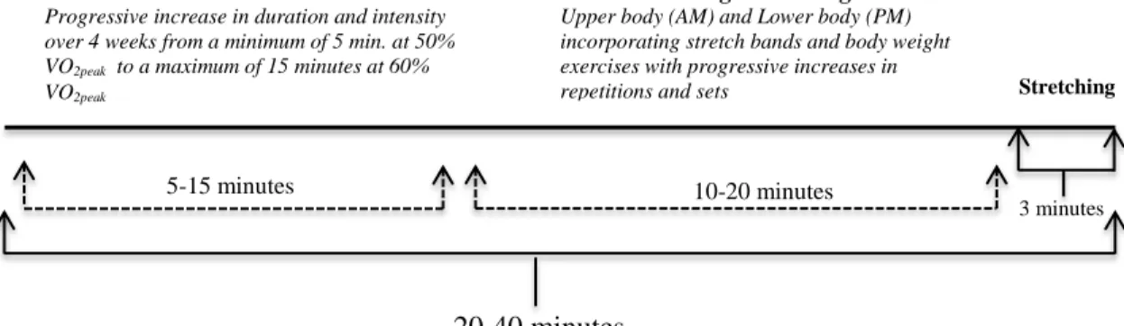
The exercise intervention encompassed both aerobic training and strength training twice a day, four days per week for 4 weeks. The time allotment for each exercise session was approximately 30-40 minutes with one trained exercise specialist to monitor each session. This intervention began the day following the baseline exercise assessment. The exercise training is summarized in Figure 2.
Figure 2. Standard Exercise Session Protocol
While adaptations were made throughout the intervention in order to
accommodate individualized physical limitation or training response, a progression of training was followed via recommendations from the American College of Sports Medicine for stimulating a training effect within interventions. The aerobic exercise segment began at 50% of VO2peak and all attempts were made to progress the intensity of the aerobic training by the last week of hospitalization to 60% VO2peak. Initially, the subject was intended to complete 5 minutes of aerobic exercise with a progression to 15 minutes by week 4. At that point, progression would be solely through intensity.
3 minutes
Strength Training
Upper body (AM) and Lower body (PM) incorporating stretch bands and body weight exercises with progressive increases in repetitions and sets
10-20 minutes
Aerobic Exercise
Progressive increase in duration and intensity over 4 weeks from a minimum of 5 min. at 50% VO2peak to a maximum of 15 minutes at 60%
VO2peak
5-15 minutes
20-40 minutes
20
On the first day during the strengthening exercises, the subject attempted each upper body exercise (wall push-up, low row, triceps extension, and a choice between lateral raises, front raises, or military press) and lower body exercise (modified squat, leg curl, leg extension, and standing calf raise) A rough estimate of intensity in terms of sets and reps were gathered with the primary focus on posture, form, and comfort. After the first day, the subject trained using the 12-6 maximum repetition methodology, 1-2 sets per exercise. A similar protocol in the exercise oncology literature has demonstrated improvements in body composition parameters and muscle hypertrophy (Galvao et al., 2010); however, modifications in execution were made because of the subjectivity of stretch band training. For instance, to increase intensity if the subject exceeded 12 repetitions, the exercise specialist would decrease the length of band to pull against or change to a thicker band.
Statistical Analysis
21 CHAPTER IV
RESULTS
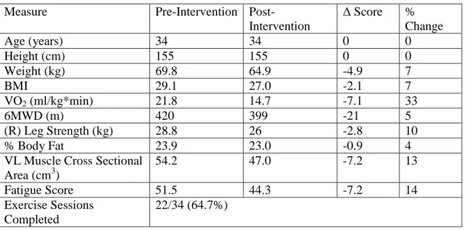
This patient was newly diagnosed with acute myelogenous leukemia recruited from the University of North Carolina Hospitals (UNC-CH Hospitals), division of Hematology/Oncology. This patient’s results are summarized in Table 1 below. Table 1- Subject Results
Measure Pre-Intervention
Post-Intervention
Δ Score % Change
Age (years) 34 34 0 0
Height (cm) 155 155 0 0
Weight (kg) 69.8 64.9 -4.9 7
BMI 29.1 27.0 -2.1 7
VO2 (ml/kg*min) 21.8 14.7 -7.1 33
6MWD (m) 420 399 -21 5
(R) Leg Strength (kg) 28.8 26 -2.8 10
% Body Fat 23.9 23.0 -0.9 4
VL Muscle Cross Sectional Area (cm3)
54.2 47.0 -7.2 13
Fatigue Score 51.5 44.3 -7.2 14
Exercise Sessions Completed
22/34 (64.7%)
22
23 CHAPTER V
DISCUSSION, CONCLUSION, AND RECOMMENDATIONS
Introduction
The standard of care for most cancer patients, including newly diagnosed AML patients is chemotherapy. The commonly used induction chemotherapy treatment plan, “7+3,” consists of a combination of cytarabine and an anthracycline and is by far the most successful at producing complete remission; however, referencing the lower than average 5-year survival rate compared to most cancers, this treatment plan is still far from optimal. In addition, commonly reported treatment-related toxicities, declines in physical function, and impairment to overall QOL result in a less than optimal post-cancer “new-normal.” There is a great need to develop interventions that can mitigate the body composition related side-effects of powerful chemotherapy drugs in order to prevent cancer-related fatigue.
24
exercise intervention to improve muscle CSA and reduce cancer-related fatigue in patients undergoing induction chemotherapy for AML.
Maintenance of VL Muscle Cross Sectional Area
According to expectations, there would not be a difference between VL CSA from pre-induction to post-induction chemotherapy. The results of the ultrasound analysis showed a decline in CSA of approximately 13%. It is difficult to compare these results to other exercise interventions in this population because panoramic ultrasound technology has not been used in the literature to measure changes in muscle size in this population. However, when comparing percent decrease in CSA in this study (14%) and the percent decrease in lean body mass from baseline to post exercise intervention in Battaglini et al., (2%), the comparison suggests the this intervention did not stimulate the vastus lateral enough to prevent losses in muscle size (2009).
Many reasons may accompany why the results suggest cachexia exists post-intervention. Primarily, the intervention may not have stimulated an environment where protein synthesis out performed protein degradation. Past research suggest that moderate intensity resistance exercise can increase protein synthesis by 50-60% (Wong and Booth, 1990). In this intervention, the patient performed 22 sessions, however, none of which provided more resistance than would be provided by a stretch band or body weight.
25 Decrease in Overall Fatigue
An important finding in this study was the reduction in fatigue levels from pre-induction to post-pre-induction chemotherapy. This result is similar to the decreases in fatigue levels detailed in Alibhai et al., (2012) and Battaglini et al., (2009). This case study only adds to the evidence that an exercise intervention may provide a relief from cancer-related fatigue in AML patients undergoing treatment. Not only is fatigue reported as the most common side-effect of chemotherapy, it is also related to many other co-morbidities and physiological systems.
AML patients are confined to hospital rooms for on average 3-5 weeks for induction chemotherapy and the subsequent recovery. During this period, many precautions are taken such that the immunocompromised patients do not develop infections that may impair recovery or shorten survival time. Essentially, patients are given the prescription of bed rest, confinement, and consequently little to no physical activity. This inactivity only leads to further decrements in body composition, and the cycle of increasing fatigue continues. The results of this exercise intervention suggest that possibly a mix of aerobic and resistance exercise may help at combating this cancer-related fatigue.
Conclusion
The common goal when treating AML is to reach complete remission and remain in complete remission. Unfortunately, the current chemotherapy regimens fail to maintain measures of body composition such as muscle CSA. The literature is clear that
26
induction phase of chemotherapy are not only safe and feasible, but also effective at mitigating the large losses in body composition and reducing the fatigue burden. Outcomes of this type of experiment can lead to future hypothesis testing.
Recommendations
Based on the results of this study and present limitations, recommendations for future research in the area of cancer and exercise, specifically, acute leukemia include:
1. The completion and analysis of the EQUAL randomized controlled study. This two arm study will allow for researchers to compare the change in muscle cross-sectional area from pre- to post-intervention with those patients participating in the normal standard of care.
2. When creating a strength maintaining program in which success is measured by vastus lateralis cross-sectional area, more emphasis should be placed on exercises that stimulate this muscle. Examples of exercises include lunges, side lunges, squats, leg extensions. As patients enter this setting with fitness levels that are across the spectrum of health, intensities should be cautiously increased using dumbbells, or alternatively decreased using body weight or modifications.
27
4. The implementation of increased physical activity based nutritional counseling in addition to standard of care nutritional counseling may improve outcomes such as weight maintenance, percent body fat changes, and maintenance of muscle cross-sectional area.
5. Providing leukemia patients with educational information in reference to the potential benefits of maintaining a moderate level of physical activity during treatment. Physical activity is a treatment choice that the patient has control over. Psychologically, this may potentially empower patients who feel powerless against the disease.
6. After completion and analysis of the EQUAL trial, extend the duration of the program to include the consolidation phase of the treatment plan. Boundaries include lack of supervision and motivation while the patient is at home between induction and consolidation; however, if possible, this extension may promote further benefits for the acute leukemia population. 7. The impact of the exercise intervention post-treatment should be recorded.
28 APPENDIX I
29 APPENDIX II
30
SCORING THE INSTRUMENT (PROMIS Fatigue SF 8a)
Short Forms: PROMIS instruments are scored using item-level calibrations. This means that the most accurate way to score a PROMIS instrument is to utilize scoring tools within Assessment Center that look at responses to each item for each participant. We refer to this as “response pattern scoring.” Response pattern scoring tools within
Assessment Center can be used even if data was collected on paper or in another software package. Because response pattern scoring is more accurate than the use of raw
score/scale score look up tables, it is preferred. However, if you aren’t able to use response pattern scoring, you can use the instructions below which rely on raw score/scale score look-up tables. For adults, each question has five response options ranging in value from one to five. To find the total raw score for a short form with all questions answered, sum the values of the response to each question. For example, for the 8-item form, the lowest possible raw score is 8; the highest possible raw score is 40 (see all short form scoring tables in Appendix 1).
A score can be approximated if a participant skips a question. If items are missing, first check how many items were answered. For short forms with at least 5 items, confirm that 4 or 50% of items, whichever is greater, were answered. For example, a 4-item short form can only be scored with complete data. A 5-item short form can be scored as long as 4 items were answered. A 10-item short form can be scored as long as the participant answered at least 5 items. For branched instruments (e.g., Alcohol Use), the screening question is not used in calculating the score and therefore shouldn’t be counted when assessing if the minimum number of items were answered. After confirming that enough responses were provided, sum the response scores from the items that were answered (not including any screening question). Multiply this sum by the total number of items in the short form. Finally, divide by the number of items that were answered. For example, if a respondent answered 5 of 8 questions and answered all items with the second lowest response option (2), you would sum all responses (10), multiply by the number of items in the short form (8) and divide by the number of items that were answered (5). Here (10x8)/5=16. If the result is a fraction, round up to the nearest whole number. This is a pro-rated raw score.
Again, the formula is:
(Raw sum x number of items on the short form) / Number of items that were actually answered
Locate the applicable score conversion table in Appendix 1 and use this table to translate the total raw score or pro-rated score into a T-score for each participant. The T-score rescales the raw score into a standardized score with a mean of 50 and a standard
31
Important: A higher PROMIS T-score represents more of the concept being measured.
For negatively-worded concepts like Fatigue, a T-score of 60 is one SD worse than average. By comparison, a Fatigue T-score of 40 is one SD better than average.
32 APPENDIX III
33 APPENDIX IV
34
REFERENCES
Ahtiainen JP, Hoffren M, Hulmi JJ, Pietikainen M, Mero AA, Avela J, Hakkinen K. Panoramic ultrasonography is a valid method to measure changes in skeletal muscle cross-sectional area. Eur J Appl Physiol. 2010; 108(2):273-9.
Alibhai SM, O'Neill S, Fisher-Schlombs K, Breunis H, Brandwein JM, Timilshina N, Tomlinson GA, Klepin HD, Culos-Reed SN. A clinical trial of supervised
exercise for adult inpatients with acute myeloid leukemia (AML) undergoing induction chemotherapy. Leuk Res. 2012; 36(10):1255-61.
al-Majid S, McCarthy DO. Cancer-induced fatigue and skeletal muscle wasting: the role of exercise. Biol Res Nurs. 2001; 2(3):186-97.
Anderson JE, Kopecky KJ, Willman CL, Head D, O'Donnell MR, Luthardt FW, Norwood TH, Chen IM, Balcerzak SP, Johnson DB, Appelbaum FR. Outcome after induction chemotherapy for older patients with acute myeloid leukemia is not improved with mitoxantrone and etoposide compared to cytarabine and daunorubicin: a Southwest Oncology Group study. Blood. 2002; 100(12):3869-76.
Ballard-Barbash R, Friedenreich CM, Courneya KS, Siddiqi SM, McTiernan A, Alfano CM. Physical activity, biomarkers, and disease outcomes in cancer survivors: a systematic review. J Natl Cancer Inst. 2012; 104(11):815-40.
Basaria S, Lieb J,2nd, Tang AM, DeWeese T, Carducci M, Eisenberger M, Dobs AS. Long-term effects of androgen deprivation therapy in prostate cancer patients. Clin Endocrinol (Oxf). 2002; 56(6):779-86.
Battaglini CL, Hackney AC, Garcia R, Groff D, Evans E, Shea T. The effects of an exercise program in leukemia patients. Integr Cancer Ther. 2009; 8(2):130-8. Berg HE, Tedner B, Tesch PA. Changes in lower limb muscle cross-sectional area
and tissue fluid volume after transition from standing to supine. Acta Physiol Scand. 1993; 148(4):379-85.
Brooks GA, Fahey T, Baldwin K. Exercise physiology: human bioenergetics and its applications. 3rd ed. In: Exercise Physiology: Human Bioenergetics and its Applications. 3rd Ed.; 2000.
Chang PH, Lai YH, Shun SC, Lin LY, Chen ML, Yang Y, Tsai JC, Huang GS, Cheng SY. Effects of a walking intervention on fatigue-related experiences of
35
Courneya KS, Segal RJ, Mackey JR, Gelmon K, Reid RD, Friedenreich CM, Ladha AB, Proulx C, Vallance JK, Lane K, Yasui Y, McKenzie DC. Effects of aerobic and resistance exercise in breast cancer patients receiving adjuvant chemotherapy: a multicenter randomized controlled trial. J Clin Oncol. 2007; 25(28):4396-404. Curt GA, Breitbart W, Cella D, Groopman JE, Horning SJ, Itri LM, Johnson DH,
Miaskowski C, Scherr SL, Portenoy RK, Vogelzang NJ. Impact of cancer-related fatigue on the lives of patients: new findings from the Fatigue Coalition.
Oncologist. 2000; 5(5):353-60.
De Backer IC, Schep G, Backx FJ, Vreugdenhil G, Kuipers H. Resistance training in cancer survivors: a systematic review. Int J Sports Med. 2009; 30(10):703-12. Dimeo FC, Stieglitz RD, Novelli-Fischer U, Fetscher S, Keul J. Effects of physical
activity on the fatigue and psychologic status of cancer patients during chemotherapy. Cancer. 1999; 85(10):2273-7.
Dimeo FC, Tilmann MHM, Bertz H, Kanz L, Mertelsmann R, Keul J. Aerobic exercise in the rehabilitation of cancer patients after high dose chemotherapy and autologous peripheral stem cell transplantation. Cancer. 1997; 79(9):1717-22. Fearon K, Strasser F, Anker SD, Bosaeus I, Bruera E, Fainsinger RL, Jatoi A,
Loprinzi C, MacDonald N, Mantovani G, Davis M, Muscaritoli M, Ottery F, Radbruch L, Ravasco P, Walsh D, Wilcock A, Kaasa S, Baracos VE. Definition and classification of cancer cachexia: an international consensus. Lancet Oncol. 2011; 12(5):489-95.
Ferrando A, Tipton K, Bamman M, Wolfe R. Resistance exercise maintains skeletal muscle protein synthesis during bed rest (L'exercice de resistance maintient la synthese des proteines des muscles du squelette lors de l'inactivite). J Appl Physiol. 1997; 82(3):807-10.
Galvao DA, Newton RU. Review of exercise intervention studies in cancer patients. J Clin Oncol. 2005; 23(4):899-909.
Galvao DA, Taaffe DR, Spry N, Joseph D, Newton RU. Combined resistance and aerobic exercise program reverses muscle loss in men undergoing androgen suppression therapy for prostate cancer without bone metastases: a randomized controlled trial. J Clin Oncol. 2010; 28(2):340-7.
36
Galvao DA, Nosaka K, Taaffe DR, Spry N, Kristjanson LJ, McGuigan MR, Suzuki K, Yamaya K, Newton RU. Resistance training and reduction of treatment side effects in prostate cancer patients. Medicine & Science in Sports & Exercise. 2006; 38(12):2045-52.
Hanson ED, Sheaff AK, Sood S, Ma L, Francis JD, Goldberg AP, Hurley BF. Strength training induces muscle hypertrophy and functional gains in black prostate cancer patients despite androgen deprivation therapy. J Gerontol A Biol Sci Med Sci. 2013; 68(4):490-8.
Hemming L, Maher D. Understanding cachexia and excessive weight loss in cancer. Br J Community Nurs. 2005; 10(11):492-5.
Hiddemann W, Kern W, Schoch C, Fonatsch C, Heinecke A, Wormann B, Buchner T. Management of acute myeloid leukemia in elderly patients. J Clin Oncol. 1999; 17(11):3569-76.
Hurley BF, Hanson ED, Sheaff AK. Strength training as a countermeasure to aging muscle and chronic disease. Sports Med. 2011; 41(4):289-306.
Jones LW, Eves ND, Haykowsky M, Joy AA, Douglas PS. Cardiorespiratory exercise testing in clinical oncology research: systematic review and practice
recommendations. Lancet Oncol. 2008; 9(8):757-65.
Khalade A, Jaakkola MS, Pukkala E, Jaakkola JJ. Exposure to benzene at work and the risk of leukemia: a systematic review and meta-analysis. Environ Health. 2010; 9:31,069X-9-31.
Klepin HD, Danhauer SC, Tooze JA, Stott K, Daley K, Vishnevsky T, Powell BL, Mihalko SL. Exercise for older adult inpatients with acute myelogenous leukemia: A pilot study. J Geriatr Oncol. 2011; 2(1):11-7.
Kyrdalen AE, Dahl AA, Hernes E, Hem E, Fossa SD. Fatigue in prostate cancer survivors treated with definitive radiotherapy and LHRH analogs. Prostate. 2010; 70(13):1480-9.
Lakoski SG, Eves ND, Douglas PS, Jones LW. Exercise rehabilitation in patients with cancer. Nat Rev Clin Oncol. 2012; 9(5):288-96.
Levy ME, Perera S, van Londen GJ, Nelson JB, Clay CA, Greenspan SL. Physical function changes in prostate cancer patients on androgen deprivation therapy: a 2-year prospective study. Urology. 2008; 71(4):735-9.
37
Mock V, Dow KH, Meares CJ, Grimm PM, Dienemann JA, Haisfield-Wolfe M, Quitasol W, Mitchell S, Chakravarthy A, Gage I. Effects of exercise on fatigue, physical functioning, and emotional distress during radiation therapy for breast cancer. Oncol Nurs Forum. 1997; 24(6):991-1000.
Newton RU, Taaffe DR, Spry N, Gardiner RA, Levin G, Wall B, Joseph D, Chambers SK, Galvao DA. A phase III clinical trial of exercise modalities on treatment side-effects in men receiving therapy for prostate cancer. BMC Cancer. 2009; 9:210,2407-9-210.
Physical Activity Guidelines Advisory Committee report, 2008. To the Secretary of Health and Human Services. Part A: executive summary. Nutr Rev. 2009; 67(2):114-20.
Poste G, Fidler IJ. The pathogenesis of cancer metastasis. Nature. 1980; 283(5743):139-46.
Roboz GJ. Current treatment of acute myeloid leukemia. Curr Opin Oncol. 2012; 24(6):711-9.
Rubnitz JE, Gibson B, Smith FO. Acute myeloid leukemia. Hematol Oncol Clin North Am. 2010; 24(1):35-63.
Saigal CS, Gore JL, Krupski TL, Hanley J, Schonlau M, Litwin MS, Urologic Diseases in America Project. Androgen deprivation therapy increases cardiovascular morbidity in men with prostate cancer. Cancer. 2007; 110(7):1493-500.
Schmitz KH, Courneya KS, Matthews C, Demark-Wahnefried W, Galvao DA, Pinto BM, Irwin ML, Wolin KY, Segal RJ, Lucia A. American College of Sports Medicine roundtable on exercise guidelines for cancer survivors. Med Sci Sports Exerc. 2010; 42(7):1409-26.
Scott JM, Martin DS, Ploutz-Snyder R, Caine T, Matz T, Arzeno NM, Buxton R, Ploutz-Snyder L. Reliability and validity of panoramic ultrasound for muscle quantification. Ultrasound Med Biol. 2012; 38(9):1656-61.
Segal RJ, Reid RD, Courneya KS, Malone SC, Parliament MB, Scott CG, Venner PM, Quinney HA, Jones LW, D'Angelo M. Resistance exercise in men receiving androgen deprivation therapy for prostate cancer. J Clin Oncol. 2003; 21(9):1653-9.
38
Siegel R, Naishadham D, Jemal A. Cancer statistics, 2013. CA Cancer J Clin. 2013; 63(1):11-30.
Smith RA, Brooks D, Cokkinides V, Saslow D, Brawley OW. Cancer screening in the United States, 2013: a review of current American Cancer Society guidelines, current issues in cancer screening, and new guidance on cervical cancer screening and lung cancer screening. CA Cancer J Clin. 2013; 63(2):88-105.
Tisdale MJ. Wasting in Cancer. J Nutr. 1999; 129(1): p. 243S.
Tsai HK, D'Amico AV, Sadetsky N, Chen MH, Carroll PR. Androgen deprivation therapy for localized prostate cancer and the risk of cardiovascular mortality. J Natl Cancer Inst. 2007; 99(20):1516-24.
van Eys J. Effect of nutritional status on response to therapy. Cancer Research. 1982; 42(2 Supplement):747s-53s.
Wong TS, Booth FW. Protein metabolism in rat tibialis anterior muscle after
stimulated chronic eccentric exercise (Metabolisme des proteines dans le muscle jambier anterieur apres un exercice excentrique chronique electrostimule). J Appl Physiol. 1990; 69(5):1718-24.
Yarasheski KE, Zachwieja JJ, Bier DM. Acute effects of resistance exercise on