Original Research Article
Closed reduction and percutaneous k-wire fixation for distal end
radius fractures
Tanveer Ali, Mohmmad Sikander Baketh, Maneer Ahmad Mir*
INTRODUCTION
The optimal management for displaced distal radius fractures has not yet been established.1,2 Although the
association between radiological and functional outcome is controversial, restoration of normal anatomy has been recommended in active patients.3 K-wire fixation is a
simple, minimally invasive technique to maintain the reduction in extra and intra articular fractures. Clinical guidelines from the American Academy of Orthopaedic Surgeons (AAOS) moderately recommend an anatomically stable surgical fixation, instead of cast fixation, to be followed by early wrist motion for treatment of patients with displaced distal radius fractures.4 Radial length is a radiological parameter that
has been shown to correlate with functional outcome.5,6
METHODS
This was a prospective study of 40 patients with distal radius fractures who were treated by K-wire fixation in Government Medical College and Hospital, Jammu for a period of one year from September 2018 to October 2019.
Patients with fractures of distal end radius of either side or both sides, with or without ulnar styloid fracture, of age group 18 - 85 years, of either sex having closed fractures of up to 3 cm from distal articular surface of radius willing for treatment were enrolled for this prospective open randomized case control comparative study. Patients less than 18 years or more than 85 years, having compound fractures associated with vascular Department of Orthopaedics, GMC, Jammu, Jammu and Kashmir, India
Received: 28 May 2020
Accepted: 10 June 2020
*Correspondence:
Dr. Maneer Ahmad Mir,
E-mail: dr.tanvirm90@outlook.com
Copyright: © the author(s), publisher and licensee Medip Academy. This is an open-access article distributed under the terms of the Creative Commons Attribution Non-Commercial License, which permits unrestricted non-commercial use, distribution, and reproduction in any medium, provided the original work is properly cited.
ABSTRACT
Background: To evaluate radiological and functional outcome in fractures of the distal radius treated by K-wire fixation.
Methods: Forty patients (16 males, 24 females) with different types of fractures of distal radius were treated. K-wire fixation was performed under axillary bolock or general anaesthesia. Anatomical restoration was evaluated by postero-anterior and lateral radiographs obtained preoperatively and at 09 months of follow up to evaluate Radial Height (RH), Radial Inclination (RI) and Volar Tilt (VT). Functional outcome was evaluated using Mayo scoring system.
Results: According to Mayo score 72.5% (n=29) of our patients had excellent to good outcome while as 17.5% (n=7) had fair outcome and 10% (n=4) patients had poor outcome.
Conclusions: Kirschner wire fixation is an inexpensive procedure that provides anatomic reduction, fracture fixation, and maintenance of reduction with an adequate method of immobilization.
Keywords: AO classification, Distal radius fracture, K-wiring, Lid strom criteria, MAYO score, Radiological evaluation
injuries or had associated multiple injuries were excluded from the study.
All fractures were classified according to AO (the Association for Osteosynthesis) classification system by getting Postero-Anterior (PA) and Lateral (Lat) views of radiographs of the wrist at the time of the initial injury.7
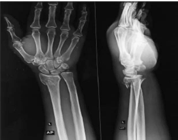
(Figure 1) Some patients needed CT scan of wrist for further evaluation of intra-articular fractures.
Figure 1: Pre-op AP and lateral X-ray.
The patients were admitted to the hospital and were operated on as soon as possible, depending on the condition of the local tissue, hematoma, tissue oedema and other associated injuries. Under axillary block or general anesthesia, the patient was placed in the supine position with the involved limb in traction with a finger trap through the index finger, and provided counter traction. An accurate reduction in the fracture was the first step in the treatment plan. The K-wires were placed under C-arm guidance (Figure 2).
Figure 2: Visualization under C-Arm.
In the post-operative period, the limb was kept strictly elevated for a period of 2 days. Patient was encouraged to begin active finger movements as soon as the effect of anaesthesia wore out. Patient at the end of 2 days was asked to mobilize his elbow. At this time the pin sites were inspected and then dressed. If pin sites and
mobilization were satisfactory, the patient was then discharged the next day.
The final results of the patients with excellent and good functional outcome were considered satisfactory. The patients underwent follow-up at our outpatient clinic at 2-week intervals following hospital discharge. The healing of the fracture was assessed both clinically and radiographically at each follow-up At the end of four weeks a check X-ray was taken (Figure 3) and if satisfactory signs of union were present, the pins were removed as was the slab and patient given a crepe bandage.
Figure 3: Post-op AP and lateral X-ray.
The treatment complications were recorded. After bony union, patients underwent further follow-up for 9 months. A check X-ray was done (Figure 4). The residual deformity and the subjective evaluation were recorded in the same way as the original scoring system.
Figure 4: AP and lateral X-ray after K-wire removal.
Radiological evaluation
measurement through Posterio-anterior and lateral radiographs. PA view provides information about RI (Radial Inclination) and RH (Radial height). RI is a measurement of the radial angle. A line is drawn along the articular surface of the radius perpendicular to the long axis of the radius, and a tangent is drawn from the radial styloid. The normal angle is 15-25º. RH is a measurement between 2 parallel lines that are perpendicular to the long axis of the radius. One line is drawn on the articular surface of the radius, and the other is drawn at the tip of the radial styloid. The normal radial height is 9.9-17.3 mm.8 In the Lateral view, the VT
(Volar Tilt) of the distal radius articular surface is measured. A line perpendicular to the long axis of the radius is drawn, and a tangent line is drawn along the slope of the dorsal-to-volar surface of the radius. The normal angle is 10-25º.8 These measurements were taken
pre operatively and at 9 months follow up. At the end of the study all the data was compiled and analyzed statistically by a one-way measure ANOVA test. p-value less than 0.05 was considered to be statistically significant.
Figure 5: Dorsiflexion and palmar flexion at 12 weeks follow-up.
Figure 6: Pronation and spination at 12 weeks follow-up.
Functional evaluation
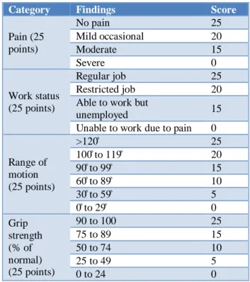
Patients were evaluated by using Modified Mayo Wrist Score (Table 1). It is a physician-rating scoring system which gives us a total score of a 100; 25 for the assessment of pain, 25 for the active extension/flexion arc
of the wrist, (Figure 5, 6) 25 for grip strength, and 25 for the ability to return to regular activities. The pain is rated according to patient’s description.
Table 1: Mayo Score.
Category Findings Score
Pain (25 points)
No pain 25
Mild occasional 20 Moderate 15
Severe 0
Work status (25 points)
Regular job 25 Restricted job 20 Able to work but
unemployed 15 Unable to work due to pain 0
Range of motion (25 points)
>120̊ 25 100̊ to 119̊ 20 90̊ to 99̊ 15 60̊ to 89̊ 10 30̊ to 59̊ 5
0̊ to 29̊ 0
Grip strength (% of normal) (25 points)
90 to 100 25
75 to 89 15
50 to 74 10
25 to 49 5
0 to 24 0
RESULTS
A total of 40 patients underwent this procedure comprising of 16 males and 24 females. The mean age and Standard Deviation (SD) at the time of the injury was 59.35±13.40years (range from 27 to 84 years). Twenty four fractures involved left side while sixteen fractures occurred on the right. Mode of injury was road traffic accident and fall in majority of the cases.
According to AO classification, most of the cases were of C (n = 16) 40.0% and A (n = 15) 37.5% type probably because of increased incidence of high velocity injuries. (Table 2).
Table 2: Types of fracture.
Radiological type (AO) Frequency Percent
A2 5 12.5%
A3 10 25.0%
B1 3 7.5%
B2 6 15%
C1 10 25.0%
C2 6 15.0%
Total 40 100.0%
Table 3: Radiological evaluations of RH, RI and VT.
K-wiring group
Radial inclination 21.1±4.56° Radial height 10.67±2.97 mm Volar tilt 9.42±3.19°
Table 4: Values on the basis of type of fracture.
Type of fracture
No. of cases
Radial inclination
Radial height
Volar tilt
Type A 15 20.3mm 11.3mm 9.1° Type B 9 21.9mm 10.9mm 9.0° Type C 16 21.4mm 9.9mm 9.9°
Authors had 15 type A, 9 type B and 16 type C fractures and obtained almost similar results in all three types. (Table 4).
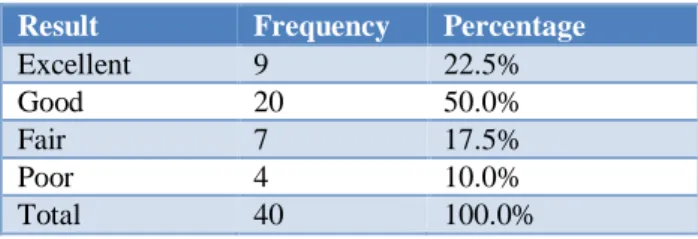
According to Mayo score 72.5% (n=29) of our patients had excellent to good outcome while as 17.5% (n=7) had fair outcome and 10% (n=4) patients had poor outcome. (Table 5).
Table 5: Mayo score following k-wire fixation.
Result Frequency Percentage
Excellent 9 22.5%
Good 20 50.0%
Fair 7 17.5%
Poor 4 10.0%
Total 40 100.0%
Complications
Six of our patients sufferd from stiffness who were send for subsequent physiotherapy. Five patients had pin site infection which needed regular dressing and antibiotics (Table 6).
Table 6: Complications seen during k-wire fixation.
Complications Frequency Percentage
Nil 29 72.5%
Stiffness 6 15.0%
Pin site infection 5 12.5%
Total 40 100.0%
DISCUSSION
Fractures of the distal radius are one of the most common fractures encountered in the emergency department. Most of the fractures are caused by a fall on the outstretched hand with the wrist in dorsiflexion. These fractures can cause significant distress to the patient and lead to disability if not managed adequately and in time. The management of fractures of distal radius has always been a debatable and challenging issue for orthopaedic
surgeons. The ultimate goal is to restore grip strength and motion and allow quick return of function and to minimize the risk for future degenerative changes in the wrist joint.
Closed reduction followed by fixation of the fracture with percutaneous pins and cast, is a widely used method. The pins may be inserted by a variety of methods; across fracture fixation or with the intrafocal Kapandji technique.9,10 Percutaneous pinning is best suited for
extra-articular fractures without substantial communition. Percutaneous pinning is associated with complications like pin site infection, radial nerve lesions, pin loosening, redisplacement and complex regional pain syndrome (CRPS). It requires about 5 weeks’ of immobilization, which often results in temporary wrist joint stiffness.
Our study comprised of 40 patients with 16 male and 24 female patients. Right side was involved in sixteen patients (40%) and left side in twenty four patients (60%). In 85% of cases, the mode of injury was fall with road traffic accident and assault compromising the other 15%. According to AO classification, most of the cases were of C (n = 16) 40.0% and A (n = 15) 37.5% type probably because of increased incidence of high velocity injuries.
Distal radius deformity was assessed by measuring RH, RI and VT at different stages of treatment. We found a mean radial inclination of21.1±4.56°, radial height10.67±2.97 mm of and volar tilt of9.42±3.19°. Our results are in concordance with the study conducted by Abulaban OA et al. They found post-operative radial inclination, radial height and volar tilt to be 22.18±4.84°, 11.65mm and 9.19±6.10° respectively in their study group managed by K wiring.11 There were minor
variations in the anatomical parameters (volar tilt, radial inclination and radial height) in our study which were comparable with the other studies but did not affect functional results significantly.12-14
According to Mayo score 72.5% (n=29) of our patients had excellent to good outcome while as 27.5% (n=11) had fair to poor outcome. Kumar P et al had a similar outcome in their study about Percutaneous kirschnerwire fixation of distal radius fractures and found excellent to good results in 75% cases and fair to poor outcomes in 25% patients.15 Most of our patients had a smooth
recovery. Six patients suffered from stiffness and five patients had pin site infection.
CONCLUSION
fixation, and maintenance of reduction with an adequate method of immobilization.
Funding: No funding sources Conflict of interest: None declared
Ethical approval: The study was approved by the Institutional Ethics Committee
REFERENCES
1. Handoll HH, Madhok R. Surgical interventions for treating distal radius fractures in adults. Cochrane Database Syst Rev. 2003;(3).
2. Koval K, Haidukewych GJ, Zirgibel BJ. Controversies in the management of distal radius fractures. JAAOS- J Am Academy Orthopaedic Surg. 2014 Sep 1;22(9):566-75.
3. Fernandez DL. Should anatomic reduction be pursued indistal radius fractures? J Hand Surg. 2000;25-B:523-7.
4. Lichtman DM, Bindra RR, Boyer MI et al. Treatment of distal radius fractures. J Am Academy Orthopaedic Surg. 2010; 18(3):180-89.
5. Melone CP. Articular fractures of the distal radius. Orthop Clin North Am 1984;15:217-36.
6. Warwick D, Prothero D, Field J, Bannister G. Radiological measurement of radial shortening in Colles’ fracture. J Hand Surg. 1993;18-B:50-2. 7. Clayton RAE, Gaston MS, Ralston SH,
Court-Brown CM, McQueen. Association between decreased bone density and severity of distal radial fractures. J Bone Joint Surg (Am). 2009;91:613-9. 8. Cooney WP, Linscheid RL, Dobyns JH. External
pin fixation for unstable Colles’ fractures. J Bone Joint Surg (Am). 1979;61:840-5.
9. Kapandji A. Intra-focal pinning of fractures of the distal end of the radius 10 years later. Ann Chir Main. 1987;6(1):57-63.
10. Harley BJ, Scharfenberger A, Beaupre LA, Jomha N, Weber DW. Augmented external fixation versus percutaneous pinning and casting for unstable fractures of the distal radius--a prospective randomized trial. J Hand Surg Am. 2004;29(5):815-24.
11. Abulaban OA, Alrifai AI, Almosallam SA. Radiological Assessment of Surgical Treatment of Distal Radius Fractures among Patients at an Academic Center in Jeddah. Egyptian J Hospital Med. 2018;72(4):4343-8.
12. Geller L, Bernstein M, Carli A, Berry G, Reindl R, Harvey E. Efficacy of different fixation devices in maintaining an initial reduction for surgically managed distal radius fractures. Canad J Surg. 2009 Oct;52(5):E161.
13. Hollevoet N, Vanhoutie T, Vanhove W, Verdonk R. Percutaneous K-wire fixation versus palmar plating with locking screws for Colles' fractures. Acta Orthopædica Belgica. 2011 Apr 1;77(2):180. 14. Chaudhary P, Shrestha BP, Khanal GK, Baral P,
Shah AB, Das J. RCT comparing closed reduction and percutaneous versus open reduction and volar locking plate in treatment of distal radius fractures in adults. Int J Orthopaed. 2017;3(2):678-81. 15. Kumar P, Kisan D. Percutaneous kirschner wire
fixation of displaced colles’ fracture. J Orthop Traumatol Rehabil. 2018;10:98-102.