Original Research Article
Evaluation of a replanted finger and measuring its perfusion changes
to predict its viability by laser speckle contrast imaging
Bilkis Akthar, Zeng Tao Wang*, Muhsin Billah Bin Khashru, Hou Zhi Dian
INTRODUCTION
While evaluating the viability of a tissue clinically, numerous sophisticated techniques, such as laser Doppler flowmetry (LDF) and laser Doppler perfusion imaging (LDPI), have appeared to quantify and qualify this method for further objectivity. However, unlike laser speckle contrast imaging (LSCI), these have been least used in recent decades to evaluate local blood flow in stressed tissues, such as flap surgery, burns, dermal
perfusion in deep tissue imbrication and wound undermining for wound closure, and also to assess retinal blood flow, liver microcirculation after hepatic transplantation, in laparoscopic surgeries, gastric tube reconstructions following esophagectomy, neurosurgery to analyze cerebral blood flow, assess oral mucosal blood flow following periodontal plastic surgery and so on.1-14
Despite this, clinical judgment by the surgeon remains the golden standard for postoperative evaluation of the tissue. However, clinical assessment is always subjective, Department of Hand and Foot Surgery, Shandong Provincial Hospital affiliated to Shandong University, Jinan,
Shandong, China
Received: 03 January 2020
Revised: 19 January 2020
Accepted: 20 January 2020
*Correspondence:
Dr. Zeng Tao Wang, E-mail: [email protected]
Copyright: © the author(s), publisher and licensee Medip Academy. This is an open-access article distributed under the terms of the Creative Commons Attribution Non-Commercial License, which permits unrestricted non-commercial use, distribution, and reproduction in any medium, provided the original work is properly cited.
ABSTRACT
Background: This research aims to monitor the microcirculation of the replanted finger, which was entirely severed by using laser speckle contrast imaging (LSCI) for early detection and revision of vascular compromise for successful finger replantation.
Methods: These six months of research was taken for a survey of 40 cases of replanted fingers of patients of distinct ages and sex. Scrutinizing was done postoperatively by LSCI, every hourly for seven days, to assess changes in blood perfusion both in replanted fingers and healthy ones and analyzed graphically.
Results: Initially, from postoperative d=0 to d=2, the perfusion value was at baseline, which ranged 40±15 perfusion unit (PU), showing a wave-like curve, then gradually increased up to 350±50 PU or above in case of those which survived successfully, showing continuous peak slope. However, a gradual drop in perfusion, <35 PU from d=2 or d=3, was seen in those despite undergoing heparinized finger pin-prick bleeding therapy and failed to thrive, showing a downslope curve. Whereas some were under meticulous observation, which flourished lately. Concurrently, a comparison was made with the healthy fingers’ of the same patient, ranging from 200±50 to as high as 400±50 or above. Clinical correlation, as well as perfusion readings of LCSI, were done simultaneously.
Conclusions: LSCI provides sensitive and reproducible finger microcirculation measurements and is reliable in predicting reductions in blood perfusion induced by venous or arterial occlusion. It is, therefore, an informative device to detect microvascular compromise during and after replantation surgery.
Keywords: Replanted finger, Replantation, Laser speckle contrast imaging, Perfusion evaluation, Microcirculation, Predictability
and tissue perfusion, also its viability cannot rely only on clinical experience.
Laser speckle contrast analysis (LASCA), also
recognized as LSCI, is a technique based on the appraisal of the imaged speckle pattern that evolves from random interference of coherent light dispersed by a media comprised of light scattering particles and visualizes blood perfusion of microcirculatory tissue instantly.15,16 It
is a camera-based method that illuminates a tissue area with 785-nm divergent laser light and analyzes the light interference pattern dispersed from the tissue composed of dark and bright fields, called a speckle pattern, which is equal to the proportion between the standard intensity deviation and the intensity mean.17 This pattern will alter
over time when there is motion in the object, such as red blood cells in a tissue. A charge-coupled device (CCD) camera with a preset exposure time will record these changes as motion blurring in the speckle pattern in this PeriCam PSI System. The amount of blurring will vary depending on the degree of motion in the picture region. The more movement in an image, the more it appears blurred, the average intensity deviation will reduce, resulting in reduced speckle contrast, and vice versa. However, this relation is independent of frame rate, number of images, and tissue perfusion.18 PeriCam PSI
system uses arbitrary units, perfusion units (PU), to record blood perfusion.
Fercher and Briersinitially proposed the biomedical use of LSCI using analog techniques in 1981.7-9 It has
developed into a non-invasive, non-contact, and rapid method for measuring microvascular blood perfusion with the growth of digital cameras and quicker computers.
Figure 1: Post-operative LSCI assessment of a replanted middle finger (rt.) of a patient in the general
ward at a working distance within 8-10 cm, at room temperature 26±2°C.
testing and can be easily handled and used on the bedside (Figure 1) or during operations (Figure 2) due to its technological simplicity; and elevated spatial and temporal resolution, making it appropriate for dynamic functionality. Blood flow (volume of blood per unit time in the vasculature) and perfusion (quantity of blood per amount of tissue per unit time) are vital clinical microcirculation parameters.19 The blood flow speed
strongly relates to the morphology of the vasculature itself (e.g., local vessel diameters and vessel density).19,20
Figure 2: Immediate intra-operative monitoring of a replanted lt. index finger by LSCI in an operating room, keeping the working distance (distance between the laser lens and the skin to be measured) between
8-10 cm.
Many reconstructions and replantation surgeries are practiced in today's plastic and reconstructive world. These have, however, so far been the least evaluated by the above techniques.21 Hence, the purpose of this
research is to assess a replanted finger by LSCI, every hour or so for seven consecutive days after surgery, and to monitor the vascularity of the replanted finger for the early detection and review of vascular compromise for successful finger replantation.
METHODS
Patients
Table 1: The number of replantation cases and their perfusion data of patients of different age groups and sexes, the levels of amputation of fingers, injury types, neurovascular structures and tendons repaired, and the number of cases where vein grafts were required.
Replantation category Total no. of fingers Average perfusion Mean age and sex (total no.) Levels of amputations (total no.) Injury types (total no.) Neurovascul ar structures anastomosed (total no.) Palmar cutaneous vein graft (total no.) Bone fixation by Kirschner wire (total no.) Flexor and extensor tendons repaired (total no.) Survived
Uncompromised 22 350±50 PU
(p<0.05)
35±15 years
M: F (30:10)
Tamai Zone I (6)
Tamai Zone II (10)
Transphalangeal [PP or DP or MP or PIPJ or MPJ or DIPJ] (24) Machinery crush (14) Electric Saw (4) Accidental Crush (13) Guillotine (9) Nerves [0.5mm-0.7mm diam.] (32) Arteries [0.6mm-1mm diam.] (40) Veins [0.6mm-0.9mm diam.] (38) (18) Single axial/ Double axial/ Arthrodesis (34) Flexor tendons (34) Extensor tendons (30) Compromised or under special Observation
3 100±50 PU
(p<0.0005)
Heparinised finger Pin-prick bleeding therapy
9 200±50 PU
(p<0.001)
Exploratory
surgery 1
300±50 PU (p<0.00005)
Necrosed 5 <35 PU
(p<0.00003)
Equipment
Laser speckle contrast imager (PeriCam PSI system, Perimed AB, Jarfalla, Sweden) LSCI has been used to evaluate the replanted finger's skin perfusion.15,22-24 The
working distance, that is, the space between the LSCI camera and the skin to be monitored, placed precisely 8-10 cm above the patient's finger. Depending on the central axis of the fingers replanted, the region of interest (ROIs) was taken as 2-5 mm², average 5 mm², and marked. The perfusion evaluated by illuminating the tissue with a divergent laser beam (invisible near infra-red) of 785-nm-wavelength, generating a pattern of speckle over the illuminated region. An advanced CCD camera recorded the speckle image, while another captured a standard color picture of the measured area. The camera exposure time for all measurements was kept constant at 30 sec. By using a frame grabber, the acquisition rate was set to 10 images/s and was procured at 50 Hz mains frequency frame rates and saved on a post-processing computer using custom software. The images transformed into pseudo-color images during this process, where the flow indicated from red (high flow) to blue (low flow).
Surgical procedure
After immediate hospitalization, emergency surgery was performed. The severed fingers were preserved at 4°C for 5-7 hours before the operation. A thorough history and necessary investigations have been done to exclude any major diseases such as cardiac, pulmonary disease, or peripheral vascular disease, so on, and precautions were taken accordingly. Replantation surgery performed by thoroughly washing the wound by hydrogen peroxide, chlorhexidine, 0.9% normal saline, and povidone-iodine. Thorough debridement of contaminated and necrotic tissues done, fixed the stump and the proximal part of the fingers with single or double axial Kirschner wires, arthrodesis done in some cases, flexor and extensor tendons were repaired respectively, digital nerves, and vessels(diameter ranging 0.6-0.9 mm) with or without palmar vein graft, anastomosis done (Table 1).23-25 The
room temperature and the patients’ vital parameters like blood pressure, heart rates, and other vital signs continuously monitored both intraoperatively and postoperatively.
Subsequently, the postoperative assessments were performed by LSCI for 24 hours for seven consecutive days to monitor the perfusion. All measurements performed at approximately 26±2°C room temperature. During this period, the patients laid supine, comfortable, and warm, and the operated hands immobilized by external plaster in a neutral position. Parenteral medications like injectable anticoagulants, antispasmodic, vasodilator, antibiotic, and analgesics set accordingly. At
Data analysis
LSCI images were processed using the system analysis software program. The perfusion values represented as PU (perfusion units) defined as an arbitrary unit calculated from the speckle contrast analysis.28 It can be
rapidly processed to generate a full-field map of the perfusion index proportional to the concentration and mean velocity of red blood cells in the tissue to be measured.29 A circular region of interest (ROIs) ranging
from 2 to 5 mm², an average 5 mm², was selected in each image. The frame rate set to 10 images per 30 sec. For each image, the average perfusion in each ROI calculated. The perfusion data of the replanted finger and that of the normal healthy finger were measured and obtained every hourly after operation for seven days; statistically analyzed and graphically represented in Microsoft Excel Worksheet.
RESULTS
Forty cases of replanted fingers (which were entirely severed) whether transphalangeal amputations (PIPJ or DIPJ or proximal or middle or distal phalanges) or fingertip amputations (Tamai zone I or II) and of distinct ages and genders are included in this six-month pilot study. Despite other general health issues, surgery performed as an emergency basis under controlled observation and care. The perfusion of these replanted fingers was compared with that of the healthy fingers of the same patient and monitored every hourly for the whole day consecutively for 7 days, both clinically and by LSCI (Figure 3). LSCI images were also captured instantly after surgery. Replanted finger microcirculation measurements were successfully made in all patients, resulting in analyzable speckle contrast images and assumption of a mean threshold value by which we can predict the viability.
The recorded perfusion values were analyzed in the Excel worksheet, and a graph was obtained. Amongst forty replanted fingers, twenty-two revived effectively. On postoperative d=0 to d=1, the average perfusion, in these uncompromised replanted digits, was found to be at baseline, from as low as 40±10 PU or 100±50 or above, depending on patients' body built (Figure 4). From d=3 to d=5, there was a gradual increase in perfusions each day, rising to 350±50 PU or more, forming a steady wave-like
curve. Nine, however, survived after enduring
undergo immediate exploratory surgery when perfusion dropped below the threshold level; however, it rose to 300±60 PU after the revision surgery (Figure 8). By this
study, we believe that when the perfusion of the replanted finger falls below the threshold level, it fails to thrive.
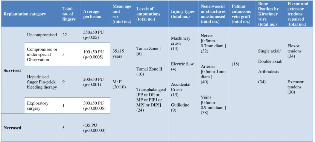
Figure 3: Respective photographs and LSCI images (A and B) showing a replanted right thumb of a 25-year-old male and that of his healthy left thumb (D and E), respectively; arrow in (B) showing minimum perfusion when ROI - 3.7×4.2 cm; the color bar below shows high blood perfusion (red) and low perfusion (blue); (C) show crinkly
changes in perfusion curve of 7 postoperative days; arrows indicating low perfusion of 144.53 PU(lt.) at 0 post-operative day and highest perfusion of 351.91 PU(rt.) on 7th post-post-operative day and later gaining stability in an
uncompromised replanted thumb. Note the perfusion values in the graph of a healthy thumb (F), the lowest perfusion of 166.08 PU (lt. arrow) at 0 operative day, and the highest 435.33 PU (rt. arrow) at 7th
post-operative day.
Figure 4: (A-D) No or mild changes in skin color and perfusion in an otherwise typically healed replanted digit of a 32-year-old male, rt. thumb (saw machine crush injury, transphalangeal amputation of proximal phalanx) on 1st, 3rd, 5th, and 10th post-operative days, respectively; (E-H) LSCI images illustrating changes in skin perfusion of 5 mm² ROI; (I) showing corresponding seven postoperative days’ steady wave-like perfusion curve; arrows showing lowest perfusion - 137.57 PU (lt.) on 0 post-operative day and highest - 301.56 PU (rt.) on 7th post-operative day,
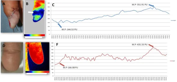
Figure 5: (A-E) Visual appearance changes of a 50-year-old male, replanted rt. thumb (machine crush injury, Tamai zone II amputation) on 0, 1st, 3rd, 5th, and 8th post-operative days, respectively, along with their corresponding LSCI images (F- J); white dash area in (C; arrow) shows skin color change due to less perfusion in
ROI of 5 mm² on 3rd post-operative day, perfusion falling to 31.22 PU (K; lt. blue arrow). Henceforth, pin-prick bleeding therapy [by a syringe needle, white arrow (D)], and adding heparin-soaked gauzes executed. Note the fresh
blood exsanguinating from the pricked area (D; black arrow), thus increasing perfusion gradually (K; red arrow), forming a sudden peak-like curve rising to 148.53 PU on 5th post-operative day, subsequently becoming steady; (E
and J) its survival on the 7th post-operative day (p<0.001).
Figure 7: (A-F) Typical visual images of an 18-year-old female replanted rt. middle fingertip (guillotine type, door crush injury, Tamai zone I amputation); despite the usual appearances of (A and B) on postoperative and 1st
post-operative day respectively, however, their corresponding contrast images (G and H) show low perfusion ROI; cautious observation without any intervention was taken; hereafter, few stitches were removed on 2nd post-operative day (C; white dotted area), the tension was decreased, showing noticeable improvement in perfusion (I), rising to 97.39 PU from as low as 51.92 PU (M) forming a saw-tooth like graph of the perfusion curve; however, the
dark tint of the skin (D-F, white dotted circles) lingered, yet, the corresponding contrast images (J-L) show tremendous improvement in perfusion, and is evident in (M) with maximum perfusion of 170.23 PU on 7th
post-operative day [p<0.0005].
Figure 8: (A) The visual appearance of a replanted rt. index finger of a 3-year-old boy (Guillotine type, knife-cut injury, transphalangeal amputation; PP) on 1st post-operative day. Note the skin color changes in (B and D; arrows) due to venous congestion on the 2nd post-operative day; corresponding perfusion graphical representation
(C) gradual downslope from 2nd post-operative day, falling to 32.62 PU; subsequently, exploratory surgery was performed immediately, excising the thrombosed segment of dorsal digital veins up to 0.01mm and reanastomosing
them (E, arrow); henceforth, gradual improvement in vascularisation is seen F and G on 0 and 3rd post-operative day, respectively, rising to 362.93 PU (I, blue arrow) on 3rd post-operative day and getting continuous afterward.
DISCUSSION
Finger amputations, whether complete or partial, are one of the significant common injuries faced in emergency departments worldwide.24,26 Despite advanced and
sophisticated microsurgical surgeries, like replantation, doubts about the outcome have not yet solved.24 These
situations prevailed over by using devices like LDF and LDPI, which are being used for decades to evaluate local blood flow in stressed tissues. However, these have not been implemented much for assessing tissue perfusion in replanted fingers.4,20,22
LASCA, also known as LSCI, is a dynamic, non-invasive, camera-based technique, that is readily applied and performed anywhere close to the bedside or operating
theatre, and visualizes blood perfusion of
microcirculatory tissue instantly due to its high spatial and temporal resolution.1-4,6-9
There are principal benefits of postoperative screening using LSCI. First, this camera-based method allows surveillance of microcirculation without touching the patient or the finger replanted. Second, tissue perfusion is measured in real-time within few seconds and can obtain a complete perfusion image which, is continually documented and makes it particularly suitable for use in restless patients who are not able to lie still for a long time. Third, it enables observation of the rapid response to arterial or venous occlusion. Fourth, skin perfusion can be quantitatively evaluated using LSCI, although it will require further clinical investigations to verify its effectiveness.
A significant drawback, however, is the technique's sensitivity to the tissue motion to be measured. It is particularly grueling in the evaluation of unsedated kids and uncooperating patients after surgery where, despite the brief procurement moment, artifacts are prevalent owing to patient motion, and breathing which can lead to diagnostic errors if clinical choices are to be made using the method. Another study limitation is, it is arduous to evaluate very tiny regions (ROIs), such as the fingertips, as most of the finger length is covered by gauze bandages and dressing pads, the possible impact on the measured perfusion of the measuring range, and tissue curvature.
There are many previous studies in which LSCI was used to assess the ability to detect microcirculation in stressed tissues, such as flap surgeries, burns, poorly healing wounds, to evaluate retinal blood flow in optic or ocular surgery, in liver transplantation, in laparoscopic surgeries, in gastric tube reconstructions following esophagectomy, neurosurgery to assess cerebral blood flow, etcetera.1-13 However, there have been only articles
about case studies published so far regarding the
microcirculation. The primary ruling in this study is that LSCI is a robust and secure method that can reliably measure changes in the perfusion of replanted fingers postoperatively, every hour, for the first week. Initially, the perfusion value curve was at baseline, from as low as 40±10 PU or 100±50 or above (depending on patients' body built), for the early two or three postoperative days, gradually increased to 350±50 PU for those fingers which persevered successfully within ten days (p<0.05). In other cases, it remained stable for the first few days ranging 35±15, then owing to special care and observation like heparinized finger pin-prick bleeding therapy, the perfusion curve had a gradual steep and raised to 200±50 PU or more from d=2 or d=3 onwards, indicating its survival (p<0.001). However, few were under meticulous observation without any intervention, perfusion ranging 100±50 PU (p<0.0005). Some needed exploratory surgery when perfusion had a sudden fall below the threshold (p<0.00005); whereas some failed, despite careful observation and care (p<0.00003).
Shortly, there are plans to study and evaluate the post-operative changes in perfusion patterns on random pattern flaps by LSCI.
CONCLUSION
In conclusion, we have found that the mean perfusion values initially during d=0 to d=2 were nearly stable and low, forming a threshold of 40±10 PU, however, when gradually increased to 350±50 PU at d=3 to d=5, the curve had a steady peak, showing the viability of an uncompromised replanted finger. When compared with a healthy finger, the perfusion values were nearly or higher than the replanted finger that thrived, about 400±50 PU or more. However, a gradual drop in perfusion, <35 PU, seen on d=2 or d=3 in those despite undergoing heparinized finger pin-prick bleeding therapy, failing to survive, had a gradual, continuous downslope. Some were undertaken particular observation in those which were seemingly congested; the perfusion values ranging from 35±5 PU to as high as 100±50 PU. Hence, LSCI is a promising predictor for the foreseeable tissue necrosis and a reliable, easily implemented technique to assess replanted finger microcirculation in real-time and lessen unavoidable complications.
Funding: No funding sources Conflict of interest: None declared Ethical approval:Not required
REFERENCES
outflow obstruction in a porcine flap model using
laser speckle contrast imaging. J Plastic
Reconstructive Aesthetic Surg. 2016;69(7):936-43. 3. Lindahl F, Tesselaar E, Sjöberg F. Assessing
pediatric scald injuries using Laser Speckle Contrast Imaging. Burns. 2013;39(4):662-6.
4. Ponticorvo A, Rowland R, Baldado M, Burmeister D, Christy RJ, Bernal NP, et al. Evaluating clinical observation versus Spatial Frequency Domain Imaging (SFDI), Laser Speckle Imaging (LSI) and thermal imaging for the assessment of burn department. Burns. 2019;45(2):450-60.
5. Krishnan NM, Brown BJ, Davison SP, Mauskar N,
Mino M, Jordan MH, et al. Reducing Wound Tension with Undermining or Imbrication-Do They
Work? Plastic Reconstr Surg Glob Open.
2016;4(7):e799.
6. Boas DA, Dunn AK. Laser speckle contrast imaging
in biomedical optics. J Biomed Opt. 2010;15(1):1-12.
7. Fercher AF, Briers JD. Flow visualization by means of single-exposure speckle photography, Optics Communications. 1981;37(5):326-30
8. Briers JD. Laser speckle contrast imaging for
measuring blood flow. Optica Applicata.
2007;37:139-52.
9. Briers D, Duncan DD, Hirst E, Kirkpatrick SJ, Larsson M, Steenbergen W, et al. Laser speckle
contrast imaging:theoretical and practical
limitations. J Biomed Optics. 2013;18(6):1-10. 10. Eriksson S, Nilsson J, Lindell G, Sturesson C. Laser
speckle contrast imaging for intraoperative
assessment of liver microcirculation:a clinical pilot study. Med Devices (Auckl). 2014;7:257-261. 11. Heeman W, Dijkstra K, Hoff C, Koopal C, Pierre J,
Bouma H, et al. Application of laser speckle contrast imaging in laparoscopic surgery. Biomed Opt Express. 2019;10(4):2010-9.
12. Milstein DMJ, Ince C, Gisbertz SS, Boateng KB, Geerts BF, Hollmann MW, et al. Laser speckle contrast imaging identifies ischemic areas on gastric tube reconstructions following esophagectomy. Medicine (Baltimore). 2016;95(25):e3875.
13. Wang Z, Hughes S, Dayasundara S, Menon RS. Theoretical and experimental optimization of laser speckle contrast imaging for high specificity to brain microcirculation. J Cereb Blood Flow Metab. 2007;27(2):258-69.
14. Molnár E, Molnár B, Lohinai Z, Tóth Z, Benyó Z, Hricisák L, et al. Evaluation of Laser Speckle Contrast Imaging for the Assessment of Oral Mucosal Blood Flow following Periodontal Plastic Surgery:An Exploratory Study. Biomed Res Int. 2017;2017:4042902.
15. Laser Speckle Contrast Analysis. Perimed. Availble
at:
https://www.perimed-instruments.com/laser-speckle-contrast-analysis. Accessed on 3 June 2019.
16. Postnov DD, Cheng X, Evren S, Erdener, Boas DA.
Choosing a laser for laser speckle contrast imaging. Scientific Reports. 2019;9(1):2542.
17. Briers JD, Webster SE. Laser speckle contrast
analysis (LASCA):a nonscanning, full-field
technique for monitoring capillary blood flow. J Biomed Optics. 1996,1 2:174 - 179.
18. Zötterman J, Mirdell R, Horsten S, Farnebo S, Tesselaar E. Methodological concerns with laser speckle contrast imaging in clinical evaluation of microcirculation. PLoS One. 2017;12(3):e0174703. 19. Nadort A. Glow with the flow:Quantifying blood
flow and photoluminescence signal in biological tissue. Semantic Scholar. 2015: 209
20. Nadort A, Kalkman K, van Leeuwen TG, Fabe DJ.
Quantitative blood flow velocity imaging using laser
speckle flowmetry. Scientific Reports.
2016;6:25258.
21. Karakawa R, Yano T, Yoshimatsu H, Harima M, Kanayama K, Iida T, et al. Use of Laser Speckle
Contrast Imaging for Successful Fingertip
Replantation. Plast Reconstr Surg Glob Open. 2018;6(9):e1924.
22. Draijer M, Hondebrink E, van Leeuwen T,
Steenbergen W. Review of laser speckle contrast techniques for visualizing tissue perfusion. Lasers Med Sci. 2009;24(4):639-51.
23. Motamedolshariati SM, Rezaei E, Dahmardehei M. Finger Replantation: A Review of Replantation of Four Fingers in Three Patients. Zahedan J Res Med Sci. 2015;17:e1941.
24. Shaterian A, Rajaii R, Kanack M, Gregory RD.. Predictors of Digit Survival following Replantation: Quantitative Review and Meta-Analysis. J Hand Microsurg. 2018;10(2):66-73.
25. Venkatramani H, Sabapathy SR. Fingertip
replantation:Technical considerations and outcome analysis of 24 consecutive fingertip replantations. Indian J Plastic Surg. 2011;44(2) 237-45.
26. Lee JY, Kim HS, Heo ST, Kwon H, Jung SN. Controlled continuous systemic heparinization increases success rate of artery-only anastomosis replantation in single distal digit amputation:A retrospective cohort study. Medicine (Baltimore). 2016;95(26):e3979.
27. Alfeky H, McArthur P, Helmy Y. Salvaging Digital Replantation and Revascularisation: Efficiency of Heparin Solution Subcutaneous Injection. Surgery Res Pract. Nov 2018;2018:1601738.
28. Mahé G, Humeau-Heurtier A, Durand S,
Leftheriotis G, Abraham P. Assessment of skin microvascular function and dysfunction with laser
speckle contrast imaging.
Circulation-Cardiovascular Imaging. 2012;5(1):155-63.
29. Thompson OB, Andrews MK. Tissue perfusion
measurements:multiple-exposure laser speckle
analysis generates laser Doppler-like spectra. J Biomed Optics. 2010;15(2):027015.