Original Research Article
Study of serum magnesium levels in diabetes mellitus and its correlation
with complications (retinopathy and HbA1c) a cross-sectional
study of one year
Nipun Saproo
1*, Roma Singh
2INTRODUCTION
Diabetes mellitus is a heterogeneous group of metabolic disorders characterized by chronic hyperglycemia with disturbance of carbohydrate, fat and protein metabolism resulting from defects in insulin secretion, insulin action or both. The effect of diabetes mellitus includes long-term damage, dysfunction and failure of various organs, eyes, kidneys, nerves and heart, and blood vessels. The
vast majority of cases of diabetes fall into two broad etiopathogenetic categories - those having little or no endogenous insulin secretory capacity (IDDM or type 1 diabetes mellitus) and those who retain endogenous insulin secretory capacity but have a combination of resistance to insulin action and an inadequate compensatory insulin secretory response (NIDDM or type 2 diabetes mellitus).1 In the year 2013, there were
382 million people living with diabetes. By the end of
ABSTRACT
Background: Diabetes mellitus is a heterogeneous group of metabolic disorders characterized by chronic hyperglycemia with disturbance of carbohydrate, fat and protein metabolism resulting from defects in insulin secretion, insulin action or both. The aim of this study was to estimate serum magnesium levels in patients with type 2 diabetes mellitus and correlate it with complications of type 2 diabetes mellitus - glycemic control and retinopathy.
Methods: A cross-sectional study of 100 patients with type 2 diabetes, attending Government Medical College and associated Hospital, Jammu over a period of one year from 1st November, 2014 to 30th October, 2015. Detailed
history including duration of diabetes, treatment mode, symptoms suggestive of retinopathy and associated diseases such as hypertension and ischemic heart disease were obtained, as per the proforma, followed by physical examination.
Results: Majority of patients (38%) of type-2 diabetes mellitus were in the age group of 51-60 years. Males constituting 71% with male to female ratio of 2.45:1. Complications were observed in 48 cases, which mainly included retinopathy 60.42% (all non-proliferative). Maximum patients (79%) had abnormal (>6.5%) glycosylated hemoglobin levels ranging from 6.5 to 12%. Patients with diabetic retinopathy had significantly higher prevalence of hypomagnesaemia compared to patients without retinopathy (58.62 % vs 18.31%).
Conclusions: Prevalence of hypomagnesaemia in type 2 diabetes was 30% in the present study. The present study illustrates that as the magnesium level decreases in type 2 diabetes mellitus patients, prevalence of retinopathy increases.
Keywords: Diabetes mellitus, Hypomagnesemia, HbA1c, Retinopathy
1Department of Medicine, Government Medical College, Jammu, India
2Department of Pathology, L. N. Medical College and Associated J.K Hospital, Bhopal, Madhya Pradesh, India
Received: 26 November 2016
Accepted: 22 December 2016
*Correspondence:
Dr. Nipun Saproo,
E-mail: saproonipun@gmail.com
Copyright: © the author(s), publisher and licensee Medip Academy. This is an open-access article distributed under the terms of the Creative Commons Attribution Non-Commercial License, which permits unrestricted non-commercial use, distribution, and reproduction in any medium, provided the original work is properly cited.
2013, diabetes had caused 5.1 million deaths. Without concerted action to prevent diabetes, in less than 25 years’ time there will be 592 million people living with the disease. Most of those cases would be preventable.2
Type 2 diabetes accounts for approximately 90 to 95% of all diagnosed cases of diabetes.3 Studies suggest that at
the time of diagnosis, the typical patient with type 2 diabetes mellitus have diabetes for at least 4 to 7 years.4
Among patients with type 2 diabetes mellitus, 25% are believed to have retinopathy, 9% nephropathy and 8% neuropathy at the time of diagnosis.5 Daruka et al has also
reported that in addition to hyperosmolar coma and ketoacidosis, patients with type 2 diabetes may have cardiovascular disease, nephropathy, retinopathy and polyneuropathy.6
Micronutrients have been investigated as potential, preventive and therapeutic agents for type 2 diabetes mellitus and their complications.7 In particular, diabetes
has shown to be associated with abnormalities in the metabolism of zinc, chromium, copper, magnesium and manganese.8 Out of these, magnesium has been
investigated as a clinically significant electrolyte, for a long term global policy to lower the burden of diabetes mellitus, with new findings and researches.
The magnesium ion has been shown to play an important role in the metabolism of carbohydrates by activating various enzyme systems and helping insulin for its action. Magnesium, the fourth most common cation in the body, is established as a central electrolyte in a large number of cellular metabolic reactions, including DNA and protein synthesis, neurotransmission, and hormone receptor binding.
It is a component of GTPase and a cofactor for Na+/ K
+-ATPase, adenylate cyclase and phosphofructokinase.9
The precise mechanism for development of microvascular changes is not fully understood, it is possible that hypomagnesaemia inhibits prostacyclin receptor function producing an imbalance between prostacyclin and thromboxane effect which has marked atherogenic potential which is responsible for microvascular complications.
Magnesium is a cofactor in more than 300 cellular enzymatic systems and has a key role in cellular metabolism, the recognition that Mg deficiency or excess may be associated with significant clinical consequences has resulted in an increased interest in the utility of serum Mg measurement.10 Magnesium is an important
intracellular cation that is distributed into three major compartments: mineral phase of bones (65%), intracellular space (34%) and extracellular fluid (1%).11
Magnesium is essential for insulin secretion, insulin receptor interaction, post receptor events (involving tyrosine kinase mediated phosphorylation) and normal carbohydrate utilization (by Mg dependent enzymes). A
compromise in these functions leads to insulin resistance in hypomagnesaemia; the latter is contributed by: a) Hyperglycemia which leads to decreased cellular Mg levels, independent of insulin levels, b) Osmotic diuresis leads to increased urinary Mg losses and c) Concomitant use of diuretics and hypolipidemic agents also increase urinary Mg loss.12-14
Hypomagnesaemia is a common feature in patients with type 2 diabetes. Although diabetes can induce hypomagnesaemia, magnesium deficiency has also been proposed as a risk factor for type 2 diabetes mellitus. Magnesium is a necessary cofactor for several enzymes that play an important role in glucose metabolism.
Researchers have found an association between magnesium levels and both cardio-vascular disease and hypertension, probably as a result of the common biochemical mechanism underlying the damage observed in each of the diseases and have a negative impact on glucose homeostasis as well as on the evolution of complications such as retinopathy, thrombosis and hypertension.14
Hypomagnesaemia may induce or worsen existing diabetes by altering cellular glucose transport, reduce pancreatic insulin secretion, defective post receptor insulin signaling, or altered receptor interactions. As the mean magnesium level decreases, severity of retinopathy increases.15
An estimated 25 to 39% people with diabetes have low concentrations of serum magnesium.16 In terms of gender
difference, independent studies have reported a higher incidence of hypomagnesaemia in women compared with men, at a 2:1 ratio.17 In addition, men with diabetes may
have higher ionized levels of Mg.18
The association between diabetes mellitus and hypomagnesaemia has wide ranging impact on diabetic control and complications. The etiology of hypomagnesaemia cannot be clearly explained and serum magnesium levels have been shown to be inversely related to the severity of diabetes.19
The release of insulin caused by a glucose challenge is partly dependent on adequate magnesium. Insulin, via its interaction with ligand activated tyrosine protein kinase associated receptors, initiates a cascade of biochemical interactions that result in several physiological, biochemical and molecular events that are involved in carbohydrate, lipid and protein metabolism.20 Although
the binding of insulin to its receptor does not appear to be altered by magnesium status, the ability of insulin once bound to receptor to activate tyrosine kinase is reduced in hypomagnesaemia states.21 As a result reduced peripheral
patients as a result of the relative magnesium deficiency.22
Diabetic retinopathy has been classified by various methods, but the most commonly accepted classification is according to International Clinical Diabetic Retinopathy Scale which classifies diabetes retinopathy into Non Proliferative (NPDR) and Proliferative Diabetic Retinopathy (PDR) (WHO, 2005).23
The exact cause of diabetic hypomagnesaemia is still unknown but an increased urinary loss of magnesium may contribute to it. Despite numerous reports linking hypomagnesaemia to chronic diabetic complications, attention to this issue is poor among clinicians.
The present study was undertaken to estimate prevalence of hypomagnesaemia in patients with type 2 diabetes mellitus and to correlate the serum magnesium concentrations with complications of diabetes glycaemic control and retinopathy Minimal work has been done in this sphere in our setup. The findings of the study will help in better management of diabetes mellitus in future.
METHODS
This is a cross-sectional study of 100 patients with type 2 diabetes, coming to Government Medical College Hospital, Jammu, India over a period of one year from 1st
November, 2014 to 30th October, 2015.
Patients with type 2 diabetes mellitus coming to Government Medical College Hospital (OPD/ In-patient), Jammu were taken for study.
Methods
Detailed history was taken including duration of diabetes, treatment mode, symptoms suggestive of diabetic retinopathy were obtained, as per the proforma, followed by physical examination.
Retinopathy was assessed by direct opthalmoscopy.
Blood samples were collected for measurement of fasting blood glucose and serum Magnesium.
Postprandial blood sugar was measured two hours after a standard meal.
Hexokinase/G6PDH enzymatic method for measuring blood glucose as per American Diabetes Association, 2014.
Fasting blood glucose of <100 mg/dL was taken as normal glucose tolerance, 100-125 mg/dL as impaired glucose tolerance and >126 mg/dL as abnormal glucose tolerance.
Postprandial blood sugar of <140 mg/dL was taken as normal glucose tolerance, 140-199 mg/dL as impaired glucose tolerance and >200 mg/dL as abnormal glucose tolerance.
HbA1c estimation was done by enzymatic method measuring N-terminal fructosyl dipeptides of the β chain of HbA1c.
HbA1c of <5.6% was taken as normal glucose tolerance, 5.7-6.4% as impaired glucose tolerance and >6.5% as abnormal glucose tolerance.
Serum Magnesium was estimated by enzymatic method by enzyme isocitrate dehydrogenase. Serum magnesium <1.8 meq/dL indicated hypomagnesaemia. Normal range was 1.8-2.6 meq/dL.
All assays were run on Abbott Architect Systems fully automatic analyser.
RESULTS
The present cross-sectional study was conducted on 100 type 2 diabetes mellitus patients over a period of one year for estimating serum magnesium levels and for correlating serum magnesium concentration with complications of type 2 diabetes mellitus. Following observations were made at the end of the study.
Table 1: Age distribution of type-2 diabetes mellitus patients (n = 100).
Age group (in years) n Percentage
<40 8 8.00
41 - 50 18 18.00
51 - 60 38 38.00
61 - 70 31 31.00
>70 5 5.00
Total 100 100.00
Mean age±standard deviation (range) = 56.81±10.04 (35 - 75) years.
Maximum patients (38%) of type-2 diabetes mellitus were in the age group of 51-60 years. Mean age of the patients was 56.81 years with a range of 35 to 75 years.
Table 2: Sex distribution of type-2 diabetes mellitus patients (n = 100).
Sex n Percentage
Male 71 71.00
Female 29 29.00
Total 100 100.00
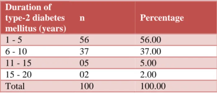
Table 3: Distribution of type-2 diabetes mellitus patients according to the duration of disease (n = 100).
Duration of type-2 diabetes mellitus (years)
n Percentage
1 - 5 56 56.00
6 - 10 37 37.00
11 - 15 05 5.00
15 - 20 02 2.00
Maximum patients (71%) of type-2 diabetes mellitus were male, with male to female ratio - 2.45:1. Maximum patients (56%) had duration of type-2 diabetes mellitus from 1 to 5 years. Mean duration of type-2 diabetes mellitus was 6.81 years with a range of 1 to 20 years.
Table 4: Distribution of type-2 diabetes mellitus patients according to the treatment mode (n=100).
Treatment mode n Percentage
Insulin 09 9.00
OHA + insulin 14 14.00
OHA 77 77.00
Total 100 100.00
Out of 100, type-2 diabetes mellitus patients 77% patients were on OHA.
Table 5: Distribution of complications in type-2 diabetes mellitus patients.
Complications n Percentage
Retinopathy 29 60.42 Neuropathy 11 22.92
Nephropathy Microalbuminuria 07 14.58 Macroalbuminuria 01 2.08
Total 48 100.00
A total of 48 cases of complications were observed in the study, which included retinopathy in 60.42% (all non-proliferative).
Table 6: Distribution of type-2 diabetes mellitus patients according to fasting blood sugar (n=100).
Fasting blood sugar (mg/dl) n Percentage
<100 (Normal) 17 17.00 100 – 125 (impaired) 41 41.00 >126 (abnormal) 42 42.00
Total 100 100.00
Maximum type-2 diabetes mellitus patients (42%) had fasting blood sugar >126 mg/dl (abnormal).
Table 7: Distribution of type-2 diabetes mellitus
patients according to post prandial blood sugar (n = 100).
Post prandial blood sugar
(mg/dl) n Percentage
<140 (Normal) 21 21.00 140 – 199 (Impaired) 68 68.00 >200 (Abnormal) 11 11.00
Total 100 100.00
Maximum type-2 diabetes mellitus patients (68%) had post prandial blood sugar in the range of 140 - 199 mg/dl (impaired).
Table 8: Distribution of type-2 diabetes mellitus patients according to serum magnesium levels
(n=100).
Serum magnesium levels
(meq/dl) n Percentage
<1.8 (hypomagnesaemia) 30 30.00 1.8 – 2.4 (normal) 70 70.00
Total 100 100.00
Hypomagnesaemia was observed in 30% patients of type-2 diabetes mellitus with a mean±standard deviation of 1.32±0.14 (range 1.2 to 1.7) mg/dl.
Table 9: Distribution of type-2 diabetes mellitus patients according to glycosylated haemoglobin
(HbAIc) (n = 100).
HbAIc levels (%) n Percentage
<5.6 (normal) 08 8.00 5. 7 - 6.4 (impaired) 13 13.00 >6.5 (abnormal) 79 79.00
Total 100 100.00
Maximum patients (79%) had abnormal (>6.5%) glycosylated hemoglobin levels ranging from 6.5 to 12%.
Table 10: Prevalence of hypomagnesaemia according to duration of diabetes.
Duration of type-2 diabetes mellitus (years)
n Patients with
hypomagnesaemia %
1 - 5 56 09 16.07
6 - 10 37 17 45.95
11 - 15 05 03 60.00
15 - 20 02 01 50.00
Total 100 30 30.00
p = 0.19 (Fisher’s exact test); Not significant
Hypomagnesaemia was present in 16.07% patients with duration of 1-5 years of type-2 diabetes mellitus, in 45.95% patients with duration of 6-10 years, in 60% patients with duration of 11-15 years and in 50% patients with duration of 15-20 years. However, there was statistically no significant difference in the prevalence of hypomagnesaemia according to duration of disease.
Moreover patients who had retinopathy were found to have lower mean serum magnesium level than those without retinopathy (1.59 mg/dl vs 1.93 mg/dl). The difference in prevalence of hypomagnesaemia in patients
with complications as compared to those without
complications was statistically highly significant (p = 0.0003).
Table 11: Prevalence of hypomagnesaemia with diabetic retinopathy.
Variable n Hypomagnesaemia (%) Normomagnesaemia (%)
Retinopathy 29 17 (58.62) 12 (41.38)
No retinopathy 71 13(18.31) 58 (81.69)
p = 0.0002 (Fisher’s exact test); highly significant
Table 12: Prevalence of hypomagnesaemia with diabetic complications.
Variable n Hypomagnesaemia (%) Normomagnesaemia (%)
Complications 30 17 (56.67) 13 (43.33)
No complications 70 13(18.57) 57 (81.43)
p = 0.0003 (Fisher’s exact test); highly significant
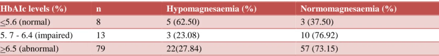
Table 13: Prevalence of hypomagnesaemia with glycosylated hemoglobin (HbAIc).
HbAIc levels (%) n Hypomagnesaemia (%) Normomagnesaemia (%)
<5.6 (normal) 8 5 (62.50) 3 (37.50)
5. 7 - 6.4 (impaired) 13 3 (23.08) 10 (76.92) >6.5 (abnormal) 79 22(27.84) 57 (73.15)
p=0.05 (Fisher’s exact test); Not significant
Serum magnesium showed no significant relationship with HbAIc with p=0.05.
DISCUSSION
Hypomagnesaemia was observed in 30% patients of type-2 diabetes mellitus with a mean±standard deviation of 1.32±0.14 (range 1.2 to 1.7) mg/dl. Mean age of the patients was 56.81 years with a range of 35 to 75 years.
Maximum patients (71%) of type-2 diabetes mellitus were male, with male to female ratio of 2.45:1. Ghafour et al also reported that the prevalence of hypomagnesemia was more in males than in females. However, Kahn et al found a higher incidence of diabetic retinopathy in females.24,25
In the present study, hypomagnesaemia was present in 16.07% patients with duration of 1-5 years of type-2 diabetes mellitus, in 45.95% patients with duration of 6-10 years, in 60% patients with duration of 11-15 years and in 50% patients with duration of 15-20 years. However, there was statistically no significant difference in the prevalence of hypomagnesaemia according to duration of disease (p = 0.19). These results are in line with other studies that suggest there is no association
between serum magnesium levels and duration of diabetes.26,27
Out of 100 type-2 diabetes mellitus patients, 9% were on insulin, 14% were on oral hypoglycemic agents (OHA)+insulin, while 77% patients were on OHA alone. There was no relation of hypomagnesemia with mode of treatment of type-2 diabetes mellitus as was found by Walti et al.26 In contrast, the serum magnesium levels
were significantly lower in the insulin treated group compared to the OHA treated in a study by Kauser et al. but this was because of the small sample size of the study.28
Maximum patients (79%) had abnormal (>6.5 mg/dl) glycosylated haemoglobin levels ranging from 6.5 to 12%. There were 13% patients who had impaired (5.7 – 6.4%) glycosylated haemoglobin levels. The present study revealed that a higher prevalence of hypomagnesaemia was found in patients having HbAIc >6.5% (79%) and FBS >126 mg/dl (42%), PPBS >140 mg/dl (68%). These results are similar to found by several investigators to correlate inversely with fasting blood glucose concentration and the percentage of HbAIc.29 But there was no statistically significant
A total of 48 cases of complications were observed in the present study, which included retinopathy in 60.42% (all non-proliferative), neuropathy in 22.92% and nephropathy in 16.66% (14.58% microalbuminuria, 2.08% macroalbuminuria).
In the present study, patients with diabetic retinopathy had significantly higher prevalence of hypomagnesaemia compared to patients without retinopathy (56.67% vs. 18.31%). The difference was statistically highly significant (p = 0.0002). These results are similar to other studies that show association of hypomagnesemia with diabetic retinopathy.30,31
The difference in prevalence of hypomagnesaemia in patients with complications as compared to those without complications was statistically highly significant (p = 0.0003). These results correlate with other studies that found an increased risk of complications of diabetes in patients with hypomagnesemia.32,33 Hypomagnesaemia is
a possible risk factor in the development and progression of diabetic retinopathy. Some studies revealed that hyperglycemia contribute to hypomagnesaemia by causing depression in the net tubular reabsorption of magnesium.34
It has been suggested that hypomagnesaemia may further impair glycemic control by inducing altered cellular glucose transport, reduced pancreatic insulin secretion, defective post receptor signalling, and/or altered insulin-insulin receptor interactions.
The present study has thus demonstrated that hypomagnesaemia is common in type-2 diabetes mellitus patients and found to be significantly associated with retinopathy, while in nephropathy and neuropathy percentage of patients with hypomagnesaemia was increased but not statistically significant.
CONCLUSION
Prevalence of hypomagnesaemia in type 2 diabetes was 30% in the present study. Hypomagnesemia was significantly associated with diabetic retinopathy (p=0.0002). The present study illustrates that as the magnesium level decreases in type 2 diabetes mellitus patients, prevalence of retinopathy increases.
Funding: No funding sources Conflict of interest: None declared Ethical approval: Not required
REFERENCES
1. ADA (American Diabetes Association). Clinical Practice Recommendations. American Diabetes Associates. Diabetes Care. 2004;6:1-16.
2. International Diabetes Federation Diabetes Atlas. Available at www.idf.org/diabetesatlas. Accessed on 14 July 2016.
3. National diabetes fact sheet. Available at: http://www.diabetes.org/diabetes-statistics.
Accessed on 14 July 2016.
4. Kim ES, Moon SD, Kim HS, Lim DJ, Cho JH, Kwon HS, et al. Diabetic peripheral neuropathy is associated with increased arterial stiffness without changes in carotid intimamedia thickness in type 2 diabetes. Diabetes Care. 2011;34(6):1403-5. 5. Frank B. Globalization of diabetes, the role of diet,
lifestyle and genes. Diabetes Care. 2011;34(6):1249-57.
6. Daruka KM. Retinopathy in type 2 diabetes mellitus and serum magnesium levels. J Evid Med Healthcare. 2015;2(6):677-85.
7. Mooradian AD, Failla M, Hoogwerf, Maryniuk M, Wylie J. Selected vitamins and minerals in diabetes. Diabetes Care. 1994;17:464-79
8. Walter RM, Bhandarkar SD. Trace elements in diabetes mellitus. J Postgrad Med. 1981;27:129-32. 9. Baig MSA, Shamshuddin M, Mahadevappa KL,
Attar AH, Shaikh AK. Serum magnesium as a marker of diabetic complications. J Evol Med Dental Sci. 2012;1(3):119-23.
10. Elin RJ. Assessment of magnesium status. Clin Chem. 1987;33:1965-70.
11. Gums JG. Clinical significance of magnesium: A review. Drug Intell Clin Pharm. 1987;21:240-6. 12. Chi TP, Mai TP, Son VP, Jaffrey MM, Thu T.
Hypomagnesaemia in patients with type 2 diabetes. Clin Am Soc Nephrol. 2007;2:366-73.
13. Hans CP, Sialy R, Bansal DD. Magnesium deficiency and diabetes mellitus. Curr Sci. 2002;83:12.
14. Badyal A, Sodhi KS, Pandey R, Singh J. Serum magnesium levels: a key issue for diabetes mellitus. JK Science. 2011;13(3):132-4.
15. Grafton G, Baxter MA. The role of magnesium in diabetes mellitus. J Diabetes Complications. 1992;6:143-9.
16. Nadler JC, Malayan S, Luong H, Shaw S, Natarajan RD, Rude RK. Intracellular free magnesium deficiency plays a key role in increased platelet reactivity in type 2 diabetes mellitus. Diabetes Care. 1992;15:835-41.
17. Sheehan JP. Magnesium deficiency and diabetes mellitus. Magnes Trace Element. 1992;10:215-9. 18. Mikhail N, Ehsanipoor K. Ionized serum
magnesium in type 2 diabetes mellitus: Its correlation with serum magnesium and hemoglobin A1c levels. South Med J. 1999;92:1162-6.
19. Nasir H, Baradaran HR. Lipids in association with serum magnesium in diabetes mellitus patients. Bratisl Lek Listy. 2008;109(7):302-6.
20. Lefebvre PJ, Scheen AJ. Improving the action of insulin. Clin Invest Med. 1995;18:342-7.
22. Laughlin MR, Thompson D. The regulatory rule for magnesium in glycolytic flux of the human erythrocyte. J Biol Chem. 1996;271:277-83.
23. WHO. Definition and Diagnoses of Diabetes, 2005. Available at http:// www.who.int/ diabetes/ publications/ Accessed on 17 august 2016.
24. Ghafour IM, Allan, Foulds WS. Common causes of blindness and visual handicap in the west of Scotland. Br J Ophthalmol. 1983;67(4):209-13. 25. Kahn HA, Hiller R. Blindness caused by diabetic
retinopathy. Am J Ophthalmol. 1974;87(1):58-67. 26. Walti MK, Zimmermann MB, Spinas GA, Hurrell
RF. Low plasma magnesium in type 2 diabetes. Swiss Med Wkly. 2003;133:289-92.
27. Antin SS, Kashinkunti M, Kataria AV, Dhananjaya M, Alevoor S. A cross sectional study of fasting serum magnesium levels in the patients with type 2 diabetes mellitus and its relation to diabetic complications. Sch J App Med Sci. 2014;2(2):502-6.
28. Kauser MM, Afreen A, Kumar SRV, Javarappa D. Study of serum magnesium in type 2 diabetes mellitus and its correlation with the modality of treatment – a south Indian study. Int J Med Sci Public Health. 2014;3(11):1398-1401.
29. Khubchandani AS, Sanghani H. Study of serum magnesium and HbAIc in diabetic patients along
with changes in their lipid profiles. Indian J Clin Pract. 2013;23(11):11.
30. Gurjar P, Kumar S, Diwan SK, Patil MM. Serum magnesium in type 2 diabetes mellitus: Case-control study in rural teaching hospital. Inter J Analy Pharma Biomed Sci. 2014;3(6):97-105.
31. Kaur P, Bal BS, Kaur I, Singh G, Singh B. Correlation of severity of diabetic retinopathy with various risk factors. Int J Res Health Sci. 2014;2(2):473-9.
32. Chauhan KP, Haridas N, Patel C. A study of serum magnesium levels in diabetic retinopathy patients. Indian J Appl Res. 2013;3(5):482-4.
33. Mohanty SS, Pinnelli VBK, Murgod R, Das R. Evaluation of serum copper, magnesium and glycated haemoglobin in type 2 diabetes mellitus. Asian J Pharma Clin Res. 2013;6(2):188-90. 34. Mcnair P, Christiansen C, Madsbad S, Lauritzen E,
Faber O, Binder C, et al. Hypomagnesemia a risk factor in diabetic retinopathy. Diabetes. 1978;27:1075-7.