Sav1
Loss Induces Senescence and Stat3
Activation Coinciding with
Tubulointerstitial Fibrosis
Janet Y. Leung,
a,bHarper L. Wilson,
aKristin J. Voltzke,
a,cLindsay A. Williams,
a,cHyo Jin Lee,
dSara E. Wobker,
eWilliam Y. Kim
a,b,fLineberger Comprehensive Cancer Center, University of North Carolina at Chapel Hill, Chapel Hill, North Carolina, USAa; Department of Medicine, University of North Carolina at Chapel Hill, Chapel Hill, North Carolina, USAb; Department of Epidemiology, Gillings School of Global Public Health, University of North Carolina at Chapel Hill, Chapel Hill, North Carolina, USAc; Department of Internal Medicine, Chungnam National University School of Medicine, Daejeon, Republic of Koread; Department of Pathology, University of North Carolina at Chapel Hill, Chapel Hill, North Carolina, USAe; Department of Genetics, University of North Carolina at Chapel Hill, Chapel Hill, North Carolina, USAf
ABSTRACT
Tubulointerstitial fibrosis (TIF) is recognized as a final phenotypic
mani-festation in the transition from chronic kidney disease (CKD) to end-stage renal
dis-ease (ESRD). Here we show that conditional inactivation of
Sav1
in the mouse renal
epithelium resulted in upregulated expression of profibrotic genes and TIF. Loss of
Sav1
induced Stat3 activation and a senescence-associated secretory phenotype (SASP)
that coincided with the development of tubulointerstitial fibrosis. Treatment of mice
with the YAP inhibitor verteporfin (VP) inhibited activation of genes associated with
senescence, SASPs, and activation of Stat3 as well as impeded the development of
fibrosis. Collectively, our studies offer novel insights into molecular events that are
linked to fibrosis development from
Sav1
loss and implicate VP as a potential
phar-macological inhibitor to treat patients at risk for developing CKD and TIF.
KEYWORDS
fibrosis, mouse, renal, senescence
C
hronic kidney disease (CKD) is currently increasing in prevalence in the United
States. The National Kidney Foundation predicts that 26 million Americans have
CKD and that millions of others are at risk. Tubulointerstitial fibrosis (TIF) occurs in
patients with CKD and is characterized by myofibroblast activation, loss of capillary
networks, and an accumulation of extracellular matrix and inflammatory cells (1–3). In
fibrotic kidneys, the cortical interstitium is generally expanded, with an increased
propensity to accumulate collagen (1–3). Epithelial cell death also occurs, resulting in
reduced expression of epithelial markers and an increase in the expression of
mesen-chymal markers (4–6).
Although it is well recognized that TIF is a final manifestation in the transition from
CKD to end-stage renal disease (ESRD), the mechanisms underlying the etiology of
renal fibrosis are poorly understood, and as a result, progress toward finding an
effective treatment has been hindered. Potential therapeutic targets for treatment of
CKD have been examined through
in vitro
and
in vivo
studies in the last decade, but the
findings from these studies have yet to be translated to viable treatment options for
patients. Inhibition of the renin-angiotensin system (RAS), for example, has been shown
to reverse CKD in animal models; however, subsequent work with RAS inhibitors or
blockers in humans has failed to show similar efficacy (7–10). Additional studies have
begun to examine targeting of nuclear transcription factors in pathways regulating lipid
metabolism and inflammation, although most candidate pharmacological inhibitors of
interest remain in preclinical stages (11). At present, dialysis or renal transplantation is
still the only solution for patients with CKD.
Received18 October 2016 Returned for modification21 November 2016 Accepted9 March 2017
Accepted manuscript posted online20 March 2017
CitationLeung JY, Wilson HL, Voltzke KJ, Williams LA, Lee HJ, Wobker SE, Kim WY. 2017.
Sav1loss induces senescence and Stat3 activation coinciding with tubulointerstitial fibrosis. Mol Cell Biol 37:e00565-16.https://doi .org/10.1128/MCB.00565-16.
Copyright© 2017 American Society for Microbiology.All Rights Reserved.
Address correspondence to Janet Y. Leung, [email protected], or William Y. Kim, [email protected].
Work in the last decade has begun to recognize the importance of genetic
altera-tions in facilitating renal fibrotic development. Genome-wide association studies have
identified loci associated with increased susceptibility to renal dysfunction in humans
(12, 13). Additionally, mouse models and genome-wide transcriptome analyses of
kidneys from patients with CKD and fibrosis have facilitated the identification of a
number of signaling pathways associated with renal fibrosis, including the Notch, Wnt,
and Hedgehog pathways (3). The further identification of signaling pathways
associ-ated with fibrosis increases the potential opportunities for targeted therapy to thwart
fibrosis.
SALVADOR I (SAV1) is a WW domain-containing protein originally identified as one
of the core components of the HIPPO signaling pathway. The HIPPO pathway is a
conserved protein kinase pathway originally identified as a regulator of organ size
(14–16). A number of cell type-dependent functions have been ascribed to HIPPO
signaling, including regulation of stem/progenitor cells, cell proliferation, apoptosis, cell
polarity, and cell-cell contact inhibition (17–21). At the center of the HIPPO pathway is
the tumor suppressor gene
Hippo
, or its mammalian counterpart,
mammalian STE-20
kinase 1 and 2
(
MstI/2
or
STK3/4
). STK3/4 phosphorylates the scaffold protein SAV1, and
together they phosphorylate and activate two other core components: LATS1/2 and its
associated protein, MOB1. LATS1/2 in turn phosphorylates the main effector molecule
of the HIPPO pathway, YAP, resulting in its inactivation and cytoplasmic sequestration
by the 14-3-3 protein. Loss of HIPPO pathway function from SAV1 loss or loss of any of
the core components has been suggested to result in YAP nuclear translocation and
its association with transcription factors to constitutively activate growth-promoting
genes involved in cancer. Specific knockout of
Sav1
in the livers of mice has been
shown to result in hepatomegaly and multifocal hepatic carcinomas (22).
YAP
amplifi-cation or constitutive overexpression has also been observed in a number of cancers
(14, 21, 23–26).
Components of the HIPPO pathway have also been implicated in tissue fibrosis. Yap
has been suggested to promote pulmonary fibrosis through its effects on
mechanosig-naling (27) and has been implicated in liver fibrosis by its role in hepatic stellate cell
activation (28). Loss of Lats1/2 or abnormal accumulation of Yap has also been
suggested to be important for kidney fibrosis after ischemic injury (29). Finally, most
recently,
Sav1
loss was also implicated in kidney fibrosis, although the role for SAV1 in
CKD had not yet been documented prior to that study (30).
In the current study, we generated a mouse model with conditional knockout of
Sav1
in the renal epithelium. We observed that
Sav1
inactivation resulted in genetic and
phenotypic alterations suggestive of renal fibrosis that were further accelerated upon
induction of acute kidney injury (AKI) by use of aristolochic acid (AA). Induction of
fibrosis by AA and
Sav1
loss was associated with an induction of senescence and a
senescence-associated secretory phenotype (SASP) that correlated with Stat3
activa-tion. Inhibition of TIF with verteporfin (VP), a Yap inhibitor, resulted in downregulation
of genes associated with senescence and SASP and in inhibition of Stat3 activation.
Collectively, these results support a role for
Sav1
loss in regulating interstitial fibrosis
and suggest that events associated with senescence and Stat3 activation are the
mechanisms underlying renal fibrosis induced by
Sav1
loss.
RESULTS
Conditional inactivation of
Sav1
in mouse renal tubule cells results in
pheno-typic changes resembling kidney fibrosis.
To determine the phenotypes associated
with conditional deletion of
Sav1
in murine renal proximal tubules
in vivo
, we crossed
Ksp-CreER
transgenic mice to
Sav1
floxed/floxed(
Sav1
f/f) mice to generate
Ksp-CreER/Sav1
fl/flmice (referred to as KS mice from this point on). Ksp-cadherin is a tissue-specific
member of the cadherin family that is expressed exclusively in the epithelial cells of the
adult kidney and the developing genitourinary tract (31).
Ksp-CreER
transgenic mice
express a tamoxifen-inducible Cre recombinase under the control of a 1.4-kb
Ksp-cadherin
promoter (31).
Sav1
f/fmice harbor
loxP
sites flanking exon 3 of
Sav1
(22).
Conditional deletion of
Sav1
in the renal tubule epithelial cells of KS mice can therefore
be achieved by administering tamoxifen by oral gavage (Fig. 1a).
Prior work has shown that specific ablation of
Sav1
in hepatocytes gives rise to
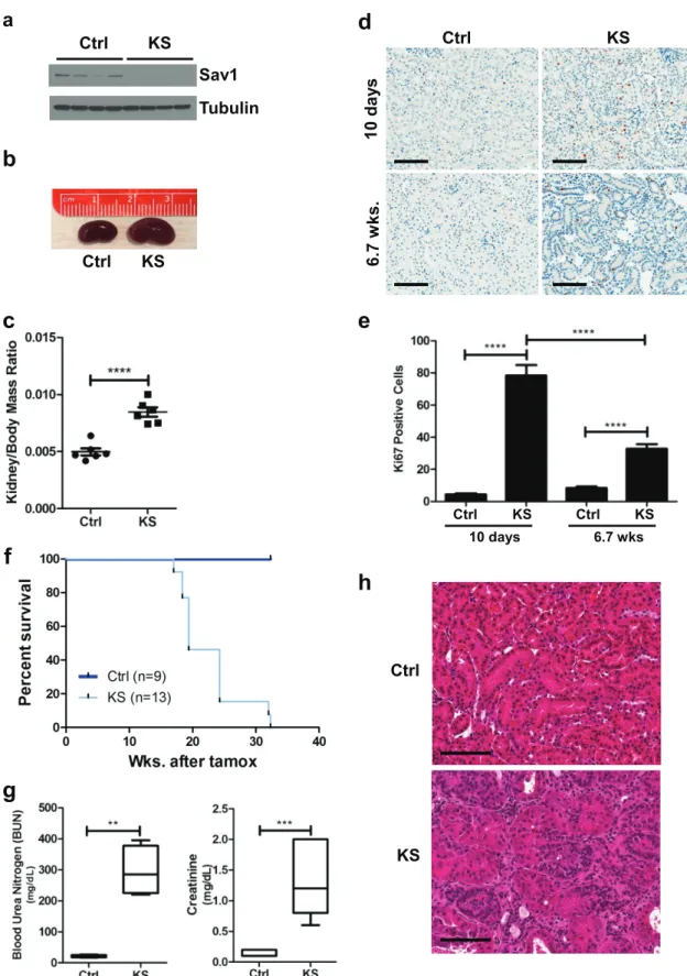
hepatomegaly, with a 10% increase in liver size by 4 months of age (22). Similar to the
phenotype observed in the liver, we observed that loss of
Sav1
at 6 weeks
post-tamoxifen treatment induced a significant increase in the kidney-to-body-mass ratio
(Fig. 1b and c). Ki67 staining of kidney sections revealed an increase in Ki67 staining in
the nuclei of renal tubule cells immediately post-tamoxifen treatment that persisted
even at 6 weeks post-tamoxifen treatment (Fig. 1d and e). We observed more
Ki67-positive cells at 10 days post-tamoxifen treatment than at 6 weeks post-tamoxifen
treatment. We speculate that this spike in proliferation may have been a nonspecific
consequence of initial Cre-ER activation, as we previously observed small increases in
Ki67 staining immediately following tamoxifen treatment that subsided at later time
points (data not shown). Collectively, these results suggest that an increase in cell
proliferation may contribute to an overgrowth of kidneys in KS mice compared to those
of age-matched controls.
KS mice had a decreased survival time (median
⫽
19.4 weeks) compared to that of
control mice (median not reached) (Fig. 1f). At the time of necropsy, significantly
elevated levels of blood urea nitrogen and creatinine were found in the plasmas of KS
mice compared to those of control mice, confirming renal failure as the likely cause of
premature death (Fig. 1g). Histological examination of KS kidney sections by
hematox-ylin and eosin (H&E) staining revealed a number of abnormalities, including dilation of
renal tubules and an expansion of the renal interstitium with increased interstitial
cellularity (Fig. 1h).
Sav1
loss in the renal epithelium results in gene expression changes associated
with renal fibrosis and senescence.
To begin to understand the consequences
associated with
Sav1
inactivation in renal tubules, we isolated RNAs from renal cortex
tissues of tamoxifen-treated
Ksp-CreER/Sav1
fl/fl(KS) mice and vehicle-treated
Ksp-CreER/
Sav1
fl/fl(control [Ctrl]) mice 6 weeks after treatment. Transcriptome sequencing
(RNA-seq) analysis revealed 4,378 genes upregulated
⬎
1.5-fold and 3,362 genes
downregu-lated
⬎
1.5-fold (with a
t
test false discovery rate [FDR] of
⬍
0.05). Examination of
differentially regulated genes by Ingenuity Pathway Analysis (IPA) showed Tgf-

1 to be
the top upstream regulator of genes activated 2-fold or more by
Sav1
loss. Tgf-

1 is a
cytokine that has long been recognized as a key mediator in the pathogenesis of renal
fibrosis (32, 33). Further analysis of the RNA-seq data showed collagen genes and genes
associated with fibrosis to be increased upon
Sav1
depletion (Fig. 2a). To further
confirm that loss of
Sav1
induces activation of profibrotic genes, we performed
real-time quantitative reverse transcription-PCR (qRT-PCR) on selected genes (34) at an
earlier time point (3 weeks) after tamoxifen/vehicle treatment. Consistent with the
results from the RNA-seq analysis, we observed significant activation of the profibrotic
genes assessed by real-time qRT-PCR (Fig. 2b). Masson’s trichrome staining and
immu-nohistochemistry (IHC) revealed accumulations of collagen and vimentin, respectively,
in KS kidneys compared to those of age-matched controls at 6 weeks posttreatment
(Fig. 2c). An increase in staining for the T-cell marker CD3 also revealed that loss of
Sav1
may trigger increased T-cell infiltration or expansion (Fig. 2c).
Aging has been linked to increased susceptibility to renal fibrosis (35–38). A
multi-tude of studies have now clearly established that aging is not merely a phenotypic
manifestation but consists of specific gene expression changes associated with cellular
senescence (39–42). Prior studies have suggested a role for genes involved in cellular
senescence in promoting CKD and fibrosis (38, 43–45). We examined KS mice for evidence
of senescence and observed upregulated expression of key senescence markers (Fig. 2d).
a
b
Ctrl
KS
c
Sav1
Ctrl
KS
Tubulin
e
Ctrl KS Ctrl KS
10 days 6.7 wks
Ctrl
KS
10 d
a
y
s
6.
7 w
k
s
.
Ctrl
KS
d
h
g
f
FIG 1Conditional knockout ofSav1 in mouse renal tubule cells results in decreased survival, renal failure, and histological
abnormalities. (a) Immunoblot of whole-cell lysates isolated from the cortex tissue ofKspCreER;Sav1f/fmice 0.8 week after treatment
with vehicle (control [Ctrl]) or tamoxifen (KS), confirming the knockout ofSav1expression. (b) Representative photomicrographs showing an enlarged kidney from a KS mouse compared to a kidney from an age-matched Ctrl mouse. Kidneys were taken from mice at 6 weeks post-tamoxifen treatment. (c) Comparison of kidney-to-body-mass ratios of KS mice (n⫽5) and age-matched control mice (n⫽6). Kidneys were taken from mice at 6 weeks post-tamoxifen treatment. (d) Representative images of Ki67-stained mouse kidney sections from mice 10 days and 6.7 weeks after treatment with vehicle or tamoxifen. (e) Bar graph showing percentages of Ki67-positive cells
(Continued on next page)
Klotho is a single-pass transmembrane protein previously termed the “anti-aging”
gene. Defects in expression of the Klotho gene in mice were originally shown to result
in a shortened life span and in aging-like phenotypes (46–48). Subsequent work later
also identified a role for defective Klotho gene expression in renal fibrosis (49). Given
that KS mice exhibit changes in gene expression previously documented to be
asso-ciated with aging, we assessed Klotho expression by real-time qRT-PCR and found
KS mice to have lower levels of Klotho gene expression than those of age-matched
littermates (Fig. 2e). Collectively, our data support the hypothesis that loss of
Sav1
in
the renal epithelium induces changes in gene expression consistent with renal fibrosis,
which is correlated with upregulated expression of genes associated with senescence.
Acute kidney injury induced by AA augments profibrotic gene expression and
an accumulation of collagen in KS mice.
Mouse models have been instrumental in
studying CKD brought forth by AKI. A number of methods are well established to
provoke injury to mouse kidneys, including unilateral ureteral obstruction (UUO),
ischemic reperfusion (IR), and aristolochic acid nephropathy (AAN) (50). Aristolochic
acid (AA) belongs to a family of carcinogenic and nephrotoxic compounds (50). AAN,
which involves intraperitoneal (i.p.) injection of AA, has been well documented to result
in tubulointerstitial damage and is one of several widely accepted models for AKI
studies (50). Given that gene expression changes observed in KS mouse kidneys
suggest a predisposition to renal fibrosis (Fig. 2), we wanted to determine if loss of
Sav1
affects the response to injury by AAN. We administered AA to Ctrl and KS mice at a dose
of 5 mg/kg of body weight, which was previously shown to result in mild to negligible
AKI in normal healthy mice (51). Intraperitoneal introduction of AA into KS mice 3 weeks
after ablation of
Sav1
expression resulted in dramatic increases in the expression levels
of all profibrotic genes (
Col1a1
,
Col3a1
,
Col4a1
,
Fn
,
Tgf

1
, and
Vimentin
) examined
compared to those in AA-treated Ctrl mice with no
Sav1
loss (Fig. 3a and b). Consistent
with an increase in profibrotic gene expression, staining with Masson’s trichrome revealed
an accumulation of collagen at 3 weeks post-tamoxifen treatment that progressively
increased at 6 and 13 weeks post-tamoxifen treatment compared to that in vehicle-treated
control mice (Fig. 3c).
Senescence and SASP genes are further activated by AAN in KS mice.
Senescent
cells have been shown to have deleterious effects on organisms by contributing to
age-related pathology through inducing expression of genes associated with
senescence-associated secretory phenotypes (SASPs), including genes encoding interleukins,
chemokines, and growth factors (52). Induction of profibrotic genes by AAN was also
associated with dramatic increases in the expression levels of all senescence genes
(
p16ink4a
,
p19arf
,
Cdkn1a
, and
Pai-1
) (Fig. 4a and b) examined. Moreover, activation of
p53 was also observed by Western blotting (Fig. 4b). To determine whether senescence
induced by
Sav1
loss is associated with induction of SASP genes, we assessed the
expression of select SASP genes after AAN in Ctrl and KS mice. We found all SASP genes
assessed to be upregulated in KS mice compared to age-matched control mice,
suggesting that
Sav1
loss induces SASP genes upon AKI (Fig. 4c).
Cytokines such as interleukin-6 (Il6) are known to activate the signal transducers and
activators of transcription (Stats) (53, 54). In particular, activation of Stat3 is known to
be achieved through phosphorylation of its tyrosine 705 site by receptor tyrosine
kinases (RTKs) and has been suggested to have a role in the progression of renal fibrosis
(53–55). Although Stat3 activation from
Sav1
loss alone was not obvious by Western
blotting (data not shown), histological examination revealed an increase in
phospho-FIG 1Legend (Continued)
a
Ctrl
KS
b
e
c
Tr
ic
h
ro
m
e
β-g
a
l
Vi
m
e
n
ti
n
Ctrl
KS
Cd
3
d
FIG 2Sav1deletion in renal tubule cells activates profibrotic and cell senescence genes. (a) Heat map representation showing increased expression
of genes associated with renal fibrosis in renal cortex tissue fromKspCreER;Sav1f/fmice 6.7 weeks after treatment with vehicle (Ctrl) or tamoxifen (KS).
(Continued on next page)
Stat3 staining in the nuclei of renal tubule cells (Fig. 4d). We next asked whether loss
of
Sav1
in KS mice affects Stat3 activation upon AAN. Intraperitoneal injection of AA
into Ctrl mice resulted in a minimal increase in phosphorylated (Tyr-705) Stat3, while AA
injection into KS mice resulted in a dramatic increase in phosphorylated Stat3
expres-sion (Fig. 4d and e). Our results suggest that
Sav1
loss induces activation of Stat3 in
association with a fibrosis phenotype that is further enhanced by AKI with AA.
Accumulation of Yap activity is observed in KS mice.
Sav1 has previously been
documented to function as a scaffold protein that brings together kinases in the HIPPO
signaling pathway to regulate phosphorylation and subsequent ubiquitin-mediated
degradation of Yap (56, 57). Loss of
Sav1
is thought to result in an accumulation of Yap
in the nucleus, where it binds to other transcription factors, such as the TEAD family of
transcription factors, to activate expression of target genes (56, 57). Previously
pub-lished work suggested that Yap also plays a role in fibrosis (27, 28, 58, 59). We assessed
Yap levels in KS mice compared to those in age-matched controls and saw progressive
increases in Yap accumulation at 6 weeks and
⬎
20 weeks post-tamoxifen treatment
(Fig. 5a and b). No changes in Yap levels were apparent between Ctrl mice of various
ages (data not shown). Consistent with an accumulation of Yap protein resulting from
Sav1
ablation, we also observed activation of Yap target genes after tamoxifen
treat-ment of KS mice (Fig. 5c). As expected, we did not see any significant change in
Yap
mRNA levels upon inactivation of
Sav1
, since Yap is primarily regulated by
ubiquitin-mediated degradation (data not shown).
VP inhibition of AAN-induced TIF in KS mice coincides with inhibition of
senescence and SASP gene activation and inhibition of Stat3 activation.
To
determine if Yap plays a central role in phenotypes associated with tubulointerstitial
fibrosis in the setting of
Sav1
loss (KS mice), we tested the efficacy of the Yap inhibitor
verteporfin (VP) in KS and Ctrl mice. VP is a small molecule belonging to the porphyrin
family of compounds that was recently identified in a drug screen for inhibitors of
YAP-TEAD activity (60). Further biochemical studies have shown that VP works by
binding YAP and preventing its interaction with other transcription factors, such as
TEAD (60). We tested the ability of VP to inhibit Yap target genes induced 3 weeks after
Sav1
loss (Fig. 5d) and confirmed that VP treatment reduced the activation of Yap target
genes (Fig. 5e).
We tested the ability of VP to suppress the activation of profibrotic genes in KS and
Ctrl mice after AAN. Consistent with our prior results, activation of profibrotic gene
expression was observed in KS mice upon AAN compared to that in Ctrl mice (Fig. 6a).
Ctrl and KS mice treated with AA but receiving VP treatment, however, showed levels
of profibrotic gene expression comparable to basal levels (Fig. 6a). Masson’s trichrome
staining revealed less collagen deposition in mice that received VP, consistent with a
reduction in profibrotic gene expression (Fig. 6b and c).
Interestingly, VP treatment also reduced the expression levels of the senescence
(Fig. 7a) and SASP (Fig. 7b) genes examined.
Pai-1
and
p16
Ink4agene expression levels
after AA-induced AKI were comparable to basal levels.
Cdkn1a
and
p19
Arfgene
expres-sion levels remained above basal levels but were still dramatically reduced even with
AA treatment (Fig. 7a). Consistent with recently published work showing that VP
inhibited Stat3 activation in colon cancer (61), VP inhibited Stat3 activation induced by
AA in KS mice (Fig. 7c). Collectively, our results suggest that kidney fibrosis and
associated changes in gene expression induced by
Sav1
loss can be inhibited by
FIG 2Legend (Continued)
(b) Real-time qRT-PCR analysis of total RNA isolated from renal cortex tissue ofKspCreER;Sav1f/fmice 3 weeks after treatment with vehicle (Ctrl;n⫽
Ct
rl
+
A
A
KS
+
A
A
13.
8 w
k
s
.
3 w
k
s.
6 w
ks.
13.
8
w
k
s
.
H&E
Trichrome
Ctrl-AA
KS-AA
C
o
l1a1
Vi
m
e
n
ti
n
b
a
c
FIG 3Acute kidney injury (AKI) with aristolochic acid (AA) drives activation of genes associated with renal fibrosis in KS mice. (a)KspCreER;Sav1f/f
mice were given vehicle (sunflower oil) (Ctrl;n⫽4) or tamoxifen (KS;n⫽4) by oral gavage. Vehicle (DMSO and PBS) or aristolochic acid was introduced by intraperitoneal injection 3 weeks later to induce acute kidney injury. Real-time qRT-PCR analysis of total RNA isolated from renal cortex tissues was used to assess profibrotic gene expression. (b) Representative images of renal sections from the mice examined for panel a, showing localization of vimentin and Col1a1. (c) Representative H&E- and Masson’s trichrome-stained images of renal sections from mice post-AAN. Mice were administered AA to induce AKI 3 weeks, 6 weeks, and 13.8 weeks after tamoxifen treatment or 13.8 weeks after vehicle (sunflower oil) treatment.**,Pⱕ0.01;****,Pⱕ0.0001. Error bars in graphs represent standard deviations. Magnification,⫻20 for all images; bars⫽100m.
verteporfin. Treatment with VP also reduced Ki67 staining and restored expression of
Klotho expression in AA-treated KS mice compared to that in AA-treated Ctrl mice,
although this was not observed in KS mice compared to Ctrl mice. These results suggest
that VP may thwart fibrosis induced by
Sav1
loss via mechanisms other than
prolifer-ation and Klotho gene expression (Fig. 7d and e).
Given that VP treatment resulted in reduced expression of genes associated with
senescence in KS mice, we determined whether VP treatment had an effect on
increased

-galactosidase activity brought on by
Sav1
loss. We previously observed that
VP was not able to reverse SA-

-Gal activity that was already initiated by
Sav1
loss (data
not shown). We modified our protocol to initiate treatment with VP prior to inducing
Sav1
loss with tamoxifen to determine if VP could instead prevent SA-

-Gal
accumu-lation (Fig. 7f). As shown in Fig. 7g, treatment with VP before and after induction of
Sav1
loss was able to prevent SA-

-Gal staining in KS mice (Fig. 7g).
a
e
b
c
d
Ctrl
+ Vehicle
Ctrl
+ AA
KS
+ AA
Phos-Stat3
(Tyr-705)
Stat3
Tubulin
p21
Ctrl-AA KS-AA
Phos-p53 (Ser-15)
Tubulin
Total p53
Ctrl
KS
Phospho-Stat3 (Tyr-705)
Vehicle
AA
FIG 4Acute kidney injury (AKI) with aristolochic acid (AA) drives activation of genes associated with senescence and SASP in KS mice.
KspCreER;Sav1f/fmice were given vehicle (sunflower oil) (Ctrl;n⫽4) or tamoxifen (KS;n⫽4) by oral gavage. Vehicle (DMSO and PBS)
DISCUSSION
Acute kidney injury resulting in chronic kidney disease (CDK) is a growing problem
in the United States. Although it is well known that fibrosis is the final manifestation in
the progression from CKD to end-stage renal disease (ESRD), the etiology underlying
renal fibrosis is poorly understood. An understanding of the molecular mechanisms
that contribute to fibrosis in the kidney is essential to the identification of potential
candidates for targeted therapy. Seo et al. (30) recently published work showing that
Sav1
loss plays a role in TIF induced by unilateral ureteral obstruction (UUO), consistent
with our observations that
Sav1
loss has a role in kidney fibrosis. Here we provide
6 w
k
s
>
20 w
k
s
Ctrl
KS
b
d
e
a
Tubulin
Phos-Yap
Total Yap
Ctrl
Tamox
6wks
Tamox
26 wks
c
FIG 5Conditional knockout ofSav1in mouse renal tubule cells results in an accumulation of Yap activity that can be inhibited with verteporfin. (a) Immunoblots
showing Yap expression in renal cortex tissue fromKspCreER;Sav1f/fmice. Mice were given vehicle (Ctrl) or tamoxifen (KS) by oral gavage, and kidneys were
collected at 6 weeks and 26 weeks posttreatment. Ctrl mice were age matched to KS mice, and kidneys were collected at 6 weeks post-tamoxifen treatment. (b) Representative images showing Yap accumulation in renal tissue from Ctrl or KS mice at the indicated time points post-tamoxifen or -vehicle (sunflower oil) treatment. (c) Real-time qRT-PCR analysis of total RNAs isolated from renal cortex tissues of Ctrl (n⫽4) and KS (n⫽4) mice. Mice were assessed for Yap target gene expression at the indicated time points. (d) Schematic showing treatment of Ctrl and KS mice with verteporfin, a Yap inhibitor, or with vehicle and induction of AKI with AA. (e) Real-time qRT-PCR analysis of total RNAs isolated from renal cortex tissues of Ctrl and KS mice after treatment with vehicle, AA, and/or VP as indicated (Ctrl,n⫽3; KS plus no VP,n⫽4; KS plus VP,n⫽6; KS plus AA plus no VP,n⫽5; KS plus AA plus VP,n⫽6) for expression of selected Yap target genes.***,Pⱕ0.001;****,Pⱕ0.0001. Error bars represent standard deviations. Magnification,⫻20 for all images; bars⫽100m.
a
VP
AA
VP
+
A
A
V
e
h
icl
e
H&E
Trichrome
b
c
FIG 6Verteporfin inhibits profibrotic gene expression induced bySav1loss. AKI was induced in Ctrl and KS mice by AAN, and mice were treated with vehicle
evidence that induction of fibrosis by
Sav1
loss is linked to induction of senescence and
Stat3 activation. Furthermore, we showed that verteporfin (VP), a Yap inhibitor, is
effective at inhibiting fibrosis and that VP’s efficacy in impeding fibrosis is correlated
with its ability to inhibit the induction of senescence gene expression, SASPs, and Stat3
activation.
a
b
Phos-Stat3
(Tyr-705)
Stat3
Tubulin
KS + AA + Vehicle KS + AA + VP
e
g
Ctrl + Vehicle
KS + Vehicle
KS + VP
c
d
f
FIG 7Inhibition of TIF by verteporfin (VP) coincides with inhibition of senescence and SASP gene expression and Stat3 activation in KS mice. (a
and b) Senescence gene (a) and SASP gene (b) expression levels were measured for Ctrl and KS mice after AAN and/or VP treatment (Ctrl,n⫽ 3; KS plus no VP,n⫽4; KS plus VP,n⫽6; KS plus AA plus no VP,n⫽5; KS plus AA plus VP,n⫽6). (c) Western blots comparing Stat3 and phosphorylated Stat3 in vehicle-treated KS mice, mice with AA-induced AKI, and VP-treated mice with AA-induced AKI. (d) Kidney sections from mice (Ctrl,n⫽3; KS plus no VP,n⫽4; KS plus VP,n⫽6; KS plus AA plus no VP,n⫽5; KS plus AA plus VP,n⫽6) were assessed forKlotho gene expression by real-time qRT-PCR. (e) Kidney sections from mice (Ctrl,n⫽3; KS plus no VP,n⫽4; KS plus VP,n⫽6; KS plus AA plus no VP,n⫽5; KS plus AA plus VP,n⫽6) were stained with Ki67. Three images were taken for each section, and Ki67-positive cells in tubules were counted and averaged. (f) Mice were administered one dosage of VP or vehicle (DMSO and PBS) followed by three consecutive daily treatments with tamoxifen or vehicle (sunflower oil). Mice were then administered VP or vehicle (DMSO and PBS) two times a week for 2 weeks before kidneys were harvested. (g) SA--Gal staining of kidney sections from Ctrl mice (n⫽2), vehicle-treated KS mice (n⫽3), and VP-treated KS mice (n⫽ 3). Error bars in graphs represent standard deviations. ns, not statistically significant;*,Pⱕ0.05;**,Pⱕ0.01;****,Pⱕ0.0001. Magnification,⫻20 for all images; bars⫽100m.
It was previously reported that knockdown of
Sav1 in vitro
in renal cell lines resulted
in increased cell proliferation and anchorage-independent growth and that
Sav1
knockout in the liver resulted in multifocal tumors (22), suggesting a role for
Sav1
as a
tumor suppressor gene (62–64). Although we did observe an increase in proliferation
as indicated by Ki67 staining, consistent with a recent study (62), KS mice did not
develop renal tumors within the time frame observed (
⬃
32.3 weeks). While this may
merely reflect a tissue-specific role of
Sav1
, with an oncogenic role in the liver but not
in the kidney, we speculate that the induction of senescence and SASPs
in vivo
that we
describe here might also serve as a significant barrier to neoplastic transformation of
renal tubular cells.
Administration of VP to KS mice was able to inhibit the genetic and phenotypic
manifestations of renal fibrosis induced by AA. Verteporfin (Visudyn; Novartis) is
currently an FDA-approved drug administered intravenously to patients in
photody-namic therapy to treat macular degeneration with minimal side effects. Our findings are
consistent with recently published work showing the efficacy of VP for inhibiting Yap
activity and renal fibrosis induced by UUO (65). Collectively, these results bring to light
the potential for VP as a candidate pharmacological inhibitor to be administered to
patients who may be at increased risk for AKI and subsequent renal fibrosis as a result
of their existing medical condition or specific medical procedures and treatments which
they are about to undergo (i.e., surgery, chemotherapy, etc.). VP also inhibited cell
proliferation, as indicated by a reduction in Ki67-positive cells and restored Klotho
expression induced by AA. Cell proliferation and reduced Klotho expression induced
solely by
Sav1
loss, however, were not affected, suggesting that VP may thwart fibrosis
induced by
Sav1
loss via other mechanisms.
Prior studies have suggested a correlation between the expression of senescence
and aging genes and renal fibrosis. Reduced expression of the
Klotho
gene, for example,
has been shown to play a role in TIF (49, 66), and repression of Klotho expression is tied
to activated
p16
Ink4aexpression (48). Moreover, it is now known that cell senescence
also accommodates an induction of specific cytokines that comprise the SASP (52).
SASP genes have been shown to play a role in inflammation and in facilitating
epithelial-to-mesenchymal transition (EMT), two characteristics that are tightly linked to TIF
progres-sion (5, 6, 52). Cytokines such as Il6 are well known to induce Stat3 activation, which is
also associated with the progression of TIF (55). In the KS mouse model, we observed
that SA-

-Gal staining was not restricted to the cortex region of the kidney but was
apparent throughout the kidney. While Ksp-cadherin is expressed to some degree in
regions of the renal medulla, such as the loops of Henle and collecting ducts (31), we
also hypothesize that induction of
Sav1
loss in the proximal and distal tubular cells in
the renal cortex results in paracrine signaling of SASPs that triggers senescence in other
parts of the kidney. Inhibition of fibrosis by use of VP correlated with downregulated
expression of senescence genes and inhibition of Stat3 activation. Collectively, our
observations support a link between genes associated with senescence, Stat3
activa-tion, and the manifestation of TIF.
open to the notion that at least in the context of kidney tissue,
Sav1
expression may not
function to promote degradation of Yap as the canonical HIPPO pathway predicts. We
hypothesize instead that immediate
Sav1
loss may irreversibly trigger a cascade of
molecular events, independent of the HIPPO pathway, to promote activation of Yap
and, in turn, to help to promote fibrosis.
Additionally, the accumulation of Yap protein as determined by Western blotting is
minimal at 3 weeks post-
Sav1
ablation. As a result, the dramatic induction of profibrotic
gene expression observed upon AAN at this time point cannot solely be attributed to
Yap overexpression. Although the efficacy of VP treatment to inhibit fibrosis in our KS
mouse model seems to suggest that Yap is a key mediator of kidney fibrosis, recent
work revealed that VP inhibited Stat3 activation in colon cancer independently of VP’s
function as a Yap inhibitor (61). This observation brings to light the possibility that VP
also inhibits fibrosis via other mechanisms in addition to its role as an inhibitor of Yap
activity.
Collectively, our findings highlight the importance of
Sav1
loss in inducing
interstitial fibrosis and offer a model system for understanding the molecular
mechanisms underlying renal fibrosis within which potential therapeutic
opportu-nities can be explored.
MATERIALS AND METHODS
Genetically engineered mouse models.KspCad-CreERT2mice have been described previously (31).
Sav1fl/flmice were a kind gift from Randy Johnson (University of Texas) and have also been described
previously (22). All experiments performed on mice have been approved by the University of North Carolina-Chapel Hill Institutional Animal Care and Use Committee (UNC-CH IACUC).
Immunoblotting conditions.Cortex tissue was homogenized in EBC buffer (50 mM HEPES [pH 7.6],
250 mM NaCl, 0.1% NP-40, 5 mM EDTA [pH 8.0]) with set I and set II phosphatase inhibitors at 1⫻ (Calbiochem) and protease inhibitors at 1⫻(Roche) by use of a TissueRuptor homogenizer (Qiagen). Protein concentrations were determined with Bio-Rad protein assay dye reagent concentrate (Bio-Rad). Proteins were resolved in SDS-PAGE gels and electrotransferred to polyvinylidene difluoride (PVDF) membranes. Western blotting was performed with the following antibodies from Cell Signaling: Yap (4912S), phospho-Yap (4911S), Sav1 (13301S), p21 (2946S), Stat3 (9139S), phospho-Stat3 (9145S), p53 (2524S), and phospho-p53 (9286S) antibodies.
Mouse RNA-seq.A total of 200 to 1,000 ng of total RNA was used to prepare RNA libraries by use
of a TruSeq Stranded mRNA sample prep kit (Illumina). Paired-end (75 bp) reads were sequenced on a NextSeq500 desktop sequencer by using a high-output flow cell kit (Illumina), yielding an average of over 28 million reads per sample. Quality control-passed reads were aligned to the mouse reference genome (mm9) by use of MapSplice (67). The alignment profile was determined by use of Picard Tools v1.64 (http://broadinstitute.github.io/picard/). Aligned reads were sorted and indexed by use of SAMtools, translated to transcriptome coordinates, and then filtered for indels, large inserts, and zero mapping quality by use of UBU v1.0 (https://github.com/mozack/ubu). Transcript abundance estimates for each sample were performed using RSEM, an expectation-maximization algorithm (68), using the UCSC known gene transcript and gene definitions. Raw RSEM read counts or all RNA-seq samples were normalized to the overall upper quartile (69).
RNA extraction and cDNA synthesis.Cortex tissue was homogenized in RLT buffer (RNeasy kit;
Qiagen) by use of a TissueRuptor homogenizer (Qiagen). RNA was extracted using an RNeasy kit (Qiagen) following the manufacturer’s protocol. cDNA was synthesized with random primers, using an Im Pro-II reverse transcription system (Promega).
TaqMan real-time qRT-PCR. TaqMan real-time qRT-PCR was performed following the standard
manufacturer’s protocol from Applied Biosystems. The following TaqMan probes were used: Col1a1 (Mm00801666_g1), Col3a1 (Mm01254476_m1), Col4a1 (Mm01210125_m1), Fn1 (Mm01256744_m1), Vim (Mm01333430_m1), Kl (Mm00502002_m1), Tgfb (Mm01178820_m1), Cdkn1a (Mm04205640_g1), Serpine1 (Mm00435860_m1), p16 (assay ID MP16-P163), p19 (assay ID MP19-P192), Ppara (Mm00440939_m1), Cpt1a (Mm01231183_m1), Birc2 (Mm00431811_m1), Birc5 (Mm00599749_m1), Areg (Mm01354339_m1), Ppargc1a (Mm01208835_m1), Csf2 (Mm1290062), Cxcl1 (Mm04207460), and Il16 (Mm00446190).
Statistical analyses.The unpaired two-tailedttest was performed in GraphPad Prism to determine
Pvalues. Error bars on all graphs, representing standard deviations, were also calculated in GraphPad Prism.
Senescence-associated-galactosidase staining.Kidney tissue was flash frozen and embedded in
OCT compound. The tissue was sectioned (5m) onto slides by the University of North Carolina (UNC) Tissue Procurement Facility and fixed in phosphate-buffered saline (PBS) (pH 6.0) containing 2% formaldehyde and 0.2% glutaraldehyde for 10 min. Slides were then rinsed in PBS (pH 6.0) and stained in a PBS (pH 6.0) solution with 0.005 M ferricyanide, 0.005 M ferrocyanide, 0.001 M MgSO4, and 1 mg/ml
X-Gal (5-bromo-4-chloro-3-indolyl--D-galactopyranoside) at 37°C overnight. Slides were rinsed in water and then dipped in eosin for⬃5 min, rinsed in water, and dehydrated using 95% ethanol (3 times; 2 min
each), 100% ethanol (3 times; 2 min each), and Histo-Clear (3 times; 2 min each). Slides were then covered with coverslips in Permount solution and dried. Tissue sectioning was performed by the UNC Tissue Procurement Facility. Images were scanned using an Aperio ScanScope by the UNC Translational Pathology Laboratory (TPL) Core Facility.
Histology.Tissue (5m) was sectioned onto slides. Hematoxylin and eosin (H&E) and Masson’s
trichrome staining was performed by the UNC Center of Gastrointestinal Biology and Disease Histology Core Facility. Ki67 and Yap (4912S; Cell Signaling) staining was performed by the UNC TPL Core Facility. Vimentin (ab92547; Abcam), Cd3 (A0452; Dako), phospho-Stat3 (9145S; Cell Signaling), and Col1a1 (ab34710; Abcam) staining was performed by the UNC Animal Histology Core Facility. All images were scanned using an Aperio ScanScope by the UNC TPL. Quantifications were performed using ImageJ.
Reagents for animal studies. (i) Tamoxifen.Conditional activation of Cre recombinase was induced
by oral administration of tamoxifen (Sigma). Tamoxifen (100 mg) was resuspended in 100l of ethanol, followed by the addition of 1 ml of sunflower oil, to a final concentration of 100 mg/ml. The solution was sonicated at 4°C in a water bath sonicator until the tamoxifen was completely dissolved. Animals (8 to 9 weeks old) were administered 50l (5 mg) of tamoxifen solution per day for 3 consecutive days by oral gavage.
(ii) AA.Aristolochic acid (AA) was suspended in 120l of dimethyl sulfoxide (DMSO) and then 880
l of PBS (pH 7.4), to a final concentration of 2 mg/ml. The AA solution was administered to mice by intraperitoneal injection at a dosage of 5 mg/kg of body weight. Tissue was extracted for analysis 1 week after AA injection unless stated otherwise.
(iii) VP.VP was resuspended in 100l DMSO and then 900l PBS (pH 7.4), to a final concentration
of 10 mg/ml. The VP solution was vortexed vigorously for at least 20 min immediately prior to use. Mice were administered VP by intraperitoneal injection at a dosage of 100 mg/kg.
BUN and creatinine analyses.Blood was collected into EDTA-free tubes. Plasma was isolated by
centrifugation for 10 min at 2,000⫻g. Blood urea nitrogen (BUN) and creatinine levels were assessed by the UNC Animal Clinical Chemistry and Gene Expression Laboratory Core Facility.
ACKNOWLEDGMENTS
We thank the UNC Center for Gastrointestinal Biology and Disease Histology Core
Facility, the UNC Translational Pathology Laboratory, the UNC Tissue Procurement
Facility, and the UNC Animal Histology Core Facility for their help with all the histology
results presented in this article.
This work was funded in part by the North Carolina University Cancer Research Fund
(UCRF).
We declare that we have no conflicts of interest.
REFERENCES
1. LeBleu VS, Taduri G, O’Connell J, Teng Y, Cooke VG, Woda C, Sugimoto H, Kalluri R. 2013. Origin and function of myofibroblasts in kidney fibrosis. Nat Med 19:1047–1053.https://doi.org/10.1038/nm.3218. 2. Zeisberg M, Neilson EG. 2010. Mechanisms of tubulointerstitial fibrosis. J Am
Soc Nephrol 21:1819 –1834.https://doi.org/10.1681/ASN.2010080793. 3. Edeling M, Ragi G, Huang S, Pavenstadt H, Susztak K. 2016.
Develop-mental signalling pathways in renal fibrosis: the roles of Notch, Wnt and Hedgehog. Nat Rev Nephrol 12:426 – 439.https://doi.org/10.1038/ nrneph.2016.54.
4. Boutet A, De Frutos CA, Maxwell PH, Mayol MJ, Romero J, Nieto MA. 2006. Snail activation disrupts tissue homeostasis and induces fibrosis in the adult kidney. EMBO J 25:5603–5613.https://doi.org/10.1038/sj .emboj.7601421.
5. Grande MT, Sanchez-Laorden B, Lopez-Blau C, De Frutos CA, Boutet A, Arevalo M, Rowe RG, Weiss SJ, Lopez-Novoa JM, Nieto MA. 2015. Snail1-induced partial epithelial-to-mesenchymal transition drives renal fibrosis in mice and can be targeted to reverse established disease. Nat Med 21:989 –997.https://doi.org/10.1038/nm.3901.
6. Lovisa S, LeBleu VS, Tampe B, Sugimoto H, Vadnagara K, Carstens JL, Wu CC, Hagos Y, Burckhardt BC, Pentcheva-Hoang T, Nischal H, Allison JP, Zeisberg M, Kalluri R. 2015. Epithelial-to-mesenchymal transition induces cell cycle arrest and parenchymal damage in renal fibrosis. Nat Med 21:998 –1009.https://doi.org/10.1038/nm.3902.
7. Boffa JJ, Lu Y, Placier S, Stefanski A, Dussaule JC, Chatziantoniou C. 2003. Regression of renal vascular and glomerular fibrosis: role of angiotensin II receptor antagonism and matrix metalloproteinases. J Am Soc Nephrol 14:1132–1144.https://doi.org/10.1097/01.ASN.0000060574.38107.3B. 8. Huby AC, Rastaldi MP, Caron K, Smithies O, Dussaule JC, Chatziantoniou
C. 2009. Restoration of podocyte structure and improvement of chronic renal disease in transgenic mice overexpressing renin. PLoS One 4:e6721.https://doi.org/10.1371/journal.pone.0006721.
9. Kavvadas P, Weis L, Abed AB, Feldman DL, Dussaule JC, Chatziantoniou C. 2013. Renin inhibition reverses renal disease in transgenic mice by shifting the balance between profibrotic and antifibrotic agents. Hyper-tension 61:901–907. https://doi.org/10.1161/HYPERTENSIONAHA.111 .00639.
10. Mauer M, Zinman B, Gardiner R, Suissa S, Sinaiko A, Strand T, Drummond K, Donnelly S, Goodyer P, Gubler MC, Klein R. 2009. Renal and retinal effects of enalapril and losartan in type 1 diabetes. N Engl J Med 361:40 –51.https://doi.org/10.1056/NEJMoa0808400.
11. Musso G, Cassader M, Cohney S, De Michieli F, Pinach S, Saba F, Gambino R. 2016. Fatty liver and chronic kidney disease: novel mechanistic in-sights and therapeutic opportunities. Diabetes Care 39:1830 –1845.
https://doi.org/10.2337/dc15-1182.
Coresh J, Schmidt R, Ferrucci L, Shlipak MG, van Duijn CM, Borecki I, Krämer BK, Rudan I, Gyllensten U, Wilson JF, Witteman JC, Pramstaller PP, Rettig R, Hastie N, Chasman DI, Kao WH, Heid IM, Fox CS. 2010. New loci associated with kidney function and chronic kidney disease. Nat Genet 42:376 –384.https://doi.org/10.1038/ng.568.
13. Okada Y, Terao C, Ikari K, Kochi Y, Ohmura K, Suzuki A, Kawaguchi T, Stahl EA, Kurreeman FA, Nishida N, Ohmiya H, Myouzen K, Takahashi M, Sawada T, Nishioka Y, Yukioka M, Matsubara T, Wakitani S, Teshima R, Tohma S, Takasugi K, Shimada K, Murasawa A, Honjo S, Matsuo K, Tanaka H, Tajima K, Suzuki T, Iwamoto T, Kawamura Y, Tanii H, Okazaki Y, Sasaki T, Gregersen PK, Padyukov L, Worthington J, Siminovitch KA, Lathrop M, Taniguchi A, Takahashi A, Tokunaga K, Kubo M, Nakamura Y, Kamatani N, Mimori T, Plenge RM, Yamanaka H, Momohara S, Yamada R, Matsuda F, Yamamoto K. 2012. Meta-analysis identifies nine new loci associated with rheumatoid arthritis in the Japanese population. Nat Genet 44: 511–516.https://doi.org/10.1038/ng.2231.
14. Chan SW, Lim CJ, Chen L, Chong YF, Huang C, Song H, Hong W. 2011. The hippo pathway in biological control and cancer development. J Cell Physiol 226:928 –939.https://doi.org/10.1002/jcp.22435.
15. Pan D. 2010. The Hippo signaling pathway in development and cancer. Dev Cell 19:491–505.https://doi.org/10.1016/j.devcel.2010.09.011. 16. Zhao B, Li L, Lei Q, Guan KL. 2010. The Hippo-YAP pathway in organ size
control and tumorigenesis: an updated version. Genes Dev 24:862– 874.
https://doi.org/10.1101/gad.1909210.
17. Ziosi M, Baena-López LA, Grifoni D, Froldi F, Pession A, Garoia F, Trotta V, Bellosta P, Cavicchi S, Pession A. 2010. dMyc functions downstream of Yorkie to promote the supercompetitive behavior of Hippo pathway mutant cells. PLoS Genet 6:e1001140.https://doi.org/10.1371/journal .pgen.1001140.
18. Cordenonsi M, Zanconato F, Azzolin L, Forcato M, Rosato A, Frasson C, Inui M, Montagner M, Parenti Anna R, Poletti A, Daidone Maria G, Dupont S, Basso G, Bicciato S, Piccolo S. 2011. The Hippo transducer TAZ confers cancer stem cell-related traits on breast cancer cells. Cell 147: 759 –772.https://doi.org/10.1016/j.cell.2011.09.048.
19. Nicolay BN, Bayarmagnai B, Islam ABMMK, Lopez-Bigas N, Frolov MV. 2011. Cooperation between dE2F1 and Yki/Sd defines a distinct tran-scriptional program necessary to bypass cell cycle exit. Genes Dev 25:323–335.https://doi.org/10.1101/gad.1999211.
20. Zhao B, Li L, Wang L, Wang CY, Yu J, Guan KL. 2012. Cell detachment activates the Hippo pathway via cytoskeleton reorganization to induce anoikis. Genes Dev 26:54 – 68.https://doi.org/10.1101/gad.173435.111. 21. Zhao B, Wei X, Li W, Udan RS, Yang Q, Kim J, Xie J, Ikenoue T, Yu J, Li L,
Zheng P, Ye K, Chinnaiyan A, Halder G, Lai ZC, Guan KL. 2007. Inactiva-tion of YAP oncoprotein by the Hippo pathway is involved in cell contact inhibition and tissue growth control. Genes Dev 21:2747–2761.https:// doi.org/10.1101/gad.1602907.
22. Lu L, Li Y, Kim SM, Bossuyt W, Liu P, Qiu Q, Wang Y, Halder G, Finegold MJ, Lee JS, Johnson RL. 2010. Hippo signaling is a potent in vivo growth and tumor suppressor pathway in the mammalian liver. Proc Natl Acad Sci U S A 107:1437–1442.https://doi.org/10.1073/pnas.0911427107. 23. Baldwin C, Garnis C, Zhang L, Rosin MP, Lam WL. 2005. Multiple
mi-croalterations detected at high frequency in oral cancer. Cancer Res 65:7561–7567.
24. Modena P, Lualdi E, Facchinetti F, Veltman J, Reid JF, Minardi S, Janssen I, Giangaspero F, Forni M, Finocchiaro G, Genitori L, Giordano F, Riccardi R, Schoenmakers EF, Massimino M, Sozzi G. 2006. Identification of tumor-specific molecular signatures in intracranial ependymoma and associa-tion with clinical characteristics. J Clin Oncol 24:5223–5233.https://doi .org/10.1200/JCO.2006.06.3701.
25. Snijders AM, Schmidt BL, Fridlyand J, Dekker N, Pinkel D, Jordan RC, Albertson DG. 2005. Rare amplicons implicate frequent deregulation of cell fate specification pathways in oral squamous cell carcinoma. Onco-gene 24:4232– 4242.https://doi.org/10.1038/sj.onc.1208601.
26. Yokoyama T, Osada H, Murakami H, Tatematsu Y, Taniguchi T, Kondo Y, Yatabe Y, Hasegawa Y, Shimokata K, Horio Y, Hida T, Sekido Y. 2008. YAP1 is involved in mesothelioma development and negatively regu-lated by Merlin through phosphorylation. Carcinogenesis 29:2139 –2146.
https://doi.org/10.1093/carcin/bgn200.
27. Liu F, Lagares D, Choi KM, Stopfer L, Marinkovic A, Vrbanac V, Probst CK, Hiemer SE, Sisson TH, Horowitz JC, Rosas IO, Fredenburgh LE, Feghali-Bostwick C, Varelas X, Tager AM, Tschumperlin DJ. 2015. Mechanosig-naling through YAP and TAZ drives fibroblast activation and fibrosis. Am J Physiol Lung Cell Mol Physiol 308:L344 –L357.https://doi.org/10.1152/ ajplung.00300.2014.
28. Mannaerts I, Leite SB, Verhulst S, Claerhout S, Eysackers N, Thoen LF, Hoorens A, Reynaert H, Halder G, van Grunsven LA. 2015. The Hippo pathway effector YAP controls mouse hepatic stellate cell activation. J Hepatol 63:679 – 688.https://doi.org/10.1016/j.jhep.2015.04.011. 29. McNeill H, Reginensi A. 2017. Lats1/2 regulate Yap/Taz to control
nephron progenitor epithelialization and inhibit myofibroblast for-mation. J Am Soc Nephrol 28:852– 861.https://doi.org/10.1681/ASN .2016060611.
30. Seo E, Kim WY, Hur J, Kim H, Nam SA, Choi A, Kim YM, Park SH, Chung C, Kim J, Min S, Myung SJ, Lim DS, Kim YK. 2016. The Hippo-Salvador signaling pathway regulates renal tubulointerstitial fibrosis. Sci Rep 6:31931.https://doi.org/10.1038/srep31931.
31. Lantinga-van Leeuwen IS, Leonhard WN, van de Wal A, Breuning MH, Verbeek S, de Heer E, Peters DJ. 2006. Transgenic mice expressing tamoxifen-inducible Cre for somatic gene modification in renal epithelial cells. Genesis 44:225–232.https://doi.org/10.1002/dvg.20207. 32. Bottinger EP, Bitzer M. 2002. TGF-beta signaling in renal disease. J Am
Soc Nephrol 13:2600 –2610.https://doi.org/10.1097/01.ASN.0000033611 .79556.AE.
33. Meng XM, Nikolic-Paterson DJ, Lan HY. 2016. TGF-beta: the master regulator of fibrosis. Nat Rev Nephrol 12:325–338. https://doi.org/10 .1038/nrneph.2016.48.
34. Kang HM, Ahn SH, Choi P, Ko YA, Han SH, Chinga F, Park AS, Tao J, Sharma K, Pullman J, Bottinger EP, Goldberg IJ, Susztak K. 2015. Defec-tive fatty acid oxidation in renal tubular epithelial cells has a key role in kidney fibrosis development. Nat Med 21:37– 46. https://doi.org/10 .1038/nm.3762.
35. Anderson S, Eldadah B, Halter JB, Hazzard WR, Himmelfarb J, Horne FM, Kimmel PL, Molitoris BA, Murthy M, O’Hare AM, Schmader KE, High KP. 2011. Acute kidney injury in older adults. J Am Soc Nephrol 22:28 –38.
https://doi.org/10.1681/ASN.2010090934.
36. Grams ME, Chow EK, Segev DL, Coresh J. 2013. Lifetime incidence of CKD stages 3–5 in the United States. Am J Kidney Dis 62:245–252.https:// doi.org/10.1053/j.ajkd.2013.03.009.
37. Rosner MH. 2013. Acute kidney injury in the elderly. Clin Geriatr Med 29:565–578.https://doi.org/10.1016/j.cger.2013.05.001.
38. Yang HC, Fogo AB. 2014. Fibrosis and renal aging. Kidney Int Suppl (2011) 4:75–78.https://doi.org/10.1038/kisup.2014.14.
39. Childs BG, Durik M, Baker DJ, van Deursen JM. 2015. Cellular senescence in aging and age-related disease: from mechanisms to therapy. Nat Med 21:1424 –1435.https://doi.org/10.1038/nm.4000.
40. Kim WY, Sharpless NE. 2006. The regulation of INK4/ARF in cancer and aging. Cell 127:265–275.https://doi.org/10.1016/j.cell.2006.10.003. 41. Krishnamurthy J, Torrice C, Ramsey MR, Kovalev GI, Al-Regaiey K, Su L,
Sharpless NE. 2004. Ink4a/Arf expression is a biomarker of aging. J Clin Invest 114:1299 –1307.https://doi.org/10.1172/JCI22475.
42. Lopez-Otin C, Blasco MA, Partridge L, Serrano M, Kroemer G. 2013. The hallmarks of aging. Cell 153:1194 –1217.https://doi.org/10.1016/j.cell .2013.05.039.
43. Clements ME, Chaber CJ, Ledbetter SR, Zuk A. 2013. Increased cellular senescence and vascular rarefaction exacerbate the progression of kid-ney fibrosis in aged mice following transient ischemic injury. PLoS One 8:e70464.https://doi.org/10.1371/journal.pone.0070464.
44. DiRocco DP, Bisi J, Roberts P, Strum J, Wong KK, Sharpless N, Humphreys BD. 2014. CDK4/6 inhibition induces epithelial cell cycle arrest and ameliorates acute kidney injury. Am J Physiol Renal Physiol 306: F379 –F388.https://doi.org/10.1152/ajprenal.00475.2013.
45. Ferenbach DA, Bonventre JV. 2015. Mechanisms of maladaptive repair after AKI leading to accelerated kidney ageing and CKD. Nat Rev Nephrol 11:264 –276.https://doi.org/10.1038/nrneph.2015.3.
46. Kuro-o M, Matsumura Y, Aizawa H, Kawaguchi H, Suga T, Utsugi T, Ohyama Y, Kurabayashi M, Kaname T, Kume E, Iwasaki H, Iida A, Shiraki-Iida T, Nishikawa S, Nagai R, Nabeshima YI. 1997. Mutation of the mouse klotho gene leads to a syndrome resembling ageing. Nature 390:45–51.
https://doi.org/10.1038/36285.
47. Kurosu H, Yamamoto M, Clark JD, Pastor JV, Nandi A, Gurnani P, McGuin-ness OP, Chikuda H, Yamaguchi M, Kawaguchi H, Shimomura I, Ta-kayama Y, Herz J, Kahn CR, Rosenblatt KP, Kuro-o M. 2005. Suppression of aging in mice by the hormone Klotho. Science 309:1829 –1833.
https://doi.org/10.1126/science.1112766.
48. Sato S, Kawamata Y, Takahashi A, Imai Y, Hanyu A, Okuma A, Takasugi M, Yamakoshi K, Sorimachi H, Kanda H, Ishikawa Y, Sone S, Nishioka Y, Ohtani N, Hara E. 2015. Ablation of the p16(INK4a) tumour suppressor
reverses ageing phenotypes of klotho mice. Nat Commun 6:7035.
https://doi.org/10.1038/ncomms8035.
49. Satoh M, Nagasu H, Morita Y, Yamaguchi TP, Kanwar YS, Kashihara N. 2012. Klotho protects against mouse renal fibrosis by inhibiting Wnt signaling. Am J Physiol Renal Physiol 303:F1641–F1651.https://doi.org/ 10.1152/ajprenal.00460.2012.
50. Debelle FD, Vanherweghem JL, Nortier JL. 2008. Aristolochic acid nephropathy: a worldwide problem. Kidney Int 74:158 –169.https://doi .org/10.1038/ki.2008.129.
51. Susnik N, Sorensen-Zender I, Rong S, von Vietinghoff S, Lu X, Rubera I, Tauc M, Falk CS, Alexander WS, Melk A, Haller H, Schmitt R. 2014. Ablation of proximal tubular suppressor of cytokine signaling 3 en-hances tubular cell cycling and modifies macrophage phenotype during acute kidney injury. Kidney Int 85:1357–1368.https://doi.org/10.1038/ki .2013.525.
52. Coppe JP, Patil CK, Rodier F, Sun Y, Munoz DP, Goldstein J, Nelson PS, Desprez PY, Campisi J. 2008. Senescence-associated secretory pheno-types reveal cell-nonautonomous functions of oncogenic RAS and the p53 tumor suppressor. PLoS Biol 6:2853–2868.https://doi.org/10.1371/ journal.pbio.0060301.
53. Chai EZ, Shanmugam MK, Arfuso F, Dharmarajan A, Wang C, Kumar AP, Samy RP, Lim LH, Wang L, Goh BC, Ahn KS, Hui KM, Sethi G. 2016. Targeting transcription factor STAT3 for cancer prevention and therapy. Pharmacol Ther 162:86 –97. https://doi.org/10.1016/j.pharmthera.2015 .10.004.
54. Chuang PY, He JC. 2010. JAK/STAT signaling in renal diseases. Kidney Int 78:231–234.https://doi.org/10.1038/ki.2010.158.
55. Bienaime F, Muorah M, Yammine L, Burtin M, Nguyen C, Baron W, Garbay S, Viau A, Broueilh M, Blanc T, Peters D, Poli V, Anglicheau D, Friedlander G, Pontoglio M, Gallazzini M, Terzi F. 2016. Stat3 controls tubulointersti-tial communication during CKD. J Am Soc Nephrol 27:3690 –3705.
https://doi.org/10.1681/ASN.2015091014.
56. Tapon N, Harvey KF, Bell DW, Wahrer DC, Schiripo TA, Haber DA, Hariharan IK. 2002. salvador promotes both cell cycle exit and apoptosis in Drosophila and is mutated in human cancer cell lines. Cell 110: 467– 478.https://doi.org/10.1016/S0092-8674(02)00824-3.
57. Wu S, Huang J, Dong J, Pan D. 2003. hippo encodes a Ste-20 family protein kinase that restricts cell proliferation and promotes apoptosis in conjunction with salvador and warts. Cell 114:445– 456.https://doi.org/ 10.1016/S0092-8674(03)00549-X.
58. Piersma B, Bank RA, Boersema M. 2015. Signaling in fibrosis: TGF-beta, WNT, and YAP/TAZ converge. Front Med (Lausanne) 2:59.https://doi .org/10.3389/fmed.2015.00059.
59. Xu J, Li PX, Wu J, Gao YJ, Yin MX, Lin Y, Yang M, Chen DP, Sun HP, Liu ZB, Gu XC, Huang HL, Fu LL, Hu HM, He LL, Wu WQ, Fei ZL, Ji HB, Zhang L, Mei CL. 2016. Involvement of the Hippo pathway in regeneration and fibrogenesis after ischaemic acute kidney injury: YAP is the key effector. Clin Sci (Lond) 130:349 –363.https://doi.org/10.1042/CS20150385.
60. Liu-Chittenden Y, Huang B, Shim JS, Chen Q, Lee SJ, Anders RA, Liu JO, Pan D. 2012. Genetic and pharmacological disruption of the TEAD-YAP complex suppresses the oncogenic activity of YAP. Genes Dev 26: 1300 –1305.https://doi.org/10.1101/gad.192856.112.
61. Zhang H, Ramakrishnan SK, Triner D, Centofanti B, Maitra D, Gyorffy B, Sebolt-Leopold JS, Dame MK, Varani J, Brenner DE, Fearon ER, Omary MB, Shah YM. 2015. Tumor-selective proteotoxicity of verteporfin inhib-its colon cancer progression independently of YAP1. Sci Signal 8:ra98.
https://doi.org/10.1126/scisignal.aac5418.
62. Kai T, Tsukamoto Y, Hijiya N, Tokunaga A, Nakada C, Uchida T, Daa T, Iha H, Takahashi M, Nomura T, Sato F, Mimata H, Ikawa M, Seto M, Matsuura K, Moriyama M. 2016. Kidney-specific knockout of Sav1 in the mouse promotes hyperproliferation of renal tubular epithelium through sup-pression of the Hippo pathway. J Pathol 239:97–108.https://doi.org/10 .1002/path.4706.
63. Matsuura K, Nakada C, Mashio M, Narimatsu T, Yoshimoto T, Tanigawa M, Tsukamoto Y, Hijiya N, Takeuchi I, Nomura T, Sato F, Mimata H, Seto M, Moriyama M. 2011. Downregulation of SAV1 plays a role in patho-genesis of high-grade clear cell renal cell carcinoma. BMC Cancer 11:523.
https://doi.org/10.1186/1471-2407-11-523.
64. Donninger H, Allen N, Henson A, Pogue J, Williams A, Gordon L, Kassler S, Dunwell T, Latif F, Clark GJ. 2011. Salvador protein is a tumor sup-pressor effector of RASSF1A with Hippo pathway-independent func-tions. J Biol Chem 286:18483–18491.https://doi.org/10.1074/jbc.M110 .214874.
65. Szeto SG, Narimatsu M, Lu M, He X, Sidiqi AM, Tolosa MF, Chan L, De Freitas K, Bialik JF, Majumder S, Boo S, Hinz B, Dan Q, Advani A, John R, Wrana JL, Kapus A, Yuen DA. 2016. YAP/TAZ are mechanoregulators of TGF--Smad signaling and renal fibrogenesis. J Am Soc Nephrol 27: 3117–3128.https://doi.org/10.1681/ASN.2015050499.
66. Sugiura H, Yoshida T, Shiohira S, Kohei J, Mitobe M, Kurosu H, Kuro-o M, Nitta K, Tsuchiya K. 2012. Reduced Klotho expression level in kidney aggravates renal interstitial fibrosis. Am J Physiol Renal Physiol 302: F1252–F1264.https://doi.org/10.1152/ajprenal.00294.2011.
67. Wang K, Singh D, Zeng Z, Coleman SJ, Huang Y, Savich GL, He X, Mieczkowski P, Grimm SA, Perou CM, MacLeod JN, Chiang DY, Prins JF, Liu J. 2010. MapSplice: accurate mapping of RNA-seq reads for splice junction discovery. Nucleic Acids Res 38:e178.https://doi.org/10.1093/ nar/gkq622.
68. Li B, Dewey CN. 2011. RSEM: accurate transcript quantification from RNA-Seq data with or without a reference genome. BMC Bioinformatics 12:323.https://doi.org/10.1186/1471-2105-12-323.