TISSUE-SPECIFIC CYTOKINE-BASED IMMUNOTHERAPY TO TREAT TYPE 1 DIABETES
Fatima Manzoor
A dissertation submitted to the University of North Carolina at Chapel Hill in partial fulfillment of the requirements for the degree of Doctor of Philosophy in the Department of Microbiology
and Immunology
Chapel Hill 2016
ABSTRACT
Fatima Manzoor: Tissue-specific cytokine-based immunotherapy to treat type 1 diabetes (Under the direction of Roland M. Tisch)
Type 1 diabetes (T1D) is characterized by the T cell-mediated destruction of the
insulin-producing β cells. Progressive loss of β cell mass results in the inability to control
blood glucose levels, leading to hyperglycemia. Although other immune cells such as B cells,
dendritic cells (DC), and macrophages also contribute to β cell destruction, the anti-islet autoimmune response is driven primarily by pathogenic effector T cells (Teff). Maintaining a
balance between Teff and suppressive Foxp3-expressing regulatory T cells (Foxp3+Treg) is important in preserving self-tolerance in the periphery. In T1D, the inflammatory response in
the islets is skewed towards proliferation and survival of pathogenic Teff, due in part to
defects within Foxp3+Treg. To reestablish tolerance and suppress the autoimmune response, the Foxp3+Treg pool needs to be augmented, both qualitatively and quantitatively.
Our first study describes the role of IL-35 in suppressing autoreactive Teff in
pre-diabetic NOD mice. Ectopically expressing IL-35 using an adeno-associated virus (AAV)
vector protected NOD female mice at a late preclinical T1D stage from diabetes onset. This
protection from diabetes was characterized by a reduction in overall islet conventional T cell
numbers and proliferative status, as well as a reduction in islet-resident DC and
contact-mediated suppression. Importantly, we found that using a pancreas-tropic capsid
serotype of AAV restricted the potent suppressive function of IL-35 to Teff in the islets;
making it a promising immunotherapy tool to treat T cell mediated immune disorders,
including T1D.
The aim of our second study was to establish an approach to enhance human
FOXP3+Treg in a tissue-specific manner. Reports demonstrate that FOXP3+Treg derived from T1D patients exhibit various defects, and sensitivity of T1D-derived Teff to
FOXP3+Treg-mediated suppression is reduced. Accordingly, we determined whether ectopic human IL-2 expression using AAV gene transfer expands FOXP3+Treg in humanized mice engrafted with T1D and non-T1D-derived T cells. NOD.RagnullIL2r
γnull (NRG) mice injected with PBMC derived from T1D and non-diabetic (ND) donors were readily reconstituted with
DEDICATION
This work is dedicated to my mother, Gul Rukh Arif, whose immeasurable strength, love,
ACKNOWLEDGEMENTS
Graduate school has been an extraordinarily challenging and rewarding endeavor, and
I would like to thank everyone who has helped me in numerous ways along this journey.
First and foremost I would like to thank my family who have always kept me grounded. To
my sister, Dr. Zubaria Iram, thank you for being my sounding board and for being there
through it all. I would especially like to thank my mother who has supported me in
everything I have done. She has been my best friend, my role model, and my guidepost for
all in life.
I consider myself incredibly lucky to have made lasting friendships throughout my
life, and graduate school has been no exception. To my friends, I have always been able to
depend on you and I am fortunate to have you all in my life. Thank you for all the memories.
This work would not have been possible without the input and guidance of several
exceptional scientists. I would like to thank all members of the Tisch Lab, who provided an
ideal and engaging environment to work in. I would especially like to thank Drs. Mark
Johnson and Y. Maurice Morillon, who were instrumental in much of the work I was able to
do in my graduate career. I would like to sincerely thank my thesis committee members for
lending me their time and for their advice throughout the years. I would like to thank the Su
Lab and especially Dr. Maureen Su for valuable input, collaboration, and for taking the time
Lastly, I am thankful for the immense time, patience, and support provided to me by
my mentor, Dr. Roland Tisch. I am profoundly fortunate to have had the opportunity to learn
TABLE OF CONTENTS
LIST OF TABLES ... XII LIST OF FIGURES ... XIII LIST OF ABBREVIATIONS ... XV
CHAPTER 1: INTRODUCTION ... 1
1.1 Type 1 diabetes mellitus ... 1
1.2 Susceptibility to T1D development ... 1
1.3 NOD mouse model of T1D ... 3
1.4 Thymic development of T cells ... 5
1.5 Immunoregulatory T cells (Treg) are important for maintaining self-tolerance in the periphery ... 7
1.6 Foxp3+Treg-mediated suppression of Teff ... 9
1.7 IL-35-mediated suppression of autoimmunity ... 11
1.8 Role of IL-2 in T1D etiology ... 12
1.9 Clinical options for T1D treatment and management ... 14
1.10 Immunotherapy approaches for T1D ... 15
1.11 Using AAV vectors for immunotherapy ... 16
1.12 Humanized mouse model to study human immune responses ... 18
1.13 Goals and findings of the dissertation ... 19
1.14 References ... 22
2.1 Summary ... 35
2.2 Introduction ... 36
2.3 Material and Methods ... 38
2.3.1 Mice ... 38
2.3.2 AAV vector engineering, packaging and vaccination. ... 38
2.3.3 Islet isolation and insulitis scoring. ... 39
2.3.4 RNA, cDNA, and quantitative real time PCR. ... 39
2.3.5 Flow cytometry. ... 40
2.3.6 Cell adoptive transfer. ... 41
2.3.7 In vitro suppressor assay. ... 41
2.3.8 Statistical analysis. ... 42
2.4 Results ... 42
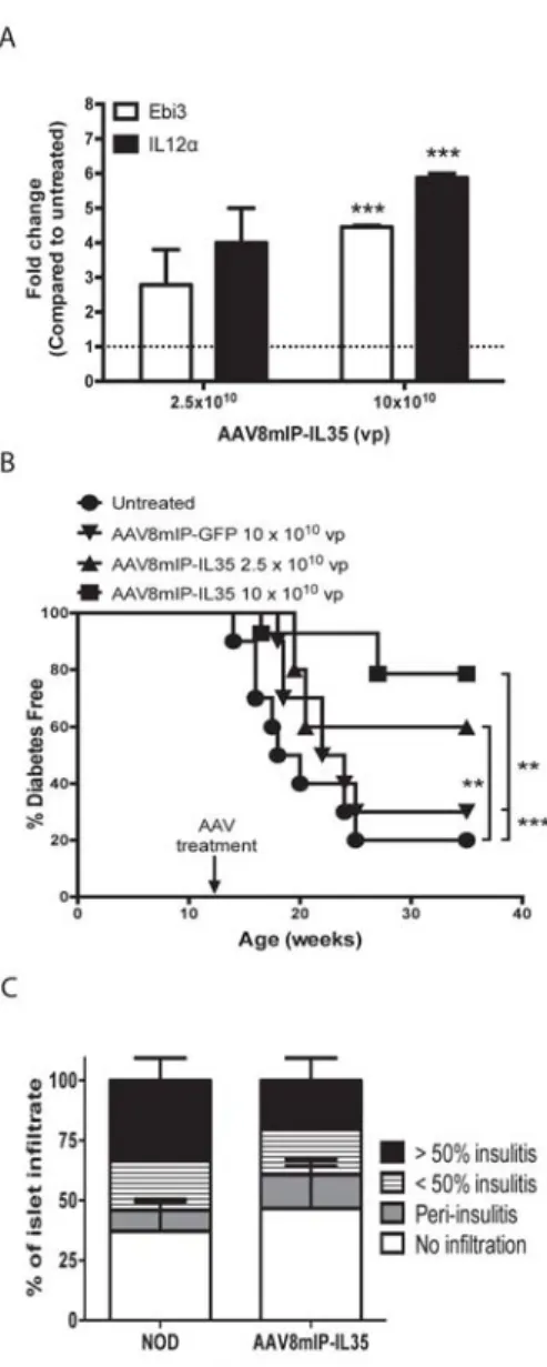
2.4.1βcell-specific IL-35 expression prevents overt diabetes at a late preclinical stage in NOD mice. ... 42
2.4.2 Ectopic IL-35 expression reduces islet infiltration. ... 43
2.4.3 Ectopic IL-35 expression suppression islet T cell expansion. ... 44
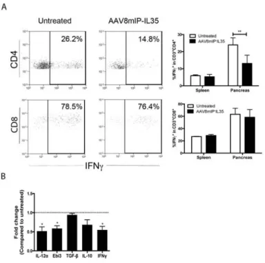
2.4.4 β cell-specific IL-35 expression reduces the proinflammatory milieu of the islets. ... 45
2.5 Discussion ... 47
2.6 References ... 65
CHAPTER 3: USING AAV-MEDIATED IL-2 THERAPY TO MANIPULATE HUMAN IMMUNOREGULATORY T CELL IN A HUMANIZED MOUSE MODEL ... 70
3.1 Summary ... 70
3.2 Introduction ... 71
3.3.1 Mice ... 74
3.3.2 PBMC isolation and engraftment ... 74
3.3.3 AAV vector engineering, packaging, and vaccination ... 74
3.3.4 Human islets ... 75
3.3.5 Flow cytometry ... 75
3.3.6 ELISA ... 75
3.3.7 In vitro suppression assay ... 75
3.3.8 Statistical analysis ... 76
3.4 Results ... 76
3.4.1 Ectopic human IL-2 expression increases pancreatic FOXP3+ Treg ... 76
3.4.2 AAV8mIP-hIL2 administration has no effect on conventional T cell pool ... 78
3.4.3 FOXP3+ Treg in T1D-derived PBMC hu-PBL-NRG mice are increased following AAV8mIP-hIL2 treatment ... 79
3.5 Discussion ... 79
3.6 References ... 90
CHAPTER 4: DISCUSSION ... 94
4.1 REFERENCES ... 99
APPENDIX 1: ISOLATION AND TRANSPLANTATION OF DIFFERENT AGED MURINE THYMIC GRAFTS ... 102
A1.1 Summary ... 102
A1.2 Introduction ... 102
A1.3 Protocol ... 104
A1.3.1. Preparation of Newborn and Adult Thymi ... 104
A1.4 Results ... 109
A1.5 Discussion ... 110
LIST OF TABLES
Table A1.1: Materials needed during surgical thymic transplantation. ... 114
Table A1.2: Total number of T cells present in peripheral blood and lymphoid
LIST OF FIGURES
Figure 2.1: β cell-specific expression of IL-35 at a late preclinical stage
prevents the onset of diabetes. ... 52
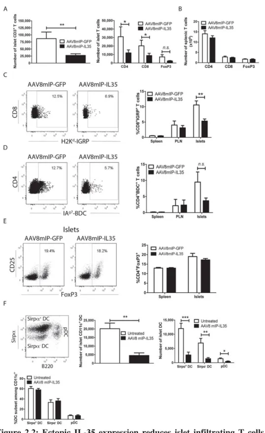
Figure 2.2: Ectopic IL-35 expression reduces islet infiltrating T cells and DC. ... 53
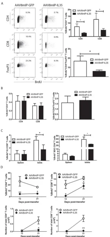
Figure 2.3: β cell-specific IL-35 reduces proliferation of islet resident
conventional T cells and Foxp3+Treg. ... 55 Figure 2.4: Ectopic IL-35 induces a qualitatively distinct pool of islet
conventional T cells. ... 57
Figure 2.5: Qualitatively distinct Foxp3+Treg are needed to suppress CD4+
Teff in AAV8mIP-IL35 treated mice. ... 58
Figure 2.S1: AAV8mIP-IL-35 treatment has no effect on T cell apoptosis. ... 60
Figure 2.S2: Proliferation and number of splenic CD4+ and CD8+ T cells is unchanged between AAV8mIP-IL35 and AAV8mIP-GFP
treated adoptive transfer recipients. ... 61
Figure 2.S3: AAV8mIP-IL35 treatment results in distinct subset of pancreatic
conventional T cells. ... 62
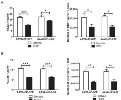
Figure 2.S4: Frequency and number of Foxp3+Treg in PC61 treated
AAV8mIP-IL35 vaccinated animals. ... 63
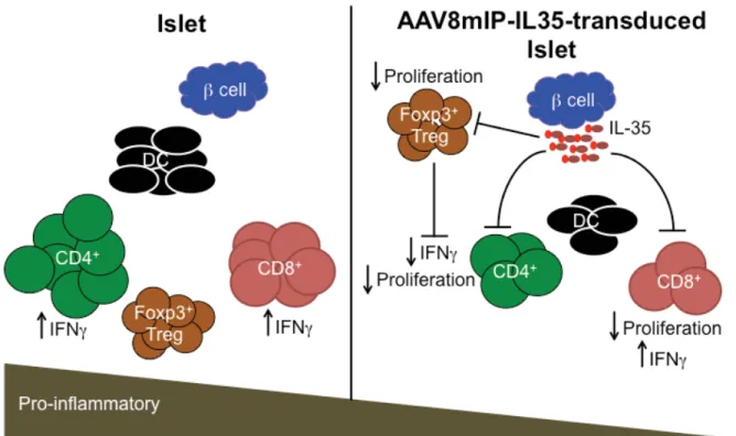
Figure 2.6: AAV8mIP-IL35 immunotherapy. ... 64
Figure 3.1: Engraftment of human T cells in hu-PBL-NRG mice. ... 83
Figure 3.2: Expression of human IL-2 and analyses of human T cells in
ND-derived hu-PBL-NRG mice treated with AAV8mIP-hIL2. ... 84
Figure 3.3: Ectopic human IL-2 expression increases the frequency and
number of FOXP3+Treg. ... 86 Figure 3.4: Ectopic human IL-2 has no effect on the number of conventional
T cells in hu-PBL-NRG mice. ... 87
Figure 3.5: AAV8mIP-hIL2 treatment increases frequency and number of
FOXP3+Treg in T1D hu-PBL-NRG mice. ... 88 Figure 3.6: Conventional T cell numbers remain unchanged in T1D hu-PBL-
NRG mice following AAV8mIP-IL2 treatment. ... 89
Figure A1.2: Position of capsular incision and placement of thymic graft. ... 115
LIST OF ABBREVIATIONS
AAV Adeno-associated virus
Ab Antibody
Aire Autoimmune regulator
ANOVA Analysis of variance
APC Antigen-presenting cell
Bcl-2 B cell lymphoma 2
BrdU 5-bromo-2’-deoxyuridine
CD Cluster of differentiation
cDC Conventional dendritic cell
cTEC Cortical thymic epithelial cell
CTLA-4 Cytotoxic T lymphocyte-associated protein 4
CTV Cell trace violet
DC Dendritic cell
DN Double negative
DP Double positive
DS Double-stranded
EAE Experimental autoimmune encephalomyelitis
ELISA Enzyme-linked immunosorbent assay
FACS Fluorescence activated cell sorting
Foxp3 Forkhead box protein 3
GFP Green fluorescent protein
GITR Glucocorticoid-induced TNF-related protein
HSC Hematopoietic stem cells
Ig Immunoglobulin
IGRP Islet-specific glucose-6-phosphatase
IL Interleukin
i.p. Intraperitoneal
IPEX Immunodysregulation polyendocrinopathy enteropathy X-linked syndrome
kb Kilobase pairs
LAG3 Lymphocyte activating gene 3
LPD Lymphoproliferative disorder
MHC Major histocompatibility complex
MIP Mouse insulin promoter
mTEC Medullary thymic epithelial cell
mTORC1 Mammalian target of rapamycin complex
nAb Neutralizing antibody
NOD Nonobese diabetic mouse
pDC Plasmacytoid dendritic cell
PBMC Peripheral blood mononuclear cells
PBS Phosphate-buffered saline
PLN Pancreatic lymph node
PTPN2 Protein tyrosine phosphatase non-receptor type 2
qPCR Quantitative polymerase chain reaction
sBDC Super BDC agonist peptide
SEM Standard error of the mean
SIRPα Signal regulatory protein alpha
SP Single positive
SS Single-stranded
STAT Signal transducer and activator of transcription
T1D Type 1 diabetes
T2D Type 2 diabetes
TCR T cell receptor
Teff Effector T cell
TGFβ Transforming growth factor beta
Th T helper cell
TLR Toll-like receptor
TNFα Tumor necrosis factor alpha
Treg Regulatory T cell
WT Wild-type
μL microliter
μM micromolar
CHAPTER 1: INTRODUCTION 1.1 Type 1 diabetes mellitus
Type 1 diabetes (T1D) is one of more than 80 autoimmune diseases affecting millions
of people worldwide. Although T1D is prevalent among all ethnic populations, a marked rise
in North America and other Western nations has been detected [1, 2]. T1D is manifested
following an interplay between genetic susceptibility and ill-defined environmental triggers.
The latter is highlighted by discordance among monozygotic twins and the development of
T1D [3]. T1D results from the destruction of insulin-producing β cells of the islets of
Langerhans. This β cell destruction is mediated primarily by pathogenic autoreactive T cells,
which leads to an inability of patients with T1D to control blood glucose levels resulting in
hyperglycemia and other co-morbidities [4]. Typically, β cell autoimmunity is initiated a number of years prior to the clinical onset of T1D. The latter is evident by the appearance of
serum islet-specific autoantibodies to insulin (IAA), glutamic acid decarboxylase (GAD65),
islet cell cytoplasm (ICA) and other β cell autoantigens in prediabetic subjects [5-9]. Most subjects progressing to overt diabetes present with at least two anti-islet autoantibodies [10].
1.2 Susceptibility to T1D development
Several genes confer susceptibility to and protection from T1D development [11, 12].
Animal models of T1D have aided in determining several susceptibility loci that in
conjunction with unknown environmental and other indeterminate triggers lead to overt
model of T1D that benefit from inbreeding, determining T1D susceptibility loci in humans
has proven challenging due in part to an inability to confirm the effect(s) of implicated genes
on mechanism of disease pathogenesis [8]. Nevertheless, studies in T1D patients and NOD
mice have shown that the most important T1D susceptibility genes are located in the major
histocompatibility complex (MHC) region, consistent with a critical role for T cells driving
disease [7]. This susceptibility region is termed the IDDM1 or idd1 locus in humans and
NOD mice, respectively [8, 13]. Although MHC loci are implicated in conferring almost
50% of genetic susceptibility to T1D, there are more than 30 non-HLA susceptibility loci, of
which at least 6 loci are shared between NOD mice and T1D patients [1, 4]. Genes in the
interleukin-2 (IL-2) and IL-2 receptor (IL-2R) signaling pathway, such as CD25, a
component of the high-affinity IL-2R, and protein tyrosine phosphatase non-receptor type 2
(PTPN2), a negative regulator of IL-2R signaling, are examples of the various genes that
have an impact on several aspects of immune regulation as well as β cell physiology in T1D
[13, 14].
The rise in T1D incidence in European and North American populations has been
used as an argument for environmental factors driving the diabetogenic response. Studies
exploring environmental triggers such as coxsackievirus infection, environmental toxins, and
diet, have not yet yielded a specific culprit. Viral infection serving as a trigger for T1D
development in susceptible individuals has been studied extensively [15]. Group B
coxsackievirus is a pancreas-tropic enterovirus that preferentially infects pancreatic β cells
and acinar cells, causing diabetes and pancreatitis. Coxsackievirus RNA has been isolated
from T1D patients at various stages of disease, and has been shown to accelerate disease
mechanisms: i) bystander activation of autoreactive T cells, ii) molecular mimicry where a
viral protein similar to an islet autoantigen epitope primes β cell-specific T cells, and iii)
direct cytolytic effects on β cells.
An environmental influence on disease development is also mirrored in NOD mice.
NOD mice housed in specific pathogen free (SPF) facilities exhibit a higher incidence of
disease than NOD mice housed in conventional “dirty” facilities. Furthermore, NOD mice
exposed to bacterial components are protected from the onset of T1D [17]. These findings
support the “hygiene hypothesis” which postulates that exposure to immune stimuli, such as
bacterial and viral microbes, have evolutionarily protected from T1D. Accordingly, enhanced
sanitation and early childhood vaccination in the Western world limits the exposure to such
stimuli, which leads to increased T1D development. Notably, NOD mice deficient for the
Myd88 gene, which is an adaptor molecule for toll-like receptor (TLR) function following
microbial stimuli, are protected from disease when housed in SPF facilities [18].
Interestingly, Myd88-deficient NOD mice housed in germ-free facilities developed disease
similar to wild-type NOD controls, indicating a protective effect of commensal microbiota
[18].
1.3 NOD mouse model of T1D
In addition to environmental modulation of disease and genetic susceptibility loci
discussed above, the NOD mouse model of T1D shares several characteristics of human
disease. Hallmark of T1D pathogenesis is the infiltration, or “insulitis”, of the islets by
immune cells, including CD4+ and CD8+ T cells, B cells, natural killer (NK) cells, dendritic cells (DC), and macrophages [19, 20]. In both T1D patients and NOD mice, β cell
infiltrate. B cells, DC, and macrophages are among the first cells infiltrating the islets. The
event that initiates trafficking of these antigen-presenting cells (APC) into the islets is
unclear. In NOD mice remodeling of the pancreas at 2 wks of age is believed to promote
APC infiltration of the islets, which is then followed by the appearance of pathogenic
effector T cells (Teff) [21].
Naïve β cell-specific T cells are first primed in the draining pancreatic lymph node
(PLN) by DC and possibly B cells that have taken up self-antigen at and have migrated from
the islets [22]. Pathogenic effector CD4+ and CD8+ T cells (Teff), typically exhibiting a type 1 phenotype, marked by secretion of interferon-γ (IFNγ), then traffic to the islets and begin
targeting the insulin-producing β cells. Initial β cell destruction is believed to be mediated by
a few select autoantigens. However, as β cells are destroyed, additional self-antigens and
epitopes within these proteins are targeted. This process, termed “epitope spreading”,
amplifies the diabetogenic response eventually leading to destruction of the majority of β cells, and the clinical onset of T1D. CD4+ and CD8+ T cells have distinct roles in driving β cell destruction. Islet resident CD4+ T cells via secretion of IFNγ and TNFα establish a proinflammatory milieu, which further enhances recruitment of Teff and other inflammatory
effectors. Notably, the combination of IFNγ and TNFα has cytolytic effects on β cells in vitro [23, 24]. On the other hand, islet resident CD8+ Teff directly kill
β cells via a classical CTL-target-mediated interaction [25]. For instance, NOD mice deficient for
β2-microglobulin (β2m), and in which β cells lack MHC class I expression, are protected from
T1D [26, 27]. Importance of CD4+ and CD8+ T cells in disease pathogenesis is demonstrated by the ability of T cells to adoptively transfer disease [28, 29]; furthermore NOD mice
1.4 Thymic development of T cells
Immune tolerance is established through central and peripheral tolerance
mechanisms. The thymus is the site of central tolerance, a process that ensures that
self-reactive T cell clones are purged from the T cell repertoire. Initiation of T1D is due to a loss
of tolerance to multiple epitopes recognizing self-antigens, demonstrating a role for defective
thymic T cell selection in conferring susceptibility to T1D. The thymus is the key site for
development of T cells and the T cell receptor (TCR) repertoire. Two important “selection”
processes occur in the thymus. Positive selection promotes development of T cells that
recognize self-MHC, whereas negative selection, or clonal deletion, ensures that T cells
bearing TCR that bind self-peptide-MHC (pMHC) complexes with moderate to high affinity
are deleted.
Initially, progenitors from the bone marrow enter the thymus as double negative (DN)
thymocytes, which lack expression of both CD4 and CD8 co-receptors. As the thymocytes
migrate through thymic microenvironments, both antigen-independent and -dependent events
drive thymocyte development and shape the TCR repertoire. At the DN stage, thymocytes
can either give rise to αβ or γδ T cells [31]. T cells committed to the αβ lineage require
expression of recombination-activation gene 1 (RAG1) and RAG2 to pair TCR β with the
pre-TCRα chain, establishing a functional pre-TCR complex [32]. αβ T cells expressing
CD4 and CD8 make up the double positive (DP) pool.
During thymic positive selection, DP that interact with pMHC presented by cortical
thymic epithelial cells (cTEC) with appropriate avidity receive survival signals, traffick to
the medulla, and mature into single-positive thymocytes (SP). DP expressing TCRs that bind
Alternatively, DP that interact with pMHC with relatively high avidity undergo apoptosis and
clonal deletion [33]. The higher the diversity of pMHC complexes presented by cTEC to DP,
the more diverse the T cell repertoire, thereby enhancing protective immunity.
Positively-selected SP reside in the medulla for 4-5 days, constantly sampling pMHC
presented by medullary thymic epithelial cells (mTEC) and thymic DC [34]. Negative
selection aims to delete SP thymocytes that are highly reactive to self-antigens. mTEC
express peripheral tissue-specific antigens (TSAs) whose expression in the thymus is
controlled by the Aire gene [35]. The transcription factor autoimmune regulator (AIRE)
controls expression of islet-specific autoantigens such as insulin in the thymus [36]. In
addition to presentation of AIRE-regulated self-antigens, SP in the medulla also recognize
self-antigens presented by thymic CD8a+ DC and migratory DC that enter the thymus from the periphery and are able to present peripheral tissue or even foreign self-antigens [34, 37].
This high diversity of self-antigens presented to SP during residency in the medulla ensures
that a high number of T cell-APC interactions take place to promote efficient deletion of
autoreactive precursors. T1D susceptibility is conferred in part by specific alleles of MHC
class II, such as IAg7 expressed by NOD mice. Evidence suggests that IAg7 binds
self-peptides poorly, which contributes to inefficient thymic negative selection [38]. Some studies
in NOD mice have shown that SP are resistant to apoptosis, which might be due to reduced
expression of Bim, a pro-apoptotic molecule [39, 40]. Meanwhile, other reports have
indicated no difference in sensitivity of NOD thymocytes to apoptosis leading to a
confounding picture of thymic selection in NOD mice [41].
thymic DC is necessary for Foxp3+Treg development. Both thymic DC and mTEC promote differentiation of CD4+SP into Foxp3+Treg that is mediated by intermediate to high TCR signaling [42]. Thymic-derived natural Foxp3+Treg therefore are skewed towards a profile of self-recognition and express the transcription factor Foxp3 [43-45].
Thymic negative selection is not absolute, and autoreactive T cells escape the thymus,
but are held in check by peripheral tolerance mechanisms. In individuals with T1D and in
NOD mice, inability of peripheral tolerance regulatory mechanisms to suppress autoreactive
T cells leads to pathology.
1.5 Immunoregulatory T cells (Treg) are important for maintaining self-tolerance in the
periphery
In T1D β cell antigen priming of naïve CD4+ T cells in the periphery preferentially skews towards type 1 or Th1 cell differentiation [20, 46]. In the early 90’s peripheral
tolerance and progression of T cell-mediated autoimmunity, such as T1D, was viewed as an
imbalance between pathogenic Th1 cells and protective Th2 cells. However, it is now
apparent that CD4+ T cells can differentiate into a variety of subsets depending on the inflammatory environment, including pathogenic Th17 and various protective Treg subsets.
Th1 cells produce IFNγ, TNFα, and promote isotype switching of immunoglobulin (Ig) to
IgG2A [46]. Th17 cells are characterized by production of IL-17, IL-21, IL-22, and GM-CSF
[47]. Th2 cells on the other hand, produce IL-4, IL-5, IL-13, and aid in antibody production
[46]. Skewing of naïve CD4+ T cell to a particular Th phenotype depends on T cell-APC interaction. IL-12, a cytokine produced by DC and macrophages, promotes robust
differentiation of a Th1-like response in vitro and in vivo, and concomitantly, has been shown
infiltration and β cell destruction in NOD mice. This is demonstrated by studies showing that
lack of IL-21 signaling was sufficient to reduce insulitis and protect NOD mice from disease
[49, 50].
Foxp3+Treg attenuate the Th1 and Th17 effector response. Unlike Foxp3+Treg, adaptive or induced Treg (iTreg) arise from naïve T cells in the periphery following antigen
encounter [51]. Naïve CD4+CD25- T cells can differentiate in to IL-10-expressing Tr1 cells or TGF-β-expressing Th3 cells that act in concert with Foxp3+Treg to maintain peripheral tolerance. Th2 cell cytokines such as IL-4, and Tr1 cells inhibit Th1 cell proliferation and
protect NOD mice from developing disease [46, 52-54]. Presence of a pro-inflammatory
milieu in the islets is likely to promote recruitment of autoreactive T cells into the islets and
exacerbate disease. Thus, maintaining a balance between pro-inflammatory and regulatory
responses, and determining factors controlling T cell plasticity, is necessary to prevent T1D.
The importance of Foxp3+Treg in curbing autoimmunity is evident in
lymphoproliferative disorders (LPDs) in mice and humans with Foxp3 gene defects [45].
Mutations in human Foxp3 gene result in immune dysregulation polyendocrinopathy
enteropathy X-linked syndrome (IPEX), characterized by multi-organ autoimmunity
including T1D, thyroiditis, and gastritis [55]. Similarly, defects in mouse Foxp3 gene in the
Scurfy mouse strain results in fatal hyperactivation of T cells [56]. In NOD mice, deletion of
Foxp3 results in hyperactivtion and proliferation of T cells as well, pointing to a critical role
for Foxp3+Treg-mediated control of Teff to maintain peripheral tolerance [57].
Based on murine studies, human FOXP3+Treg were identified through expression of surface CD25 (IL-2Rα chain) and intracellular FoxP3, as a Treg-specific marker. However,
FoxP3 in humans, thus confounding the use of FoxP3 as a lone marker for FOXP3+Treg. Recent studies have shown that lack of expression of CD127 (IL-7Rα) on CD4+ T cell enriches for a suppressive FOXP3+Treg population [58]. Human FOXP3+Treg are therefore characterized by high expression of CD25 and FOXP3, and low expression of CD127.
1.6 Foxp3+Treg-mediated suppression of Teff
Human and mouse Foxp3+Treg mediate suppression of Teff in a variety of ways, including contact-mediated suppression and via cytokine secretion. In addition, the make-up
of the local inflammatory environment where Foxp3+Treg are present also affects Foxp3+Treg suppressive function. Defects in Foxp3+Treg, and factors that render Teff resistant to suppression by Foxp3+Treg contribute to development of T1D in mouse and human and are discussed herein [59].
Contact-dependent suppression by Foxp3+Treg relies on upregulation of surface molecules such as cytotoxic T lymphocyte antigen 4 (CTLA-4), lymphocyte activating gene
3 (LAG3), granzyme A, and CD95 (Fas) [60]. Recent studies have shown that LAG3 is
necessary for optimal Foxp3+Treg suppressor function, although exact mechanisms by how LAG3, granzyme A and CD95 mediate Foxp3+Treg suppression in vivo have yet to be determined [61, 62]. In addition to CD25, Foxp3+Treg constitutively express CTLA-4. CTLA-4 is a negative regulator of T cell activation and interacts with CD80 and CD86 on
DC. Importantly, mice deficient for CTLA-4 develop spontaneous autoimmune disease and
Foxp3+Treg derived from CTLA-4-deficient mice show reduced suppression of Teff in vitro and in vivo [63, 64]. Notably, mutations in the ctla4 gene are mapped to T1D susceptibility
Immunoregulatory cytokines, such as IL-10 and TGF-β, have been the focus of
numerous studies of Foxp3+Treg suppressor activity. There is clearly a requirement of IL-10 and TGF-β for development of Tr1 and Th3 cells in vivo. For example, IL-10 and TGF-β
production by Foxp3+Treg is required for preventing colitis in IBD-prone mice [60, 65]. Of interest, a recently discovered immunoregulatory cytokine, IL-35, is expressed by
Foxp3+Treg and is essential for suppression of pathogenic Teff [66, 67]. IL-35 is a
heterodimer of Epstein-Barr-virus-induced gene 3 (Ebi3) and interleukin-12 alpha (IL-12α)
subunits, both of which are expressed in Foxp3+Treg but not in activated Teff [68]. Additionally, Ebi3- and IL-12α-deficient Foxp3+Treg are unable to suppress T cell proliferation in vitro and in vivo [68]. The role these immunoregulatory cytokines play in
regulating function and maintenance of Foxp3+Treg in vivo is largely unknown. NOD mice exhibit an age-dependent decrease in Foxp3+Treg. Whether islet
FOXP3+Treg number or function in T1D patients decreases following disease onset has yet to be conclusively determined. This is hindered by lack of availability of pancreases from
T1D patients correlating with various stages of disease. However, a recent study examining
pancreases of 29 recent-onset diabetics demonstrated that FOXP3+Treg were present in only one patient, and even then at very low numbers [19]. Similarly, decreased numbers of islet
Foxp3+Treg over time correlates with the progression of β cell destruction [69]. Studies in NOD mice and in T1D patients have shown that pathogenic Teff are
SLE [59, 70, 72, 73]. Notably, NOD mice can be rescued from T1D through adoptive
transfer of Foxp3+Treg, as well as by treatment with IL-2, a cytokine critical for Foxp3+Treg development, maintenance, and function [69, 74].
1.7 IL-35-mediated suppression of autoimmunity
Cytokine-mediated suppression by Foxp3+Treg relies on secretion of immunoregulatory cytokines such as the newly discovered IL-35. IL-35 is an
immunosuppressive cytokine required for maximal Foxp3+Treg mediated suppression in vitro and in vivo [66]. Treatment of naïve T cells with IL-35 results in differentiation of a
FoxP3- regulatory population, known as Tr35 cells [66]. Tr35 cells are characterized by secretion of IL35, but not IL-10 or TGFβ. Mechanistically, IL-35 inhibits T cell proliferation,
Th1 and Th17 cell differentiation, and has previously been shown to have therapeutic effects
in various models of T cell-mediated pathology, including IBD and experimental
autoimmune encephalomyelitis (EAE) [75]. Importantly, generation of transgenic NOD mice
with β cell-restricted expression of IL-35 results in significantly reduced diabetes incidence
compared to control wild-type NOD mice [76]. Protection was primarily mediated by
reduced proliferation of islet resident CD4+ and CD8+ T cells that corresponded to reduced frequencies of IFNγ-producing Teff and β-cell antigen-specific T cells [76]. Whether IL-35
therapy prevents or delays diabetes in recent onset or pre-diabetic NOD mice is unknown and
is the focus of Chapter 2 in this dissertation.
IL-35 signals through a heterodimer of IL-12Rβ2 and gp130, although it can also
alternatively signal through gp130 and IL-12Rβ2 homodimers [77]. While gp130 is
in the IL-12Rβ2 gene have been linked to development of Crohn’s disease, psoriasis, and
even T1D [76, 82-84]. IL-35 is unique in that it shares the gp130 subunit with IL-27, leading
to downstream phosphorylation of STAT1 and STAT4 molecules [77]. Data suggests that
signaling through both STAT1 and STAT4 molecules is required to mediate IL-35-dependent
differentiation of T cells into Tr35 cells and that signaling through gp130 or IL-12Rβ2
homodimers instead limits the suppressive function of IL-35 [77].
1.8 Role of IL-2 in T1D etiology
In addition to reduced Foxp3+Treg numbers, pathogenesis of T1D in NOD mice is marked by reduced numbers of NKT cells and impaired costimulation of naïve T cells by
APCs [20]. Costimulation is necessary not only for conventional T cell activation but also for
development and maintenance of Foxp3+Treg as alluded to earlier. This is exhibited by the reduced Treg pool in CD28-deficient NOD mice, which leads to exacerbated T1D
development [85, 86]. As discussed above, IL-2 therapy in NOD mice reverses diabetes [87].
To better understand the use of IL-2 therapy to protect spontaneous T1D development, it is
imperative to first discuss Foxp3+Treg dependency upon IL-2.
IL-2 signals through the IL-2R, which consists of IL-2Rα (CD25), IL-2Rβ (CD122), and the common γ chain (γc) (CD132) [51]. CD25, a component of the high-affinity IL-2R, is required for Foxp3+Treg development and maintenance in the periphery. Deficiencies in IL-2, IL-2Rα, or IL-2Rβ in mouse and human lead to fatal LPD and organ-specific
autoimmunity [88-90]. IL-2 deficiency results in accelerated T1D in NOD mice,
characterized by reduced number and proliferation of Foxp3+Treg in the thymus and
periphery [91]. IL-2 administration, on the other hand, induces Foxp3+Treg proliferation and
mice display reduced expression of CD25, FoxP3, and the anti-apoptotic molecule Bcl-2,
suggesting that IL-2 is necessary to maintain and prevent apoptosis of Foxp3+Treg in vivo [69]. These defects can be attributed to impaired IL-2 production and IL-2/IL-2R signaling.
IL-2 is produced by conventional T cells and DC and acts in a paracrine manner to
maintain the Foxp3+Treg pool. Therefore it has an essential role in maintaining immune homeostasis [92]. Administration of low dose IL-2 increases survival and proliferation of
Foxp3+Treg, whereas higher doses of IL-2 preferentially activate Teff and NK cells [69, 87]. This is due to the fact that Foxp3+Treg are highly responsive to IL-2 at lower doses than are needed to activate Teff, primarily due to constitutive expression of CD25 by Foxp3+Treg. IL-2/IL-2R engagement initiates signaling through the Jak/STAT pathway and activation of
STAT5. These downstream signaling events lead to upregulation of IL-2-dependent genes
such as Foxp3, which in turn increases CD25 expression that sustains increased IL-2R
signaling in a feedback loop [93]. Impaired maintenance of Foxp3 expression stemming from
defects in IL-2R signaling is characterized by reduced phosphorylation of STAT5 and
increased expression of PTPN2 in T1D patients [94]. Inhibitors of IL-2R signaling, including
protein tyrosine phosphatases such as PTPN2, are critical in regulating the IL-2R signaling
cascade. Defects in the 2/2R signaling pathway, and polymorphisms in the 2,
IL-2Rα, and PTPN2 genes are associated with T1D development in both NOD mice and
humans [94]. For instance, the IL-2 gene maps to idd3 in NOD mice [95, 96], and NOD mice
congenic for the C57BL/6-derived idd3 locus are protected from diabetes [97].
The central role IL-2 plays in T1D pathogenesis makes it an attractive and promising
target for immunotherapy. Low dose IL-2 has been used in the clinic for treatment of patients
to selectively expand Teff to prevent suppression of anti-tumor Teff. Advances in preventing
GvHD have demonstrated a protective role for FOXP3+Treg [98, 99]. GvHD develops when donor T cells recognize host MHC antigens and is a concern in both allogeneic and
mismatched recipients even with the administration of immunosuppressive drugs. Therefore,
suppressing donor Teff may spare target tissues from destruction. In fact, in mouse studies
both Foxp3+Treg and induced Treg subsets prevent disease following bone marrow
transplantation. In addition to low dose IL-2 therapy, targeting the IL-2R signaling pathway
has also been beneficial in NOD mice [92]. As mentioned above, dosage of IL-2 was
important in preferentially increasing Foxp3+Treg and preventing disease. However, IL-2 therapy in humans either alone or in combination with rapamycin, an agonist of mammalian
target of rapamycin complex (mTORC1), led to increased eosinophilia, NK cell function, and
exacerbated β cell autoimmunity [100]. These adverse effects on β cell function could be due to systemic toxicity associated with the pleiotropic nature of IL-2.
1.9 Clinical options for T1D treatment and management
To date, there is no therapy that can prevent T1D. Although risk of T1D development
increases up to 90% in first-degree relatives of T1D patients, more than 80% of newly
diagnosed patients do not have any family history of the disease, making it difficult to enroll
T1D susceptible individuals in prevention trials [1]. Diagnosis of T1D in the clinic is usually
made on the basis hyperglycemia in conjunction with presence of one or more anti-islet
autoantibodies, such as for insulin or GAD65 [1]. These diagnostic criteria preclude
erroneous diagnosis of individuals with insulin-resistant type 2 diabetes (T2D). Once
diagnosed, administration of daily exogenous insulin is necessary to maintain normal blood
in T1D patients with varying degrees of success, which is measured by a period of insulin
independence following transplantation. Islet transplantation is hampered by the need for
multiple donors to achieve adequate number of islets for transplantation. Transplantation
requires allogeneic donors and a regimen of immunosuppressive agents to prevent rejection
of the transplanted organ. Life-long use of immunosuppressants to prevent graft rejection and
to keep autoimmunity in check necessitates better treatments and a cure for T1D.
1.10 Immunotherapy approaches for T1D
Antigen-specific therapies involving GAD65 or insulin administration have failed to
effectively prevent T1D in at risk subjects [101, 102]. On the other hand, non-mitogenic
anti-CD3 antibody (Teplizumab), engineered to prevent binding to Fc receptors, maintained
residual β cell mass for up to 5 years in some patients, although diabetes reversal was not
achieved [102-104]. Success of non-mitogenic anti-CD3 antibody in NOD mice was
characterized by restoring the balance between Teff and Foxp3+Treg via depletion of Teff and expansion of Foxp3+Treg [4]. Similar immunotherapy approaches in recent-onset diabetic subjects treated with anti-human CD20 antibody (rituximab), or CTLA-4Ig have
yielded modest success in clinical trials characterized by short-term preservation of inulin
responses [105, 106].
In NOD mice, cytokine-mediated immunotherapies have been successful in disease
prevention. Notably, IL-4 and IL-10 treatment of NOD mice expand Treg and prevent T1D
development [107, 108]. As stated above, low dose IL-2 therapy expands Foxp3+Treg and prevents disease development but not without complications associated with systemic
administration of IL-2. Success of cytokine based immunotherapy approaches rely on strict
and the off-target effects associated with systemic cytokine delivery hinder development of
successful long-term outcomes for T1D treatment in the clinic. As opposed to systemic IL-2
delivery, targeting IL-2 expression to β cells in NOD mice leads to an islet-specific
expansion of a qualitatively distinct pool of Foxp3+Treg in NOD mice, which suppresses ongoing β cell autoimmunity and prevents diabetes onset in NOD mice [109]. The effect of
tissue-specific expression of human IL-2 on human FOXP3+Treg however remains unknown and is the topic of Chapter 3 of this dissertation.
To date, the lack of long-term suppression of β cell autoimmunity and/or reversal of
diabetes further highlights the need for effective immunotherapies in the clinic.
Tissue-specific gene therapy provides a promising approach to restore or preserve islet-function in
T1D patients. Utilization of adeno-associated virus (AAV) vector-mediated gene therapy
approach is a safe and robust tool to achieve this goal to treat T1D.
1.11 Using AAV vectors for immunotherapy
AAV vectors have become the preferred tools for gene therapy. The initial discovery
that AAV showed limited pathogenicity in infected humans presented the potential for
exploiting AAV as a vehicle for gene therapy [110]. Successful infection of AAV in humans
requires co-infection with a helper virus such as herpes simplex virus (HSV) or adenovirus
[110, 111]. Thus, despite a large proportion of AAV serotype-positive individuals
worldwide, AAV infection has not been associated with disease in humans [111]. AAV is a
parvovirus that consists of a single-stranded (ss) genome 4.7 kilobases (kb) in length. Upon
injection, ssAAV vector is converted to transcriptionally active double-stranded (ds) DNA,
vectors have been engineered to permit rapid transgene expression in vivo [112]. Both ss and
ds AAV vectors are used effectively for gene transfer in vivo.
One of the major strengths of using AAV vectors is that various cell types can be
targeted in vivo, depending on the AAV capsid protein employed for packaging [110, 113,
114]. Up to 13 distinct AAV serotype capsids have been isolated which exhibit tropism for
particular tissues in the body [110]. Notably, AAV vector serotype 8 shows strong tropism
and high transduction efficiency for the pancreas [110, 112, 115, 116]. In addition, efforts are
ongoing to engineer novel capsid proteins using “DNA shuffling’ strategies for instance, to
enhance tropism for given tissues while limiting immunogenicity [117].
Additional advantages of AAV vector gene delivery include limited pre-existing
AAV-specific immunity, establishment of stable long-term transgene expression, and ability
to transduce both replicating and non-replicating cells [118]. Additionally, safety and
efficacy of gene therapy using AAV vectors in the clinic has been established [119, 120].
AAV vectors have been used to treat a multitude of disorders in the clinic, such as
hemophilia, cystic fibrosis, Parkinson’s disease, and muscular dystrophy [114, 121-124].
AAV vectors have also been utilized to successfully prevent disease in NOD mice as well as
in STZ-induced model of diabetes [52, 53, 109, 115, 125]. Successful diabetes prevention in
mouse models have relied on AAV vector serotype 8 capsid for efficient transduction and the
use of a murine insulin-II promoter (mIP) to specifically target transgene expression to β
cells in vivo [52, 109, 115]. This approach limits the possibility of systemic transgene
1.12 Humanized mouse model to study human immune responses
Failure to translate efficacy induced by immunotherapies in NOD mice to the clinic
has been a major hurdle within the field. This is attributed to intrinsic differences between the
human and murine immune systems. Accordingly, there is a need to study the direct effects
of immunotherapies on human immune effectors in vivo. The latter has led to the
development and use of “humanized” mice [126-128].
Immunodeficient mice can be “humanized” via injection of peripheral blood
mononuclear cells (PBMC), hematopoietic stem cells (HSC) alone, or in conjunction with
implantation of autologous fetal liver and thymus tissues (BLT) [129, 130]. Early reports
demonstrating engraftment of human PBMC in immunodeficient NOD.scid mice resulted in
minimal engraftment of human immune cells, and the need for prohibitively large numbers of
PBMC [129, 131-134]. Development of NOD.scid (NSG) and NOD.Ragnull (NRG) mice bearing a null mutation of the IL-2R γc allowed for increased engraftment of human PBMC
and HSC, particularly human T cells [126, 135, 136]. IL-2, IL-4, IL-7, IL-9, IL-15, and IL-21
all signal through γc and the lack of γc in these mice results in severe defects in innate and
adaptive arms of the immune system [92].
NRG mice engrafted with human PBMC (hu-PBL-NRG) show robust engraftment of
T cells but not B cells, NK cells, NK T cells, DC or macrophages [127]. Islet autoantibodies
can be detected in peripheral blood of hu-PBL-NRG mice injected with PBMC derived from
T1D donors, but these mice do not demonstrate T cell infiltration of the islets [129].
However, a report showed that NSG mice transgenic for human HLA-2.1 expression and
engrafted with PBMC derived from T1D but not healthy donors exhibited islet-infiltration by
Humanized mice can also be used to model hyperglycemia. Hyperglycemia in T
cell-engrafted NRG or NSG mice induced chemically by streptozotocin (STZ) is reversed by
transplantation of human islets [135]. Mouse models of non-immune spontaneous
hyperglycemia have also been generated [138, 139]. These mice harbor a mutation in the
mouse insulin 2 gene (Ins2Akita) that results in unstable insulin expression and spontaneous β cell apoptosis. Spontaneously diabetic NRG.Ins2Akita mice support engraftment with human PBMC, and transplantation of human islets in these mice restores euglycemia [138].
The advantages of using mice engrafted with a fully functional human immune
system, including T cells, should aid in developing more effective clinical therapies. Despite
the many advantages outlined here, there do exist some limitations that need to be taken into
account in studies utilizing humanized mice. Hu-PBL-NRG mice, for example, exhibit
xGvHD 30-40 days following injection of PBMC characterized by pancreatitis and
infiltration of the liver and skin among other organs, which limits the window of time in
which experimental studies can be performed [133]. Recently generated NSG mice lacking
MHC class I and II, NSG.β2mnull and NSG.Abnull, respectively, exhibit significantly delayed progression of xGvHD following engraftment, and should help overcome some of these
limitations [133].
1.13 Goals and findings of the dissertation
The autoimmune response of T1D is marked by infiltration of the pancreatic islets
with β cell-specific Teff and protective Foxp3+Treg. While it is widely accepted that the balance between Foxp3+Treg and Teff is important in determining the risk and kinetics of disease development, mechanisms regulating immunoregulatory responses to autoreactive T
mechanisms to suppress Teff in the periphery, including cytokines that have potent
immunoregulatory effects. IL-35 is one such cytokine secreted by Foxp3+Treg that inhibits T cell activation, proliferation, and promotes Tr35 differentiation. A recent study demonstrated
that transgenic NOD mice expressing β cell-specific IL-35 exhibit reduced incidence of T1D
due, in part, to a reduction in pathogenic CD4+ and CD8+ Teff in the islets. The
immunotherapeutic potential of IL-35, however, is ill-defined. In the first part of our studies,
we examined the therapeutic potential of IL-35 in NOD female mice at a late preclinical
stage of T1D. NOD mice vaccinated with AAV serotype 8 vector packaged with mIP-driven
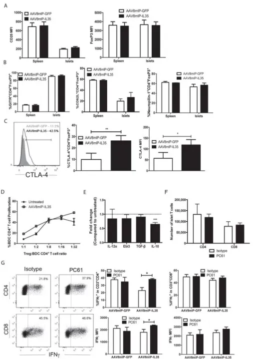
IL-35 (AAV8mIP-IL35) exhibited reduced islet CD4+ and CD8+ T cells numbers, which corresponded with reduced islet T cell proliferation and islet-resident DC numbers.
Furthermore, ectopic IL-35 expression led to a qualitatively distinct pool of Teff and
Foxp3+Treg. Islet CD4+ T cells exhibited reduced expression of IFNg+ and islet Foxp3+Treg displayed high levels of CTLA-4. The effects of ectopic IL-35 were islet-specific since the
frequency, number and activation status of splenic T cells were unchanged in AAVmIP-IL35
treated animals. Islet-restricted IL-35 expression effectively blocked progression to overt
diabetes in the majority of NOD mice. Together, these findings demonstrate that
islet-restricted IL-35 expression is sufficient to suppress ongoing β cell autoimmunity and that
immunotherapy using IL-35 is a promising approach to treat T1D as well as other T
cell-mediated pathologies.
Not unlike murine Foxp3+Treg, human FOXP3+Treg are potent mediators of
peripheral tolerance. IL-2R signaling is crucial for maintaining the Foxp3+Treg pool. Studies using NOD mice have shown that reduced IL-2 levels result in diminished survival of islet
demonstrated that FOXP3+Treg from T1D patients exhibit impaired responsiveness to IL-2 thereby reducing survival and function. Furthermore, defects in the IL-2R signaling pathway
have been implicated in conferring susceptibility to T1D in humans. In the second part of our
studies, we investigated the effects of ectopic IL-2 expression on human FOXP3+Treg. We utilized hu-PBL-NRG “humanized” mice to determine the response of T1D and non-T1D
derived FOXP3+Treg to AAV8mIP-hIL2 immunotherapy in vivo. AAV8mIP-hIL2 treatment expanded non-T1D and T1D-derived pancreatic FOXP3+Treg in hu-PBL-NRG mice. These early findings indicate a promising approach to study the effect of IL-2 on human
1.14 REFERENCES
1 Atkinson, M. A. and Eisenbarth, G. S., Type 1 diabetes: new perspectives on disease pathogenesis and treatment. Lancet 2001. 358: 221-229.
2 Onkamo, P., Vaananen, S., Karvonen, M. and Tuomilehto, J., Worldwide increase in incidence of Type I diabetes--the analysis of the data on published incidence trends. Diabetologia 1999. 42: 1395-1403.
3 van Belle, T. L., Coppieters, K. T. and von Herrath, M. G., Type 1 diabetes: etiology, immunology, and therapeutic strategies. Physiol Rev 2011. 91: 79-118.
4 Bluestone, J. A., Herold, K. and Eisenbarth, G., Genetics, pathogenesis and clinical interventions in type 1 diabetes. Nature 2010. 464: 1293-1300.
5 Kent, S. C., Chen, Y., Bregoli, L., Clemmings, S. M., Kenyon, N. S., Ricordi, C., Hering, B. J. and Hafler, D. A., Expanded T cells from pancreatic lymph nodes of type 1 diabetic subjects recognize an insulin epitope. Nature 2005. 435: 224-228.
6 Nakayama, M., Abiru, N., Moriyama, H., Babaya, N., Liu, E., Miao, D., Yu, L., Wegmann, D. R., Hutton, J. C., Elliott, J. F. and Eisenbarth, G. S., Prime role for an insulin epitope in the development of type 1 diabetes in NOD mice. Nature 2005. 435: 220-223.
7 Wucherpfennig, K. W. and Eisenbarth, G. S., Type 1 diabetes. Nat Immunol 2001. 2: 767-768.
8 Davies, J. L., Kawaguchi, Y., Bennett, S. T., Copeman, J. B., Cordell, H. J., Pritchard, L. E., Reed, P. W., Gough, S. C., Jenkins, S. C., Palmer, S. M. and et al., A genome-wide search for human type 1 diabetes susceptibility genes. Nature 1994. 371: 130-136.
9 Lohmann, T., Leslie, R. D. and Londei, M., T cell clones to epitopes of glutamic acid decarboxylase 65 raised from normal subjects and patients with insulin-dependent diabetes. J Autoimmun 1996. 9: 385-389.
10 Concannon, P., Rich, S. S. and Nepom, G. T., Genetics of type 1A diabetes. N Engl J Med 2009. 360: 1646-1654.
11 Castano, L. and Eisenbarth, G. S., Type-I diabetes: a chronic autoimmune disease of human, mouse, and rat. Annu Rev Immunol 1990. 8: 647-679.
12 Tisch, R. and McDevitt, H., Insulin-dependent diabetes mellitus. Cell 1996. 85: 291-297.
14 Atkinson, M. A., Eisenbarth, G. S. and Michels, A. W., Type 1 diabetes. Lancet 2014. 383: 69-82.
15 Coppieters, K. T., Boettler, T. and von Herrath, M., Virus infections in type 1 diabetes. Cold Spring Harb Perspect Med 2012. 2: a007682.
16 Serreze, D. V., Ottendorfer, E. W., Ellis, T. M., Gauntt, C. J. and Atkinson, M. A., Acceleration of type 1 diabetes by a coxsackievirus infection requires a
preexisting critical mass of autoreactive T-cells in pancreatic islets. Diabetes 2000. 49: 708-711.
17 Qin, H. Y. and Singh, B., BCG vaccination prevents insulin-dependent diabetes mellitus (IDDM) in NOD mice after disease acceleration with cyclophosphamide. J Autoimmun 1997. 10: 271-278.
18 Wen, L., Ley, R. E., Volchkov, P. Y., Stranges, P. B., Avanesyan, L.,
Stonebraker, A. C., Hu, C., Wong, F. S., Szot, G. L., Bluestone, J. A., Gordon, J. I. and Chervonsky, A. V., Innate immunity and intestinal microbiota in the
development of Type 1 diabetes. Nature 2008. 455: 1109-1113.
19 Willcox, A., Richardson, S. J., Bone, A. J., Foulis, A. K. and Morgan, N. G., Analysis of islet inflammation in human type 1 diabetes. Clin Exp Immunol 2009. 155: 173-181.
20 Anderson, M. S. and Bluestone, J. A., The NOD mouse: a model of immune dysregulation. Annu Rev Immunol 2005. 23: 447-485.
21 Serreze, D. V., Fleming, S. A., Chapman, H. D., Richard, S. D., Leiter, E. H. and Tisch, R. M., B lymphocytes are critical antigen-presenting cells for the initiation of T cell-mediated autoimmune diabetes in nonobese diabetic mice. J Immunol 1998. 161: 3912-3918.
22 Turley, S., Poirot, L., Hattori, M., Benoist, C. and Mathis, D., Physiological beta cell death triggers priming of self-reactive T cells by dendritic cells in a type-1 diabetes model. J Exp Med 2003. 198: 1527-1537.
23 Campbell, I. L., Iscaro, A. and Harrison, L. C., IFN-gamma and tumor necrosis factor-alpha. Cytotoxicity to murine islets of Langerhans. J Immunol 1988. 141: 2325-2329.
24 Suk, K., Kim, S., Kim, Y. H., Kim, K. A., Chang, I., Yagita, H., Shong, M. and Lee, M. S., IFN-gamma/TNF-alpha synergism as the final effector in autoimmune diabetes: a key role for STAT1/IFN regulatory factor-1 pathway in pancreatic beta cell death. J Immunol 2001. 166: 4481-4489.
mechanisms in type 1 diabetes: the islet is both target and driver of disease. Rev Diabet Stud 2012. 9: 148-168.
26 Hamilton-Williams, E. E., Palmer, S. E., Charlton, B. and Slattery, R. M., Beta cell MHC class I is a late requirement for diabetes. Proc Natl Acad Sci U S A 2003. 100: 6688-6693.
27 Katz, J., Benoist, C. and Mathis, D., Major histocompatibility complex class I molecules are required for the development of insulitis in non-obese diabetic mice. Eur J Immunol 1993. 23: 3358-3360.
28 Wong, F. S., Visintin, I., Wen, L., Flavell, R. A. and Janeway, C. A., Jr., CD8 T cell clones from young nonobese diabetic (NOD) islets can transfer rapid onset of diabetes in NOD mice in the absence of CD4 cells. J Exp Med 1996. 183: 67-76.
29 Christianson, S. W., Shultz, L. D. and Leiter, E. H., Adoptive transfer of diabetes into immunodeficient NOD-scid/scid mice. Relative contributions of CD4+ and CD8+ T-cells from diabetic versus prediabetic NOD.NON-Thy-1a donors. Diabetes 1993. 42: 44-55.
30 Mora, C., Wong, F. S., Chang, C. H. and Flavell, R. A., Pancreatic infiltration but not diabetes occurs in the relative absence of MHC class II-restricted CD4 T cells: studies using NOD/CIITA-deficient mice. J Immunol 1999. 162: 4576-4588.
31 Germain, R. N., T-cell development and the CD4-CD8 lineage decision. Nat Rev Immunol 2002. 2: 309-322.
32 von Boehmer, H. and Fehling, H. J., Structure and function of the pre-T cell receptor. Annu Rev Immunol 1997. 15: 433-452.
33 Klein, L., Kyewski, B., Allen, P. M. and Hogquist, K. A., Positive and negative selection of the T cell repertoire: what thymocytes see (and don't see). Nat Rev Immunol 2014. 14: 377-391.
34 Klein, L., Hinterberger, M., Wirnsberger, G. and Kyewski, B., Antigen
presentation in the thymus for positive selection and central tolerance induction. Nat Rev Immunol 2009. 9: 833-844.
35 Gabler, J., Arnold, J. and Kyewski, B., Promiscuous gene expression and the developmental dynamics of medullary thymic epithelial cells. Eur J Immunol 2007. 37: 3363-3372.
37 Li, J., Park, J., Foss, D. and Goldschneider, I., Thymus-homing peripheral dendritic cells constitute two of the three major subsets of dendritic cells in the steady-state thymus. J Exp Med 2009. 206: 607-622.
38 Kanagawa, O., Martin, S. M., Vaupel, B. A., Carrasco-Marin, E. and Unanue, E. R., Autoreactivity of T cells from nonobese diabetic mice: an I-Ag7-dependent reaction. Proc Natl Acad Sci U S A 1998. 95: 1721-1724.
39 Kishimoto, H. and Sprent, J., A defect in central tolerance in NOD mice. Nat Immunol 2001. 2: 1025-1031.
40 Liston, A., Lesage, S., Gray, D. H., O'Reilly, L. A., Strasser, A., Fahrer, A. M., Boyd, R. L., Wilson, J., Baxter, A. G., Gallo, E. M., Crabtree, G. R., Peng, K., Wilson, S. R. and Goodnow, C. C., Generalized resistance to thymic deletion in the NOD mouse; a polygenic trait characterized by defective induction of Bim. Immunity 2004. 21: 817-830.
41 Villunger, A., Marsden, V. S. and Strasser, A., Efficient T cell receptor-mediated apoptosis in nonobese diabetic mouse thymocytes. Nat Immunol 2003. 4: 717; author reply 718.
42 Wirnsberger, G., Mair, F. and Klein, L., Regulatory T cell differentiation of thymocytes does not require a dedicated antigen-presenting cell but is under T cell-intrinsic developmental control. Proc Natl Acad Sci U S A 2009. 106: 10278-10283.
43 Fontenot, J. D., Gavin, M. A. and Rudensky, A. Y., Foxp3 programs the
development and function of CD4+CD25+ regulatory T cells. Nat Immunol 2003. 4: 330-336.
44 Hori, S., Nomura, T. and Sakaguchi, S., Control of regulatory T cell development by the transcription factor Foxp3. Science 2003. 299: 1057-1061.
45 Khattri, R., Cox, T., Yasayko, S. A. and Ramsdell, F., An essential role for Scurfin in CD4+CD25+ T regulatory cells. Nat Immunol 2003. 4: 337-342.
46 Liblau, R. S., Singer, S. M. and McDevitt, H. O., Th1 and Th2 CD4+ T cells in the pathogenesis of organ-specific autoimmune diseases. Immunol Today 1995. 16: 34-38.
47 Li, Y., Liu, Y. and Chu, C. Q., Th17 Cells in Type 1 Diabetes: Role in the Pathogenesis and Regulation by Gut Microbiome. Mediators Inflamm 2015. 2015: 638470.
48 Trembleau, S., Penna, G., Bosi, E., Mortara, A., Gately, M. K. and Adorini, L., Interleukin 12 administration induces T helper type 1 cells and accelerates
49 Spolski, R., Kashyap, M., Robinson, C., Yu, Z. and Leonard, W. J., IL-21 signaling is critical for the development of type I diabetes in the NOD mouse. Proc Natl Acad Sci U S A 2008. 105: 14028-14033.
50 Sutherland, A. P., Van Belle, T., Wurster, A. L., Suto, A., Michaud, M., Zhang, D., Grusby, M. J. and von Herrath, M., Interleukin-21 is required for the
development of type 1 diabetes in NOD mice. Diabetes 2009. 58: 1144-1155.
51 Sakaguchi, S., Ono, M., Setoguchi, R., Yagi, H., Hori, S., Fehervari, Z., Shimizu, J., Takahashi, T. and Nomura, T., Foxp3+ CD25+ CD4+ natural regulatory T cells in dominant self-tolerance and autoimmune disease. Immunol Rev 2006. 212: 8-27.
52 Rehman, K. K., Trucco, M., Wang, Z., Xiao, X. and Robbins, P. D., AAV8-mediated gene transfer of interleukin-4 to endogenous beta-cells prevents the onset of diabetes in NOD mice. Mol Ther 2008. 16: 1409-1416.
53 Yang, Z., Chen, M., Wu, R., Fialkow, L. B., Bromberg, J. S., McDuffie, M., Naji, A. and Nadler, J. L., Suppression of autoimmune diabetes by viral IL-10 gene transfer. J Immunol 2002. 168: 6479-6485.
54 Mueller, R., Krahl, T. and Sarvetnick, N., Pancreatic expression of interleukin-4 abrogates insulitis and autoimmune diabetes in nonobese diabetic (NOD) mice. J Exp Med 1996. 184: 1093-1099.
55 Bennett, C. L., Christie, J., Ramsdell, F., Brunkow, M. E., Ferguson, P. J., Whitesell, L., Kelly, T. E., Saulsbury, F. T., Chance, P. F. and Ochs, H. D., The immune dysregulation, polyendocrinopathy, enteropathy, X-linked syndrome (IPEX) is caused by mutations of FOXP3. Nat Genet 2001. 27: 20-21.
56 Brunkow, M. E., Jeffery, E. W., Hjerrild, K. A., Paeper, B., Clark, L. B., Yasayko, S. A., Wilkinson, J. E., Galas, D., Ziegler, S. F. and Ramsdell, F., Disruption of a new forkhead/winged-helix protein, scurfin, results in the fatal lymphoproliferative disorder of the scurfy mouse. Nat Genet 2001. 27: 68-73.
57 Chen, Z., Benoist, C. and Mathis, D., How defects in central tolerance impinge on a deficiency in regulatory T cells. Proc Natl Acad Sci U S A 2005. 102: 14735-14740.
58 Liu, W., Putnam, A. L., Xu-Yu, Z., Szot, G. L., Lee, M. R., Zhu, S., Gottlieb, P. A., Kapranov, P., Gingeras, T. R., Fazekas de St Groth, B., Clayberger, C., Soper, D. M., Ziegler, S. F. and Bluestone, J. A., CD127 expression inversely correlates with FoxP3 and suppressive function of human CD4+ T reg cells. J Exp Med 2006. 203: 1701-1711.
60 Vignali, D. A., Collison, L. W. and Workman, C. J., How regulatory T cells work. Nat Rev Immunol 2008. 8: 523-532.
61 Do, J. S., Visperas, A., Sanogo, Y. O., Bechtel, J. J., Dvorina, N., Kim, S., Jang, E., Stohlman, S. A., Shen, B., Fairchild, R. L., Baldwin Iii, W. M., Vignali, D. A. and Min, B., An IL-27/Lag3 axis enhances Foxp3+ regulatory T cell-suppressive function and therapeutic efficacy. Mucosal Immunol 2016. 9: 137-145.
62 Huang, C. T., Workman, C. J., Flies, D., Pan, X., Marson, A. L., Zhou, G., Hipkiss, E. L., Ravi, S., Kowalski, J., Levitsky, H. I., Powell, J. D., Pardoll, D. M., Drake, C. G. and Vignali, D. A., Role of LAG-3 in regulatory T cells. Immunity 2004. 21: 503-513.
63 Oderup, C., Cederbom, L., Makowska, A., Cilio, C. M. and Ivars, F., Cytotoxic T lymphocyte antigen-4-dependent down-modulation of costimulatory molecules on dendritic cells in CD4+ CD25+ regulatory T-cell-mediated suppression. Immunology 2006. 118: 240-249.
64 Tivol, E. A., Borriello, F., Schweitzer, A. N., Lynch, W. P., Bluestone, J. A. and Sharpe, A. H., Loss of CTLA-4 leads to massive lymphoproliferation and fatal multiorgan tissue destruction, revealing a critical negative regulatory role of CTLA-4. Immunity 1995. 3: 541-547.
65 Asseman, C., Mauze, S., Leach, M. W., Coffman, R. L. and Powrie, F., An essential role for interleukin 10 in the function of regulatory T cells that inhibit intestinal inflammation. J Exp Med 1999. 190: 995-1004.
66 Collison, L. W., Chaturvedi, V., Henderson, A. L., Giacomin, P. R., Guy, C., Bankoti, J., Finkelstein, D., Forbes, K., Workman, C. J., Brown, S. A., Rehg, J. E., Jones, M. L., Ni, H. T., Artis, D., Turk, M. J. and Vignali, D. A., IL-35-mediated induction of a potent regulatory T cell population. Nat Immunol 2010. 11: 1093-1101.
67 Collison, L. W., Pillai, M. R., Chaturvedi, V. and Vignali, D. A., Regulatory T cell suppression is potentiated by target T cells in a cell contact, IL-35- and
IL-10-dependent manner. J Immunol 2009. 182: 6121-6128.
68 Collison, L. W., Workman, C. J., Kuo, T. T., Boyd, K., Wang, Y., Vignali, K. M., Cross, R., Sehy, D., Blumberg, R. S. and Vignali, D. A., The inhibitory cytokine IL-35 contributes to regulatory T-cell function. Nature 2007. 450: 566-569.
70 Schneider, A., Rieck, M., Sanda, S., Pihoker, C., Greenbaum, C. and Buckner, J. H., The effector T cells of diabetic subjects are resistant to regulation via CD4+ FOXP3+ regulatory T cells. J Immunol 2008. 181: 7350-7355.
71 D'Alise, A. M., Auyeung, V., Feuerer, M., Nishio, J., Fontenot, J., Benoist, C. and Mathis, D., The defect in T-cell regulation in NOD mice is an effect on the T-cell effectors. Proc Natl Acad Sci U S A 2008. 105: 19857-19862.
72 Gregori, S., Giarratana, N., Smiroldo, S. and Adorini, L., Dynamics of pathogenic and suppressor T cells in autoimmune diabetes development. J Immunol 2003. 171: 4040-4047.
73 Monk, C. R., Spachidou, M., Rovis, F., Leung, E., Botto, M., Lechler, R. I. and Garden, O. A., MRL/Mp CD4+,CD25- T cells show reduced sensitivity to
suppression by CD4+,CD25+ regulatory T cells in vitro: a novel defect of T cell regulation in systemic lupus erythematosus. Arthritis Rheum 2005. 52: 1180-1184.
74 Tang, Q., Henriksen, K. J., Bi, M., Finger, E. B., Szot, G., Ye, J., Masteller, E. L., McDevitt, H., Bonyhadi, M. and Bluestone, J. A., In vitro-expanded
antigen-specific regulatory T cells suppress autoimmune diabetes. J Exp Med 2004. 199: 1455-1465.
75 Choi, J., Leung, P. S., Bowlus, C. and Gershwin, M. E., IL-35 and Autoimmunity: a Comprehensive Perspective. Clin Rev Allergy Immunol 2015.
76 Bettini, M., Castellaw, A. H., Lennon, G. P., Burton, A. R. and Vignali, D. A., Prevention of autoimmune diabetes by ectopic pancreatic beta-cell expression of interleukin-35. Diabetes 2012. 61: 1519-1526.
77 Collison, L. W., Delgoffe, G. M., Guy, C. S., Vignali, K. M., Chaturvedi, V., Fairweather, D., Satoskar, A. R., Garcia, K. C., Hunter, C. A., Drake, C. G., Murray, P. J. and Vignali, D. A., The composition and signaling of the IL-35 receptor are unconventional. Nat Immunol 2012. 13: 290-299.
78 Betz, U. A. and Muller, W., Regulated expression of gp130 and IL-6 receptor alpha chain in T cell maturation and activation. Int Immunol 1998. 10: 1175-1184.
79 Hibbert, L., Pflanz, S., De Waal Malefyt, R. and Kastelein, R. A., IL-27 and IFN-alpha signal via Stat1 and Stat3 and induce T-Bet and IL-12Rbeta2 in naive T cells. J Interferon Cytokine Res 2003. 23: 513-522.
80 Liao, W., Lin, J. X., Wang, L., Li, P. and Leonard, W. J., Modulation of cytokine receptors by IL-2 broadly regulates differentiation into helper T cell lineages. Nat Immunol 2011. 12: 551-559.
composed of two beta-type cytokine receptor subunits. Proc Natl Acad Sci U S A 1996. 93: 14002-14007.
82 Bossini-Castillo, L., Martin, J. E., Broen, J., Gorlova, O., Simeon, C. P., Beretta, L., Vonk, M. C., Callejas, J. L., Castellvi, I., Carreira, P., Garcia-Hernandez, F. J., Fernandez Castro, M., Spanish Scleroderma, G., Coenen, M. J., Riemekasten, G., Witte, T., Hunzelmann, N., Kreuter, A., Distler, J. H., Koeleman, B. P., Voskuyl, A. E., Schuerwegh, A. J., Palm, O., Hesselstrand, R., Nordin, A., Airo, P., Lunardi, C., Scorza, R., Shiels, P., van Laar, J. M., Herrick, A., Worthington, J., Denton, C., Tan, F. K., Arnett, F. C., Agarwal, S. K., Assassi, S., Fonseca, C., Mayes, M. D., Radstake, T. R. and Martin, J., A GWAS follow-up study reveals the association of the IL12RB2 gene with systemic sclerosis in Caucasian
populations. Hum Mol Genet 2012. 21: 926-933.
83 Cargill, M., Schrodi, S. J., Chang, M., Garcia, V. E., Brandon, R., Callis, K. P., Matsunami, N., Ardlie, K. G., Civello, D., Catanese, J. J., Leong, D. U., Panko, J. M., McAllister, L. B., Hansen, C. B., Papenfuss, J., Prescott, S. M., White, T. J., Leppert, M. F., Krueger, G. G. and Begovich, A. B., A large-scale genetic
association study confirms IL12B and leads to the identification of IL23R as psoriasis-risk genes. Am J Hum Genet 2007. 80: 273-290.
84 McGovern, D. P., Rotter, J. I., Mei, L., Haritunians, T., Landers, C., Derkowski, C., Dutridge, D., Dubinsky, M., Ippoliti, A., Vasiliauskas, E., Mengesha, E., King, L., Pressman, S., Targan, S. R. and Taylor, K. D., Genetic epistasis of IL23/IL17 pathway genes in Crohn's disease. Inflamm Bowel Dis 2009. 15: 883-889.
85 Salomon, B., Lenschow, D. J., Rhee, L., Ashourian, N., Singh, B., Sharpe, A. and Bluestone, J. A., B7/CD28 costimulation is essential for the homeostasis of the CD4+CD25+ immunoregulatory T cells that control autoimmune diabetes. Immunity 2000. 12: 431-440.
86 Tang, Q., Henriksen, K. J., Boden, E. K., Tooley, A. J., Ye, J., Subudhi, S. K., Zheng, X. X., Strom, T. B. and Bluestone, J. A., Cutting edge: CD28 controls peripheral homeostasis of CD4+CD25+ regulatory T cells. J Immunol 2003. 171: 3348-3352.
87 Grinberg-Bleyer, Y., Baeyens, A., You, S., Elhage, R., Fourcade, G., Gregoire, S., Cagnard, N., Carpentier, W., Tang, Q., Bluestone, J., Chatenoud, L., Klatzmann, D., Salomon, B. L. and Piaggio, E., IL-2 reverses established type 1 diabetes in NOD mice by a local effect on pancreatic regulatory T cells. J Exp Med 2010. 207: 1871-1878.
88 Horak, I., Lohler, J., Ma, A. and Smith, K. A., Interleukin-2 deficient mice: a new model to study autoimmunity and self-tolerance. Immunol Rev 1995. 148: 35-44.