Changes in Lower Extremity Movement Patterns Following Exercise-induced Fatigue and Verbal Feedback
Melanie L. McGrath
A dissertation submitted to the faculty of the University of North Carolina at Chapel Hill in partial fulfillment of the requirements for the degree of Doctor of Philosophy in the
Department of Human Movement Science.
Chapel Hill 2009
iii Abstract
Melanie L. McGrath: Changes in Lower Extremity Movement Patterns Following Exercise-induced Fatigue and Verbal Feedback
(Under the direction of Dr. Darin A. Padua)
The present study investigated how exercise-induced fatigue and verbal feedback altered lower extremity coordination, variability, and kinetic variables in male and female athletes. Sixty-one healthy, club level athletes were divided into two groups: one that received a verbal feedback intervention post-fatigue, and one that did not. All subjects performed an unanticipated side-step cut, agility task, and vertical jump pre-fatigue, then completed an intense, intermittent, multi-directional fatigue protocol. Subjects in the feedback group received a quick verbal feedback intervention, focusing on landing technique. All subjects then repeated the pre-fatigue testing. The results indicated that fatigue caused subjects in the non-feedback group to change their coordination pattern in the sagittal plane, while subjects in the feedback group maintained their pre-fatigue pattern in all but one segment pairing (thigh-trunk frontal plane decreased post-fatigue in the feedback group). Fatigue caused all subjects to decrease their variability in the foot-shank and shank-thigh pairings in both the frontal and sagittal plane. Subjects in the non-feedback group also increased their anterior tibial shear force and vertical ground reaction force (VGRF), while the feedback group decreased their VGRF and knee extension moment. Fatigue also decreased vertical jump, and increased the score in the agility task, in both groups post-fatigue. These results suggest that an intermittent, multi-directional fatigue protocol causes a more in-phase, stiffer, less
variable movement pattern, but that a simple verbal feedback intervention can prevent many of these changes from occurring. While the implications of these results on actual injury risk are unknown, these changes do suggest that verbal feedback may be an effective method for acutely altering some proposed risk factors for lower extremity injury, particularly anterior cruciate ligament injury. These results also suggest that muscular fatigue may be an
iv Dedication
To Scott, the love of my life, and the reason I was able to pursue this dream. Equal credit for this project goes to you, for your patience, love, and the occasional kick in the rear.
To my parents Richard and Diane, who provided me with a true passion for learning and academia. Your love and support have carried me my entire life.
To my brother Andrew, my partner-in-crime who can understand the pain that comes with long excel spreadsheets and faulty MatLab code.
To my labmates (Cathy, Chuck, Dan, Michelle, Andy, Jason, Lindsay, Dave, Shana, Johna, Saki, Marc, and Ben), who provided endless assistance, ideas, feedback, and laughter.
To Darin, the source of so much inspiration and encouragement, who was exactly the type of mentor I needed during this process.
To my friends, who gave me the breaks I needed to keep my sanity.
To Steve and Taffy, and Quinn, who gave Scott and I tremendous support during our adventure in North Carolina.
v
Acknowledgements
This research was supported by two research grants: The National Athletic Trainers Association District 3 Research Award, in the amount of $960.00, and the University of North Carolina Smith Graduate Research Grant, in the amount of $985.00. This funding was critical to the recruitment of subjects and purchasing of research equipment. I wish to thank these two groups for their support of this dissertation.
Special acknowledgement needs to be given to the four undergraduate research assistants whom volunteered their time and effort to this project. Russell Hennessey,
vi
Table of Contents
List of Tables ... x
List of Figures ... xii
List of Abbreviations and Symbols... xiv
CHAPTER 1 ... 1
Introduction ... 1
Operational Definitions ... 6
Limitations/assumptions ... 9
Delimitations ... 10
Statement of Problem ... 10
Independent Variables ... 11
Dependent Variables ... 11
Research Questions ... 12
Hypotheses ... 14
CHAPTER 2 ... 17
Section One: Injury Epidemiology and Kinetic Risk Factors ... 18
Epidemiology of ACL Injury ...18
Suggested Risk Factors for Non-Contact ACL Injury ...21
Environmental Risk Factors ...22
Anatomic Risk Factors ...23
Hormonal Risk Factors ...24
Section Two: Muscular Fatigue ... 34
Muscular Fatigue ...35
Fatigue as a risk factor ...40
Quantifying Muscle Fatigue during Athletic Activity ...41
Impact of Fatigue on Knee Laxity ...43
Impact of Fatigue on Lower Extremity Biomechanics ...44
Repetitive CKC Exercises...45
Repetitive Jumping and/or Sprinting ...49
Intermittent Shuttle Runs ...52
Graded Treadmill Tests...53
General Conclusions ...55
Section Three: Movement Coordination/Variability ... 57
Methods of Quantifying Coordination and Variability ...58
Movement Coordination and Variability in the Lower Extremity ...62
Section Four: Verbal Feedback ... 65
Feedback and Decreased Landing Forces ...66
Feedback as an Injury Prevention Modality ...69
CHAPTER 3 ... 71
Rationale ... 71
Population ... 72
Subjects ...72
Group Assignment ...74
Power Analyses ...74
Data Collection ... 75
viii
Procedure ...77
Warm-up ...77
Testing battery ...78
Fatigue protocol ...80
Feedback delivery ...81
Post-test ...82
Data Processing and Reduction ... 82
Marker Identification and Processing ...82
Joint Center Calculation ...83
Importing and Aligning Files ...83
Kinetic Calculations ...84
Data Reduction ...84
Dependent Variable Calculation ...85
Coordination and Variability ...85
Peak Kinetics ...87
Physical Testing ...87
Statistical Analysis ... 88
CHAPTER 4 ... 90
Results ... 90
Coordination ... 91
Variability ... 92
Kinetics ... 93
Time-to-peak Kinetics ... 93
Vertical Jump and Motor Skill Test ... 94
ix
CHAPTER 5 ... 96
Discussion ... 96
Fatigue and Coordination and Variability ... 97
Fatigue and Kinetics ... 101
Feedback and Coordination and Variability ... 104
Feedback and Kinetics ... 106
Ancillary Data ... 108
General Conclusions ... 112
Limitations ... 114
Future Research Directions ... 116
Conclusion ... 117
APPENDIX 1 ... 160
APPENDIX 2. ... 162
APPENDIX 3. ... 165
REFERENCES ... 181
x List of Tables
Table
1. Feedback Power Analyses for all Dependent Variables ...139 2. Fatigue Power Analyses for all Dependent Variables ...140 3. Within-day Intraclass Correlation Coefficients (ICC2,k) for
MARP and DP ... 141 4. Participant Demographics (Mean (sd)) ...142 5. Pre-fatigue t-tests to determine equivalency of groups
(Mean(sd))...143 6. Fatigue Protocol Statistics (mean (sd)) ...144 7. Pre- and Post-Fatigue Results for Sagittal-plane Coordination
Variables (MARP), by Feedback Group and Gender ...145 8. Pre- and Post-Fatigue Results for Frontal-plane Coordination
Variables (MARP), by Feedback Group and Gender ...146 9. Summary of coordination (MARP) ANOVA analyses. F-values,
p-values, partial eta-squared (2), and observed power for all analyses ...147
10. Pre- and Post-Fatigue Results for Sagittal-plane Variability
(DP), by Feedback Group and Gender ...148 11. Pre- and Post-Fatigue Results for Frontal-plane Variability
(DP), by Feedback Group and Gender ...149 12. Summary of variability (DP) ANOVA analyses. F-values,
p-values, partial eta-squared (2), and observed power for all analyses ...150 13. Pre- and Post-Fatigue Results for Kinetic Variables, by
Feedback Group and Gender ...151 14. Summary of kinetic ANOVA analyses. F-values, p-values,
xi
16. Physical Testing Results Pre- and Post-Fatigue...154 17. Summary of Mean Changes and Effect Sizes (Cohen’s d)
for Sagittal Plane MARP Values ...155 18. Summary of Mean Changes and Effect Sizes (Cohen’s d)
for Frontal Plane MARP Values ...156 19. Summary of Mean Changes and Effect Sizes (Cohen’s d)
for Sagittal Plane DP Values...157 20. Summary of Mean Changes and Effect Sizes (Cohen’s d)
for Frontal Plane DP Values ...158 21. Summary of Mean Changes and Effect Sizes (Cohen’s d)
xii List of Figures
Figure
1. Diagram of the Motor Skill Test (MST) ...119
2. Illustration of the 25-marker markerset ...120
3. Pictures of the sidestep cutting task ...121
4. Fatigue protocol course diagram ...122
5. Diagram of the testing protocol ...123
6. Main Effects for Fatigue on Coordination (MARP) values ...124
7. Interaction of fatigue and feedback group on foot-shank sagittal plane coordination (MARP) ...125
8. Interaction of fatigue and feedback group on shank-thigh sagittal plane coordination (MARP) ...126
9. Interaction of fatigue and feedback group on shank-thigh frontal plane coordination (MARP) ...127
10. Interaction of fatigue and feedback group on thigh-trunk sagittal plane coordination (MARP) ...128
11. Interaction of fatigue and feedback group on thigh-trunk frontal plane coordination (MARP) ...129
12. Main Effects for Fatigue on Variability (DP) values ...130
13. Interaction of fatigue, gender, and feedback group on shank-thigh frontal plane variability (DP) ...131
14. Main Effects for Fatigue on Moments ...132
15. Main effects for fatigue on ATSF ...133
16. Main effects for fatigue on VGRF ...134
17. Interaction of fatigue and feedback group on ATSF ...135
xiii
19. Interaction of fatigue and feedback group on KEM ...137 20. Theoretical Model of the Effects of Fatigue on
xiv
List of Abbreviations and Symbols
ACL: Anterior Cruciate Ligament ANOVA: Analysis of Variance ASIS: Anterior Superior Iliac Spine ATSF: Anterior Tibial Shear Force DP: Deviation Phase
DST: Dynamic Systems Theory IRB: Institutional Review Board KEM: Knee Extension Moment KVM: Knee Valgus Moment
L5-S1: 5th lumbar vertebral spinous process-1st sacral vertebral spinous process MARP: Mean Absolute Relative Phase
OA: Osteoarthritis
PAR-Q: Physical Activity Readiness Questionnaire RPE: Rating of Perceived Exertion
CHAPTER 1
Introduction
Injuries to the Anterior Cruciate Ligament (ACL) are one of the most costly and debilitating injuries suffered by athletes, both recreational and competitive. Researchers estimate that approximately 112,500 ACL injuries occur in the United States per year, leading to over 500,000 physician visits and an estimated cost of over $2 billion. 1-3 While the majority of athletes are able to return to their respective sport activities within 6-9 months following injury or surgery, follow-up studies suggest that up to 70% of ACL injured athletes no longer participate in the high-risk activity that lead to the initial injury in as few as three years post-injury. 4, 5 Of increasing concern are the recent studies that suggest that the risk of osteoarthritis (OA) increases dramatically within 10-15 years post-injury. Several authors report that a history of knee injury is one of the strongest predictors of knee OA, and that radiographic changes that suggest the development of OA are present as early as 10 years post-injury. 6-9 Most researchers and clinicians agree that, in light of the current body of research, finding ways of preventing ACL injury is vitally important for both the short-term performance and long-term health of athletes and other active individuals.
2
latter stages of rugby practices. 10, 11 Hawkins reported that noncontact knee injuries occur most often during the final 15 minutes of the first half, and final 30 min of the second half, of soccer matches. 12, 13 It seems feasible that player fatigue may play a role in these statistics, but this conclusion is far from definitive. Several other factors, in addition to fatigue, may explain these results (including playing intensity, psychological factors, or training changes). However, most researchers agree that player fatigue is one of the major factors contributing to the increased incidence of injury during the later stages of games or practices.
A significant body of research has been developed in the last 20 years by researchers hoping to pinpoint the risk factors associated with ACL injury. Initial studies, using cadaver models, demonstrated that the most direct method of inducing strain on the ACL is to apply a linear shear force at the proximal tibia, causing translation of the tibial plateau anteriorly relative to the femur. 14, 15 The application of either a valgus moment, or an internal rotation moment, further increases the strain on the ACL and decreases the shear load necessary to cause failure of the ligament. 14-16 The researchers associated with these studies all concluded that combination loading, specifically an anterior shear force at the tibial plateau combined with a valgus and/or internal rotation moment, represent a high-risk joint loading situation for the ACL.
3
increased ground reaction forces along the vertical, medial-lateral, and anterior-posterior axes, excessive quadriceps activation compared to the hamstrings, and poor neuromuscular control over the lower extremity and trunk, may be responsible for non-contact ACL injuries.
17-30
However, most of these gender-related factors may only indirectly cause non-contact ACL injury. For instance, many researchers have demonstrated that contraction of the quadriceps musculature, particularly with the knee flexed less than 30 degrees, created a significant amount of anterior tibial shear force (ATSF) at the tibial plateau, which may be offset by increased force production by the hamstring musculature. 31-33 Thus, the proposed relationship of sagittal plane knee position, quadriceps muscle activity, and/or hamstring muscle strength to non-contact ACL injury may be largely due to their influence on the forces responsible for increasing ACL strain. This is further supported by recent regression analyses that found posterior ground reaction forces, vertical ground reaction forces, knee flexion angle, and quadriceps EMG activity, are all correlated and predictive of ATSF. 28, 32 A recent prospective cohort study found that knee valgus moment was significantly related to non-contact ACL injury in female athletes. 34 While the precise mechanism of ACL injury still remains elusive, the current research seems to strongly suggest that multi-planar loading of the knee joint is the most direct cause of non-contact ACL injuries.
4
entire lower extremity over a full cycle of movement. However, researchers have begun using tools from Dynamic Systems Theory (DST), which allow for the study of the behavior of the neuromuscular system in simpler variables. 35-37 Using the relative phasing of the segments of the lower extremity, single variables can be calculated to represent the coordination and variability present during a movement cycle. 36 These variables, mean absolute relative phase (MARP) and deviation phase (DP) have been used in multiple studies examining coordination and variability in diseased or injured populations. 38-40 Different relative phase patterns have been observed during gait between ACL-reconstructed patients and controls, with ACL-reconstructed subjects demonstrating more out-of-phase patterns during walking between the foot and shank, but more in-phase patterns between the shank and thigh during walking and between the foot and shank during running. 39 Decreased variability has been observed in injured runners with patellofemoral pain syndrome. 38 Despite the growing body of literature in motor control with regard to phase dynamics, coordination, and variability, these methods have not been utilized as part of the current ACL injury literature. Using these variables may provide insight into the organization of the movement patterns in the lower extremity, and how these patterns may relate to both the forces that cause ACL injury, as well as ACL injury itself.
One area of research that has received increased interest in the past 5 years is the effects of fatigue on the proposed biomechanical risk factors for ACL injury. Many
5
researchers have also found that sagittal plane angles at the knee and hip decrease post-fatigue, or have found that women do not change knee flexion angle post-fatigue while men increase their flexion angle. 41, 42, 44, 45 This suggests that the development of ATSF may be enhanced post-fatigue. Accordingly, two studies have demonstrated that anterior tibial shear force (ATSF) changes post-fatigue and suggest that females do not attenuate landing forces effectively pre- or fatigue, leading to higher ATSF compared to males pre- and post-fatigue. 42, 44 Together, these studies suggest that whole-body, or functional, fatigue alters lower extremity biomechanics in a potentially negative fashion, and may be partially responsible for the higher rate of injury observed in the latter stages of games. 10-13
Despite the extensive research into the possible risk factors for ACL injury, there has been little research on how to quickly and acutely modify these risk factors. Promising injury prevention protocols have been developed and researched at various institutions across the country that a combination of plyometric, strength, balance, and flexibility training,
occasionally combined with visual and/or verbal feedback. Many studies have demonstrated a reduction in ACL injury rates following intervention; 46, 47 however, these programs typically last 6-8 weeks and require adherence to the protocol and education about how and when exercises should be progressed. Quick, simple interventions that may be implemented in a few minutes, and that have an immediate impact on movement, have yet to be studied extensively as a method for injury prevention. This may be particularly important when an athlete is experiencing fatigue. Of the multiple components of injury prevention protocols previously studied, verbal feedback appears to be the best suited for this “quick, immediate” intervention. Prior studies have demonstrated that verbal feedback on proper landing
6
48-50
In addition, augmented feedback (both visual and verbal) is effective for decreasing landing forces and increasing peak knee flexion angle. 51 These studies suggest that feedback can immediately change both lower extremity motion and landing forces in a manner that is associated with decreased risk of ACL injury.
The existing literature on ACL injuries is extensive, but several significant gaps remain with regard to the coordination and variability profiles of men and women that may represent groups at different risk of ACL injury, the effect of fatigue on these movement patterns as well as lower extremity kinetics, and how feedback may change movement coordination, variability, and kinetics. The overall goal of this study is to examine how lower extremity coordination, variability, and kinetics associated with increased ACL strain,
change after a functional fatigue protocol, to see if differences exist between men and women in these variables, and to examine the ability of a quick verbal feedback protocol to alter coordination, variability and kinetics post-fatigue. A group of 61 subjects was recruited in order to better understand the organization of the neuromuscular system both pre- and post-fatigue during a dynamic, athletic task, and to examine whether a quick feedback protocol can change these variables in a favorable manner.
Operational Definitions
1. Muscular fatigue: Reduction in the maximum force that a muscle can produce as the result of physical exertion.
7
under 150% of the first time recorded three times in a row. This protocol has been validated and shown to produce substantial muscular fatigue that is both central and peripheral in nature.
3. Feedback: Augmented externally-provided information about the kinematics of the side-step cutting task, combined with auditory information about how the foot should sound when contacting the ground. This type of feedback is considered “knowledge of performance”, or KP.
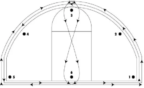
4. Unanticipated Sidestep Cutting Task: Subjects stand a distance equal to 50% of their body height from the front edge of a forceplate. A 17cm high hurdle was placed a distance equal to 25% of their body height from the front of the forceplate. The subject jumped with both legs over the hurdle. As they jumped, they cued the unanticipated task cueing system to provide the direction of the cut (sidestep=cut to contralateral side of dominant leg, crossover=cut to ipsilateral side of dominant leg). They land on their dominant leg (defined as the leg used to kick a ball for maximum distance) and cut in the cued direction.
5. Initial Contact: The first time point during each trial where the vertical ground reaction force recorded by the forceplate registers over 10N.
6. Toe-off: The first time point after Initial Contact where the vertical ground reaction force recorded by the forceplate registers less than 10N.
8
8. Segment Angular Position: The instantaneous angular position of the segment of interest relative to the world horizontal axis, expressed in degrees (°).
9. Segment Angular Velocity: The instantaneous angular velocity of the segment of interest, calculated using the change in angular position across 5 points divided by the change in time:
where X= segment angular position, and y= time (in seconds). Expressed as degrees per second (°·s-1). Five data points were used as pilot testing revealed that a longer window (10 points) over-smoothed the velocity data. Stance phases are typically less than 100 data points in length, thus the 5 second window provided accurate data. 10.Phase Portrait: A graphical representation of the current state of a dynamic system,
produced by plotting a segment’s angular position (x-axis) versus its angular velocity (y-axis) 36.
11.Phase Angle: The angle () formed between the radius and the horizontal (x) axis, when the Cartesian (x,y) coordinates of the phase portrait are transformed into polar coordinates (radius, ) 36.
12.Relative Phase: Representation of the interaction and coordination between two segments. Calculated as relative phase = distal segment – proximal segment, where relative phase is
the relative phase angle, distal segment is the phase angle of the distal segment, and proximal segment is the phase angle of the proximal segment. Calculated for each time
point during the stance phase 36.
9
while lower values indicate a more in-phase relationship. Calculated as the average of the sum of the absolute values of each relative phase angle, 36
14.Deviation Phase: A single term to quantify the stability of the organization of the neuromuscular system. High values indicate a highly variable and unstable
coordination pattern, while low values indicate a stable and low-variability pattern. Calculated as the average of the standard deviations of the ensemble relative phase curve points 36,
15.Knee extension moment: The combined contribution of the soft tissue surrounding the knee joint producing a moment in the direction of knee extension.
16.Knee valgus moment: The combined contribution of the soft tissue surrounding the knee joint producing a moment in the direction of knee valgus.
17.Anterior tibial shear force: The net force applied in the anterior direction at the Tibiofemoral joint, causing translation of the tibia anteriorly relative to the femur.
Limitations/assumptions
1. Segment kinematics calculated from the motion analysis system and biomechanical software were accurate and reliable.
2. Subjects performed the unanticipated sidestep cutting task in the lab in the same way they would perform the task on the field of play.
10
4. The effects of the fatigue protocol lasted long enough to complete the post-fatigue and post-feedback testing.
5. Subjects accurately followed directions in the feedback group.
Delimitations
1. 61 subjects (31 men and 30 women) were recruited from the local university population.
2. All subjects were between 18-30 years of age.
3. All subjects were healthy with no history of lower extremity or lumbar spine surgery in the past year, no history of knee surgery, and no history of lower extremity injury in the past 6 months.
4. All subjects were current participants in the University of North Carolina club sports teams (soccer, lacrosse, basketball, volleyball, or handball).
5. Segment kinematic data were collected from the trunk, thigh, shank and foot using an infrared video camera motion capture system.
6. Ground reaction force data were collected using a conductive forceplate.
Statement of Problem
11
research in sports medicine. This research project provides critical insights on how
neuromuscular fatigue, a condition commonly associated with athletic performance, impacts some of the accepted risk factors for ACL injury. The additional information provided by the use of a verbal feedback intervention may give clinicians a new method of correcting
movement patterns, and potentially preventing injury, in a quick and simple manner.
Ultimately, this research project aims to further the understanding of how the lower extremity responds to fatigue and feedback, and how these changes may be used to prevent injury and protect the long-term joint health of active individuals.
Independent Variables
1. Fatigue (pre-fatigue vs. post-fatigue) 2. Gender (men vs. women)
3. Feedback (feedback vs. non-feedback)
Dependent Variables
1. Mean Absolute Relative Phase (MARP) for the following segment pairs: a. Foot-shank sagittal plane
b. Foot-shank frontal plane c. Shank-thigh sagittal plane d. Shank-thigh frontal plane e. Thigh-trunk sagittal plane f. Thigh-trunk frontal plane
12 a. Foot-shank sagittal plane
b. Foot-shank frontal plane c. Shank-thigh sagittal plane d. Shank-thigh frontal plane e. Thigh-trunk sagittal plane f. Thigh-trunk frontal plane 3. Selected knee kinetic measures
a. Peak knee extension moment b. Peak knee valgus moment
c. Peak Anterior Tibial Shear Force d. Peak Vertical Ground Reaction Force
Research Questions
1. How does a functional fatigue protocol alter the coordination, variability, and kinetics of the lower extremity during the stance phase of an unanticipated sidestep cutting task in a healthy, athletic population?
a. Compare the Mean Absolute Relative Phase (MARP) values pre- and post-fatigue for the following segment pairs: foot-shank sagittal plane, foot-shank frontal plane, shank-thigh sagittal plane, shank-thigh frontal plane, thigh-trunk sagittal plane, thigh-trunk frontal plane.
13
shank-thigh sagittal plane, shank-thigh frontal plane, thigh-trunk sagittal plane, thigh-trunk frontal plane.
c. Compare the peak knee extension moment, knee valgus moment, peak anterior tibial shear force, and peak vertical ground reaction force, during the first 40% of the stance phase pre- and post-fatigue.
2. How does an acute intervention (verbal feedback) affect the post-fatigue
coordination, variability, and kinetics of the lower extremity during the stance phase of an unanticipated sidestep cutting task in a healthy, athletic population?
a. Compare the pre- and post-feedback Mean Absolute Relative Phase (MARP) values between the feedback and non-feedback groups for the following segment pairs: foot-shank sagittal plane, foot-shank frontal plane, shank-thigh sagittal plane, shank-thigh frontal plane, thigh-trunk sagittal plane, thigh-trunk frontal plane.
b. Compare the pre- and post-feedback Deviation Phase (DP) values between the feedback and non-feedback groups for the following segment pairs: foot-shank sagittal plane, foot-foot-shank frontal plane, foot-shank-thigh sagittal plane, shank-thigh frontal plane, thigh-trunk sagittal plane, thigh-trunk frontal plane. c. Compare the pre- and post-feedback peak knee extension moment, knee
14
3. Do men and women exhibit different lower extremity coordination, variability, and kinetics pre- and post-fatigue during the stance phase of an unanticipated sidestep cutting task?
a. Compare the pre-fatigue Mean Absolute Relative Phase (MARP) and Deviation Phase (DP) values between men and women for the following segment pairs: foot-shank sagittal plane, foot-shank frontal plane, shank-thigh sagittal plane, shank-thigh frontal plane, trunk sagittal plane, and thigh-trunk frontal plane.
b. Compare the pre-fatigue peak knee extension moment, knee valgus moment, peak anterior tibial shear force, and peak vertical ground reaction force, during the first 40% of the stance phase between men and women. c. Compare the post-fatigue Mean Absolute Relative Phase (MARP) and
Deviation Phase (DP) values between men and women for the following segment pairs: foot-shank sagittal plane, foot-shank frontal plane, shank-thigh sagittal plane, shank-thigh frontal plane, trunk sagittal plane, and thigh-trunk frontal plane.
d. Compare the post-fatigue peak knee extension moment, knee valgus moment, peak anterior tibial shear force, and peak vertical ground reaction force, during the first 40% of the stance phase between men and women.
Hypotheses
15
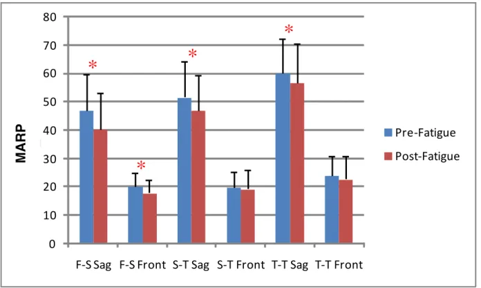
a. Mean Absolute Relative Phase (MARP) values will become more in-phase (decrease in value) in the sagittal plane, and more out-of-phase (increase in value) in the frontal plane, post-fatigue.
b. Deviation Phase (DP) values will increase post-fatigue.
c. Peak knee extension moment, knee valgus moment, peak anterior tibial shear force, and peak vertical ground reaction force during the first 40% of the stance will increase post-fatigue.
2. An acute intervention (verbal feedback) will affect the post-fatigue coordination, variability, and kinetics of the lower extremity during the stance phase of a sidestep cutting task in the following ways:
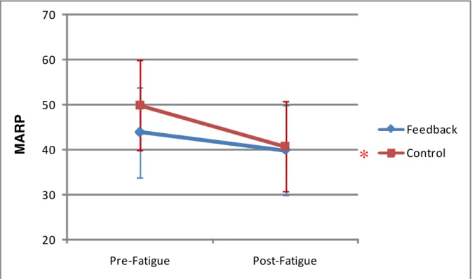
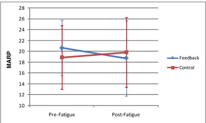
a. Post-feedback Mean Absolute Relative Phase (MARP) values will return to pre-fatigue values in the feedback group, but will remain at post-fatigue values in the non-feedback group.
b. Post-feedback Deviation Phase (DP) values will return to pre-fatigue values in the feedback group, but will remain at post-fatigue values in the non-feedback group.
c. Post-feedback peak knee extension moment, knee valgus moment, peak anterior tibial shear force, and peak vertical ground reaction force during the first 40% of the stance phase will return to pre-fatigue values in the feedback group, but will remain at post-fatigue values in the non-feedback group. 3. Men and women will exhibit different lower extremity coordination, variability, and
16
a. Pre-fatigue Mean Absolute Relative Phase (MARP) values in women will be more in-phase (decrease in value) in the sagittal plane, and more out-of-phase (increase in value) in the frontal plane, when compared to men.
b. Pre-fatigue Deviation Phase (DP) values will be higher in women than in men. c. Pre-fatigue peak knee extension moment, knee valgus moment, peak anterior
tibial shear force, and peak vertical ground reaction force during the first 40% of the stance phase will be higher in women than in men.
d. Post-fatigue Mean Absolute Relative Phase (MARP) and Deviation Phase (DP) values will change more in women than in men.
CHAPTER 2
Injuries to the ACL are a costly and debilitating injury. The mechanisms and potential risk factors for this injury have been extensively studied, yet we still do not have enough understanding about these factors to effectively prevent injury. One of the primary limitations of most ACL-related research is the performance of movements in controlled, laboratory environments that may not mimic the competitive environments where injuries occur. There has been little research that examines how perturbations in the environment, such as muscular fatigue or inability to anticipate movement direction, may alter proposed risk factors for injury. Additionally, there has been no research that has examined the potential role for movement coordination and variability to explain the differences between genders, or how different external constraints will change movement patterns. The
overarching purpose of this study was to examine these factors in a collegiate athletic population, in order to better understand how proposed risk factors may change due to fatigue or anticipation.
This review will focus on four primary areas. First, an introduction to the
18
change movement patterns and kinetics in order to prevent injury. The current concepts in each area are discussed, as well as the gaps in the literature that this research addresses. The final paragraph will summarize the information in this review, and serve as the rationale behind the methods described in chapter 3.
Section One: Injury Epidemiology and Kinetic Risk Factors
Injuries to the Anterior Cruciate Ligament (ACL) are a costly and debilitating injury to both competitive and recreational athletes. Approximately 112,500 ACL injuries occur every year in the United States, which places a substantial burden on the healthcare system. These athletes can expect to lose 6-9 months of competition, and many athletes decide to stop participation in their sport altogether. However, the greatest concern may be the long-term joint health for these individuals. Several studies have confirmed that joint injury, and specifically ACL injury, may be a leading cause of osteoarthritis (OA). These concerns have led researchers and clinicians to study the factors that may increase the risk of suffering ACL injury, as well as methods of preventing this injury in high-risk groups. This section will focus on the factors that are associated with ACL injury, specifically the kinetic variables that have been suggested to be the most critical in the development of non-contact ACL injuries.
Epidemiology of ACL Injury
19
in persons aged 15-25.52 Typical recovery time from surgery is 6-9 months, dependent on the presence of other joint injury (meniscal damage, concomitant damage to other ligaments, etc) and the type of rehabilitation program employed.53 While surgical procedures have improved significantly in the past 10 years, many athletes decide to cease participation in the sport where the injury occurred. Myklebust and Bahr report that a significant majority of athletes can return to sport within the first year of the injury (65-88%).5 However, in their review of the literature, Myklebust and Bahr also found that athletes who have suffered an ACL injury tend to retire from sports participation, participate in lower-risk activities, or play at lower competitive levels that prior to the injury at a higher rate than non-injured athletes.5 In a separate study, Roos found that 70% of ACL injured athletes had stopped participation in soccer within 3 years of the injury, compared to 20% of a control group. 4 While research is still largely retrospective, it does appear that ACL injury may lead to a significant decline in athletic participation.
20
research in this field has focused on non-contact ACL injuries, and the factors that are associated with this injury.
Participation in certain sports or activities can increase the risk of suffering non-contact ACL injury. Recent studies published from the National Collegiate Athletic Association’s (NCAA) Injury Surveillance System (ISS) have found that women’s gymnastics, soccer, basketball, and lacrosse, and men’s football, are the sports associated with the highest risk of ACL injury.55-58 All of these sports involve quick changes of direction, cutting and/or pivoting movements, and landing from jumps, motions that have been associated with non-contact ACL injury in the literature.54, 59-61 While the specific mechanism of injury for ACL injury is still not clearly understood, it is apparent that
performing cutting, pivoting, or landing activities places an athlete at higher risk of suffering ACL injury.
Perhaps the factor that has received the greatest amount of attention from researchers and clinicians is gender. Numerous studies have demonstrated that females in certain sports are at higher risk of non-contact ACL injury than their male counterparts.55, 56, 58, 62, 63 A systematic review published in 2007 found that females were at 1.5-4.3 times the risk of non-contact ACL injury than their male counterparts.3 Typically, researchers have found that soccer and basketball demonstrate the greatest gender disparity in ACL injury rates.3, 55, 63 These statistics have lead to a considerable body of literature comparing proposed risk factors between genders, which has formed the theoretical basis for many injury prevention programs.
21
osteoarthritis (OA). Studies by several researchers have demonstrated that injury to the knee joint is one of the most significant predictors of knee OA. 6, 7, 9, 64 Specifically, the study by Thelin found that a history of knee injury explained why participation in sport has been linked to the development of knee OA. A significant relationship existed for sport participation (specifically, soccer and ice hockey) prior to any adjustment for other confounding variables, but when adjusted for history of knee injury the relationship disappeared. 9 The study by Gelber demonstrated prospectively that knee injury during adolescence and young adulthood substantially increased the risk of OA (relative risk = 5.17, 95% CI: 3.07, 8.71). 6 This trend towards an increased risk of knee OA after knee injury is also apparent when ACL injury is considered. Salmon found that 79% of patients had radiographic changes within 13 years of surgical reconstruction, and that 50% of patients with an isolated ACL injury (no meniscus damage) had signs of the development of OA.8 These results are similar to other reports using different surgical techniques to repair the ACL.65, 66 These studies all demonstrate that a history of knee injury, and specifically ACL injury, lead to a substantial increase in the risk of OA development. Thus, prevention of these injuries is paramount in the prevention of future knee joint pathology, particularly for young individuals.
Suggested Risk Factors for Non-Contact ACL Injury
22
recently, prospective and computer modeling studies have been used to confirm some of these proposed risk factors, independent of gender. However, despite the substantial body of research that has been accumulated, the precise mechanism and most predictive risk factors for ACL injury remain elusive. Researchers are continuing to develop research
methodologies and study designs that can more definitively study the factors that lead to ACL injury in athletes.
Environmental Risk Factors
The study of risk factors for ACL injury began with footwear design in the late 1960’s and 1970’s. The development of artificial playing surfaces and new cleat designs coincided with a marked increase in the number of non-contact injuries, particularly ACL injuries. Researchers began to study how the external environment, specifically the playing surface and the footwear design, influenced injury rates. Torg and Quendenfeld provided the first evidence that footwear may influence injury rates.69, 70 In these studies, American football players who wore footwear with a lower release coefficient had substantially lower knee injury rates than players who wore traditional football cleats. This lower release coefficient essentially prevented excessive fixation of the foot with the ground, which is associated with increased torques and forces transmitted to the lower extremity of the
23
The influence of playing surface and other environmental factors (precipitation, ground hardness, temperature) has also received attention from researchers in the past. Orchard and colleagues in Australia have published several studies demonstrating higher injury rates in general, and ACL injury rates in particular, when athletes play on hard and dry surfaces.76-78 Orchard has also found that knee injury rates in American football players decrease when the ambient temperature was colder.78 The authors all conclude that the external playing environment can significantly alter the traction developed between the shoe and the playing surface, and that fields with lower traction development are beneficial for the prevention of injuries.
Anatomic Risk Factors
Anatomical features of the lower extremity are also believed to play a role in the development of non-contact ACL injuries. In one of the first prospective studies on ACL injuries, Uhorchak and colleagues found that a narrow intercondylar notch was predictive of ACL injury in a cohort of United States Military Academy cadets.79 The authors
24
tibial plateau) and increased risk of ACL injury.85, 86 Both of these factors require additional study and prospective confirmation, but show promise as possible risk factors for injury.
Ligamentous laxity and anatomic alignment of the lower extremity has been
extensively studied as a potential risk factor for ACL injury. Uhorchak and colleagues found that generalized joint laxity was predictive of noncontact ACL injury in their prospective study of military cadets.79 Laxity of the knee joint, specifically in the anterior direction, has also been suggested as a factor for ACL injury. Uhorchak and colleagues found that anterior knee laxity predicted ACL injury in his prospective study, while Woodford-Rogers
retrospectively found that anterior knee laxity could predict status as ACL injured in high school athletes.79, 87 However, both general ligament laxity and anterior knee laxity require substantial further study to confirm their role as risk factors for injury. Additionally, anatomic alignment variables such as genu recurvatum, genu valgum, pronation at the subtalar joint, excessive Q-angle, pelvic tilt, tibial torsion, and femoral anteversion have all been suggested as possible reasons for the female predisposition towards noncontact ACL injuries.87-90 While some studies have shown a possible link between these alignment variables and injury risk, the nature of the relationship is still unclear. To this point no prospective studies have found a significant link between lower extremity alignment and ACL injury risk.
Hormonal Risk Factors
25
studies have not found a definitive link between a specific phase of the menstrual cycle (which is correlated to certain hormone concentrations) and increased likelihood of ACL injury.74, 95-98 It does appear that fluctuations in the concentration of female hormones may play a role in the laxity of the knee joint, and that this relationship is highly variable between female subjects.99-101 Researchers have also suggested that the changes in serum hormone concentrations may have a delayed action on ligamentous properties, which may help explain the equivocal results from prior studies.102 Currently, while most researchers agree that hormones likely play a role in ACL injury in female athletes, the precise mechanism behind this effect is unclear.
Biomechanical & Neuromuscular Risk Factors
The influence of lower extremity biomechanics and neuromuscular control on
noncontact ACL injury risk is one of the most widely studied phenomena in sports medicine. A substantial body of literature has been published, examining which movements, forces, moments, and neuromuscular functions are related to ACL injury. Most of these studies have used a comparison of male and female athletes in order to draw conclusions about noncontact ACL injury risk. In general, females have been found to display less knee and hip flexion, more knee valgus and transverse plane movement, greater knee extension moments, greater knee valgus (or adduction) moments, higher anterior tibial shear forces, greater quadriceps muscle activation (particularly when compared to hamstring activation) during landing or cutting maneuvers, and lower hamstring muscular strength compared to males.17-27, 103-111 While there is a general consensus that these factors likely play a role in the development of ACL injury, it is unclear from these gender-comparison studies exactly how each
26
Sagittal plane biomechanical and neuromuscular factors may provide the most direct loading on the ACL. A great deal of cadaveric research has confirmed that the most direct way of inducing strain on the ACL is by applying a linear force in the anterior direction, causing anterior translation of the tibia on the femur.14, 15, 31, 75, 112 This force, often referred to as anterior tibial shear force (ATSF), is believed to be caused in vivo by a combination of external ground reaction forces as well as quadriceps muscle force, particularly when the knee is near full extension.28, 31, 33, 113 When the knee is between 0-20° of flexion, the angle formed between the patellar tendon and the shaft of the tibia is at its highest value. This angle, called the patellar tendon-tibial shaft angle, is a critical determinant of the amount of quadriceps muscle force that is directed anteriorly.33, 113 At high patellar tendon-tibial shaft angles, the force the quadriceps produces will create a significant anteriorly-directed force at the tibia which will draw the tibia forward relative to the distal femur. Recent studies have demonstrated that the quadriceps is capable of inducing significant ATSF upon the knee joint, and that aggressive contraction of the quadriceps is capable of rupturing the ACL in cadaver models.31, 33 Sell and colleagues examined the impact of various biomechanical and neuromuscular factors on the development of ATSF, and determined that quadriceps EMG activity was a significant predictor of ATSF during a landing task,32 These studies all support the theory that ATSF is strongly influenced by quadriceps muscle force, specifically when the knee is near full extension.
27
ground reaction forces in the 3 cardinal directions (vertical, anterior-posterior, and medial-lateral) to calculate the transfer of moment and force to the most proximal joint to the floor.114 These moments are then used to calculate the moments and forces in the next proximal joint, continuing in this fashion up the kinetic chain. Thus, the application of
ground reaction forces will have a strong influence upon the kinetics of the joints in the lower extremity. Recent studies by Sell and colleagues, as well as Yu and colleagues, have
demonstrated strong correlations between posterior ground reaction force and ATSF.28, 32 The study by Yu demonstrated a very high correlation (r=0.85) between peak posterior ground reaction force and ATSF, and a smaller but significant correlation (r=0.51) between peak vertical ground reaction force and ATSF during jump landing maneuvers.28 Sell examined several kinematic and kinetic variables as predictors of ATSF, and found that peak posterior ground reaction force was a significant predictor of ATSF and had a significant correlation to ATSF (r=-0.276). These studies highlight the importance of externally applied ground
reaction forces when studying the kinetics of the lower extremity during athletic movements. While ground reaction forces have a strong relationship to ATSF on their own, they may exert greater influence on the development of moment at the knee joint, which has the greatest influence on ATSF found in the literature. The study by Yu reported that both peak posterior ground reaction force (r=0.86) and peak vertical ground reaction force (r=0.57) had significant correlations to peak knee extension moment, which represents the internal
28
simultaneously during the landing phase of the jump-landing task.28 Sell found that the knee flexion moment (measured at the time of peak posterior ground reaction force) was the strongest predictor of ATSF and had the highest pairwise correlation (r=-0.8986) of any variable tested.32 The use of knee flexion moment reflects an external moment applied to the knee joint. Researchers often report either external or internal moments, as both represent the demands placed upon a joint (internal moments represent the needed soft tissue requirements in order to respond to external moments).114 Thus, while the type of moment represented in these two studies is different, the conclusion is the same. The sagittal plane moment at the knee joint is strongly related to the development of ATSF, and it appears that this moment is the result of posterior ground reaction force and quadriceps muscle force acting at the tibiofemoral joint.
29
ATSF alone. Considerable debate exists regarding this conclusion. Several studies have demonstrated differences in ATSF between genders, particularly during tasks that generate a high level of posterior ground reaction force, such as a jump-landing.18, 28, 42, 118 These researchers believe that this gender disparity clearly highlights the importance of ATSF as a risk factor for ACL injury. However, the highest amount of ATSF measured in these inverse dynamics studies was 0.79 multiples of body weight, which is still below the theoretical ACL injury threshold of 2000N.117 Thus, most researchers have examined how other motions in the frontal and/or transverse plane, may influence ACL strain and potentially lead to ACL injury.
Cadaver studies provided the first evidence that combined, multi-planar loading led to higher ACL strain than any single direction alone. Combining ATSF with valgus or varus moment dramatically increased ACL strain in several cadaver studies.14, 15, 119 Researchers also demonstrated that a combination of ATSF and internal rotation moment elevated ACL strain.14, 15, 120 Using computer modeling, Chaudhari and Andriacchi demonstrated that the threshold for injury to the ACL decreases as the knee moves into either valgus or varus, indicating that lower levels of ATSF would be needed to cause injury to the ACL.16 The results of these studies demonstrated that forces and moments applied in the frontal and transverse planes may play a critical role in the development of ACL injury. Between-gender research, using females to represent a high-risk group of subjects, concluded that females display greater knee valgus moments during athletic movements than males.17, 18, 21, 24, 26, 27, 41,
105, 108
30
However, the extent to which valgus moment influences ACL injury risk is still unclear from these cross-sectional studies.
Perhaps the best evidence that frontal plane moments are associated with increased risk of ACL injury comes from a prospective study by Hewett and colleagues. This study prospectively collected biomechanical data from 205 female basketball, soccer, and
volleyball athletes. These athletes were then followed by study personnel over the course of two competitive seasons. Females that suffered a non-contact ACL injury were then
compared to the remaining healthy athletes to determine if any biomechanical measures were different between groups. Females that injured their ACL had greater knee valgus angles at both contact with the ground (8.4° more than uninjured) and maximum value (7.6° more than uninjured). They also demonstrated less maximum knee flexion (10.5° less) than uninjured athletes. However, the greatest difference was detected in knee abduction moment. Injured females had 2.5 times the knee abduction moment than their uninjured counterparts. When a logistic regression analysis was performed, knee abduction moment was a significant
predictor of ACL injury status, and demonstrated a sensitivity of 78% and a specificity of 73%.34 The findings from this prospective study provide the strongest evidence to date that knee valgus (or abduction) moment is significantly related to ACL injury, and may be a critical risk factor for female athletes.
31
hip abduction) in females.28, 108, 121-123 However, other studies have shown no difference in hip joint kinematics or kinetics.19, 24 While evidence is building that the hip may play a role in ACL injury, the precise mechanism is unclear. Several researchers believe that a stiff landing style, which is associated with both decreased knee and hip flexion angles, is responsible for generating large joint resultant forces that may be responsible for ACL injury.28, 108, 122, 123 Associations between frontal plane hip movement, specifically increased hip adduction angle, and knee valgus angle have been found in previous literature.34, 121 Thus, hip motion may influence both knee valgus angle and moment, both of which are associated with increased risk of injury. Research is on-going on how hip motion may influence the risk of injury to the ACL.
Less research is available about how motion at the ankle may influence knee joint loading. Between-gender research has demonstrated that ankle excursions are greater in females than males, in both the frontal and sagittal plane.19, 20, 121 Decker demonstrated that females have a greater reliance on an “ankle” energy absorption strategy, utilizing ankle motion and musculature to a greater degree than males.19 This may not be the most
advantageous strategy to utilize, as the ankle musculature has limited ability to absorb energy when compared to the larger musculature of the knee and hip joints.19 While little research has been performed on how ankle and foot mechanics relate to ACL injury risk, it does appear to influence the development of motion and forces at the knee.
The trunk has received recent attention as another body segment that may have an influence on knee joint motion and moment. Several studies have demonstrated that
32
Knee valgus moment increases significantly when the trunk leans to the opposite side of a sidestep cut. In addition, rotating the trunk during the cutting maneuver increases the internal rotation moment at the knee.124 A study by Gupta also demonstrated an correlation between trunk obliquity and knee valgus moment, where increased frontal plane trunk movement was associated with higher knee valgus moment.125 Thus, isolated changes in trunk movement can have a significant impact on moments at the knee that are associated with increased risk of ACL injury. Blackburn and Padua studied how isolated trunk flexion impacts knee kinematics, and found that increasing trunk flexion during a landing task creates a greater peak knee flexion angle when compared to a normal, preferred landing style.126 While only a few studies exist that examine how trunk movement influence knee joint loading, these studies demonstrate that a link does exist between trunk position and lower extremity forces and moments.
Two recent studies published by Zazulak and colleagues concluded that poor neuromuscular control over the trunk is associated with knee injury. In one study,
33
analyzing trunk angular displacement after a sudden force release into flexion, extension, and lateral bending. Logistic regression revealed that increased displacement was predictive of all knee injuries, as well as ligamentous or meniscal injuries, and ACL injuries specifically.29 Lateral displacement was the strongest predictor of injury in athletes, with odds ratios of 1.9 (all knee injuries), 2.0 (ligamentous/meniscal injuries), and 2.2 (ACL injuries). These two studies suggest that the ability to control the movement of the trunk, particularly in the lateral direction, may be critical in the prevention of knee injuries. While this study was performed under relatively artificial conditions (highly controlled laboratory testing of trunk
proprioception and control), the nature of the results do suggest that researchers should incorporate an analysis of trunk movement into injury risk factor studies.
34
research in these areas is still progressing. Thus, future research that adds to the knowledge base on the kinetics of the lower extremity during athletic tasks, as well as how movement of other body segments influences the potential for ACL injury, is warranted.
This research project was designed to address these key kinetic variables that have been suggested to directly produce strain on the ACL, thus leading to injury. This project was designed to investigate how these variables respond to different external conditions which likely occur during game-play (fatigue and unanticipated movements). The sidestep cutting task is commonly performed in athletics and is also associated with a high risk of injury to the ACL, which makes it well-suited to study how different perturbations affect joint kinetics during athletic movements associated with ACL injury. The results of this study provide needed insight on how these selected kinetic variables may change, and potentially increase strain on the ACL, during an athletic task under various external constraints designed to mimic actual game-play.
Section Two: Muscular Fatigue
35
project aims to answer some of these questions, and provide a more clearly researched fatigue protocol to elicit fatigue.
Muscular Fatigue
Fatigue is a general term used to describe the experience of muscular weakness, loss of strength, or psychological exhaustion. Because this term can be interpreted in several ways, many researchers prefer that the term “fatigue” be more precisely defined.
Experimentally, “muscular fatigue” refers to the loss of maximum force applied by a
muscle.127 Researchers also prefer that the perceived endpoint of performance, or the point of maximal exertion during physical activity, be referred to as “exhaustion” and not fatigue.127 The term “muscular fatigue” does not apply to the loss of muscle force capability or
perceived weakness due to disease or another medical condition. Thus, the definition of “muscular fatigue” should refer to a loss of peak force production by a muscle as a result of physical exertion.128
36
in muscular function distal to the neuromuscular junction.128 Different types of activities will produce different amounts of both central and peripheral fatigue. Understanding which “location” is most affected by a given exercise or type of physical activity can help researchers understand not only the physiological processes that may be involved in the development of muscular fatigue, but also how to target interventions to reduce fatigue-related impairments in the neuromuscular system.
Several authors have suggested that central fatigue is unlikely to occur during most types of athletic activity.129-131 Kirkendall hypothesized that under normal conditions, when a subject is sufficiently motivated, central fatigue is unlikely to occur.130 Several more recent studies have found that fatigue-related loss of muscular strength could be largely explained by peripheral changes in the muscle cell, specifically in the excitation-contraction
coupling.129, 131 However, the Lattier study examined uphill running on a treadmill, which eliminates the eccentric contribution of the leg musculature during activity. While this prevents muscular damage from contaminating the results, it also fails to simulate athletic activity which will always employ some eccentric muscle activity. The Skof study used a run at a set distance and intensity (6k at anaerobic threshold) in highly trained athletes. This may have prevented the athlete from reaching a true “exhaustion”, which may have prevented the authors from seeing central contributions to the muscular fatigue observed from this protocol. Finally, both of these studies utilized continuous activities, which do not reflect the
37
Evidence of central fatigue during full-body functional fatigue protocols has been found by several authors. A recent study by Theurel compared variable intensity exercise to a continuous bout of exercise.134 The authors found that exercising at a variable intensity induces significantly greater central fatigue, due to a substantial decrease in voluntary activation level post-fatigue, when compared to continuous cycling. This result may help explain why some prior research has failed to find significant central fatigue during
continuous running. Additionally, this study found that perceived exertion, blood lactate, and heart rate, were all higher in the variable exercise protocol, despite controlling for the total average power output. Thus, this fatigue protocol appears to produce greater physiological responses, as well as a higher reported experience of fatigue. This study highlights not only the presence of central fatigue during intermittent exercise, but that physiological and perceptual changes are also different following this type of exercise.
The physiological changes that occur during fatiguing bouts of exercise may play a crucial role in the development of central fatigue. Thomas and colleagues 135 found that a progressive exhaustive cycling protocol produced evidence of peripheral fatigue,
38
The prospect that peripheral fatigue may influence the development of central fatigue has been suggested by other researchers as well. Amann 136-138 and his colleagues have published several studies investigating how respiratory factors (oxygenation, fatigue of respiratory muscles) influence the development of fatigue during cycling. Amann concludes that feedback to the central nervous system from muscles experiencing peripheral fatigue is the major determinant of central motor drive. Both Amann and Dempsey 139, 140 suggest that the body tightly regulates the amount of peripheral fatigue that can occur during exercise by changing central drive in response to sensory feedback from muscles. When peripheral fatigue reaches a critical threshold, then the body responds by decreasing central motor drive to prevent muscles from continuing to full exhaustion. Thus, exhaustive exercise likely results in both peripheral and central fatigue, due to the interaction of these two mechanisms.
An additional way that athletic activities likely produce both peripheral and central fatigue is via modulation of spinal reflexes. In the Theurel study, the authors postulate that some of the central fatigue was due to inhibition of alpha motor neurons.134 This hypothesis is in accordance with conclusions drawn from fatigue studies using stretch-shortening cycle (SSC) exercise. SSC exercise involves the use of eccentric muscle contractions to augment the development of force during concentric contraction.141 Many researchers believe that most human activities involve SSC muscular activity, and these researchers advocate
39
may be responsible for this mechanism.141 Again, these discussions suggest that peripheral factors may play a role in the central fatigue developed during exercise, and support the notion that both central and peripheral fatigue occur during normal athletic activities.
Since the current research supports the hypothesis that central fatigue does occur during exercise, specifically exercise that simulates intermittent athletic activity, some important questions have been raised about how fatigue affects other tasks performed by the central nervous system. Of these, the possible role of fatiguing exercise on the ability to perform cognitively demanding tasks has received the most attention. Several studies have been performed that examine how fatiguing exercise influences reaction time, cognitive function, attentional focus, and other features of cognition. Tomporowski 142 performed an extensive review on how acute bouts of exercise may influence these factors, and concluded that there is no consensus on how exhaustive, high-intensity anaerobic-type exercise affects cognition. In this review, the author suggests that the studies that did show decreased cognitive functioning only found small, transitory effects. The only type of physical fatigue that produced consistent degradation in cognitive function were long, continuous bouts of physical activity that lead to dehydration or energy depletion (typically 2 hours or longer).142 This review suggests that further research that specifically defines “fatigue” (a definition that is often missing in these studies) and uses measures that are sensitive to smaller changes in function, may produce more consistent results.
40
of these two types of fatigue, and whether the central fatigue is due to spinal or supraspinal changes, remains unknown. Second, exhaustive high-intensity fatigue does not produce consistent changes in cognitive function, despite the evidence that central fatigue exists. Methodological constraints may play a large role in these findings, but there is currently no evidence that cognitive function is significantly altered after this type of fatigue. Thus, it appears that the central mechanisms that are affected by fatigue may be independent of cognitive function, so long as energy supply is not adversely affected. However, this area remains an intriguing area for further research, particularly in regard to decision-making during athletic activities (such as deciding a movement direction), which is a necessary cognitive skill for athletes.
Fatigue as a risk factor
Anecdotally, many clinical sports medicine staff will report a higher incidence of injury when a player is experiencing muscular fatigue. However, there is still no good scientific evidence to support this point. Most researchers will use recent epidemiologic studies to support the hypothesis that player fatigue plays a role in injury incidence. Gabbett, using injury surveillance in South African Rugby matches and practices, found that injuries are more likely to occur during the second half of matches, or in the latter states of
41
incidence of injury. Psychological factors may also play a significant role in injury
occurrence. Additionally, fatigue was not defined or measured in any of these studies, so it is unknown how fatigued these players were, or if the injured players had greater fatigue than non-injured players. Thus, while muscular fatigue may be an important risk factor for knee injuries, these initial studies can make only broad suggestions about this point.
Quantifying Muscle Fatigue during Athletic Activity
More recent studies have attempted to quantify the level of muscular fatigue that may occur during athletic activity. Two studies 132, 133 have used treadmill protocols that are based on the relative “average” workload and activity level of soccer field players during a game, while one study simulated soccer activity in a gymnasium.143 These protocols were both based on prior research that had quantified the type and intensity of activity performed by soccer players during a match.144, 145 Rahnama 133 used 13 male soccer players from the university setting to perform a 90-minute fatigue protocol with a “halftime” given to more accurately simulate an actual game. An isokinetic dynamometer was used to quantify concentric and eccentric hamstring and quadriceps peak torque pre-fatigue, at halftime, and at the conclusion of the fatigue protocol. The authors found that, at all testing speeds, both eccentric and concentric hamstring and quadriceps peak torque declined between the pre-test and the post-test at the end of the full protocol. Most of the tests also declined between the pre-test and halftime, and between halftime and the end of the protocol. Concentric
quadriceps torque dropped between 8.5-15.5% after the full fatigue protocol, while
42
fatigued more than the quadriceps.133 The authors concluded that both the quadriceps and hamstrings exhibited significant fatigue after a simulated soccer game.
Greig 132 also used a soccer-game simulation on a treadmill to study the effects of fatigue on muscular strength in ten male professional soccer players. Subjects performed 6-15 minute periods of activity (with a 6-15 minute halftime), with muscle strength tested after each 15 minute session. Isokinetic strength was tested concentrically for the knee extensors, and concentrically and eccentrically for the knee flexors at three testing speeds. The authors reported that concentric quadriceps and hamstrings strength were maintained during the simulated game, whereas eccentric hamstrings torque decreased significantly at 180 and 300 °/s during the fatigue protocol. Peak torque decreased 18.8% from pre-test to the end of the protocol at 180 °/s, and 24% at 300 °/s. Dynamic hamstring:quadriceps strength ratio
(eccentric Hamstrings: concentric Quadriceps) also tended to decrease as the fatigue protocol continued.132 The authors concluded that eccentric hamstring strength was particularly affected by a soccer simulation fatigue protocol, which may present a risk of injury to the ACL.
43
suggest that most intermittent, athletic-type fatigue protocols that simulate actual game play intensity result in significant muscular fatigue.
Impact of Fatigue on Knee Laxity
Since authors have reported increased risk of injury, and specifically knee ligament injury, during the latter stage of games, several researchers have attempted to study how fatigue may impact factors that are hypothesized to increase the risk of injury to the ACL. A prospective study by Uhorchak demonstrated that increased knee laxity, particularly in the anterior direction, is a risk factor for subsequent ACL injury.79 Thus, several studies have studied how muscular fatigue affects knee joint laxity. Both Wojtys and Rozzi 146, 147 used isokinetic protocols to induce quadriceps and hamstring muscular fatigue, performed until subjects were unable to sustain torque output of 50% (Wojtys) and 25% (Rozzi) of the peak pre-testing torque achieved. In the Wojtys study, anterior knee laxity increased significantly post-fatigue, an average of 32.5%. However, Rozzi failed to find any significant increase in anterior knee joint laxity, which may be due to the difference in cutoff torque output used to define fatigue.