INVESTIGATION OF AIRWAY ACCESS TECHNIQUES IN MEN’S LACROSSE WITH RELATION TO HELMET FIT
Alexis Ann Altier
A thesis submitted to the faculty of the University of North Carolina at Chapel Hill in partial fulfillment of the requirements for the degree of Masters of Arts in the Department of Exercise and Sport Science (Athletic Training).
Chapel Hill 2014
Approved by:
Meredith Petschauer
Kevin Guskiewicz
Barnett Frank
Ashley Littleton
ABSTRACT
ALEXIS ANN ALTIER: Investigation of Airway Access Techniques in Men’s Lacrosse with Relation to Helmet Fit
(Under the direction of Meredith Petschauer and Kevin Guskiewicz)
Objective: Determine effect of helmet fit (athletic trainer-AT vs. player-PF) and airway access technique (helmet removal-HR vs. facemask removal-FR) on cervical
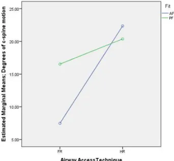
spine (C-spine) motion. Results: Interaction effect for integrated motion in frontal plane
(F1,17= 8.052, P=0.011) and peak displacement in sagittal (F1,17= 12.336, P=0.003) and
transverse planes (F1,17= 11.118, P= 0.004). Main effect of airway access technique in all
planes for peak displacement and integrated motion; HR resulted in more motion than
FR. Main effect of fit for transverse plane peak displacement and frontal plane integrated
motion; AF resulted in more motion than PF. Conclusion: These findings suggest an
increase in c-spine motion with HR compared to FR; HR is a faster method of
airway-access. FR is the current guideline for airway access technique, but HR should be
ACKNOWLEDGEMENTS
To my parents, brother, and fiancé who continually believe in me even when I fail
to believe in myself. Without your consistent support I am confident in the fact that I
TABLE OF CONTENTS
LIST OF TABLES ... ix
LIST OF FIGURES ... x
LIST OF ABBREVIATIONS ... xi
CHAPTER 1 ... 1
INTRODUCTION ... 1
VARIABLES ... 5
Independent ... 5
Dependent ... 5
RESEARCH QUESTIONS ... 6
HYPOTHESES ... 7
Alternate ... 7
Research ... 8
OPERATIONAL DEFINITIONS ... 8
ASSUMPTIONS ... 10
DELIMITATIONS ... 11
LIMITATIONS ... 11
CHAPTER 2 ... 12
INTRODUCTION ... 12
EPIDEMIOLOGY ... 13
Normal Anatomy ... 14
Pathological Anatomy ... 17
MECHANISM OF INJURY ... 19
GUIDELINES FOR SUSPECTED C-SPINE INJURIES ... 20
Management of Cervical Spine Injury ... 20
Men’s Lacrosse Airway Access Guidelines ... 21
Upon Reaching Hospital... 22
AIRWAY ACCESS TECHNIQUE ... 23
Facemask Removal With Cordless Screwdriver ... 23
Men’s Lacrosse as Compared to Football and Ice Hockey ... 23
DISSENSION REGARDING CURRENT GUIDELINES ... 25
Spinal Cord Involvement When Helmet is Removed ... 28
LACROSSE HELMET DESIGN ... 29
Helmet Fit ... 29
Helmet Choice ... 30
Difficulty in other helmet designs ... 31
MEASUREMENT OF C-SPINE MOTION ... 32
SIGNIFICANCE OF STUDY ... 33
CHAPTER 3 ... 35
METHODOLOGY ... 35
SUBJECTS ... 35
EQUIPMENT ... 36
PROTOCOL ... 36
DATA REDUCTION ... 41
CHAPTER 4 ... 43
OVERVIEW ... 43
MANUSCRIPT ... 44
METHODS ... 47
Participants ... 47
Equipment ... 47
Protocol ... 48
DATA REDUCTION ... 50
STATISTICAL ANALYSIS ... 50
RESULTS ... 51
Change in Peak Displacement and Integrated Motion ... 51
Time to Completion ... 56
Helmet Fit ... 57
DISCUSSION ... 57
Change in Peak Displacement and Integrated Motion ... 58
Time to Completion ... 60
Limitations... 62
Future Research ... 62
Conclusion... 63
LIST OF TABLES
1. Table 3.1 Counterbalance Design of Data Collection………...37
2. Table 3.2 Visual Representation of ANOVA Study Design………..42
3. Table 3.3 Visual Representation of time t-test comparison………...42
LIST OF FIGURES
1. Figure 3.1 Cascade® R Lacrosse Helmet and HardTail SPRfit™ Techonology...38
2. Figure 3.2 Helmet Removal …..………40
3. Figure 3.3 Facemask Removal ………..40
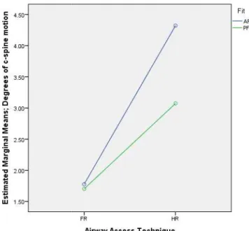
4. Figure 4.1 Interaction effect in the sagittal plane for change in
peak displacement …………..………...54
5. Figure 4.2 Interaction effect in the transverse plane for change in peak
displacement ………...………...…………...54
6. Figure 4.3 Interaction effect in the frontal plane for integrated motion……...….55
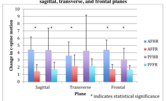
7. Figure 4.4 Change in peak displacement for all four testing conditions
in the sagittal, transverse, and frontal planes ………...…………...………55
8. Figure 4.5 Integrated motion in each plane for all four testing conditions
in the sagittal, transverse, and frontal planes ………..…………..56
9. Figure 4.6 Time to Completion………..57
LIST OF ABBREVIATIONS AT Certified athletic trainer
C-spine Cervical spine
AF AT fit helmet condition
PF Player fit helmet condition
HR Helmet Removal
CHAPTER 1 INTRODUCTION
Lacrosse is a rapidly growing sport in the United States, with 41% expansion in
collegiate men’s lacrosse teams since 1988 (Dick, Romani, Agel, Case, & Marshall,
2007). An increase in participation represents a call for establishing evidence-based
practice regarding proper emergency management of a potential catastrophic event, such
as a head or neck injury, which can result in permanent disability or potential fatality as a
consequence of improper care. Due to specialized training and experience in equipment
removal in addition to nature of professional role; athletic trainers (AT) and other sports
medicine professionals remain as the primary party responsible for proper management
of potential cervical spine (c-spine) injuries in competitive athletic events. Unfortunately,
very few studies have examined emergency airway access procedures specific to men’s
lacrosse equipment, (Bradney & Bowman, 2013; Higgins, Tierney, Driban, Edell, &
Watkins, 2010; Petschauer, Schmitz, & Gill, 2010; Sherbondy, Hertel, & Sebastianelli,
2006; Waninger, Richards, Pan, Shay, & Shindle, 2001) with limited evidence regarding
best practice for proper removal technique to ensure athlete safety and c-spine integrity.
This is alarming due to the fact that since 1982, 22 catastrophic neck and spine injuries
have been recorded in high school men’s lacrosse and 13 catastrophic injuries have been
recorded in collegiate men’s lacrosse (Mueller, 2011). Thus, to ensure that future injuries
Men’s lacrosse is a fast paced, and high contact sport, (Diamond & Gale, 2001)
with 11.7% of recorded game injuries and 6.2% of recorded practice injuries involving
the head and neck from 1988 to 2004 (Dick et al., 2007). Improper handling of c-spine
injuries can result in outcomes as dire as spinal cord disruption such as transverse
myelopathy, which results in a loss of spinal function below the level of injury. Although
improvement of one spinal level above the lesion may be seen when primary swelling
subsides, subsequent loss of function is seldom reversed. Furthermore, spinal cord
injuries can transpire even when spinal cord continuity is maintained; hemorrhage or
ischemia can block impulse transmission, thus it is imperative that correct management
procedures are established to minimize potential for injury due to subsequent
mishandling of a c-spine injury (Bailes, Petschauer, Guskiewicz, & Marano, 2007).
To minimize additional hemorrhage or ischemia associated with c-spine injuries,
current guidelines are in place in regards to airway access and emergency management to
prevent excessive movement (Bailes et al., 2007). These guidelines are critical to
individuals who will be first to the scene of a catastrophic injury. US Lacrosse Sports
Science and Safety states that current guidelines are; “the helmet and shoulder pads of an
injured lacrosse athlete should be left in place until they can be removed in a controlled
environment” (Lacrosse Helmet Facemask/Chinguard Removal Hints for Certified
Athletic Trainers) based upon findings from Sherbondy, Hertel, and Sebastianelli’s 2006
study (Sherbondy et al., 2006). These guidelines have given rise to controversy for many
reasons. The first debate stems from the fact that often times the medical personnel most
sports medicine team immediately involved at the scene have a better understanding of
equipment removal procedures than emergency room clinicians who may have no
training in helmet removal methods (Banerjee, Palumbo, & Fadale, 2004b). The second
argument is due to that fact that the research upon which these guidelines are based only
found minimal movement in the area of occiput-C2 in a helmet removed airway access
situation and found no movement in the area of C2-C7, where most injuries occur
(Higgins et al., 2010). Additionally, previous research has revealed that airway access in
a men’s lacrosse athlete via helmet removal, while leaving shoulder pads in place, did not
affect space available for the spinal cord, (Higgins et al., 2010) which is critical in
avoiding tissue disruption. Lastly, it was revealed that a men’s lacrosse athlete laying
supine experiences an increase in cervical spine extension of 6 degrees as compared to an
athlete with no equipment on while placed in the neutral position (Sherbondy et al.,
2006).
Another concern regarding the current guidelines in place is the fact that
improperly fit men’s lacrosse helmets do not provide the adequate security needed for
airway access using an in-line stabilization of the c-spine with clinicians securing the
helmet (Petschauer et al., 2010; Sherbondy et al., 2006). Previous literature states that
equipment should be removed if the helmet and chin straps do not stabilize the head
securely such that immobilization does not also immobilize the head (Bailes et al., 2007;
Kleiner, 2003; Swartz et al., 2009). Evidence suggests that a large population of men’s
lacrosse athletes do not wear properly fitted helmets (Petschauer et al., 2010), thus the
clinical utility of the current guidelines established using properly fit helmets may be
helmets do not afford the same customizable fitting options seen in those of football
helmets, and even with a properly fitted men’s lacrosse helmet it has been observed that
the helmet does not effectively stabilize the athletes head to a spine board (Petschauer et
al., 2010). Furthermore, the same study reported that only limited prevention of neck
flexion and extension was afforded with the addition of extra padding provided to adjust
fit with no additional restrictions in the other planes of motion (Petschauer et al., 2010).
Petschauer et al.’s 2010 findings afford the notion that removing the helmet to access an
airway in an emergent situation may be the optimal technique to access a men’s lacrosse
player’s airway safely, resulting in the least c-spine motion, until a men’s lacrosse helmet
that properly stabilizes the head can be designed and proper fit can consistently be
ensured (Petschauer et al., 2010).
The issue of airway access in men’s lacrosse is consistently debated. While there
are guidelines in place, they do not go uncontested. Although the current guidelines
suggest that it is the least deleterious to leave the athlete in their helmet and simply
remove the facemask to gain airway access prior to reaching the controlled environment
of the hospital (Sherbondy et al., 2006), the concern that the head is not adequately
stabilized in the helmet despite the status of its fit gives rise to concern over establishing
practices to safely stabilize the athlete’s head and neck (Petschauer et al., 2010). In
addition, it has been observed that there is not adverse cervical extension in a helmet
removed condition with men’s lacrosse as seen in football due to the much less dramatic
thoracic elevation provided by the much slimmer men’s lacrosse shoulder pads as
compared to bulky football shoulder pads (Higgins et al., 2010). Lack of adequate in-line
athletic trainers and the sports medicine staff immediately involved at the scene of the
injury, in general, have more specialized training in equipment removal as compared to
emergency medical technicians or emergency room technicians lends the question: why
shouldn’t the helmet be removed on-site if there are qualified personnel available?
However, the effect of c-spine motion in the act of removing the helmet to access the
airway has not been researched (Higgins et al., 2010). Therefore the purpose of this study
is to evaluate the effects of helmet fit and equipment removal technique on c-spine
motion during airway access in collegiate men’s lacrosse players.
VARIABLES
Independent
1. Airway access technique
a. Facemask removal (FR)
b. Helmet removal (HR)
2. Fit
a. AT Fit (AF)
b. Player Fit (PF)
Dependent
1. Angular Motion
a. Head-to-thorax cervical rotation in the transverse plane
b. Head-to-thorax cervical flexion/extension in the sagittal plane
c. Head-to-thorax cervical lateral flexion in the frontal plane
1. Change in peak displacement
2. Integrated motion in Each Plane
2. Time to completion
RESEARCH QUESTIONS
RQ1: What is the interaction effect between helmet fit and airway access
technique on c-spine change in peak displacement during airway access in collegiate
men’s lacrosse players?
RQ1A: What is the effect of helmet fit on c-spine change in peak
displacement during airway access in collegiate men’s lacrosse players?
RQ1B: What is the effect of airway access technique on c-spine change in
peak displacement during airway access in collegiate men’s lacrosse players?
RQ2: What is the interaction effect between helmet fit and airway access
technique on total c-spine motion in each plane during airway access in collegiate men’s
lacrosse players?
RQ2A: What is the effect of helmet fit on total c-spine motion in each
plane during airway access in collegiate men’s lacrosse players?
RQ2B: What is the effect of airway access technique on total c-spine
motion in each plane during airway access in collegiate men’s lacrosse players?
HYPOTHESES
Alternate
AH1: There will be an interaction effect on c-spine motion involved in airway
access technique in player fit men’s lacrosse helmet as compared to airway access
technique in AT fit men’s lacrosse helmet.
AH1A: There will be less c-spine motion during airway access in a men’s
lacrosse athlete wearing a player fit men’s lacrosse helmet as compared to an AT
fit men’s lacrosse helmet.
AH1B: There will be a greater effect on c-spine motion during facemask
removal than during helmet removal.
AH2: There will be an interaction effect on total c-spine motion in each plane
involved in airway access technique in player fit men’s lacrosse helmet as compared to
airway access technique in AT fit men’s lacrosse helmet.
AH2a: There will be a smaller effect on total c-spine motion in each plane
during airway access in a men’s lacrosse athlete wearing a player fit men’s
lacrosse helmet as compared to an AT fit men’s lacrosse helmet.
AH2b: There will be a greater effect on total c-spine motion in each plane
during facemask removal than during helmet removal.
AH3: Time to completion will not be significantly different based upon airway
Research
RH1: There will be no interaction effect of helmet fit and equipment removal
technique on c-spine motion during airway access in collegiate men’s lacrosse players.
RH1A: There will be no effect of helmet fit on c-spine motion during
airway access in men’s lacrosse athletes.
RH1B: There will be no effect of helmet removal technique on c-spine
motion during airway access in men’s collegiate lacrosse athletes.
RH2: There will be no interaction effect of helmet fit and equipment removal
technique on total c-spine motion in each plane during airway access in collegiate men’s
lacrosse players.
RH2a: There will be no effect of helmet fit on total c-spine motion in each
plane during airway access in men’s lacrosse athletes.
RH2b: There will be no effect of helmet removal technique on total c-spine
motion in each plane during airway access in men’s collegiate lacrosse athletes.
RH3: Time to completion will be significantly shorter during the helmet removal
airway access technique.
OPERATIONAL DEFINITIONS
1. AT Fit (AF) Helmet- helmet that meets all of the following qualifications per
a. The back of helmet should be in uniform firm contact with the back of the
athlete’s head.
b. The skin of the athlete’s forehead should move with helmet when helmet is
moved anterior to posterior and side-to-side; helmet should not be able to slip
over head.
c. The helmet should not gap at athlete’s forehead when anterior force is applied
to occiput segment of helmet.
d. When pressure is applied anteromedially and posteromedially to either
parietal area of helmet, skin on the athlete’s forehead should move with
helmet and liner should bunch cheeks. The helmet should not slide towards
the athlete’s nose.
e. Clearance from end of the athlete’s nose to facemask should be at least 2-3
finger widths.
2. Player fit (PF) helmet- fit of helmet in which subject arrives wearing and wears
consistently at practice as well as in games
3. C-spine motion- degrees of motion of the head relative to the thorax, in the sagittal,
frontal, and transverse planes (Mihalik, Beard, Petschauer, Prentice, & Guskiewicz,
2008; Toler et al., 2010); measured by change in peak displacement and integrated
motion in each plane.
4. Helmet Removal- the act of in-line stabilization and two-person helmet removal with
towel placed under athlete’s head
5. Facemask removal- the act of in-line stabilization and facemask removal with a
6. Change in peak displacement- the absolute difference between maximum values of
rotation of sensor on left temple in one direction and rotation in the other as compared
to sensor on the sternum
7. Integrated motion in each plane- the absolute difference between maximum values of
rotation in one direction and rotation in the other using Simpson’s integration
normalized to time
8. Time to completion- time in which it takes to complete each airway access technique
trail based upon the following:
a. Each helmet removal trial will begin when AT secures head and will end
when the research assistant places towel under head after complete helmet
removal.
b. Each facemask removal trial began as soon as AT secured head in in-line
stabilization and ended when the facemask was placed on the ground next to
the subject.
ASSUMPTIONS
1. Flock of Birds is reliable and valid in modeling c-spine motion through analyzing
motion between the head and the thorax.
2. The Cascade® R is a widely used helmet.
3. The movement of the head relative to the thorax accurately represents cervical
motion.
4. The subjects will follow the instructions given.
5. The subjects and researchers will be consistent in conducting airway access
DELIMITATIONS
1. Only the Cascade® R men’s lacrosse helmet is used.
2. No goalie helmets were tested.
3. This study only studied c-spine motion in relation to airway access
4. The only measurement of c-spine motions was head motion in relation to the
thorax.
LIMITATIONS
1. College aged athletes may not represent all athletes helmet fit.
2. Measurements taken in lab are representative of on-field c-spine motion that
would occur during the airway access techniques being used in the study.
3. There may be inconsistencies in conductance of airway access techniques.
4. Even in the properly fit condition, not all helmets may fit exactly the same.
5. Study limited to evaluation of facemask and helmet removal airway access
CHAPTER 2 INTRODUCTION
Although uncommon, catastrophic injury is an unfortunate risk associated with
participating in contact sports like men’s lacrosse. Although catastrophic injury is not
completely preventable, ensuring adequate immediate treatment of cervical spine injuries
lends to a more positive outcome (Banerjee, Palumbo, & Fadale, 2004a; Banerjee et al.,
2004b). It is important that immediate treatment of potential c-spine injuries be handled
in a manner in which unnecessary head and neck motion is avoided in order to decrease
chances of exacerbating a potential injury already sustained (Bailes et al., 2007;
Waninger et al., 2001). Incorrectly managed c-spine injuries have the potential to lead to
devastating outcomes including compromised cardiac and respiratory status as well as
irreversible neurologic damage leading to permanent disability (Banerjee et al., 2004a,
2004b).
With the consequences of improper potential c-spine injury management being so
deleterious, it is important that competent health care professionals establish a
comprehensive pre-hospital protocol prior to the initiation of a men’s lacrosse athletics
program. It is essential that this plan entail specifics in regards to airway access. Prior to
transportation of an athlete with suspected c-spine injury to an emergency facility, access
to an unobstructed airway needs to be maintained in case of respiratory status
standard emergency action plan with the most beneficial course of actions for the
athletes’ health. (Banerjee et al., 2004b)
EPIDEMIOLOGY
Catastrophic injuries in athletics are most prevalent in contact sports (Bailes et al.,
2007). Men’s lacrosse is a contact sport that has exhibited 45.9% of injuries stemming
from contact with another player (Dick et al., 2007). Additionally, it has been recorded
that 11.7% of recorded game injuries and 6.2% of recorded practice injuries involve the
head and neck (Dick et al., 2007). The men’s lacrosse rate of head injury prevalence
comes secondary only to football (Lincoln, Caswell, Almquist, Dunn, & Hinton, 2013). It
was found that men’s lacrosse athletes sustain concussions 47% of the time in a head
down position while attempting to pick up a ground ball (Lincoln et al., 2013). This is
concerning due to the compromised position of the c-spine in a flexed neck arrangement.
Men’s lacrosse is also a rapidly growing sport yielding an expansion of 71 NCAA
programs from the years of 1988-2004. In conjunction, the number of NCAA students
participating grew from 4805 to 7100 in those years as well (Dick et al., 2007). With this
increase in participation comes an increase in need for evidence-based practice regarding
proper emergency management of a potential catastrophic event, such as a head or neck
injury that can result in permanent disability or potential fatality as a consequence of
NORMAL AND PATHOLOGICAL ANATOMY
Normal Anatomy
A catastrophic injury is defined by Mueller and the National Center for
Catastrophic injury as a sport injury that results in a brain, spine, spinal cord, or skull
injury (Mueller, 2011). The c-spine is a critical element of human anatomy. It is made up
of precise segments that each offer a unique contribution to movement and stabilization.
In addition, c-spine anatomy is organized in such a manner that small deviations from
normality can result in adverse effects in spinal anatomy and surrounding structures. For
these reasons, there are increasingly specific guidelines that must be followed to ensure
the best outcome possible when caring for potential c-spine injures.
The human c-spine consists of 4 bony sections (Bogduk & Mercer, 2000). The
most superior segment is the atlanto-occipital joint. This joint is made up by the
articulation of the occiput of the skull and the superior facets of the atlas. The convex
shape of the occiput in relation to the concave superior surface of the atlas allows for
movement in the sagittal plane to occur (Bogduk & Mercer, 2000). Moving inferiorly, the
next section of the c-spine is created by the atlanto-axial joint. The atlanto-axial joint is
made up of the superior projection of the axis, or the dens, articulating superiorly through
the atlas. The shape of the dens and its positioning within the axis allow for rotational
head movements to occur (Bogduk & Mercer, 2000). The next section of the human
c-spine is identified at the C2-C3 joint. This joint is the joint at which motion begins to be
classified as c-spine motion rather than head movement (Bogduk & Mercer, 2000). This
joint also marks the start of uniformity among c-spine vertebrae. With that being said; the
C2-C3 joint is not, in it of itself, uniform. The C2-C3 joint is made up of the inferior
unique such that it not only extends superiorly into the atlas, but also extends inferiorly to
articulate C3 in a distinct manner. This inferior projection works to serve as an anchor for
head movement. The inferior anchor of the axis also creates a unique facet joint between
the C2-C3 vertebrae; affording the joint a medial orientation in addition to the superior
and posterior orientation revealed in all other c-spine facets. The non-uniformity at this
joint lends to a difficulty in determining the articulation’s specific function (Bogduk &
Mercer, 2000). Following the C2-C3 joint inferiorly to the C6-C7 joints, uniform bony
segments are found. Typical cervical segments are made up of vertebral bodies and
intervertebral discs (Bogduk & Mercer, 2000). The cervical intervertebral discs are
oriented obliquely in relation to the long axes of the vertebral bodies due to the surface of
vertebral bodies; a unique feature of the c-spine. The vertebral bodies in the c-spine are
also curved laterally and medially, which give them qualities similar to that of an
ellipsoid joint. This arrangement allows for sagittal plane rocking motion. Frontal plane
motions is blocked by the oblique angulation previously mentioned (Bogduk & Mercer,
2000). Due to the motion afforded by these unique bony elements the importance of
considering special precautions in the care suspected c-spine bony pathologies is
warranted.
Aside from a unique bony anatomy and the resultant arthrokinematics and
osteokinematics, the human c-spine possesses soft tissue mechanisms to resist forces that
are also unique from other musculoskeletal structures. Initially, the human c-spine is
protected circumferentially starting at the foramen magnum by osseoligamentous
structures, which continue inferiorly to cover the entirety of the c-spine (Banerjee et al.,
in which the annulus fibrosus is situated in the intervertebral disc. Whereas in other
aspects of the spine, the annulus fibrosus forms concentric rings surrounding the entire
nucleus pulposus to form an intervertebral disc; in the c-spine the annulus fibrosus is
nearly absent laterally and posteriorly (Bogduk & Mercer, 2000). Although the c-spine
vertebral discs are different than other intervertebral discs, they still work to resist
compressive loads in the spine. An unequal annulus fibrosus leads to an unequal force
distribution. In conjunction with the role of resisting and dispersing compressive loads,
the annulus fibrous is also the c-spine’s main barrier to tensile forces (Banerjee et al.,
2004a). The longitudinal ligaments, supraspinous ligaments, and interspinous ligaments
offer additional resistance to tensile forces to aid the annulus fibrosus. The paraspinal
ligaments and musculature aid in resisting shear forces as well as distraction (Banerjee et
al., 2004a).
The individual characteristics of each c-spine segment lend themselves to an
organization pattern that is only found in the c-spine. Most easily observed, is the lordotic
curve that the annulus fibrosus, supporting ligaments, and supporting musculature create
in the human c-spine. This lordotic posture is the position in which all stabilizing and
force distributing structures are in their optimal alignment. Not so easily observed is the
intrinsic organization of the c-spine. The c-spine vertebrae possess the largest vertebral
openings most superiorly; the vertebral opening decreases in diameter between the levels
of C4 and C7. This natural stenosis is complicated by the fact that the spinal cord itself
increases in diameter as it moves inferiorly through c-spine segments. The average
diameter of the spinal cord at mid-cervical levels ranges between 8 and 9 mm whereas
Between the levels of C4 and C7 the spinal cord fills approximately 75% of all space
available in the vertebral space (Banerjee et al., 2004a). This characteristic anatomically
explains the phenomenon that spinal cord damage rarely occurs in the upper cervical
spine (C1-C4); there is greater space available within the vertebral canal (Banerjee et al.,
2004a).
Pathological Anatomy
There are a variety of different maladies that can arise from injuries to and/or
around the cervical spinal cord. Despite the subsequent symptoms and impairment,
neurological injury does not take place only when direct damage to the spinal cord is
caused. Neurological injury can be caused by disruption of the spinal cord transmission in
the form of ischemia stemming from hemorrhage or edema from an alternate injury (i.e.
c-spine vertebrae fracture or dislocation) or from damaged vessels that supply the spinal
cord with blood and nutrients. Compression and ischemia of the spinal tracts contents can
be predicted when the vertebral canal’s diameter becomes less than 10mm (Banerjee et
al., 2004a). This physiological secondary injury can cause the same extensive injuries as
primary anatomic injury to the spinal cord and is more common (Bailes et al., 2007).
The most extreme case of spinal cord injury is a transverse myelopathy in which
the entirety of the spinal cord is affected at a specific cross section. A transverse
myelopathy results in complete loss of spinal function below the level of spinal injury
(Bailes et al., 2007). An array of other spinal cord injuries result from a partial blockage
of neural transmission and partial loss of spinal function. Central cord syndrome is a
condition in which loss of motor function in upper extremities is more severe than that in
spinal cord that is responsible for voluntary control of muscle contraction (Martini,
Timmons, & Tallitsch, 2009). It is thought that the upper extremity function is more
severely impacted than that of the lower extremity function due to the more medial
placement of upper extremity motor neurons (Bailes et al., 2007). Another spinal cord
malady is anterior spinal cord syndrome, which is classified as injury to the anterior
section of the spinal cord that’s blood supply is controlled by the anterior spinal artery.
Neurologic deficits include complete loss of spinal motor function at every level inferior
to that of injury as well as sensory deficits, because the anterior spinal artery provides
nourishment for both the corticospinal tract and spinothalmic tract (Bailes et al., 2007).
The spinothalmic tract is responsible for transmission of sensation signals such as pain
and temperature (Martini et al., 2009). Anterior spinal cords syndrome’s mirror image is
posterior spinal cord syndrome in which the area of the spinal cord supplied by the
posterior spinal artery is affected. Posterior spinal cord syndrome is observed clinically to
a lesser extent than that of anterior spinal cord syndrome. Posterior spinal cord syndrome
is also objectively less traumatic than anterior spinal cord syndrome due to the entities
that the posterior spinal artery serves; the corticospinal tract and spinothalmic tract do not
rely on blood from the posterior spinal artery and are thus unaffected (Bailes et al., 2007).
Finally, Brown-Sequard Syndrome results from damage to a sagittal half of the spinal
cord; lateral corticospinal tracts and spinothalmic tracts. Resulting motor function loss is
seen on the ipsilateral half of the body as compared to the hemisection of damage to the
spinal cord, whereas resulting sensory function loss is seen on the contralateral side
(Bailes et al., 2007). This is due to the fact that crossover in the central nervous system
Spinothalmic crossover occurs at the axon of the second-order neuron located in the
spinal cord or brain stem meaning that any damage superior to that spinothalmic
crossover will affect the contralateral side (Martini et al., 2009). Lateral corticospinal
tracts crossover occurs in the medulla oblongata of the brain; therefore, any lateral
damage occurring in the spinal cord will affect the same side (Martini et al., 2009).
Physiological and/or anatomical damage to the spinal cord can take place in many
forms and cause different neurological outcomes. The aforementioned syndromes have
been found clinically independent of one another as well as in conjunction with one
another (Bailes et al., 2007). Minimizing c-spine movement in emergent care is essential
not only to prevent anatomical spinal cord damage, but also prevent further injury to the
surrounding structures limiting the risk of secondary injury to the greatest capacity
possible.
MECHANISM OF INJURY
In athletic activities there are a number of ways in which the spinal cord can be
harmed. However, in contact sports there has been a distinct mechanism observed in
which c-spine injuries are most prevalent. In football and ice hockey serious cervical
injury occurs when a large compression vector is applied to the top of the head and
slightly less often when a large flexion vector is applied to the head (Banerjee et al.,
2004a). However, the most common c-spine injury is seen when a compression and
flexion vector are applied at once (Bailes et al., 2007; Banerjee et al., 2004a).
Cervical flexion increases the severity of a compressive load on the c-spine
because it decreases the effectiveness of force distributing mechanisms in the c-spine by
normal length tension relationship in paraspinal muscles and limits the function of the
surrounding stabilizing musculature; leaving the spinal column to withstand forces all on
its own (Banerjee et al., 2004a). Additionally, the spine is most stable anteriorly due to
the situation of the annulus fibrosus; flexion stresses the annulus fibrosus posteriorly
where it is most weak (Bogduk & Mercer, 2000).
When the ability to distribute force is decreased, an increased amount of stress is
placed on bony structures. This is why a compression-flexion mechanism has the
capacity to lead to c-spine vertebrae fracture and/or dislocation. C-spine fractures and/or
dislocations are the leading causes of spinal cord trauma in athletics. Unstable fractures
are often times the most severe because they cause the c-spine to become unable to
support even physiological loads without potentially damaging the spinal cord or nerve
roots (Banerjee et al., 2004a). Compression, or “burst”, fractures may also compromise
the spinal cord. When a “burst” fracture occurs, osseous fragments have the potential to
infiltrate the vertebral canal and damage the spinal cord (Banerjee et al., 2004a).
Unlike other athletic injuries, individuals are not at a predetermined risk based
upon anatomical factors for spine fractures/dislocations. Individuals are at risk for
c-spine injuries based upon nature of sport and use of technique. Hitting an opponent or
being hit on the crown of the head while in a cervical flexion position is the main
predictor of c-spine injury (Bailes et al., 2007). Therefore, education in any contact sport
program is key in avoiding c-spine injuries (Banerjee et al., 2004b).
GUIDELINES FOR SUSPECTED C-SPINE INJURIES
Management of Cervical Spine Injury
head movement in order to refrain from exacerbating any possible current injury when a
c-spine injury is suspected. Initial arrival to the scene of a suspected c-spine injury should
begin with the primary survey consisting of: airway access and maintenance, ventilatory
assessment and treatment if necessary, and circulatory assessment and treatment if
necessary (Bailes et al., 2007; Banerjee et al., 2004b). All actions taken during the
primary survey need to be conducted with the individual held in manual c-spine neutral
(in-line stabilization) in order to minimize head motion and the potential for secondary
injury.
If no immediate life-threatening condition is detected, then a neurological
screening can commence to determine c-spine involvement. Mid-line neck pain, altered
sensation, paresthesia, and weakness should all be evaluated in a conscious individual
(Bailes et al., 2007). If any of the above signs and symptoms are in the neurologic
screening, or the individual is unconscious, transportation to the hospital will be required
and should be done so very carefully while maintaining in-line stabilization in order to
prevent further c-spine injury (Bailes et al., 2007; Banerjee et al., 2004a). In a conscious
individual a cognitive and cranial nerve screening can take place while waiting for
emergency personnel to arrive at the scene (Bailes et al., 2007).
Men’s Lacrosse Airway Access Guidelines
In helmeted sports, such as men’s lacrosse, it is essential that an unobstructed
airway is established in individuals with suspected c-spine injury prior to transportation
to emergency facility regardless of respiratory status at the time of transportation
removed prior to transportation in emergency vehicle and all other equipment should be
left in place until taken off upon arrival to hospital (Lacrosse Helmet
Facemask/Chinguard Removal Hints for Certified Athletic Trainers). Men’s lacrosse
helmets also add the additional challenge of a chinguard as an airway obstruction. US
Lacrosse Sport and Safety instructs that the chinguard must also be removed prior to
transportation (Lacrosse Helmet Facemask/Chinguard Removal Hints for Certified
Athletic Trainers).
Upon Reaching Hospital
In making a plan for a men’s lacrosse c-spine emergency, planning does not stop
when the injured individual leaves the field in emergency vehicle. Prior to emergent
situation, emergency transportation that will take individual to medical facility capable of
treating c-spine injuries needs to be identified (Banerjee et al., 2004b). If possible, a team
physician or athletic trainer should accompany the individual to medical facility to
provide continuity of care and assistance in further equipment removal. Equipment
removal is routinely a part of the sideline team physicians’ and athletic trainers’ annual
training, therefore, the task is more familiar to them as compared to emergency room
employees. As the guidelines currently stand, all equipment except for the facemask will
be in place when individual arrives at emergency medical facility and emergency medical
clinicians may not be familiar with proper removal, whereas the sports medicine staff
AIRWAY ACCESS TECHNIQUE
Facemask Removal With Cordless Screwdriver
There are a multitude of different tools that can be used to remove a men’s
lacrosse facemask. Tools include a: cordless screwdriver, the Face Mask Extractor®, the
Trainer’s Angel®, and modified pruning shears (Bailes et al., 2007; Bradney & Bowman,
2013; Lacrosse Helmet Facemask/Chinguard Removal Hints for Certified Athletic
Trainers). Bradney and Bowen (2013) found that of these four tools, the cordless
screwdriver is the fastest and easiest to use for men’s lacrosse facemask removal
(Bradney & Bowman, 2013). It was discovered that although the cordless screwdriver
and the pruning shears are statistically the most efficient tools to use, practically, the
cordless screwdriver far surpassed the pruning shears in efficiency by taking an average
of 32 seconds to remove a facemask to the pruning shears 68 seconds average (Bradney
& Bowman, 2013). Additionally, the cordless screwdriver was given the lowest rate of
perceived exertion by individuals operating all 4 possible implements (Bradney &
Bowman, 2013). Therefore, if looking only at the measurements of time and difficulty of
use, the cordless screwdriver is the most beneficial tool for facemask removal purposes.
Men’s Lacrosse as Compared to Football and Ice Hockey
Football and ice hockey are two other contact sports in which participants wear
helmets. Football equipment removal and airway access is the most researched realm of
equipment removal to date. Current guidelines for football airway access are consistent
with that of men’s lacrosse helmet removal (Decoster et al., 2012; Swartz, Belmore,
In football it has been proven that complete helmet removal with the shoulder
pads still in place produces an adverse c-spine lordosis and is discouraged while not at a
medical facility (Decoster et al., 2012). If, however, a situation arises in which it is
completely necessary for the helmet to be removed, it has been found that placing a towel
underneath the individuals head will limit lordosis associated with helmet removal
(Decoster et al., 2012). Further equipment removal should be avoided in the pre-hospital
setting (Decoster et al., 2012).
Facemask removal in football, like men’s lacrosse, is recommended to be
performed with a cordless screwdriver. In the case of faulty equipment it is recommended
that sports medicine personnel be prepared with a back-up cutting tool in the event of
cordless screwdriver failure, such as the Trainer’s Angel®, FMX Extractor®, and/or anvil
pruning shears (Swartz et al., 2010). Additionally, if football helmets are equipped with a
Quick Release system, that has been found to be just as efficient as using a cordless
screwdriver; taking 15 seconds less on average to perform the task. It has been displayed
that the Quick Release system does not increase head motion or difficulty of task
completion when compared to use of a cordless screwdriver; therefore, the Quick Release
system’s ability to decrease time to facemask removal makes it more favorable (Swartz et
al., 2010; Toler et al., 2010).
In relation to ice hockey airway access and equipment removal there is
significantly less research in which to base emergency equipment removal and airway
access practice. Although research is limited, one study observed that helmet removal in
ice hockey similarly results in antalgic c-spine lordosis if shoulder pads are left in place
reported that ice hockey helmets are unable to ensure that the head and the helmet will
move as one entity; even in a manufacturer recommended fit condition (Mihalik et al.,
2008).
Due to the fact that there is a significantly less amount of airway access and
equipment removal research pertaining specifically to ice hockey and men’s lacrosse, it is
evident that recommendations for both sports are established based upon research
conducted in relation to football equipment. This is problematic due to the fact that both
ice hockey and lacrosse helmets have very different designs than that of a football
helmet. Additionally, shoulder pads worn in all three sports are of different widths and
make, with football shoulder pads commonly displaying an increased width. This
shoulder pad discrepancy is notable due to the lordosis that is subsequently caused in a
helmet removed situation; this phenomenon has been shown to be not as extreme while
equipped with lacrosse shoulder pads (Higgins et al., 2010). These disparities alone give
rise to the fact that findings within one sport’s equipment should not be generalized
among all three.
DISSENSION REGARDING CURRENT GUIDELINES
There is currently dissension in the men’s lacrosse emergent care community as to
airway access guidelines as they currently stand. Current guidelines state that emergency
airway access should be obtained by facemask removal only (Lacrosse Helmet
Facemask/Chinguard Removal Hints for Certified Athletic Trainers). Many arguments
stem from the fact that the issue of helmet fit is not addressed in the guidelines and there
is no assurance during play of individual’s wearing properly fitted helmets. Additionally,
manner in which the facemask is removed inconsistent, the integrity of the helmet in a
facemask removed condition changes based upon helmet design.
Issue of fit
A component that is largely missing from the current men’s lacrosse airway
access guidelines is the course of action that needs to be taken if an individual is wearing
an incorrectly fit helmet. Even though the current guidelines recommend helmet removal
if immobilization of the helmet does not result in immobilization of the head, the
guidelines fail to address how to assess for head immobilization. (Bailes et al., 2007).
Additionally, it has been found that men’s lacrosse athletes do not wear their helmets
fitted to manufacturer’s standard (Evan Boyd Allen, 2010; Petschauer et al., 2010); in
two different studies conducted on men’s lacrosse athletes it was found that 100% of
subjects reported with incorrectly fit helmets. Most men’s lacrosse athletes fail to
adequately tighten chinstraps and/or insert additional padding when necessary to improve
the fit of the helmet (Evan Boyd Allen, 2010; Petschauer et al., 2010).
In conjunction, even when men’s lacrosse helmets are properly fitted per
manufacturer’s guidelines, they do not provide adequate head stabilization. A thesis
project conducted by Boyd et al. (2010) investigated helmet-to-thorax and head-to-thorax
motion in a prone log roll technique in order to assess disparities between helmet motion
and head motion within the helmet in three different helmet conditions: 1) Competition
fit, 2) Properly fit, and 3) Helmet removed. This study found that a men’s lacrosse athlete
wearing a properly fit helmet displayed greater head-to-thorax transverse plane head
degrees respectively. Another finding in this study was that head to helmet movement
was statistically significant in both the properly fitted helmet condition and competition
during the prone log roll tasks despite the fact that all of the competition fit helmets were
classified as “improperly fit” per manufacturer guidelines. This speaks to the fact that
although men’s lacrosse helmets may be fit to the manufacturers guidelines, they do not
adequately stabilize the athlete’s head inside the helmet.
Lastly, Petschauer et al. (2010) investigated the effects of three helmet conditions
(improperly fit, properly fit, and helmet removed) on available c-spine range of motion
when secured to a spine board. The results from this study revealed that in both the
improperly fit condition and properly fit condition, available c-spine range of motion was
greater in the sagittal, transverse, and frontal planes than in the helmet removed
condition. The only plane of motion in which differences were found between the
improperly fit helmet and the properly fit helmet was the sagittal plane. Both of these
finding signify that the aforementioned condition of necessary helmet removal is met
even when men’s lacrosse helmets are properly fitted.
The only research available to dispute the findings of Boyd et al. (2010) and
Petschauer et al. (2010) is a study done by Waninger et al. in 2001. They studied the
relative motion between the head and the helmet in a properly fitted football helmet, ice
hockey helmet, and men’s lacrosse helmet while secured to a spine board. Their study did
not observe any significance in allowed motion between the 3 helmet types. However,
their study did not actual look at any c-spine motion and did not take into account an
Spinal Cord Involvement When Helmet is Removed
A distinct point of contention when men’s lacrosse helmet removal is discussed is
the fact that antalgic c-spine lordosis may be created as a result of thorax elevation
stemming from shoulder pads. However, Higgins et al. (2010) used magnetic resonance
imaging to investigate the difference in space available for the spinal cord as well as
cervical thoracic angle in supine men’s lacrosse athlete under three conditions: 1) helmet
and shoulder pads worn, 2) shoulder pads only, and 3) no equipment. It was observed that
while in a shoulder pads only condition, space available in the vertebral canal for the
spinal cord remained unchanged as compared to normal (Higgins et al., 2010). Although
through visual observation and MRI measure the cervical thoracic angles changes in a
helmet removed condition as opposed to helmet worn condition, c-spine movement is
kept in mid-range due to the minimal thoracic elevation provided by men’s lacrosse
shoulder pads; 5.23 + 1.3 mm in a no equipment condition at the level C7 as compared to
5.29 + 1.5 mm in a shoulder pads only condition (Higgins et al., 2010). This proves that
c-spine motion is not nearly as deleterious in men’s lacrosse helmet removal as what has
been found in football helmet removal studies.
A study performed by Sherbondy et al. (2006) used a CT scan to investigate
men’s lacrosse athletes’ sagittal c-spine alignment, at C0-C7, C0-C2, and C2-C7, in the
supine position in three conditions: 1) helmet and shoulder pads in place, 2) helmet
removed and shoulder pads in place, and 3) no equipment. Their findings were
unexpected with the no significant effect between the lower cervical angle in condition 1
or 2 (16.3 and 17.2 degrees respectively) and actually a significantly smaller angle was
the helmet and shoulder pads in place condition (59.2 and 63.9 degrees respectively).
Although it was found that removing the helmet in a supine men’s lacrosse athlete may
put the athlete in a more optimal c-spine position, the authors argue that any movement in
the c-spine may be deleterious and should be avoided. However, helmet removal will not
remain avoidable forever. At some point the athletes’ helmet will have to be removed in
order to receive imaging or further medical care, having the most qualified personnel
perform this task would prove to be most beneficial for outcome; current guidelines
prevent that.
Although these studies on fit and cervical spine alignment provide evidence that
removing the lacrosse helmet rather than just the facemask, may be more effective in an
emergent situation, guidelines remain unchanged. This is because a significant piece of
information is missing; how is the c-spine affected in the act of helmet removal. This
study will help fill that void.
LACROSSE HELMET DESIGN
Helmet Fit
Companies that manufacture lacrosse helmets set forth guidelines for correct
fitting of their helmet. Fitting guidelines are in place to ensure that helmet is providing
maximal protection for athlete. Cascade® is a widely used men’s lacrosse helmet
company, and their helmets will be used in this study. Cascade’s current fit guidelines are
as follows (Cascade, 2013b):
a. Back of helmet should be in uniform firm contact with the back of the
b. The skin of the athlete’s forehead should move with helmet when helmet is
moved anterior to posterior and side-to-side; helmet should not be able to slip
over head.
c. The helmet should not gap at athlete’s forehead when anterior force is applied
to occiput segment of helmet.
d. When pressure is applied anteromedially and posteromedially to either
parietal area of helmet, skin on the athlete’s forehead should move with
helmet and liner should bunch cheeks. The helmet should not slide towards
the athlete’s nose.
e. Clearance from end of the athlete’s nose to facemask should be at least 2-3
finger widths.
If an athlete is wearing a helmet that does not satisfy all of the above guidelines then they
are wearing a helmet deemed incorrectly fit per manufacturers standards and are not
being offered optimal protection.
Helmet Choice
There are several different men’s lacrosse helmets available for purchase and use.
The individual designs of the helmets may affect the ability to access the airway in a
suspected c-spine injury. In a study done by Bradney and Bowen (2013) the Brine
Triumph and the Cascade CPX were more quickly removed than the other models (Onyx
Lacrosse Riddell Revolution, Cascade CPX, Warrior Venom, and Cascade Pro7) with
times of 72.89 + 70.17 seconds and 72.75 + 74.67 seconds respectively. Ease of facemask
removal was also ranked similarly for the two models, rated as 3.84 + 1.21 and 3.66 +
failure rate with the Brine Triumph registering 8 out of 56 as failed attempts and the
Cascade CPX only registering 3 out of 56 as failed attempts. This study revealed that the
Cascade CPX can be most quickly and efficiently removed in an airway access situation.
This disparity in findings between helmets strengthens contention with the current
recommendations for airway access due to the fact that practitioner familiarity with every
make plays a large key in successful completion.
Difficulty in other helmet designs
Bradney and Bowen (2013) observed that the Cascade Pro7 helmet required the
most time for successful facemask removal (159.57 + 132.30), over twice as long as both
the Cascade CPX and Brine Triumph. The authors cite many difficulties in removal based
upon helmet and facemask design. The Cascade Pro7’s chinguard is pop riveted to the
shell of the helmet; this prevents the facemask and the helmet from being removed as one
unit using a cordless screwdriver as is possible in other helmet designs. In alternate
designs of the Cascade Pro7 the chinguard can be removed with a cordless screwdriver
because screws are used instead of pop rivets; however, they require 5 screws to be
removed whereas most other helmets require only 3. Additionally, this discontinuity
between the exact same helmet model increases the obscurity of airway access in men’s
lacrosse. Another design difficulty in the Pro7 is the placement of the T-nut that holds the
side loop strap. In the Pro7 the T-nut is placed extremely close to the helmet shell, which
makes it difficult to remove once the screw holding the side loop strap is removed.
Finally, there is a metal ball on each side of the facemask in the Cascade Pro7 that is
placed there to hold the side loop straps in place, however, this makes using a cutting tool
Due to the difficulty in removing the Cascade Pro7 facemask, full helmet removal
may be warranted if managing an athlete wearing a Cascade Pro7 in attempting airway
access. Equipment removal is warranted if the facemask cannot be removed to gain
airway access (Bailes et al., 2007).
MEASUREMENT OF C-SPINE MOTION
Measurement of c-spine motion in this study will be obtained by using the Flock
of Birds with Motion Monitor Software. This system has the capability to measure
movement at a rate of 144 Hz in 6 degrees of freedom with accuracy of 0.5 degrees in
relation to angular acceleration and 0.07 degrees in static posture ("Ascension
Technology Corporation,"). This measurement tool has been validated and found reliable
(Koerhuis, Winters, van der Helm, & Hof, 2003). Also, there have been past studies that
use a landmark on the head and landmark on the sternum in order to adequately assess
cervical motion (Koerhuis et al., 2003; Toler et al., 2010).
Koerhuis, Winters, van der Helm, and Hof (2003) found that after appropriate
calibration the Flock of Birds system is able to properly measure 3-D angles involved in
neck mobility. They cited that subjects’ movements were minimally obstructed so angles
could be adequately measured and translated into practical tasks. Their study matched
actual human subjects with ‘dummy heads’ in order to assess reliability. A receiver was
mounted on the human subjects sternum and forehead while their nosebridge, chin
midpoint, xiphoid process, internal jugular, external occipital protuberance, spinous
process of C7, and spinous process of T8 were digitized using a stylus. Koerhuis et al.
observed that the Flock of Birds system is able to accurately quantify neck motion with a
Toler et al. (2010) similarly used the Flock of Birds as a measurement tool to
collect c-spine motion data. In this study the MotionMonitor® software V8.0 (Innovative
Sport Training, Inc, Chicago, IL) calculated not only head-to-thorax range of motion,
which was used to quantify c-spine motion, but also head to helmet motion. A sensor was
placed on the subjects’ left temple and distal sternum as well as on the crown of the
helmet.
The purpose of this study is to evaluate c-spine motion in relation to airway
access technique and helmet fit condition on men’s lacrosse athletes. The measurements
used in order to obtain c-spine motion will be angular motion in the frontal, sagittal, and
transverse planes of the subjects’ head in relation to their thorax. The Flock of Birds with
MotionMonitor software has been shown to not only adequately measure angular motion
("Ascension Technology Corporation,"), but also be reliable in the representation of
c-spine motion (Koerhuis et al., 2003).
SIGNIFICANCE OF STUDY
There is a limited amount of research currently available that specifically pertains
to men’s lacrosse airway access. Current guidelines instruct on-field personnel to remove
the facemask as the sole means of gaining airway access. However, it has been illustrated
that some men’s lacrosse helmets have high failure rates with facemask removal and
inadequate necessary quickness of removal (Bradney & Bowman, 2013). Additionally, it
has been demonstrated that even properly fit men’s lacrosse helmets fail to provide
adequate in helmet stabilization during emergency procedures (Evan Boyd Allen, 2010;
evidence that a supine men’s lacrosse athlete in a helmet removed and shoulder pads in
place condition does not experience antalgic c-spine angles in resting position (Higgins et
al., 2010; Sherbondy et al., 2006).
The only information missing in the argument that complete helmet removal
should be the standard of care in men’s lacrosse airway access rather than facemask
removal is the c-spine motion that takes place during the actual act of removing the
helmet. The aim of this study is to determine if deleterious motion occurs in the c-spine
of a men’s lacrosse athlete during best practice helmet removal. Correct helmet removal
maintains spinal immobilization (Kleiner, 2003; Swartz et al., 2009) and we hypothesize
that this study will identify best practice in men’s lacrosse emergency airway access and
CHAPTER 3 METHODOLOGY
The purpose of this study is to evaluate the effects of helmet fit and equipment
removal technique on c-spine motion and time to removal during airway access in
collegiate men’s lacrosse players. This is pertinent to current clinical practice due to the
fact that deleterious motion occurring in the c-spine of a men’s lacrosse athlete during
correct helmet removal as compared to facemask removal in order to access an airway
has not yet been studied. This information may lead to a change in current guidelines and
standard of on-field emergency care. This study used a two-way within subject design.
The independent variables are airway access technique (facemask removal vs. helmet
removal) and fit condition (AT Fit vs. Player Fit). The dependent variables are time to
completion and angular c-spine motion in the transverse, sagittal, and frontal plane
measured in change in peak displacement and integrated motion in each plane.
SUBJECTS
A total of 18 subjects participated in this study in order to counterbalance testing.
This method was used in previous research (Evan Boyd Allen, 2010; Mihalik et al., 2008;
Petschauer et al., 2010). Subjects were members of the University of North Carolina at
Chapel Hill’s men’s lacrosse team and ranged in ages from 18-22 years old (height =
184.46 ± 6.15 cm, mass = 90.49 ± 6.81 kg). Subjects were excluded if they were
informed consent form approved by the Institutional Review Board of The University of
North Carolina at Chapel Hill.
EQUIPMENT
The helmet used in all testing scenarios was a Cascade® R (Cascade Lacrosse,
Liverpool, NY). The subjects were asked to bring the helmet and shoulder pads worn
during lacrosse practices and games. Subjects practiced in helmets for at least 3 weeks
prior to data collection to ensure that they had time to make personal adjustments to their
helmets. Players’ helmets were used for the player fit (PF)_conditions, while a separate
Cascade® R helmet provided by the researchers was used for the athletic trainer fit (AF)
conditions.
A TrackStar (Ascension Technologies, Burlington, VT) electromagnetic motion
analysis system, controlled by the Motion Monitor software (Innovative Sports Training
Inc Chicago, IL), was used to collect data. Kinematic data was collected at 144 Hz.
For facemask removal a cordless screwdriver with the ability to orient at 90
degrees or 180 degrees was used and a manual screwdriver was available if additional
torque was necessary to remove screw.
PROTOCOL
Subjects arrived to lab with personal Cascade® R helmet and shoulder pads.
Subjects were tested using a repeated measure, counterbalanced design in one of two
helmet conditions and one of two airway access techniques (Table 3.1). For the AF
condition, a Cascade® R helmet (separate from the PF helmet) was fitted by a research
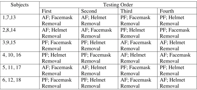
Table 3.1 Counterbalance Design of Data Collection
Subjects Testing Order
First Second Third Fourth
1,7,13 AF; Facemask
Removal AF; Helmet Removal PF; Facemask Removal PF; Helmet Removal
2,8,14 AF; Helmet
Removal AF; Facemask Removal PF; Helmet Removal PF; Facemask Removal
3,9,15 PF; Facemask
Removal PF; Helmet Removal AF; Facemask Removal AF; Helmet Removal
4, 10, 16 PF; Helmet
Removal PF; Facemask Removal AF; Helmet Removal AF; Facemask Removal
5, 11, 17 AF; Facemask
Removal AF; Helmet Removal PF; Facemask Removal PF; Helmet Removal
6, 12, 18 PF; Facemask
Removal PF; Helmet Removal AF; Facemask Removal AF; Helmet Removal
The AT left the room for all PF helmet assessment and AF helmet adjustment for
blinding purposes. For the AF conditions, the subject was asked to place the research
assistant provided helmet on their head. Once in place, the research assistant ensured that
the back of the helmet was in uniform contact with the back of the head. If the helmet
was too loose, the HardTail SPRfit™ technology (Cascade Lacrosse, Liverpool, NY) was
tightened until uniform contact around the entire head was reached. After making this
adjustment, the research assistant applied an anterior pressure over the occiput of the
helmet to ensure that there was not gapping at the subjects’ forehead. In addition, the
research assistant applied rotational forces on either side of the athlete’s head in order to
assess if the skin on the subjects’ forehead moved with the helmet, verifying fit per
manufacturer’s guidelines (Cascade, 2013b). If the helmet moved independently of the
subjects’ head in during any of the fitting assessment the HardTail SPRfit™ technology
Finally, the facemask was inspected to ensure a 2-3 finger width clearance from the
subjects’ nose (Cascade, 2013b).
The same inspection was conducted on the PF helmet. Data would not have been
collected if the PF helmet fit all the conditions necessary in the AF condition, but none of
our subjects presented with PF helmets that fit the AF helmet criteria. The AT returned to
the room following PF helmet assessment and AF helmet adjustment and assessed both
helmet conditions with subject supine in order to assess ability to judge difference in
helmet fit.
Figure 3.1 Cascade® R Lacrosse Helmet and HardTail SPRfit™ Technology
Three electromagnetic sensors were fit to each of the subjects. One was fit on the
crown of the helmet, left temple, and distal sternal notch of the thorax. Similar receiver
arrangement has been used in previous research studies (Toler et al., 2010). After the
receivers were properly secured, the subjects sat upright in order to digitize anatomical
landmarks with a wooden stylus. The anatomical landmarks identified include: T12/L1,
xiphoid process, proximal sternal notch, T8, C7, chin, bridge of nose, and occiput. After
digitization the subjects lay supine.
The starting position was standardized; supine with subject instructed to lie
motionless at all times. Subjects were instructed not to assist in maintaining head posture
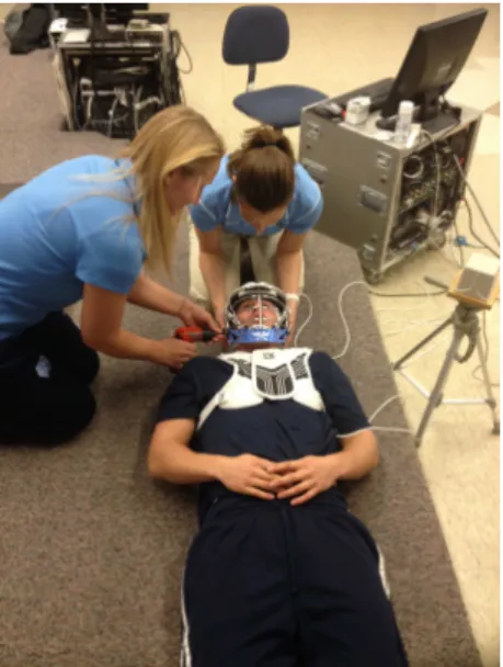
in any way. One certified athletic trainer (AT) and one research assistant who had been
taught and practiced proper airway access techniques for managing on-field men’s
lacrosse spine injuries, performed both helmet removal and facemask removal
techniques. For each helmet removal technique, the AT maintained control of the
subjects’ head inferiorly as the research assistant removed the chinstrap followed by the
helmet; following complete helmet removal the research assistant placed towel under
subjects head (Figure 3.2). Each helmet removal trial began when the AT secured the
head and will end when the research assistant placed a towel under the head after
complete helmet removal. In the facemask removal technique, the research assistant
performed the facemask removal and the AT stabilized the head superiorly (Figure 3.3).
Each facemask removal trial began as soon as AT secured head in in-line stabilization
and ended when the facemask was placed on the ground next to the subject. Initiation of
task was signified by verbal cue of “stabilized” and termination of task was signified by
task. Each airway access technique was performed three times under both helmet fit
conditions.
Figure 3.2 Helmet Removal
Figure 3.3 Facemask Removal
In order to prevent a learning effect, both the AT and research assistant performed