Characterizing Ku’s Role as an AP lyase in
Nonhomologous End Joining
Natasha Tiffany Strande
A dissertation submitted to the faculty of the University of North Carolina at Chapel Hill in partial fulfillment of the requirements for the degree of Doctor of Philosophy in the Department of Biochemistry and Biophysics.
Chapel Hill
2013
Approved by:
Dale A. Ramsden
Jean Cook
Dorothy Erie
Thomas A. Kunkel
iii
ABSTRACT
NATASHA TIFFANY STRANDE: Characterizing Ku’s Role as an AP lyase in
Nonhomologous End Joining
(Under the direction of Dale A. Ramsden)
Nonhomologous end joining (NHEJ) is important for the repair of ionizing
radiation and radiomimetic drug-generated DSBs, which are often associated with
ligation-obstructing nucleotide damage. To facilitate ligation at such breaks, NHEJ
employs a host of processing factors (i.e. nucleases, polymerases, etc.) that prepare DNA
ends for joining. While this mechanism is efficient at joining broken chromosomes, it
can frequently be inaccurate (i.e. loss of sequence at the DSB), because repair is
mediated without the assistance of a template. My dissertation demonstrates how
NHEJ-mediated repair of DSBs with associated abasic sites is an exception to this
phenomenon. I show that abasic sites at DSB termini severely block NHEJ’s ligation
step and must be excised for joining to proceed. Despite the many processing enzymes
associated with NHEJ, none are capable of excising this damage. Instead we found that
the NHEJ core factor, Ku, has intrinsic lyase activity that removes these abasic sites.
Analysis of Ku’s substrate specificity reveals that lyase activity is restricted to abasic
sites near a 5’ terminus that directly block ligation. Furthermore, sequence 5’ of abasic
sites embedded in double stranded DNA (+4 bps) is mostly preserved due to Ku’s limited
activity in this context. By characterizing Ku’s active site I identified eight lysine
is within the N-terminus of Ku70 (K31). These amino acids reside on the outer surface
of the Ku heterodimer nearest the DNA end – an optimal position for interacting with
abasic sites closest to the break terminus. My results provide mechanistic insight into
how NHEJ deals with one type of damage induced by ionizing radiation and may
explain why loss of Ku leads to severe radiation sensitivity. Additionally, my results
suggest NHEJ is more than a simple ligation machine but rather it is a sophisticated
v
ACKNOWLEDGEMENTS
Firstly, I am forever grateful to my advisor, Dale, for his continuous support and
encouragement throughout my graduate career. He has believed in my scientific
abilities and given me encouragement and perspective when I needed it most. I am also
extremely grateful for my fantastic committee members, Thomas A. Kunkel, Dorothy
Erie, Jean Cook, and Brian Strahl, for their engaging discussions, interest in my
project, and insightful input.
I am also thankful for the companionship of the Ramsden lab members past and
present. I am especially thankful to Christina for her unending patience, willingness to
answer questions (no matter how random they are), and friendship over the past five
years. I would also like to specifically thank Crystal for her support, friendship, and
keeping me motivated; graduate school would not have been the same without them.
My family has played a huge role in my success and I am grateful for their
gracious care and support over the years. I am especially grateful to my parents,
Jackie, Steve, Mark, Sherry, John and Jean for believing in me and encouraging me. I
also thank my brothers, Josh and Thaddeus, and sister Rachel for their love and
support. My church has been my family away from home and I am forever indebted to
them for their support, love, and perspective. I am particularly grateful to my friend
Ryan Hallett for introducing me to my church family and for showing me that being a
scientist and believing in God do not have to be mutually exclusive. This concept has
importantly, I am grateful for my husband, John, as he has providing constant
encouragement and support over the last two years. His ability to keep me motivated
and focused on my goals has been extremely helpful in the last stretch of graduate
school. He is an amazing man that I cannot imagine my life without.
Additional acknowledgements specific to each chapter:
Chapter 1
This chapter was entirely written by Natasha Strande and was modified for
publication in Genome Integrity (1). Figures 1.3 and Table 1.1 were generated by
Natasha Strande for publication in (1).
Chapter 2
This chapter has been modified from its original version appearing in Nature
volume 464, 2010(2). I have rearranged the figures so as to incorporate supplementary
information in a logical manner. Additionally, the text was partitioned into sections, for
the sake of consistency with the rest of my dissertation. N.S designed and conducted
experiments determining kinetic rates of Ku’s lyase activity (Figure 2.5A). All other
experiments were designed by S.A.R. and D.A.R. In vitro experiments were performed
by S.A.R, N.S. (specifically Figures 2.3, 2.4B,C,F, 2.5 , and 2.6B,C,D), M.D.B, and D.A.R.
Mutagenesis and protein purification were performed by S.A.R. and D.A.R. S.A.R. C.S.,
J.M.H., and M.D.B. performed cellular experiments. P.H. provided Ku70 knockout
vii
scholar award to DAR, as well as PHS grants R01 CA76317-05A1 and P01 AG17242 to
PH.
Chapter 3
This chapter originally appeared in an issue of The Journal of Biological
Chemistry in 2012 (3). N.S. designed and conducted all experiments. S.A.R.
contributed preliminary observations of Ku’s substrate specificity on a subset of shorter
oligonucleotide substrates. S.O. and E.A.H provided the HCT116 cell lines used in
Figure 3.4. We thank Crystal Waters and Kenjiro Asagoshi for critical reading of the
manuscript, and Kenjiro Asagoshi for analysis of HCT116 transfections by flow
cytometry. This work was supported by NIH grant R01 CA 84442 to DAR, and RO1
GM088351 and RO1 CA154461 to EAH.
Chapter 4
Natasha Strande conducted all experiments in this section and was solely
responsible for the writing within this chapter. Jody Havener generated the plasmid
constructs used to make Ku70 mutants: K31,160A (2A) and K160A as well as plasmids
for Ku80 mutants: K543-545A and K565,566,568A. Steven Roberts was responsible for
TABLE OF CONTENTS
LIST OF TABLES ... xi
LIST OF FIGURES ... xii
LIST OF ABBREVIATIONS AND SYMBOLS ... xiv
1. Introduction ... 1
1.1 Double Strand Break Repair ... 1
1.2 Nonhomologous End Joining Mechanism ... 2
1.3 Complex End Structures ... 3
1.4 Damage Processing and Associated Factors ... 5
1.4.1 Protein Occlusions ... 5
1.4.2 Secondary Structures – Resolution by V(D)J Recombination ... 6
1.4.3 Distorted/Damaged Nucleotide Removal ... 7
1.4.4 Class Switch Recombination and Abasic Sites ... 8
1.5 Overview of Dissertation ... 9
2. Ku is a 5’dRP/AP lyase that excises nucleotide damage near broken ends ... 17
2.1 Introduction ... 17
ix
2.2.4 In Vitro NHEJ Assays ... 21
2.2.5 Cellular NHEJ Assays ... 21
2.2.6 PCR Assays ... 22
2.3 Results ... 22
2.4 Discussion ... 27
3. Specificity of the dRP/AP lyase of Ku promotes non-homologous end joining (NHEJ) fidelity at damaged ends ... 34
3.1 Introduction ... 34
3.2 Materials and Methods ... 36
3.2.1 Proteins ... 36
3.2.2 Oligonucleotide Assays ... 36
3.2.3 NHEJ Assays ... 38
3.3 Results ... 39
3.3.1 Ku’s Substrate Specificity ... 39
3.3.2 Ku’s 5’dRP/AP Lyase Activity and NHEJ’s Ligation Step ... 42
3.4 Discussion ... 44
4. Ku has a defined AP lyase active site that determines Ku’s substrate specificity in NHEJ ... 54
4.1 Introduction ... 54
4.2 Materials and Methods ... 56
4.2.1 Protein Purification ... 56
4.2.2 DNA Substrates ... 56
4.2.3 Lyase Reaction ... 57
4.2.4 Trapping Assay ... 58
4.3 Results ... 58
4.3.1 Ku70 K31 is the Primary Nucleophile in Ku’s Lyase Reaction ... 58
4.3.2 Ku80 Residues Compensate in the Absence of Ku70’s Lyase Activity ... 60
4.3.3 DNA-PKcs Has Weak Lyase Activity that is Negligible for Ku’s Lyase ... 61
4.4 Discussion ... 63
5. Discussion ... 71
5.1 DSBs with Abasic Sites ... 72
5.1.1 DSBs Generated by Radiation ... 72
5.1.2 DSB Intermediates of Class Switch Recombination ... 74
5.2 Enzymes that Cleave Abasic Sites ... 75
5.2.1. Class II (hydrolytic) AP Endonucleases ... 76
5.2.2. Class I AP Endonucleases (lyase) ... 76
5.2.3. Deoxyribophosphodiesterases ... 77
5.3 NHEJ Overlap with BER ... 78
5.4 Ku in the Context of Cancer Therapies ... 79
5.5 Concluding Remarks ... 80
xi
LIST OF TABLES
Table 1.1 End processing factors ... 10
LIST OF FIGURES
Figure 1.1 NHEJ Mechanism ... 11
Figure 1.2 Crystal structure of the Ku heterodimer ... 12
Figure 1.3 Biological sources of DSBs generate complex end structures ... 13
Figure 1.4 Nucleotide damage and generation of abasic sites ... 14
Figure 1.5 V(D)J recombination ... 15
Figure 1.6 Class switch recombination generates DSBs terminated by 5’ dRP residues ... 16
Figure 2.1 In vitro NHEJ of ends with abasic sites ... 28
Figure 2.2 Cellular NHEJ of ends with abasic sites ... 29
Figure 2.3 5’dRP/AP lyase activity of purified NHEJ factors ... 30
Figure 2.4 Characterization of the Ku70 3A mutant ... 31
Figure 2.5 Analysis of kinetics and substrate specificity ... 32
Figure 2.6 AP lyase activity of cell extracts with or without Ku ... 33
Figure 3.1 Activity on AP sites within 5’ overhangs ... 48
Figure 3.2 Activity on AP sites within 3’ overhangs ... 49
Figure 3.3 Activity on AP sites within double-stranded DNA ... 50
Figure 3.4 Joining of ends with near terminal abasic sites in vitro ... 51
xiii
Figure 4.2 Contributing nucleophiles from Ku80 ... 67
Figure 4.3 DNA-PKcs’s weak lyase activity does not rescue
Ku mutant activity in vitro ... 68
Figure 4.4 Structure of Ku’s lyase active site modeled on
DNA with an abasic site ... 69
Figure 4.5 Sequence alignment of Ku nucleophiles ... 70
LIST OF ABBREVIATIONS AND SYMBOLS
aa, amino acid
Alt-EJ, alternate end joining
AID, activation-induced-deamniase
AP, apurinic/apyrimidinic
APE1, AP endonuclease 1
APTX, Aprataxin
BER, base excision repair
Bio-TEG, biotin-tetra-ethylene glycol
Bp, base pair
C, constant region
CHO, Chinese hamster ovary
CSR, class switch recombination
dL, 2-deoxyribonolactone
DNA-PKcs, DNA dependent Protein Kinase catalytic subunit
dRP, deoxyribose phosphate
dRPase, deoxyribophosphodiesterase
ds, double-stranded
DSB, double-strand break
xv
Fpg, formadopyrimidine glycosylase
hOGG1, human 8-oxoguanine-DNA glycosylase
hNTHL1, human thymine glycol-DNA glycosylase
HR, homologous recombination
IR, ionizing radiation
Kd, dissociation constant
kDa, kilo dalton
MRN, Mre11/Rad50/Nbs1
MMR, mismatch repair
Neil1/Neil2, endonuclease VIII-like
NER, nucleotide excision repair
NHEJ, nonhomologous end joining
PAGE, polyacrylamide gel electrophoresis
PBS, phosphate buffered saline
PG, 3’-phosphoglycolate esters
PNK, polynucleotide kinase/phosphatase
Pol, polymerase
qPCR, quantitative polymerase chain reactions
RAG, recombination activation gene
ROS, reactive oxygen species
RSS, recombination signal sequence
S, switch region
SDS, sodium dodecyl sulfate
ss, single-stranded
SSBR, single strand break repair
TBE, tris/borate/EDTA buffer
TEG, tetra-ethylene glycol
Tdp, tyrosyl DNA phosphodiesterase
Top, topoisomerase
UDG, uracil DNA glycosylase
V(D)J, Variable, Diversity, and Joining
WRN, Werners syndrome protein
XLF, XRCC4-like factor/Cernunnos
XRCC1, X-ray cross-complementary gene 1
XRCC4, X-ray cross-complementary gene 4
α, alpha
β, beta
ε, epsilon
γ, gamma
ι, iota
CHAPTER 1
Introduction
1.1
Double Strand Break Repair
Protecting our genome from the constant threat of DNA damage is essential to
organismal survival. Of the various forms of DNA damage that jeopardize genomic
integrity, the double strand break (DSB) poses the greatest danger to cellular viability.
Failure to repair these breaks results in genomic instability, premature replicative
senescence, and a predisposition to immunodeficiency and cancer. Repair of DSBs
occurs by one of two main pathways: (1) homologous recombination (HR) or (2)
nonhomologous end joining (NHEJ). These pathways primarily differ in the extent of
resection necessary for repair and the ensuing accuracy of the junction (reviewed in (4)).
HR requires a template, either a homologous chromosome or sister chromatid, to
direct synthesis after extensive resection at the DNA ends. While this pathway ensures
sequence integrity, it is limited by the availability of a template and thus only occurs
during S and G2 of the cell cycle. Conversely, NHEJ can join DSBs during any phase of
the cell cycle given that repair occurs in the absence of a template (5). Although this
makes NHEJ more flexibility than HR, it also leads to potential inaccuracies in the
repair product. Despite this disadvantage, NHEJ is the preferred pathway for DSB
resolution in mammals (reviewed in (6)).
NHEJ is particularly important for the repair of DSBs induced by ionizing
radiation (IR) and some chemotherapeutic drugs. Moreover, after exposure to IR,
Breaks resulting from such damage are frequently associated with additional nucleotide
lesions or altered end structures that can block ligation (detailed discussion in section
1.3). This added complexity presents a particularly challenging scenario for NHEJ,
where damage must be removed and replaced without the assistance of a template. My
thesis illustrates the sophisticated manner in which NHEJ avoids extensive sequence
loss at particular DSBs (i.e. those with abasic sites) despite the lack of a template.
1.2
Nonhomologous End Joining Mechanism
The NHEJ pathway is defined by the requirement of four core factors to detect
and join broken chromosomes: Ku, DNA Protein Kinase catalytic subunit (DNA-PKcs),
XRCC4-like factor (XLF) and the X-ray cross-complementary gene 4 (XRCC4)/DNA
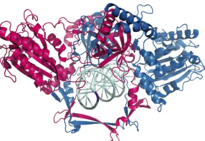
Ligase IV complex (Figure 1.1) (reviewed in (17)). The heterodimeric protein, Ku,
recognizes DSBs and loads onto the DNA initiating the first step of the pathway. This
ring-structured protein is comprised of two intimately associated subunits (70 and 86
kDa) that tightly bind DNA ends (Kd ranges from 40-800 pM) (Figure 1.2) (18-22). Once
loaded, Ku is not restricted to the ends and can slide along the length of the DNA, due
to predominantly electrostatic interactions with the DNA phosphodiester backbone (
22-24). With Ku bound at the DNA break, the remaining end-joining factors can then be
recruited to the site of damage.
The 460 kDa protein, DNA-PKcs, associates with Ku and the DNA to form a
complex, known as DNA-PK, that bridges the DSB ends to facilitate end alignment
3
clamping action of XLF (31-36), some damaged DNA ends are an impediment to direct
joining and thus require processing (discussed in section 1.4). For processing factors to
gain access to the DNA ends, the DNA-PK complex must be remodeled through the
auto-phosphorylation of DNA-PKcs(26, 27).
1.3
Complex End Structures
DNA damaging agents can generate a multitude of DSB end structures, which
may impede ligation depending on the severity of the damage. These structures can
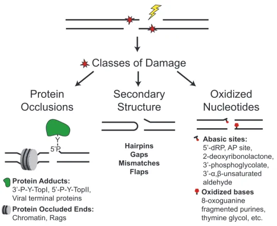
loosely be categorized into three classes of damage: protein occlusions, secondary
structures, and distorted/damaged nucleotides (Figure 1.3). The most common source of
these “complex” end structures is from exogenous agents, such as ionizing radiation,
chemotherapeutic drugs, or UV radiation. However, some endogenous sources also
create complex DSB breaks (e.g., reactive oxygen species (ROS) from metabolic
processes, aborted base excision repair (BER), etc.) (reviewed in (37))(38, 39).
Protein occlusions and secondary structures at DSB ends are often the product of
endogenous sources. The compacted structure of chromatin, comprised of DNA-protein
complexes (i.e. nucleosome), can impair several cellular processes to varying degrees,
including BER (40) and in vitro nucleotide excision repair (NER) reactions (41)
(reviewed in (42)). NHEJ, however, is not impaired by the nucleosome structure due to
Ku’s unique ability to peel DNA ends from the histone octamer (43).
Topoisomerase-adducted DNA ends, on the other hand, impede NHEJ and require removal for ligation
to occur (reviewed in (44)). Secondary structures at DSB ends, such as hairpins, are
another type of altered end structure incurred from endogenous cellular processes such
recombination). The hairpins generated during these events must be processed to free
the DNA ends for ligation (reviewed in(45)).
Distorted/damaged nucleotides can result from both endogenous and exogenous
sources of damage. The removal of such damage during NHEJ will be the focus of this
dissertation. Nucleotides can be altered at the base or the deoxyribose through
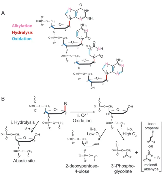
hydrolysis, alkylation, oxidation, etc., with the most common sites of alteration depicted
in Figure 1.4A (46, 47). One of the most severe and commonly occurring types of
nucleotide damage is the loss of a base, which can arise in a number of ways (48, 49).
Hydrolysis of the N-glycosyl bond between the C1’ atom of the sugar and the N1’ or N9’
atom of the base (pyrimidine and purine respectively) directly generates an
apurinic/apyrimidinic (AP or abasic) site (Figure 1.4B) (47, 50). Alkylated DNA
drastically increases the rate at which hydrolytic cleavage occurs, especially for purines,
making these lesions more susceptible to base loss (reviewed in(51)). Several carbons of
the deoxyribose chain can be oxidized, but C4’ is the major target with its oxidation
yielding an oxidized abasic site (2-deoxypentose-4-ulose) or a 3’
phosphoglycolate-terminated single strand break (Figure 1.4B) (52, 53). Additionally, abasic sites arise
during BER as an intermediate of glycosylation of altered bases (i.e. oxidized). An
individual non-bulky nucleotide lesion within the chromosome presents little threat to
genomic integrity due to its efficient repair through BER. However, when multiple
lesions cluster together at one site in the genome secondary lesions can arise (i.e. DSBs)
5
to “indirect” nucleotide damage nearby directly induced lesions creating “cluster
damage.” If multiple lesions exist in opposing DNA strands, it is likely that a DSB will
be produced in the process of repairing the damage – especially when these lesions are
abasic sites (64, 65).
1.4
Damage Processing and Associated Factors
Some of the aforementioned DNA end structures impede the ability of the
XRCC4/Ligase IV complex to complete ligation. These structures require end processing
for the ligation step to take place. As such, NHEJ is associated with a host of proteins
possessing a wide array of enzymatic activities that can prepare ends for ligation as
necessary (Table 1.1) (from (1)). Below I discuss some of the better characterized
processing factors associated with the three aforementioned classes of damage at DSB
ends: protein occlusions, secondary structures, and distorted/damaged nucleotides.
1.4.1 Protein Occlusions
DNA topoisomerases are required for facilitating changes in the topology of DNA
that occur during important DNA-mediated cellular processes (e.g. replication). These
proteins enable manipulation of DNA from one state to another, for example going from
supercoiled to relaxed. This process requires breaking the phosphodiester backbone of
each DNA strand and the subsequent re-ligation of the strands. A phosphotyrosyl
linkage between the DNA phosphate backbone and a tyrosine within the topoisomerase
active site generates the DNA strand break (reviewed in (44, 66)).
Topoisomerase I (top1) breaks the DNA strands one at a time, whereas
Topoisomerase II (top2) breaks both strands simultaneously creating a DSB.
Disruption of either mechanism results in a covalently linked topoisomerase-DNA end
phosphodiesterase enzymes, Tdp1 and Tdp2, that cleave 3’ and 5’ phosphotyrosyl
linkages to Top1 and Top2, respectively(67, 68). Tdp1 is predominantly implicated in
the repair of damage at SSBs, but possesses additional enzymatic activities that could
be beneficial to NHEJ repair: removal of 3’ phosphoglycolate termini, 3’ abasic sites
within bubble substrates, 3’ phosphohistidine linkage, and exonuclease activity
restricted to a single nucleotide from the 3’ OH end (69-72). Unlike Tdp1, the substrate
specificity of Tdp2 appears to be significantly more restricted. Biochemical
characterization of Tdp2 shows that its preferred substrates are 5’ phosphotyrosines
within ssDNA and dsDNA with a 4 nucleotide 5’ overhang (73), consistent with the
biological substrate for Top2 induced damage (74, 75). Most recently Tdp2 activity was
shown to be important for repair of Top2 induced DSB repair by NHEJ (76).
1.4.2 Secondary Structures – Resolution by V(D)J Recombination
As previously mentioned, normal cellular processes such as programmed
recombination events can result in DSBs with secondary structures. V(D)J
recombination (reviewed in (77-80)) creates hairpin intermediates during the generation
of the exons encoding the diverse antigen receptors important for adaptive immunity.
V(D)J recombination derives its diverse products by rearranging the immunoglobulin
gene segments (Variable, Diversity, and Joining). The lymphoid specific recombination activation gene (RAG) proteins bind to the recombination signal sequences (RSSs)
associated with each V, D, and J gene segments. RAG-mediated cleavage at these sites
7
and subsequently processed by the endonuclease activity of the DNA-PKcs:Artemis
complex, which intentionally creates diverse products (81).
1.4.3 Distorted/Damaged Nucleotide Removal
The multitude of possible nucleotide alterations and the alarming frequency with
which they occur present a need for mechanisms to remove these lesions. Excision of
nucleotide damage in the context of intact chromosomes occurs through the
well-characterized and conserved BER pathway (82). BER repairs base damage in a
step-wise fashion initiated by recognition and removal of the lesion by one of the eleven
mammalian glycosylases (reviewed in (83, 84)). Excision of the base results in an abasic
site that is cleaved by AP endonuclease 1 (APE1) or the glycosylase itself (if bifunctional
with lyase activity). The latter lyase activity cuts the DNA 3’ of the abasic site creating
a 5’ phosphate and a 3’ blocking lesion. Alternatively, APE1 nicks the DNA 5’ of the
abasic site creating a 3’ OH and a 5’ deoxyribose phosphate (5’ dRP) residue and can
also remove the 3’ lesions left by a bifunctional glycosylase (or lyase). The final step of
the pathway involves removal of the 5’ dRP (if present) and replacement of the missing
nucleotide by DNA polymerase β (reviewed in (85)). Repair of nucleotide lesions at
DSBs, however, is not nearly as well understood. It is often assumed that “complex”
breaks joined by NHEJ are imprecisely resolved using a combination of nuclease and
polymerase activity (86). While some types of damage are likely resolved in this
non-specific manner, others are purposely targeted and removed by enzymes associated with
NHEJ (for extensive discussion see review (1)).
Aprataxin (APTX) and polynucleotide kinase/phosphatase (PNK) are repair
proteins that interact with the NHEJ protein XRCC4 as well as the BER protein
proteins are important for generating ligatable ends (i.e. generate a 5’ phosphate and 3’
hydroxyl). APTX is particularly important for removing the 5′ adenylate adducts
produced during aborted ligation (91-93), which may occur after attempted ligation of a
“complex” break. PNK has both 5’ kinase and 3’ phosphatase activities of which the
phosphatase has been shown to act coordinately with TDP1 by removing the 3’
phosphate product of 3’ phosphoglycolate excision by TDP1 (69, 94). The involvement of
these proteins in NHEJ is supported by their interaction with the NHEJ protein
XRCC4, as well as, the varying degrees of radiation sensitivity exhibited in the absence
of these proteins (88, 95, 96).
NHEJ is also associated with several nucleases that can iteratively remove
nucleotides until an appropriate end is generated for ligation. Artemis, Werners
syndrome protein (WRN) and the Mre11/Rad50/Nbs1 (MRN) complex are relatively
well-characterized nucleases known to interact with the core NHEJ factors. In addition
to Artemis’s role in V(D)J recombination, it has 5’>3’ exonuclease activity (81) and is
important for protecting cells from IR (97-99), suggesting Artemis may be important for
resolving the “complex” breaks induced by IR. Both WRN and MRN have 3’>5’
exonuclease activity (100-102) that could be important for resolving breaks with damage
clusters. The extensive resection mediated by these nucleases is likely a reason why
NHEJ is considered to be a “simple and error-prone” pathway. My dissertation,
however, demonstrates that NHEJ is capable of precise repair of abasic sites at DSBs as
9
in (103)). A direct intermediate of this process is a DSB with a 5’ dRP residue at the
terminus, similar to an IR product. These intermediates are generated by a deletional
recombination event at “switch” (S) regions that precede the C regions of the
immunoglobulin gene (reviewed in (104, 105)). This process is initiated when the
activation-induced-deamniase (AID) extensively deaminates cytosines within these S
regions to yield deoxyuracils. The BER proteins, uracil DNA glycosylase (UDG) and
APE1 work in concert to convert the uracils to abasic sites and then 5’ dRP SSBs. Due
to the extent of AID demamination, subsequent repair by BER generates a high
frequency of closely residing SSBs with 5’ dRP residues. The close proximity of these
SSBs results in the formation of DSBs with 5’ dRP groups, which are subsequently
joined by the NHEJ pathway (Figure 1.6). As I will demonstrate in following chapters,
the NHEJ pathway is particularly adept at joining such DSBs.
1.5
Overview of Dissertation
Given the severity of abasic sites in the genome and the likelihood of their
occurrence at DSB ends (i.e. IR and CSR), we were interested in determining how
NHEJ resolves DSBs with associated abasic sites. Chapter 2 of this dissertation shows
that terminal abasic sites at DSB ends indeed block NHEJ’s ligation step.
Furthermore, we determined that NHEJ instead of utilizing one of the many
end-processing proteins it is associated with, the NHEJ core factor, Ku, is capable of
removing such damage at DNA ends (2). In Chapter 3, I describe Ku’s restricted
substrate specificity for removing abasic sites and illustrate how this restricted activity
is beneficial to preservation of sequence(3). Finally in Chapter 4, I will elaborate on the
Table 1: NHEJ End processing factors
Factor* Activity
APTX Removes 5’-adenylate adducts (91)
PNKP Removes 3’ phosphates and phosphorylates 5’ hydroxyls (106) APLF Histone chaperone (107) 3’-5’ exonuclease, endonuclease
(108, 109)
TDP1 Removes Top I adducts (67), 3’ deoxyribose fragments (69, 70,
94)
TDP2 Removes Top II adducts (68)
XRCC5,XRCC6 (Ku) Removes 5’-dRP residues and abasic sites (110)
POLM (Pol µ) Fills in gaps when ends align with no complementarity (111) POLL (Pol λ) Fills in gaps when ends are partly complementary (111, 112) DCLRE1C (Artemis) Endonuclease, 5’-3’ exonuclease (81)
WRN 3’-5’ exonuclease (100, 101) and 3’-5’ helicase (113) MRE11/RAD50/NBN
(MRN) 3’-5’ exonuclease, endonuclease (102, 114) SETMAR (Metnase) Endonuclease/exonuclease (115)
11
13
Figure 1.3: Biological sources of DSBs generate complex end structures. Examples include protein occlusions, such as topoisomerase-adducts, secondary structures generated by V(D)J recombination, and oxidized nucleotides as might arise from IR.
Classes of Damage
Protein
Occlusions
Y 5’P
Protein Adducts: 3’-P-Y-TopI, 5’-P-Y-TopII, Viral terminal proteins Protein Occluded Ends: Chromatin, Rags
Secondary
Structure
Hairpins Gaps Mismatches
Flaps
Oxidized
Nucleotides
Abasic sites: 5’-dRP, AP site, 2-deoxyribonolactone, 3’-phosphoglycolate,
Figure 1.4: Nucleotide damageand generation of abasic sites. A. Sites of most common nucleotide modification of guanine, cytosine, thymine, and adenine (from top) Major sites of modification are represented either in boldface font or as thick bonds. B. Abasic sites can form directly from hydrolysis at the C1’ atom releasing the base (B) (i) or as a result of oxidation (ii) (with the most frequent site (C4’) targeted depicted here). Under low oxygen conditions the oxidized abasic site 2-deoxypentose-4-ulose is generated (ii-a.), while high concentrations of oxygen lead to a 3’-phosphoglycolate and either a base propenal or a malondialdehyde depending on the source of oxidation (ii-b.).
A
O O P O O-O CH2O O
P
O-O CH2
-N N NH O N N NH2 N N NH2 O P O O-O CH2
O N NH O P O O-O CH2
O OH NH2 O O O CH3 1 1 3 5 9 7 1 3 5 9 7 3 5 1 3 5 1’ 2’ 4’ 1’ 2’ 4’ 1’ 2’ 4’ 1’ 2’ 4’ N N N
B
B ii. C4’ Oxidation i. Hydrolysis OH O O P O O-O CH2O P
O-O CH2
1’ Abasic site O O P O O-O CH2
O P O
-O CH2 OH
HO
O P O
O-O CH2
O P O
-O CH2
O O
ii-a. Low O2
ii-b. High O2
2-deoxypentose-4-ulose O O P
O-O CH2
O - 3’-Phospho-glycolate
+
B H O OR B H O H O + base propenal malondi-aldehyde B 1’ O O P O O-O CH2O P O
-O CH2
15
Figure 1.5: V(D)J recombination. Rearrangement of immunoglobulin gene segments (V, D, and J depicted by colored rectangles) is initiated by RAG-mediated (grey circle is Rag1/2 complex) cleavage (i) at the recombination signal sequences (RSSs) (triangles) associated with each gene segment. This cleavage event generates 2 DSBs resulting in hairpin-coding ends and blunt RSS ends that are joined by NHEJ (ii) through two different mechanisms. The hairpin-coding ends require additional processing mediated by the DNA-PK:Artemis complex for NHEJ ligation to generate the coding junction (ii-a.). The excised segment terminated by the RSSs is circularized through a blunt-ended ligation (ii-b.) and likely lost from the genome.
V V V D D J J J S C
S C
J
V V V D
J J D
Rag1/2
i. Cleavage at RSS
ii. NHEJ
D
J
J
ii-b. Blunt Ligation
S C
J
V V V D
ii-a. DNA-PK:Artemis hairpin processing
S C
J
V V V D
End processing
Figure 1.6: Class switch recombination generates DSBs terminated by 5’ dRP residues. Once V(D)J recombination generates the variable region of an antigen receptor, a deletional recombination event via class switch recombination (CSR) at the switch regions (S depicted as purple or red ovals) that precede the constant regions (yellow rectangles). This deletion event changes the constant region of the immunoglobulin altering the antibody’s class distinction. The initial step of CSR is mediated by extensive cytosine deamination by the activation-induced-deaminase (AID). The resulting uracils are converted to abasic sites by the uracil DNA glycosylase (UDG). AP endonuclease 1 (APE1) removes the abasic sites resulting in closely spaced SSBs with associated 5’dRP residues that result in DSBs that can be joined by NHEJ.
V D J S
CSR
V D J
+
Circularized by NHEJ
DSB generation at S regions
c c
c c c c
AID
u u
u u u u
UDG
5’
3’
dR dR
dR dR dR dR
APE1 Recombined Ig gene
Excised gene segment
5’
3’
dR
dR 3’
5’ DSB
CHAPTER 2
Ku is a 5’dRP/AP lyase that excises nucleotide damage near broken
ends
2.1
Introduction
Mammalian cells require nonhomologous end joining (NHEJ) for efficient repair
of chromosomal DNA double strand breaks(116). A key feature of biological sources of
strand breaks is associated nucleotide damage, including base loss (abasic or AP sites)
(117). At single strand breaks, 5’ terminal abasic sites are excised by pol β’s 5’dRP lyase
activity (118-121): we show here in vitro and in cells that accurate and efficient repair
by NHEJ of double strand breaks with such damage similarly requires 5’dRP/AP lyase
activity (Figure 2.1A). Classically defined NHEJ is moreover uniquely effective at
coupling this end-cleaning step to joining in cells, helping distinguish this pathway from
otherwise robust alternate NHEJ pathways. Surprisingly, the NHEJ factor Ku can be
identified as an effective 5’dRP/AP lyase. Similar to other lyases (122), Ku nicks DNA 3’
of an abasic site by a mechanism involving a Schiff base covalent intermediate with the
abasic site. We demonstrate using cell extracts that Ku is essential for efficient removal
of AP sites near double strand breaks and, consistent with this result, joining of such
breaks is specifically reduced in cells complemented with a lyase-attenuated Ku mutant.
Ku had previously been presumed only to recognize ends and recruit other factors that
processed ends; our data supports an unexpected direct role for Ku in end processing
2.2
Materials and Methods
2.2.1 Methods Summary
For in vitro end joining and lyase reactions recombinant purified Ku,
XRCC4-LigaseIV, XLF/Cernunnos, and/or DNA-PKcs purified from Hela cells were incubated
with radiolabeled substrates before analysis by electrophoresis. For cellular NHEJ
assays a dermal fibroblast line from a mouse deficient in Ku70 and p53 (123) was
engineered by retroviral transduction to express wild type mouse Ku70, Ku70 3A, or an
empty vector control. Expressing cells were then purified by selection for puromycin
resistance. NHEJ substrates were introduced into these cells by electroporation,
harvested, and sample recovery and NHEJ efficiency assessed by qPCR. NHEJ
accuracy was further assessed by digestion of amplified junctions with a restriction
enzyme diagnostic for predicted products.
2.2.2 Constructs and Protein Preparations
Recombinant purified human Ku, XRCC4-LigaseIV, pol β and pol λ were
expressed and purified as previously described (111). The Ku 70 3A mutant involved
substitution for alanine of K31, K160, and K164 (human cDNA) or K29, K158, and K162
(mouse cDNA) by the quickchange method (Stratagene). For XLF, a cDNA (the gift of
K. Meek, MSU) was introduced into pFASTBAC1 with a C-terminal hexahistidine tag
and purified by successive chromatography on HisTrap and Mono Q columns (GE
biosciences). DNA-PKcs was purified from HeLa cells as described in (26). For whole
19
supernatant to phosphocellulose (Sigma), and hydroxyapatite (Biorad). Extracts were
then dialysed or diluted until equivalent to 10 mM TRIS pH 8.0, 250 mM KCl, 0.1%
NP40, 10% glycerol, and 1 mM EDTA. HeLa cell extracts were further immunodepleted
by two sequential adsorptions to protein A Sepharose beads loaded with either
pre-immune serum (Mock depleted) or serum from rabbits immunized against human Ku
(Ku-depleted). HeLa and CHO cell extracts were then analyzed by Western blotting
using antibodies against Ku (raised against purified human Ku heterodimer), pol β
(ab26343, Abcam), actin (A2066, Sigma), and DNA-PKcs (Ab4, Neomarkers). In Figure
2.6A comparison of the recombinant Ku standard (rKu; 5 ng) to Ku in mock depleted
extracts (1.5mg) allows for the estimation of the concentration of Ku in HeLa cell
extracts as 4ng Ku/mg extract.
2.2.3 DNA Substrates
NHEJ substrates were generated with varied end structures as previously
described (124). Briefly, substrates with 2, 3, or 4 nucleotide overhangs were created
using polymerase chain reaction (PCR) to append the restriction sites Smu I, Bsp QI,
or Bsa I (respectively) to either a 250 bp core sequence from the mouse Jk1 germline
(used in Figure 2.1B) or a 300 bp variant modified to allow development of qPCR
primers (used in all subsequent Figures). The resulting PCR products were cloned
(TOPO-TA, Invitrogen) to generate plasmid templates used for further amplification.
Amplified DNA fragments were then digested with appropriate restriction enzymes, and
resulting substrate purified using a Qiaquick PCR clean up kit (Qiagen). Amplifications
included 32P-α-dATP when substrates were used for in vitro reactions. Oligonucleotides
for substrates were obtained from Integrated DNA Technologies, labelled at the 3’ end
form duplex substrates. The labeled strands were
5’Phos-UGGAAATCAAACGTAAGTAG for 5’dRP-DSB and 5’dRP-SSB,and
5’Phos-GUGGAAATCAAACGTAAGTAGAATCCAAAGTCTCTTTCTTCCG for AP-DSB. The
appropriate labelled strands for 5’dRP-DSB, 5’dRP-SSB, and AP-DSB were annealed to
5’biotin-TEG-TCTACTTACGTTTGATTTC,
5’biotin-TEG-TCTACTTACGTTTGATTTCCAGCTTGGTGCCTCCA and
5’biotin-TEG-TGGAGGCACCAAGC, and finally
5’biotin-TEG-TCGGAAGAAAGAGACTTTGGATTCTACTTACGTTTGATTTC, respectively. A variant
AP-DSB with the biotin terminal 22 bp deleted was employed in experiments described
in Supplemental Figure 6a. All biotinylated substrate ends were blocked by
pre-incubation of the substrate with 1 mM streptavidin (Pierce) for 5 minutes. The
tetrahydrofuran containing substrate was generated by substituting dU in the labelled
strand in AP-DSB with tetrahydrofuran (“dSpacer”; Integrated DNA Technologies).
Bleocin damaged substrates were generated by annealing the oligonucleotide
5’TCTACTTACGTTTGATTTCCAGCTTGGTGCCTCCA to
5’TGGAGGCACCAAGCTGGAAATCAAACGTAAGTAG, and incubating 100 fmol of the
resulting duplex with 100 pmol Bleocin (EMD Biosciences) for 10 minutes at 37oC. For
all other substrates, abasic sites were made by inclusion of dU at the appropriate site
and incubation with 0.02 units uracil DNA glycosylase (NEB) per fmol substrate for 5
minutes at 37oC. Reduced AP site substrates were made by treating these glycosylated
21 2.2.4 In Vitro NHEJ Assays
5 nM of 250 bp radiolabeled substrates with noted end structures were
pre-incubated with 20 nM recombinant Ku, 10 nM DNA-PKcs, 40 nM XRCC4-LigaseIV, and
80 nM XLF in a standard reaction buffer (25 mM NaPO4 pH 7.4, 125 mM KCl, 0.1 mM
EDTA, and 1 mM DTT) supplemented with 10% polyethylene glycol for 5 minutes at
25oC. Reactions were started by addition of 2 mM MgCl2 and 100 mM ATP and
incubated at 37oC, stopped by deproteinization, and analyzed by native 5%
polyacrylamide gel electrophoresis (PAGE).
2.2.5 Cellular NHEJ assays
Dermal fibroblasts from Ku70-/- p53-/- mice were infected with retroviruses with
a wild type mouse Ku70 cDNA, the mouse Ku70 3A mutant, or empty vector
(pBABE-puro) using standard techniques and selected for at least 4 days using 2 mg/ml
puromycin (this dose and time was sufficient to fully kill uninfected cultures treated in
parallel). 1X10⁶ puromycin resistant cells were then transfected with 5 ng substrate
and 1.5 mg pMAX-GFP tracer using the Amaxa nucleofector II, a MEF-2 kit, and
program A-023 (Lonza). Transfected cells were harvested 5 hrs later and small
molecular weight DNA recovered in a Hirt supernatant (125). Joining was evaluated
both by semi-quantitative and realtime PCRs (qPCR).
Expression levels of wild type and Ku 70 3A could not be accurately assessed by
western analysis for technical reasons. We therefore used undamaged control substrate
to ensure Ku 70 3A complemented cells were not generally defective (e.g. due to
insufficient complementation, for whatever reason). Highly efficient and consistent
complementation was further validated by also assessing joining efficiencies with
undamaged substrate in control Ku70 deficient cells (infected with empty vector) in
cells were only 5.4%, +/-2.2 (mean for the 4 transfections, +/- s.e.m.) as active, or only
6.0%, +/-2.5 as active relative to cells complemented with Ku 70 3A. These experiments
were repeated twice for each of two different virus preparations/infections
(complementations). In agreement with consistent complementation, independent viral
preps/infections did not contribute significantly to the overall experimental variation
observed in results with either wt or Ku70 3A construct. Two-tailed t-tests were used
for tests of statistical significance (Prism, Graphpad).
2.2.6 PCR assays
Semi-quantitative PCRs (30 cycles) were performed with 2.5 mCi 32P-α-dATP
(without dye). Products were analyzed after digestion as appropriate (to characterize
junctions) and electrophoresis on 8% polyacrylamide gels under native conditions.
qPCRs were performed with Power-SYBR mix and an ABI 7300 (Applied Biosystems) to
quantify both substrate and head-to-tail junctions. Levels of head-to-tail junctions were
adjusted according to the results of substrate qPCR to account for differences in
transfection and sample recovery (though substrate-specific qPCRs rarely varied by
more than 30% from the mean for a given experiment).
2.3
Results
Abasic sites are frequently associated with double strand breaks generated by
ionizing radiation, treatment with radiomimetic drugs (117, 126), after aborted base
excision repair (127, 128), or as an intermediate in class switch recombination (129,
23
lyase (Figure 2.1B, lanes 2 and 5). Nevertheless, characterization of junctions indicated
ligation occurred after precise excision of the abasic site (Figure 2.1C), and ligation was
negligible if 5’dRP/AP lyase activity was blocked (119) by prior reduction of substrate
abasic sites (Figure 2.1B, “R”). NHEJ must therefore employ lyase activity for excision
of 5’terminal abasic sites in a manner similar to short patch base excision repair (BER)
(Figure 2.1A). Additionally, our purified NHEJ core factor preparations are
surprisingly sufficient to perform this function.
We next asked if a 5’dRP/AP lyase was also important in NHEJ of such
substrates in cells. We used Ku70 and p53 deficient mouse dermal fibroblasts (123)
either complemented by expression of a mouse Ku70 cDNA (+Ku70) or an empty vector
control (+vector), and introduced into these cells variations of the abasic site-containing
substrates described above as well as undamaged control versions of these substrates.
Quantitative polymerase chain reactions (qPCR) were then used to assess efficiency of
overall recovery as well as head-to-tail joining (substrate and junction qPCRs; Figure
1c).
Cells proficient in classically-defined NHEJ (+Ku70) were equally effective in
joining substrates with an embedded normal AP site (5’AAT) as they were in joining
undamaged control substrates (5’ATAT, 5’AT) (Figure 2.2B; columns 1, 3 and 7). Using
restriction enzyme digestions diagnostic for specific junctions (Figure 2.2D) we further
determined undamaged ends (5’ATAT, 5’AT) were usually joined without deletions or
additions (Figure 2.2E), as expected. By comparison, the 5’AAT substrate was
typically joined after precise excision of the AP site, but with no further addition or
deletion (Figure 2.2E), consistent with in vitro data and the model in Figure 2.1A. Also
consistent with in vitro data, reduction of the AP site (and consequent blocking of
columns 3 and 5). The majority of junctions that were formed contained deletion of
DNA flanking the reduced AP site (Figure 2.2E,F). Excision of AP sites near DSBs by a
5’dRP/AP lyase is thus critical for efficient and accurate resolution of such ends by
cellular NHEJ (Figure 2.1A).
Consistent with other studies (131, 132) (reviewed in (133)) an alternative to
NHEJ (Alt-NHEJ) allows for significant joining of undamaged ends in Ku70 deficient
cells (Figure 2.2B, columns 1 and 2, 7 and 8). In comparison to classical NHEJ however,
a near-terminal abasic site is a clear barrier to Alt-NHEJ and reduction of the AP site
no longer has a significant impact (Figure 2.2B, columns 2, 4, 6). Even the intrinsically
less stable terminal abasic site (5’dRP) is a strong barrier to Alt-NHEJ, but not
Ku-dependent NHEJ (Figure 2.2C). We conclude Ku-Ku-dependent NHEJ is uniquely able to
couple 5’dRP/AP lyase activity to joining (Figure 2.1A), and that this activity is critical
for efficient and accurate resolution of ends with such damage.
The experiment described in Figure 2.1B indicated that one of the known NHEJ
core factors might be a 5’dRP/AP lyase. Of the four core factors employed in this assay
only Ku had significant activity on its own: Ku excised both penultimate (AP sites,
Figure 2.3A) and terminal (5’dRP, Figure 2.3B) abasic sites, resulting in species that
co-migrate with alkali-cleaved controls (Figure 2.3A, lane 8). This is consistent with
cleavage 3’ of the abasic site and production of a 5’ phosphorylated terminus. Activity
was also blocked by substitution of abasic sites with an analogue resistant to lyase
25
(122). Addition of NaBH4 reduces this intermediate to instead make a stable
protein-DNA adduct - if the protein-DNA substrate is radioactive, NaBH4 treatment radiolabels the
active lyase. As expected, adducted species required both the abasic site and NaBH4
and were consistent with DNA adducted to Ku70 and Ku80 (confirmed by mutant
analysis, Figure 2.4B). Importantly, NaBH4 trapping identifies even weak nucleophiles.
In this regard DNA-PKcs, though also capable of forming an adduct (Figure 2.3D),
typically does little to directly impact activity of the Ku heterodimer (e.g. Figure 2.3A)
whether DNA-PKcs is active as a kinase or not (Figure 2.3E).
We further characterized Ku70’s contribution to activity by systematic
mutagenesis of 21 candidate catalytic lysines. Mutation of K31 alone diminished
Ku70’s ability to form a Schiff base (unpublished data), but additional mutation of two
nearby (22) lysines (K160, K164; Figure 2.4A) was required to completely ablate adduct
formation (Ku70 3A, Figure 2.4B). However, the Ku 70 3A mutant only reduces
5’dRP/AP lyase activity 2 fold (Figure 2.4C) and promotes adduction with Ku80 (Figure
2.4B), indicating nucleophiles within Ku80 can at least partly compensate. Similar
observations of compensating activity have been observed after mutation of the primary
nucleophiles in other biologically important 5’dRP lyases (e.g. Pol4 KK247::248AA
(134), Pol β K72A (135)). The modest lyase defect observed with the Ku70 3A mutant
in vitro was nevertheless sufficient to cause a comparable defect in NHEJ of 5’dRP
containing substrates in cells (Figure 2.4D, Figure 2.4E). Importantly, the mutant
heterodimer is not significantly defective for DNA end binding in vitro (Figure 2.4F) or
joining of the undamaged control substrate in cells (Figure 2.4D, Figure 2.4E). Notably,
general loss of function was observed with a more severe perturbation of the candidate
consistent with a significant and non-redundant role for Ku’s 5’dRP/AP lyase activity in
NHEJ of ends with associated abasic sites.
Is Ku a robust 5’dRP/AP lyase? 5’dRP and AP sites are excised by Ku with half
lives of 2.7 and 7.0 minutes, respectively (Figure 2.5A). These rates are readily
accommodated within the typical half life of radiation-induced double strand breaks in
cells (136) and are in the range of rates of dRP removal as performed during BER/SSBR
(120, 137, 138). However, proximity of an abasic site to a DSB can present a barrier for
canonical dRP/AP lyases. For example, while Pol β is proficient at this step at single
strand breaks (5’dRP-SSB) (where Ku is not significantly active) (139) (also Figure
2.5B), its activity is greatly reduced at DSBs. Ku, however, is active at DSB ends -
approximately 10 fold more active than is pol β (Figure 2.4B). Another pol X member,
pol λ, is implicated in NHEJ, but its 5'dRP lyase activity (140) is even more restricted to
single strand breaks than pol β (Figure 2.4C). These results are consistent with genetic
studies arguing against an important role for lyase activity of pol 4, the only pol X
member in S. cerevisiae, in cellular NHEJ of ends with nearby abasic sites (141).
We next tested whole cell extracts to more comprehensively assess the
importance of Ku’s 5’dRP/AP lyase activity. Strikingly, extracts specifically deficient in
Ku (Figure 2.5A) were 70 fold (human cell extracts) or 5 fold (rodent cell extracts) less
active than matched control extracts in excising DSB terminal abasic sites (Figure 2.6B
and C). Moreover, human cell extracts have much higher specific activity than rodent
27
damaged in vitro with the radiomimetic drug bleocin (Figure 2.4E), consistent with an
important role for Ku in cleaning termini with abasic site damage derived directly by
oxidation (126) as well as after glycolysis (Figures 1-3). We therefore conclude that Ku
easily accounts for the majority of 5’dRP/AP lyase activity in cell extracts when AP sites
are near 5’ termini of double strand breaks.
2.4
Discussion
We show both in vitro and in cells that NHEJ can and must “clean” termini of
abasic sites, a class of nucleotide damage commonly associated with strand breaks,
before such broken ends can be joined. Notably, abasic sites are a strong barrier to
Alt-NHEJ, suggesting a general inability of Alt-NHEJ to couple end cleaning steps to
joining might help explain why mammalian cells deficient in classical NHEJ are so
radiosensitive (116, 143). That NHEJ utilizes a 5’dRP/AP lyase for excision of near
terminal abasic sites is a striking parallel to short patch BER/SSBR (Figure 2.1A), and
has clear advantages: the ability to specifically excise damage allows for more
conservative resolutions than less directed end processing mechanisms. We further
surprisingly identify the “scaffolding” factor Ku as the 5’dRP/AP lyase NHEJ employs
Figure 2.1: In vitro NHEJ of ends with abasic sites. Substrate cartoons show position of abasic site (open circle; if reduced, closed circle) relative to intact nucleotides (blocks/letters). A. Repair of strand breaks (single strand, SSB; double strand, DSB) with associated abasic sites. B. Radiolabeled 250 bp substrates were incubated with purified Ku, DNA-PKcs, XRCC4-LigaseIV, and XLF at 37oC for 20 minutes (5’dRP-EJ)
or 60 minutes (AP-EJ) and products detected by electrophoresis and phosphorimaging. R; abasic sites reduced. C, Ligation of substrates without excision of abasic sites will generate junctions sensitive to alkali treatment; alternatively, joining of ends after precise excision of abasic sites will generate junctions sensitive to a diagnostic unique restriction enzyme (Ase I for 5’dRP-EJ, and Nco I for AP-EJ). D. In vitro ligation products were digested with a restriction site internal to ends (Hinf I) to resolve concatemers, and either mock treated or treated with 80mM NaOH for 20 minutes at 95oC. Shown are head-to-tail junctions after 5% denaturing PAGE. Resistance to alkali
reflects the absence of abasic sites in products. Denaturing 15% PAGE confirmed alkali treatment was sufficient to completely cleave abasic sites present at un-reacted substrate ends (unpublished data). E. Head-to-tail junctions were PCR amplified and
B
Abasic
site + + R + + R
1 2 3 4 5 6
NHEJ + + + +
Sub.
5'dRP-EJ AP-EJ
A c-NHEJ
(DSBR) BER
(SSBR)
?
pol dRPase
AseI NcoI
1 2 3 4
NaOH + +
Direct Ligation Excision
NaOH AseI
Direct Ligation Excision
NaOH NcoI
C
D
E
29
Figure 2.2: Cellular NHEJ of ends with abasic sites. A, B, and C. Substrates with end structures varied as noted in cartoons were introduced into Ku70 -/- fibroblasts complemented with Ku70 or empty vector standard. Recovered DNA was then assessed for efficiencies of substrate recovery and joining by qPCRs using substrate and junction primer pairs, respectively. Error bars reflect the standard error of the mean (s.e.m.) from 4 independent transfections. D. Direct joining or joining after lyase-dependent excision generates junction structures with diagnostic restriction sites as noted. Junction qPCR primers detect junctions containing up to 10 bp deletion from either side (21 bp total). E. Cellular junctions were amplified by standard (dye-free) semi-quantitative PCR and mock digested (-) or digested with Eco RV (E) or Mbo I (M) as noted. Shown are results from samples pooled from 4 independent transfections. The fraction of junctions sensitive to noted restriction enzymes is indicated below the gel image (%D). na; joining was too infrequent to accurately characterize junctions. F. Mbo I sensitive junction sequence at top, compared to 10 representative Mbo I resistant junctions formed after joining of substrates with reduced abasic sites. An apparent non-templated addition is italicized.
RE:- E - E - M - M - M - M - M - M
-%D: 90 83 90 na 15 na 94 91 na
5'ATAT 5'AT
Ku70: Sub.: + - + -+ - + -- -E C 5' A?AT TA?A Nuclease A Mbo I Lyase ATAT Eco RV No processing
qPCRs: Junction Substrate
A TATA AT TA AT 5' 5' 5' TA
5'A AT 5'A AT
F
GGGTGTGCTG AT
GGGTGTGCTG GGGTGTGCT GGGTGTGCTG
GGGTGTGC A
GGGTGTGCTG AT GGGTGTGCTG GGGTGTGCT GGGTGT x2 CCAGCTTAGCT CCAGCTTAGCT CCAGCTTAGCT CAGCTTAGCT CCAGCTTAGCT CTTAGCT CTTAGCT TAGCT GCT x2 Mbo I A Joining ef ficienc y 103 102 101 100 10-1 +Ku70 +vector
Substrate end structure
5'1ATAT2 5'A AT3 4 5'A AT5 6 5'7AT 8
B 5'A?AT +Ku70 or vector Ku70 -/-5' A?AT TA? A
qPCRs: Junction Substrate
5'
+
Substrate end structure
5'AT 5' AT
Joining
e
fficienc
y102
101
100
10-1
+Ku70 +vector
1 2 3 4
Figure 2.3: 5’dRP/AP lyase activity of purified NHEJ factors. A. 1 nM radiolabeled 40 bp AP-DSB substrate with abasic site (open circle, lanes 1-8) or lyase-resistant analogue (tetrahydrofuran; filled circle, lanes 9-11) were incubated with 2.5 nM purified proteins as indicated at 37oC for 30 minutes and reactions analyzed by
electrophoresis. B. 20 nM of each indicated NHEJ factor was incubated with 5nM of a 300bp radiolabeled 5’dRP containing substrate for 10 minutes. Reaction products analyzed as described in Methods. C. Addition of NaBH4 at the start of the reaction
reduces the Schiff base covalent intermediate, forming a stable adduct between an abasic site within a radiolabeled DNA substrate and the 5’dPR/AP lyase. D. Trapped Schiff-base intermediates between purified NHEJ factors and AP-DSB were analyzed as described in C. E. 2.5 nM Ku or DNA PKcs or both proteins were incubated with 1nM radiolabeled AP-DSB for 10 minutes with or without 5 mM Mg2+ and 0.5 mM ATP as
noted, and reactions analyzed by gel electrophoresis.
A
*
1 2 3 4 5 6 7 8 9 10
Al l K u PKc s Ku+PKc s X4-LI V XL F Alkal i Al l K u 1 1 -
-*
AP-DSB*
*
THF-DSB B NH * K * +NH * K Schiff Base+NaBH4 Adduct
elimination *
All Ku PKcs X4-LIV XLF - All, -Ku
S P D All (-AP) 190 64 51
All Ku PKcs Ku+PKcs X4-LIV XLF
All (-NaBH 4 ) * B
-K u PKc sBoth Both +ATP/Mg
31
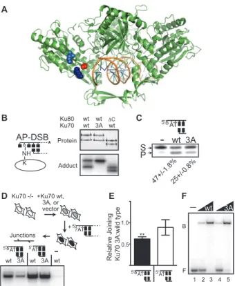
Figure 2.4: Characterization of the Ku70 3A mutant. A. Space filling representations identify K160 (dark blue) and K164 (light blue) in a structure of Ku (green ribbon representation) bound to DNA (1JEY) (22). K31 is not resolved in this structure; instead the proximal G34 is depicted in red space-filling representation. B. Adducts were formed as described in Figure 2.3C with purified wild type heterodimer (wt), a heterodimer of Ku70 3A (Ku70 with K31A, K160A, and K164A substitutions) and wild type Ku80, and a heterodimer of wild type Ku70 and Ku80 lacking the C terminal 162 amino acids (ΔC). Top panel: purified heterodimers and bottom panel: protein-DNA
adducts. C. Reactions were performed with wild type and Ku 70 3A mutant heterodimer on the 300 bp 5’dRP containing substrate as in Figure 2.3B. The proportion of product generated, +/- the s.e.m. for triplicate reactions, is noted below a representative experiment. D and E. Ku70 -/- fibroblasts were complemented with wild type Ku70, Ku70 3A, or empty vector, and then transfected with various substrates as indicated. Joining was evaluated by (D) semi-quantitative PCR (shown for a pool of samples from 4 independent transfections) or E. qPCR. The proportion of joining in Ku70 3A relative to wild type complemented cells was determined for a substrate with a terminal 5’dRP (filled bars), as well as a control substrate with undamaged ends (open bars). Error bars represent the s.e.m. from 4 independent experiments. Ku70 3A complemented cells were significantly less efficient than wild type complemented cells in joining 5’dRP ends; p<0.01 (**). Ku70 3A complemented cells were not significantly different from wild type complemented cells in joining undamaged ends; p=0.59 thus cells were consistently complemented F. 1 or 2.5 nM of wild type or Ku 70 3A heterodimers were incubated with 1 nM radiolabeled 5’dRP-DSB at 25oC for 15 minutes.
Ku-bound DNA complexes (B) were separated from free probe (F) by electrophoresis on a 6% polyacrylamide gel under native conditions.
A
F
- wt 3A
B
F
D
5'AT
5' AT
-Junctions
wt 3A wt 3A wt
5' ?AT +Ku70 wt, 3A, or vector Ku70 -/-+ B Protein Adduct Ku80
Ku70 wtwt wt3A wtC AP-DSB NH * K C 3A wt -5' AT S P 47+/-1.8%25+/-0.8% E 5'AT 5'AT 0.5 1.0 **
Relative Joining Ku70 3A:wild type
Figure 2.5: Analysis of kinetics and substrate specificity. A. Saturating concentrations of Ku were incubated with 5’ dRP-DSB or AP-DSB (50 nM Ku and 10 nM substrate) at 37°C and the fraction of product (y) generated at various times (x) was determined by electrophoresis. Values for kobs were determined by a non-linear
regression fitting of the data to the equation y=ymax(1-e-kobs*x) (Prism, Graphpad
software), and half lives were determined as ln(2)/kobs B. The specificity of Ku’s lyase
activity was directly compared to that of pol β by incubating 1, 2.5, or 5 nM protein with
three noted AP-containing, radiolabeled DNA substrates at 1 nM for 1 minute (5’dRP-SSB), 2.5 minutes (5’dRP-DSB), or 10 minutes (AP-DSB). Products were analyzed by
A
B
Fraction cleaved
Time (min)
C
0 10 20 30 40 50
0.0 0.2 0.4 0.6 0.8 1.0 * B * B
kobs=0.26 min-1
kobs=0.1 min-1
Ku
1 2 3 4 5 6 7 8 9 S P S P S P Substrate 0.04 0.03 0.02 0.01 0.00 Ku V elocity (min -1 )
33
Figure 2.6: AP lyase activity of cell extracts with or without Ku. A. Mock depleted (Mock) or Ku-depleted (α-Ku) HeLa (human) cell extracts, recombinant Ku
(rKu) and wild type (K1), Ku-deficient (xrs6), or Ku-complemented (xrs6+Ku80) hamster (rodent) cell extracts were analyzed by western blotting with antibodies as noted to the left of each row. B, C, D. 1 nM AP-DSB at 37oC was incubated with 1 µg Mock, α-Ku, or
Ku depleted + 4 ng recombinant Ku (α-Ku+rKu) human (HeLa) cell extracts, or 10 µg K1, xrs6, or xrs6+Ku80 rodent (CHO) cell extracts. B, C. Activity assays were performed in triplicate, stopped after 5, 10, 20, and 40 minutes, and products analyzed by gel electrophoresis. Velocities were determined by quantification of the time course and linear regression. Velocities of extracts with Ku were compared to extracts without Ku by two tailed t-test; p<.01, **; p<.0001,***). D. Adduct formation was performed as in Figure 2.3C,D and Figure 2.4B. Top panel is a total protein stain of the noted extracts, while the bottom panel is a phosphorimage of the corresponding protein-DNA adducts. E. HeLa cell extracts were incubated with 10 nM Bleocin (Calbiochem) damaged duplex and 5 mM NaBH4 as in D and Figures 2.3C,D and 2.4B. Total proteins
were detected in the top panel, and DNA-protein adducts detected in the bottom panel. DNA*
Ku
1 2 3 4 5 6 7 8
B v (fmol/min/ g) 0.05 0.10 0.15
Mock -Ku -Ku +rKu
*** *** * AP-DSB HeLa xt: C K1
xrs6 xrs6+ Ku80
1 2 3 v (fmol/min/mg) ** *** CHO xt:
Mock -Ku Xrs6 rKu
-BH 4 K1 Xrs6+Ku80 -Ku+rKu D 28 39 51 64 190
Mock -Ku K1 Xrs6
HeLa CHO
1 2 3 4
Ku pol Actin X6+Ku 5 rKu DNA-PKcs E A kDa
Mock -Ku Recom
-BH 4 20 30 40 50 60 220 80 120 -Bleocin Protein
1 2 3 4 5
Ku DNA*
Adduct
Mock -Ku Recom
-BH
4
-Bleocin
1 2 3 4 5
1 2 3 4 5 6 7 8
Mock -Ku Xrs6 rKu
-BH
4
K1 Xrs6+Ku80