Haploinsufficiency for Core Exon Junction
Complex Components Disrupts Embryonic
Neurogenesis and Causes p53-Mediated
Microcephaly
Hanqian Mao1☯, John J. McMahon1☯, Yi-Hsuan Tsai2, Zefeng Wang2, Debra L. Silver1,3,4,5*
1Department of Molecular Genetics and Microbiology, Duke University School of Medicine, Durham, North Carolina, United States of America,2Department of Pharmacology, University of North Carolina, Chapel Hill, North Carolina, United States of America,3Department of Cell Biology, Duke University School of Medicine, Durham, North Carolina, United States of America,4Department of Neurobiology, Duke University School of Medicine, Durham, North Carolina, United States of America,5Duke Institute for Brain Sciences, Duke University, Durham, North Carolina, United States of America
☯These authors contributed equally to this work. *[email protected]
Abstract
The exon junction complex (EJC) is an RNA binding complex comprised of the core compo-nents Magoh, Rbm8a, and Eif4a3. Human mutations in EJC compocompo-nents cause neurodeve-lopmental pathologies. Further, mice heterozygous for eitherMagohorRbm8aexhibit aberrant neurogenesis and microcephaly. Yet despite the requirement of these genes for neurodevelopment, the pathogenic mechanisms linking EJC dysfunction to microcephaly remain poorly understood. Here we employ mouse genetics, transcriptomic and proteomic analyses to demonstrate that haploinsufficiency for each of the 3 core EJC components causes microcephaly via converging regulation of p53 signaling. Using a new conditional allele, we first show thatEif4a3haploinsufficiency phenocopies aberrant neurogenesis and microcephaly ofMagohandRbm8amutant mice. Transcriptomic and proteomic analyses of embryonic brains at the onset of neurogenesis identifies common pathways altered in each of the 3 EJC mutants, including ribosome, proteasome, and p53 signaling compo-nents. We further demonstrate all 3 mutants exhibit defective splicing of RNA regulatory proteins, implying an EJC dependent RNA regulatory network that fine-tunes gene expres-sion. Finally, we show that genetic ablation of one downstream pathway, p53, significantly rescues microcephaly of all 3 EJC mutants. This implicates p53 activation as a major node of neurodevelopmental pathogenesis following EJC impairment. Altogether our study reveals new mechanisms to help explain how EJC mutations influence neurogenesis and underlie neurodevelopmental disease.
a11111
OPEN ACCESS
Citation:Mao H, McMahon JJ, Tsai Y-H, Wang Z,
Silver DL (2016) Haploinsufficiency for Core Exon Junction Complex Components Disrupts Embryonic Neurogenesis and Causes p53-Mediated Microcephaly. PLoS Genet 12(9): e1006282. doi:10.1371/journal.pgen.1006282
Editor:Paul A Trainor, Stowers Institute for Medical
Research, UNITED STATES
Received:January 21, 2016
Accepted:August 8, 2016
Published:September 12, 2016
Copyright:© 2016 Mao et al. This is an open access
article distributed under the terms of theCreative Commons Attribution License, which permits unrestricted use, distribution, and reproduction in any medium, provided the original author and source are credited.
Data Availability Statement:Relevant data are
within the paper and its Supporting Information files. All genomic analyses have been deposited in GEO database record GSE85576.
Funding:This research was funded by an NIH R01
Author Summary
The mammalian neocortex is the brain structure responsible for higher cognition, abstract thought, and language. One process critical for brain development is neurogenesis, in which neural stem cell populations generate neurons. Alterations in neurogenesis can lead to neurodevelopmental disorders affecting brain size and function, such as microcephaly, in which the brain is significantly smaller than normal. Therefore, understanding the genes and processes controlling normal brain development is of strong clinical relevance. Here we studied proteins of the RNA binding exon junction complex, which are strongly implicated in several neurodevelopmental pathologies, but whose functions in brain devel-opment remain largely unknown. Using mouse models, we find that reduced levels of any of three essential proteins of this complex results in altered embryonic neurogenesis and microcephaly. We demonstrate that mutant mice show common alterations in p53 activa-tion, expression of ribosomal components and splice variants for RNA processing factors. Interestingly we find that genetic suppression of p53 significantly rescues microcephaly in mutant mice. Given that patients harboring mutations in exon junction complex compo-nents present with neurodevelopmental deficits, our findings highlight molecular path-ways which could underlie disease pathogenesis.
Introduction
Proper function of the cerebral cortex, our brain structure responsible for higher cognitive functions, relies upon embryonic neurogenesis. During neurogenesis, neural stem cells (NSCs) generate excitatory neurons [1,2]. In mice the onset of neurogenesis is embryonic day (E) 10.5, when NSCs consist of neuroepithelial cells that primarily undergo self-renewal divisions. As development proceeds, neuroepithelial cells are replaced by radial glial cells that generate neu-rons either directly, or indirectly via new NSCs and intermediate progenitors (IPs) (Fig 1A) [3– 5]. Defective neurogenesis impacts neuron production and can cause neurodevelopmental dis-orders such as microcephaly, in which brain size is severely reduced. To elucidate causes for such diseases requires a comprehensive understanding of how NSCs mediate proper brain development.
One level of control increasingly implicated in NSC function and disease is post-transcrip-tional regulation [6–8]. In particular, a set of RNA binding proteins associated with develop-mental pathologies of the cerebral cortex is the exon junction complex (EJC). The core EJC, composed of Rbm8a (Y14), Magoh, and Eif4a3 (Ddx48), influences mRNA splicing, transla-tion, mRNA localizatransla-tion, and nonsense mediated decay (NMD), via direct interactions with both RNA and auxiliary proteins in the nucleus and cytoplasm [9–15]. Copy number variations ofRBM8A,EIF4A3, and peripheral EJC components, are each strongly associated with neuro-developmental phenotypes [16–18]. MoreoverRBM8AandEIF4A3mutations cause TAR syn-drome and Richieri-Costa-Pereira synsyn-drome, respectively, both of which are associated with neurological deficits [19–22]. While altered EJC levels are significantly linked to neurodevelop-mental diseases, the pathogenic mechanisms by which EJC impairment causes these disorders are largely unknown.
Recent studies from our lab have helped shed light on this question, with the discoveries that haploinsufficiency for eitherMagohorRbm8a, disrupts mouse cortical development. In these mouse models, both NSCs and IPs are depleted, neurons are ectopic, and there is massive apoptosis of neurons and progenitors, all leading to severe microcephaly [23–26]. We recently discovered that inMagohmutants these neurogenesis phenotypes may be due in part to
Competing Interests:The authors have no conflicts
prolonged mitosis of NSCs [26]. Moreover, we identified Lis1 as one relevant Magoh down-stream target during neurogenesis [23]. While these studies showMagohandRbm8aare essen-tial for corticogenesis, it remains unknown if impairment of the third major EJC constituent, Eif4a3, causes microcephaly. Additionally, if all EJC components are required in the developing brain, it is unclear whether they function via common regulatory pathways. This information is critical to understand how EJC genes regulate brain development.
In this study we examined mice haploinsufficient forMagoh,Rbm8a, orEif4a3, to expose mechanisms by which EJC dysfunction impacts cortical development. First, we generated a NSC-specific conditionalEif4a3mouse model to demonstrate thatEif4a3haploinsufficiency phenocopies the aberrant neurogenesis and microcephaly seen inRbm8aandMagohmutants. We then utilized transcriptomic and proteomic analyses to uncover common genetic pathways controlled by all 3 EJC components at the onset of neurogenesis. These include expression of factors associated with the ribosome, proteasome, and p53 signaling pathway. All 3 EJC mutants showed splicing alterations in RNA processing factors, implicating the EJC in regulat-ing a network of RNA metabolism factors. Finally, we focus on one of these downstream path-ways, p53, and show thatp53ablation significantly rescues microcephaly of all 3 EJC mutants. Altogether our study reveals novel mechanisms to help explain how EJC deficiency disrupts Fig 1. EJC components are co-expressed in neurogenesis.(A) Schematic of embryonic neurogenesis of the dorsal telencephalon. NSC, neural stem cell; IP, intermediate progenitor. (B) Two main questions posed in this study. 1. DoesEif4a3haploinsufficiency cause
microcephaly? 2. Do EJC components regulate common pathways during neurogenesis? (C) qPCR ofMagoh,Eif4a3, andRbm8amRNA levels in developing neocortices of indicated ages. qPCR was performed using a standard curve, withMagohrelative expression at E10.5 set to 1.0, and all expression levels normalized toGapdh. (D-I) Immunofluorescence of E10.5 dorsal neocortices for Hoechst (blue), Magoh (D, E), Rbm8a (F, G), and Eif4a3 (H, I). (E, G, I) are high magnification images of D, F, H, respectively. Student’sttest, Error bars, S.D.,**, p<0.01, ns = not significant. n = 3 biological replicates each age. Scale bars, D, F, H; 50μm; E, G, I, 25μm.
neurogenesis, implicating elevated p53 signaling in the etiology of EJC-mediated neurodeve-lopmental pathologies.
Results
Eif4a3
haploinsufficiency causes aberrant neurogenesis and
microcephaly
We previously showed that NSC-specific haploinsufficiency for eitherMagohorRbm8acauses microcephaly in mice [23–25]. To understand whether common mechanisms contribute to microcephaly following depletion of EJC core components, we first sought to address the role of the third core EJC component,Eif4a3, in brain development (Fig 1B). We examined the expression profile ofEif4a3relative toMagohandRbm8aat early stages of cortical develop-ment. RT-qPCR showed thatMagoh,Eif4a3, andRbm8aare expressed in the developing neo-cortex and show parallel increases in expression as neurogenesis proceeds (Fig 1C).In situ hybridization revealed enrichedEif4a3expression in the proliferative ventricular and sub-ven-tricular zones of the E14.5 neocortex, where NSCs reside, in a similar pattern toRbm8aand Magoh[23,24,27] (S1A Fig). Immunostaining showed that at the onset of neurogenesis (E10.5), EIF4A3 protein is expressed at detectable levels and is primarily localized within the nucleus, similar to MAGOH and RBM8A (Fig 1D–1I). Together, these analyses indicate that Eif4a3,MagohandRbm8aare co-expressed spatially and temporally in the developing mouse neocortex.
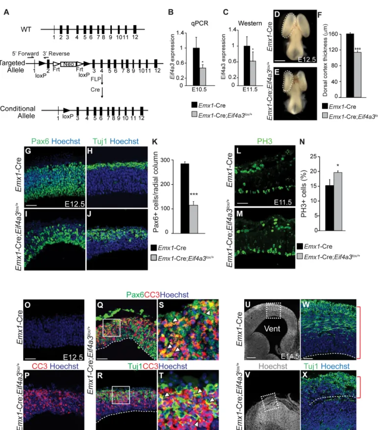
We generated a conditional mouse carrying a floxed allele ofEif4a3(Eif4a3lox/+) to assess the phenotype ofEif4a3deficiency in the developing brain (Fig 2A).Eif4a3lox/+mice were crossed toEmx1-Cre, which drives Cre expression in NSCs of the dorsal neocortex beginning at E9.5 [28,29] (cre.jax.org). Genotyping of genomic DNA fromEmx1-Cre;Eif4a3lox/+mice confirmed the presence of predicted bands for bothwildtypeandloxalleles (S1B Fig). Follow-ing Cre recombination, exon 2 is excised to generate a transcript predicted to undergo NMD-mediated degradation. Consistent with this,Eif4a3mRNA and protein were reduced by about 50% inEmx1-Cre;Eif4a3lox/+neocortices (Fig 2B and 2C). These data demonstrateEif4a3can be efficiently depleted in the conditional haploinsufficient mouse model.
cortex was extremely thinned and neurons were disorganized (Fig 2W and 2X). These pheno-types are highly similar toEmx1-Cre;Rbm8alox/+brains [24].
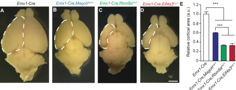
Surprisingly, despite the prevalent disruption of the developing neocortex,Eif4a3,Rbm8a, andMagohconditional mutant mice survive into adulthood [24,25]. We evaluated postnatal (P) brain sizes of each EJC mutant. Comparison of P12 whole mount brains demonstrated sig-nificant reductions in all 3 EJC mutants (Fig 3A–3E). BothEif4a3andRbm8a haploinsuffi-ciency caused severe microcephaly, with an average 70% reduction in cortical area of whole mount brains [24] (Fig 3E). The microcephaly phenotype ofRbm8aandEif4a3mutant mice was significantly worse thanMagohhaploinsufficient mice, which exhibited a 40% reduction [24,25]. This phenotypic difference may be due to redundant expression of a secondMagoh homolog, whereas the other EJC components do not have identifiable homologs [30]. Together with our previous studies, these analyses indicate that Eif4a3, Magoh, and Rbm8a each control similar aspects of neurogenesis (NSC proliferation, number and apoptosis), and ultimately brain size.
Transcriptome analysis of EJC mutants reveals alterations in expression
levels of ribosomal, proteasome, and p53 signaling components
Given the overlapping expression patterns, common neurogenesis phenotypes, and vast litera-ture connecting Magoh, Rbm8a, and Eif4a3, we hypothesized that these EJC components work is noticeably smaller in theEif4a3mutant. (F) Quantification of cortical thickness of E12.5Emx1-Cre andEmx1-Cre;Eif4a3lox/+dorsal neocortices. (G-J) 4
different coronal sections from E12.5Emx1-Cre (G,H) andEmx1-Cre;Eif4a3lox/+(I,J) neocortices stained for Hoechst (blue), Pax6 (green, G,I) or Tuj1 (green, H,J). (K) Density of Pax6+ cells within 200μm wide radial columns spanning the E12.5 cortices of indicated genotypes. (L, M) Images of E11.5 Emx1-Cre (L) orEmx1-Cre;Eif4a3lox/+(M) cortices stained for PH3 (green). (N) Graph depicting percentage of all cells which are PH3-positive for
indicated genotypes at E11.5. (O-T) E12.5 coronal sections fromEmx1-Cre (O) andEmx1-Cre;Eif4a3lox/+brains (P-T) stained for Hoechst (blue), CC3
(red), Pax6 (green, Q, S), and Tuj1 (green, R,T). S and T are high-magnification views of Q and R, respectively, as indicated. Arrowheads depict cells co-labeled for apoptotic and cell fate markers. (U-X) Coronal sections of E14.5Emx1-Cre (U,W) andEmx1-Cre;Eif4a3lox/+(V,X) cortices stained for Hoechst (white or blue) and Tuj1 (green). W and X are high-magnification images of U and V, respectively as indicated. Red brackets denote cortical thickness. Vent, ventricle. Student’sttest,*,p<0.05,***,p<0.001. Error bars, S.D. n = 3 biological replicates each. Scale bars, D, E, 1 mm; G-J, L,M,O-R, W, X, 50μm; S,T, 20μm; U,V, 200μm.
doi:10.1371/journal.pgen.1006282.g002
Fig 3. Haploinsufficiency of EJC components causes microcephaly.(A-D) Images of whole mount brains at P12 from indicated genotypes. Dotted lines denote dorsal cortex. (E) Quantification of relative dorsal cortical area in P12 brains of indicated genotypes. The area ofEmx1-Cre brains was set to 1.0. ANOVA with Tukey posthoc,***,p<0.001, Error bars, S.D. n = 3–4 biological replicates, Scale bar, A-D, 2 mm.
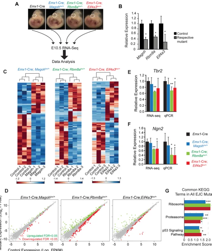
together to influence cortical development. To test this, we aimed to identify molecular changes associated with early neurogenesis in each of the three EJC mutants. We performed transcrip-tome profiling of E10.5 neocortices from the following genotypes:Emx1-Cre,Emx1-Cre; Rbm8alox/+,Emx1-Cre;Magohlox/+, andEmx1-Cre;Eif4a3lox/+(n = 3 biological replicates each) (Fig 4A). We focused on E10.5 for several reasons. This stage marks the beginning of neurogen-esis when the neocortex is composed primarily of self-renewing neuroepithelial NSCs [4]. Moreover, it is just prior to the onset of severe defects in EJC mutants, and a stage when all 3 genes are reduced in their respective mutants, as evidenced by RT-qPCR of the RNA-sequenc-ing samples (Fig 4B).
We examined global RNA changes in the 3 mutants relative to the control and to each other. Amongst the 18,465 detectable coding and non-coding transcripts expressed in the E10.5 control cortex, 2.9% were altered inEmx1-Cre;Rbm8alox/+, 0.9% were altered inEmx1 -Cre;Eif4a3lox/+, and 0.4% were altered inEmx1-Cre;Magohlox/+(FDR, q<0.05) (S1 Table). Hierarchical clustering of these significantly altered transcripts revealed segregation of control and mutant biological replicates for all 3 EJC mutants, as evidenced in heat maps (Fig 4C). Equivalent proportions of transcripts were upregulated and downregulated within individual EJC mutants (Fig 4D,S1 Table). We validated expression for several differentially expressed transcripts,Tbr2,Ngn2,NeuroD6, andGtse1, using RT-qPCR, which showed similar trends to RNA-seq data (Fig 4E and 4F,S2B Fig). Despite the fact that the EJC binds a large fraction of expressed transcripts in immortalized cells [31–33], these experiments suggest EJC haploin-sufficiency does not broadly impair transcript levels of E10.5 neocortices. This observation ech-oes previous microarray studies of germlineMagohMos2/+mutant brains [23],Eif4a3silenced Xenopus[34], and EJCDrosophilamutants [35].
We next assessed the extent to which transcripts overlapped amongst the EJC mutants, focusing only on the fraction of alterations which were highly significant (q<0.05). We noted extensive overlap in pairwise comparisons between individual mutants (S2A Fig). Of the 70 Magohdependent transcripts, 87% were altered inRbm8amutants and 46% were altered in Eif4a3mutants. Of the 172 transcripts altered inEif4a3mutants, 19% overlapped withMagoh mutants and 46% overlapped withRbm8amutants. Fisher’s exact tests demonstrated these overlapping changes were highly significant. In all 3 mutants, 31 transcript changes overlapped, which represents 6%, 18%, and 44% of all altered transcripts in theRbm8a,Eif4a3, andMagoh mutants, respectively. As noted by Venn diagram, some transcript alterations were specific to individual mutants (S2A Fig). This was especially evident inRbm8aandEif4a3mutants, and suggests there could be roles for EJC components outside of the complex. Yet, taken together, these data support the notion that EJC components also work together to selectively affect mRNA levels at the onset of neurogenesis.
11.4%, 6.5% and 7.5% ofMagoh,Rbm8a, andEif4a3mutants, respectively. This indicates altered protein homeostasis pathways, including the ribosome, are shared early consequences of EJC haploinsufficiency.
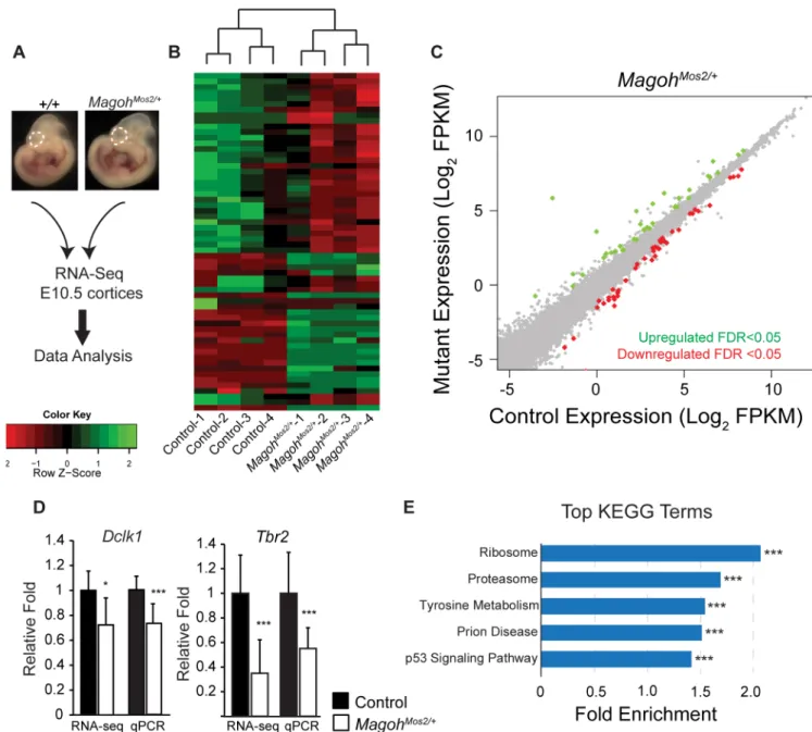
To assess transcript regulation in an independent EJC model not reliant on Cre, we per-formed RNA-sequencing on E10.5 neocortices from control (C57BL/6J) and germlineMagoh haploinsufficient mice (MagohMos2/+) (n = 4 biological replicates each) (Fig 5A). Hierarchical analysis revealed consistent expression changes inMagohMos2/+compared to control litter-mates (Fig 5B). Amongst the 23,577 genes detected, only 80 (0.3%) transcripts were differen-tially expressed (q<0.05), and these were equivalently upregulated and downregulated (Fig 5C, S1 Table). RT-qPCR validation confirmed alterations in two transcripts,Dclk1andTbr2, with similar trends to RNA-seq (Fig 5D). Changes were more dramatic than inEmx1-Cre; Magohlox/+, consistent with a more severe impact of theMagohgermline deletion [23,25]. GSEA KEGG analysis of all detectable transcripts revealed significant enrichment for ribosome, proteasome, and p53 signaling components, amongst additional regulators of protein metabo-lism (Fig 5EandS4A Fig). GO analysis also detected ribosomes as a top altered category (S4C Fig). Of note, we observed overlap betweenEmx1-Cre;Magohlox/+andMagohMos2/+transcripts within the ribosome, proteasome, and p53 categories (S4B Fig). Altogether, these transcrip-tome analyses from 4 independent mouse lines, including 2 models ofMagoh, demonstrate that EJC haploinsufficiency influences a few common pathways including ribosome, proteo-some and p53 signaling.
Haploinsufficiency for
Magoh
,
Eif4a3
, and
Rbm8a
causes aberrant
splicing of RNA regulatory proteins
Given the requirement of the EJC in splicing and NMD, we next assessed splicing isoforms in the transcriptome data. Consistent with a published study in human cell lines [36], wide-spread splicing changes were evident in all 3 EJC mutants compared to control (S3 Table). We measured specific splicing events relative to all annotated alternative splicing (AS) events using Mixture-of-Isoforms (MISO) software [37,38]. Comparing the changed AS events to all annotated AS events, the distribution of AS types was significantly altered, with a 2–3 fold enrichment in retained intron (RI) events in all 3 EJC mutants (p<0.001) (Fig 6A). Amongst the RI events, 61%, 70%, and 23% were increased inMagoh,Rbm8a, andEif4a3 haploinsuffi-cient mutants, respectively (Fig 6B,S3 Table). InEmx1-Cre;Rbm8alox/+, 91% of RI events introduced a premature stop codon, which presumably leads to mRNA degradation through NMD (S3 Table). We validated several events, includingMapk13in E11.5Emx1-Cre; Rbm8alox/+brains andFusinMagohMos2/+brains, noting alterations consistent with predic-tions (Fig 6EandS5A Fig). Thus, the enrichment of RI events could be due to inefficient NMD activity [12,14]. Consistent with previous findings that EJCDrosophilamutants cause increased RI events [39–41], our data suggest EJC components influence mRNA splicing in NSCs.
We next used bioinformatics analysis to determine if there are overlapping classes of splic-ing variants in the 3 EJC mutants. We performed KEGG analysis on those genes with signifi-cant alterations in splice variant expression (Bayes factor>20) using the Database for plots of transcripts significantly upregulated (green dots) and downregulated (red dots) in E10.5Emx1-Cre;Magohlox/+,Emx1-Cre;Rbm8alox/+, and
Emx1-Cre;Eif4a3lox/+cortices (q<0.05). (E, F) qRT-PCR validation at E11.5 compared to relative RNA-seq values ofTbr2(E) andNgn2(F) in the indicated genotypes. For RNA-seq and qPCR, each control was normalized to 1.0 and compared to mutants. (G) Graph depicting common KEGG terms identified by GSEA analysis that were significant in all 3 EJC mutants, showing corresponding enrichment score. Student’sttest (B,E,F), Error bars, S.D.*,p<0.05,**,p<0.01,***,p<0.001.
Annotation, Visualization and Integrated Discovery (DAVID). These analyses showed com-mon terms acom-mongst all EJC mutants, including a significant enrichment of spliceosome (Fig 6F). GO analysis reinforced this finding, with enrichment of RNA regulatory categories includ-ing ribonucleoproteins and ribosomes (S4 Table). 51 identical alternative splicing events were predicted among all 3 EJC mutants. String analyses of these genes revealed two clusters for Fig 5. Transcriptome analyses of E10.5Magohgermline haploinsufficient brains identifies alterations in ribosome and p53 signaling pathways.(A) Diagrammatic overview of RNA sequencing analysis of E10.5 neocortices (dotted lines) from indicated genotypes. (B) Heatmaps showing z-score transformed normalized expression for control andMagohMos2/+. Genes and samples were clustered using correlation distance with complete linkage. (C) Scatter plot of transcripts significantly upregulated (green dots) and downregulated (red dots) in E10.5MagohMos2/+ cortices (q<0.05), n = 4 biological replicates each. (D) Validation and RNA-seq values forDclk1andTbr2in indicated E11.5 mutant dorsal neocortices. Controls were normalized to 1.0. (E) Graph depicting top ranked KEGG terms by GSEA analysis inMagohMos2/+showing
corresponding fold enrichment. Student’sttest (D), Error bars, S.D.*,p<0.05,***,p<0.001.
ribosome regulation and splicing regulation (S5B Fig). These data suggest that in addition to influencing transcript expression, EJC components have been co-opted to impact splicing of RNA regulators. Together, this implies an EJC dependent regulatory network that fine-tunes gene expression at the RNA level.
Fig 6. Haploinsufficiency for EJC components alters mRNA splicing of splicing regulators.(A) Bar graph showing alternative splicing events for each mutant relative to the control. (B-D) Clustered column graphs of the distribution ofψvalues of all identified intron retention (RI) events in E10.5 dorsal cortices for each EJC mutant, using a threshold of 20 for Bayes factor.Ψ<0 indicates higher probability for the mutant to have intron retention when compared to the control. (E) Top: IGV view of increasedMapk13intron 6–7 reads in red frame. Primers indicated as arrows. Bottom: RT-PCR showing increasedMapk13RI isoforms inEmx1-Cre;Rbm8alox/+E11.5 dorsal cortices compared to the control. (F) Bar graph of common KEGG terms that were significant in all 3 EJC mutants, showing corresponding enrichment score. ANOVA (A), Modified fisher’s exact test (F),*,p<0.05,**, p<0.01,***,p<0.001.
Proteomic analyses reveal core EJC components influence protein
levels of ribosomal components and RNA processing factors at the
onset of neurogenesis
We next measured the proteomes of control,Magoh,Rbm8a, andEif4a3haploinsufficient E11.5 neocortices using quantitative proteomic liquid chromatography/mass spectrometry (LC_MS/MS) analyses (n = 3 biological replicates each) (Fig 7A,S5 Table). We detected 3,587 proteins in the control and assessed relative levels of these proteins in each of the mutants. Magoh,Eif4a3, andRbm8ahaploinsufficiency led to significant alterations in 3.8%, 1.5%, and 4.3% of the detectable proteome, respectively (p<0.05). Consistent with our transcriptome analysis, the proteomes of the various mutants showed both overlapping and independent alterations (S6A Fig).
We next asked if there were common alterations amongst those proteins significantly altered in the 3 EJC mutants. Using KEGG DAVID analysis to examine only significant protein changes (p<0.05), we identified ribosomes as the only pathway enriched in all 3 EJC mutants, significant in 2 of the mutants (Fig 7B). GO analysis showed components of ribosomes and ribonucleoprotein complexes amongst the most significantly enriched categories (Fig 7C,S6 Table). We performed STRING analysis of all altered proteins in the EJC mutants within the largest GO term,“Ribonucleoprotein Complex,”which included ribosome components and splicing factors (Fig 7D). This analysis reinforced strong regulatory networks present amongst proteins downstream of the EJC, and the consistent directional changes evident in all 3 mutants. Closer inspection of all significant protein changes within each mutant showed that ribosomal proteins made up 7.9%, 5.8%, and 7.5% ofMagoh,Rbm8a, andEif4a3mutant changes, respectively. A large fraction of ribosomal proteins changed consistently across all 3 mutants, showing up and down regulation at the protein level (S6B Fig). Altogether these geno-mic and proteogeno-mic analyses support the notion that ribosome and ribonucleoprotein alter-ations are major early defects associated with EJC deficiency in the developing brain.
Activation of p53 is a major contributor to microcephaly of EJC mutant
mice
The omics analyses pointed to several common pathways that are dysregulated at the onset of neurogenesis, and suggested candidate molecules that could be relevant for EJC mutant pheno-types. We hypothesized that p53 signaling, in particular, was a major contributor to EJC-medi-ated microcephaly. ActivEJC-medi-ated p53 is a key regulator of apoptosis and defective cell cycle progression [42], two major phenotypes of EJC mutant brains. Moreover, p53 target transcripts were upregulated in all 3 conditional EJC mutants andMagohgermline mutant (Figs4and5, S1 Table). Additionally, a correlation has been previously observed in p53 transcript changes inMagohgermline brains and induced radiation [43]. Altogether these data suggest p53 activa-tion may be a common critical node in disease pathogenesis following EJC impairment. We thus probed the relationship between EJC haploinsufficiency and p53 signaling, by assessing p53 nuclear accumulation in embryonic brain sections, as a proxy for pathway activation [26]. Haploinsufficiency forMagoh,Rbm8a, andEif4a3led to a significant accumulation of p53 in the VZ compared to control brains, which showed no evidence of p53 accumulation (Fig 8A– 8J). Western blotting confirmed accumulation of p53 protein inEif4a3mutant cortices (S7A Fig). P53 activation was evident in E11.5Rbm8amutants (Fig 8E and 8F), prior to the onset of apoptosis [24], and was specifically enriched in PAX6-positive NSCs (Fig 8I and 8J). This dem-onstrates that p53 is activated in EJC haploinsufficient NSCs.
Fig 7. Proteomic analysis of E11.5 EJC mutant brains reveals alterations in levels of ribosome-associated proteins and
Emx1-Cre;Eif4a3lox/+, onto ap53lox/loxnull background. We collected E18.5 embryos and mea-sured cortical area. Compared to control,Emx1-Cre;p53lox/loxdid not alter brain size (Fig 9A, 9C, 9F, 9H, 9K and 9M). As expected, cortical area was significantly reduced in mice haploin-sufficient forMagoh,Rbm8a, orEif4a3, to a similar degree seen in adults (CompareFig 9B, 9G and 9LtoFig 3). Strikingly, for all 3 EJC mutants the microcephaly was significantly, albeit par-tially, rescued in ap53mutant background (Fig 9D, 9I and 9N). Amongst the 3 EJC mutants, the extent of p53-mediated rescue varied and was most effective in the least severe microceph-aly mutant,Magoh(Fig 9E, 9J and 9O). These data indicate that p53 activation is a major cause of microcephaly in all 3 EJC mutants. Our data also suggest that forRbm8aandEif4a3, addi-tional p53 independent factors likely contribute to the reduced brain size.
To elucidate the nature of the p53-mediated rescue we examined apoptosis and neuron number. Amongst the 3 core EJC components, reducedRBM8Alevels are the most strongly associated with human microcephaly [19,21,44]. Given this clinical relevance, we focused our analysis on theRbm8amutant. As p53 is essential for induction of apoptosis, we first assayed whetherp53ablation rescued apoptosis in theRbm8amutant. As predicted, CC3 immunos-taining revealed complete rescue of apoptosis in E12.5Emx1-Cre;Rbm8alox/+;p53lox/loxbrains (S7B–S7D Fig). Thus p53 activation promotes apoptosis downstream ofRbm8a.
We next examined neuronal layers of E18.5 brains (Fig 10A–10D). As we have previously shown [24],Rbm8amutant brains are missing most of their pallium (Fig 10B). In the Emx1-Cre;Rbm8alox/+;p53lox/loxbrains, the pallium is restored, consistent with the rescue of apoptosis (Fig 10D). We asked ifp53loss impacts neuronal layers, focusing on the tissue adjacent to the pallial-subpallial boundary which is still present inRbm8amutants [24]. We quantified both deep and superficial neuronal markers which are generated at early and late stages of neuro-genesis, respectively [4]. As predicted, Cux1+ neurons (layer II/III) were nearly ablated in Emx1-Cre;Rbm8alox/+brains, compared to control orp53alone (Fig 10E–10G). In contrast, in p53;Rbm8acompound mutant brains Cux1+ neuron number was largely rescued (Fig 10H and 10M). Another marker of both superficial and some deep layer neurons, Satb2, was reduced in and level of significance. Two networks of splicing regulators and ribosome-associated proteins are detected. Modified fisher’s exact test,*, p<0.05,**,p<0.01,***,P<0.001.
doi:10.1371/journal.pgen.1006282.g007
Fig 8. EJC haploinsufficiency induces P53 activation.(A-J) Coronal sections of cortices from E13.5Emx1-Cre (A), E13.5Emx1-Cre;Magohlox/+(B), E12.5Emx1-Cre (C, G, I), E12.5Emx1-Cre;Eif4a3lox/+(D), E11.5Emx1-Cre (E), E11.5Emx1-Cre;Rbm8alox/+(F), and E12.5Emx1-Cre;Rbm8alox/+(H,J) embryonic cortices stained for Hoechst (blue), P53 (green), and Pax6 (red), with co-localization indicated in yellow. Sections were demarcated with dotted lines. Each image is representative of at least 3 independent biological samples. Scale bar, A-J, 50μM.
Fig 9. Loss ofp53partially rescues microcephaly ofMagoh,Rbm8a, andEif4a3haploinsufficient mutants.(A-D, F-I, and K-N) Whole mount brains of E18.5 embryos with indicated genotypes. (E, J, O) Quantification of cortical area in E18.5 embryonic brains with indicated genotypes. Dotted lines demarcate the dorsal cortical areas measured. The surface area of littermate control brains was set to 100. ANOVA with Tukey posthoc,**, p<0.01,***,p<0.001, NS, not significant. Error bars, S.D. n = 3–9 biological replicates each. Scale bars, A-D, E-I, and K-N, 1 mm.
Emx1-Cre;Rbm8alox/+, but partially rescued in ap53null background (S7E–S7I Fig)[45,46]. We also examined earlier born deep layer Tbr1+ neurons (Fig 10I–10L). As we previously described [24], inEmx1-Cre;Rbm8alox/+brains Tbr1 number is normal but distribution is skewed basally (Fig 10I, 10J, 10K and 10N–10P). This is consistent with our previous finding that at early stages of development, Tbr1 density is increased inRbm8amutants, perhaps due to increased neuron production [24]. InEmx1-Cre;Rbm8alox/+;p53lox/loxbrains, aberrant Tbr1-+ neuron distribution was restored to normal (Fig 10L, 10O and 10P). These analyses show that inRbm8amutants, p53 activation influences the number and distribution of neurons gen-erated at different stages of neurogenesis, and plays a particularly important role in genesis of upper layer neurons. Taken together, our data implicate p53 activation as a key node in the microcephaly pathology following EJC impairment.
Discussion
The EJC is a central regulator of mRNA metabolism, yet how its molecular roles translate into physiological functions relevant for disease has been poorly understood. Here we used mouse haploinsufficiency models for the 3 core EJC components to demonstrate their common requirements for neurogenesis and proper brain size. We employed transcriptomics and prote-omics to identify converging molecular pathways regulated by the EJC at the onset of neuro-genesis. Our unbiased analyses demonstrate that reduced levels ofMagoh,Eif4a3, orRbm8a lead to altered expression of ribosomal components, splicing changes, and aberrant p53 signal-ing. We focused on the p53 pathway, demonstrating that aberrant p53 activation is a major contributor to EJC-mediated microcephaly. Given that human mutations in EJC components are associated with neurodevelopmental diseases, our study suggests these pathologies may be due in part to aberrant p53 activation.
EJC controls gene expression in neural stem cells
Our study elucidates several layers of EJC-dependent gene expression in the developing neo-cortex. Whereas EJC-dependent targets are known inDrosophilaand immortalized cells [35,36], our study is the first to discover EJC-dependent gene expression in a mammalian stem cell population. We demonstrate that EJC haploinsufficiency alters only a small fraction of the transcriptome, and these changes are disproportionately enriched for ribosomal proteins, pro-teasome components, and p53 signaling. Thus, the EJC may be especially important in regula-tion of protein homeostasis machinery. We also find the 3 core EJC proteins converge in regulating alternative splicing events. In particular we identify aberrant intron retention events which are suggestive of roles in mRNA splicing and NMD, and are consistent with genomic studies of EJC depletion inDrosophilaand mammalian cells [36,39–41]. Notably these splicing changes are enriched for both spliceosomal and ribosomal components. Alterations in ribo-somes are also observed at the protein level. Altogether, these analyses indicate the EJC is inte-gral to an RNA regulation network controlling neurogenesis.
These findings raise several fascinating questions. Although we focused on common EJC regulatory pathways, our data also highlight there are unique targets of individual EJC compo-nents. In future studies it will be of interest to consider potential independent roles for EJC components outside of the complex in neurogenesis. Another interesting question is how the EJC differentially regulates its targets in individual cells. For example, although we measured with Tukey posthoc*,p<0.05,**,p<0.01,***,p<0.001, ns, not significant. Error bars, S.D. n = 2–3 biological replicates each. Scale bars, A-L, 50μm.
genomic changes in tissue that is mainly composed of 1 cell type, neuroepithelial progenitors, observed transcript and splicing differences could be attributed to progenitors in different cell cycle states. Moreover, it will be of interest to determine if the same pathways are regulated by the EJC in non-Emx1-derived cell types.
P53 attenuation rescues microcephaly caused by EJC
haploinsufficiency
We demonstrate that EJC mutant mice all exhibit profound microcephaly, which is signifi-cantly rescued byp53deletion. Detailed analysis ofRbm8amutants reveal thatp53attenuation partially restores superficial neuron number and distribution of deep layer neurons. Thus, the dramatic loss of upper layers inRbm8amutants is due, in part, to p53 activation. At least 2 sce-narios could explain this rescue. P53 induction of apoptosis may severely reduce both neuron and progenitor number, particularly at later stages when upper layers are produced. Aberrant p53 activation may also influence stem cell divisions and thus their progeny. Our lab previously showed increased mitotic index inMagohandRbm8amutant NSCs [23,24,26]. Mitotically delayedMagohmutant NSCs preferentially produce neurons and apoptotic progeny, at the expense of NSCs [26]. We find that p53 activation is evident at E10.5, which precedes the onset of mitotic defects at E11.5 and E12.5. Given this sequence of events, it is tempting to speculate that aberrant p53 activation may influence progenitor (and ultimately neuron) number by delaying mitosis. Future experiments will be useful for evaluating if this relationship is correla-tive or causal.
How might p53 be activated by EJC dysfunction? It is plausible that ribosomal alterations contribute to p53 activation, as evidenced in many examples from the literature for genes con-trolling ribosome biogenesis [42,47–51]. Alternatively, p53 could be activated independent of the ribosome, as seen in the pancreas [52]. The EJC could also directly regulate RNA metabo-lism of p53 pathway components, as has been observed in splicing of apoptotic regulators [53]. The mechanisms contributing to p53 activation in EJC models are a topic of future interest.
Beyond p53: other alterations associated with EJC haploinsufficiency
ForEif4a3andRbm8amutants, p53 rescue was incomplete, suggesting there must be addi-tional EJC-dependent pathways mediating early stages of neurogenesis. Our analyses implicate several promising candidates. Reduced expression of canonical neurogenesis regulators, includingNgn2,Tbr2, andNeuroD6, could contribute to cell fate changes in the neocortex. All 4 EJC mutants also showed alterations in components of the proteasome, indicating that the EJC could influence neurogenesis by regulating protein homeostasis. Our data also identify ribosomal alterations at the transcriptome, splicing, and proteomic level, suggesting ribosome regulation could contribute to EJC-dependent microcephaly. Indeed, human genetic studies suggest that ribosome biogenesis defects cause neurodevelopmental diseases [54]. Of the signif-icant (FDR<0.05) changes in 3 different mutants, ribosomal transcripts made up 5–11%, well above the fraction expressed in progenitors, a finding which is reinforced with unbiased GSEA analysis.
that ribosomal transcripts at E10.5 were nearly universally upregulated, whereas one day later the proteins were differentially altered. This could be due to differences in RNA versus protein regulation or could suggest compensatory responses to restore ribosomal levels in the brain. Understanding the nature of how the EJC influences ribosome stoichiometry, and how this may influence microcephaly, will be an important question for the future.
Roles for the EJC in neurogenesis and neurodevelopmental disorders
Mutations and copy number variations in core and peripheral EJC components are strongly associated with neurodevelopmental deficits in humans, yet the etiology of these pathologies is poorly understood. Microdeletions and duplications of a 15-gene locus containingRBM8Aare associated with microcephaly, macrocephaly, autism, and epilepsy [19,20]. Compound inheri-tance of this deletion and a regulatoryRBM8Amutation is responsible for TAR syndrome, a congenital malformation of blood and skeletal systems which can also present with neurolog-ical deficits [21]. Moreover, regulatoryEIF4A3mutations cause a craniofacial disorder
presenting with learning and language disabilities [22]. Intriguingly, both craniofacial and neu-rodevelopmental anomalies are associated with disruption of p53 signaling and ribosomal impairments [49,50,59,60]. It is notable the EJC downstream splicing changes include several genes, such asRPL10, which are mutated in patients with neurodevelopmental disorders [60]. Thus, it is interesting to consider whether some of the expression changes we have identified in mouse models may contribute to EJC disease etiology.
Altogether, based on our discoveries, we propose aberrant p53 signaling contributes to the pathology of EJC related disorders and that modifications of p53 signaling may be of potential therapeutic interest. It is tempting to speculate that EJC diseases could be considered as riboso-mopathies. Going forward, the EJC haploinsufficient mouse mutants we have generated pro-vide valuable models for understanding the etiology of microcephaly and dissecting cell autonomous requirements in NSCs. Future studies using ubiquitous knockout of EJC compo-nents may help to further model other disease manifestations. In summary, our findings dem-onstrate new mechanisms to explain how EJC haploinsufficiency causes microcephaly, which has implications for understanding physiological functions of the EJC in the developing brain and in disease pathogenesis.
Materials and Methods
Ethics statement
All experiments were performed in agreement with the guidelines from the Division of Labora-tory Animal Resources from Duke University School of Medicine and IACUC.
Mouse husbandry and generation of conditional
Eif4a3
allele
min (1X); 94°C X 15 s, 62°C X 20 s, 72°C X 30s (30X); 72°C X 10 min (1X). 5’forward: CTTGCAGTTGTCTTTCTGCGG; 3’Reverse: CACACATGGCGATCCGCTCG. The follow-ing strain was acquired from Jackson labs:Emx1-Cre(B6.129S2-Emx1tm1(cre)Krj/J).
Western blot and RT-qPCR analyses
E10.5 neocortices and E11.5 dorsal cortices were collected fromEmx1-Cre,Emx1-Cre; Eif4a3lox/+,Emx1-Cre;Rbm8alox/+, andEmx1-Cre;Magohlox/+mice and lysed in RIPA lysis buffer with protease inhibitors (Pierce, Rockford, IL). Cortical lysates were run on 4–20% pre-casted SDS–Polyacrylamide gels (Bio-Rad). For Pax6 and P53 blots, stain free gels were used for total protein normalization. Gels were transferred onto nitrocellulose membranes and blotted using the following primary antibodies: rabbit anti-Eif4a3 (1:200, Santa Cruz), rabbit anti-Pax6 (1:1,000, Millipore), rabbit anti-p53 (1:1,000, Leica) and mouse anti-α -Tubu-lin (1:10,000, Sigma). Blots were developed using ECL reagent (Pierce). Densitometry was performed using ImageJ. Final values were quantified by normalizing EJC protein levels to loading controls (1:10,000, Tubulin, Sigma) or UV-induced Stain-free pre-casted gel (Bio-Rad), and analyzed for significance using a Student’sttest. For qPCRs, whole neocortices from E10.5 and dorsal neocortices of E11.5, and E12.5 and E14.5 embryos were collected from C57BL/6J (wild-type),Emx1-Cre,Emx1-Cre;Eif4a3lox/+,Emx1-Cre;Rbm8alox/+, and Emx1-Cre;Magohlox/+embryos and RNA was extracted using Trizol reagent (Invitrogen) followed by the RNeasy kit (Qiagen). cDNA was prepared according to the iScript kit (Bio-Rad). qPCR was performed in triplicates using Taqman probes (Life Technologies):
Rbm8a(Mm04214345_s1),Eif4a3(Mm00836350_g1),Magoh(Mm00487546_m1),Ngn2
(Mm00437603_g1),Tbr2(Mm01351984_m1),Dclk1(Mm00444950_m1) andGapdh
(4352339E). Sybr Green iTaq (Biorad) was performed with primers designed forGste1(5’ For-ward-CCAGAGCAAAGAGGACCAAG and 3’Reverse-CCGTGAGAACTTTGGGGTTA),
NeuroD65’Forward-GCCTCAATGATGCTCTGGACAA and 3’Reverse- CTCTTGCCA
ATCCTCAGAATTTCAG), andβ-Actin(5’Forward- CCTTCTTGGGTATGGAATCCTG and 3’Reverse- GTTGGCATAGAGGTCTTTACGG). For wild-type samples at different developmental stages, semi-quantitative qRT-PCR was performed. A standard curve was gen-erated with a 5 serial 10-fold dilution of cDNA from an independent E14.5 wildtype embryo. Final values were normalized toGapdhloading control. For E10.5 control and conditional mutant samples, comparative qRT-PCR was performed. Values were normalized toGapdh control. For each genotype, 3 embryos were examined, a student’sttest was run to determine the significance. For all experiments, 3 biological samples for each genotype were used.
Immunohistochemistry and quantification of tissue sections
Fluor 594 (1:200–400; Invitrogen) and Hoechst (1:1000; Invitrogen). High magnification images were captured using a Zeiss Axio Observer Z.1 microscope coupled with an apotome. Cortical thickness was measured with Zen software. Quantifications were performed using ImageJ. A minimum of 3 sections from anatomically comparable regions per embryo and 3 biological replicates from control and mutants were measured/quantified.
RNA-Seq, splicing and bioinformatics
Control and EJC mutant embryonic neocortices were dissected at E10.5. Samples were flash-frozen in liquid nitrogen and stored at -80°C until further treatment. RNA was extracted with Trizol (Invitrogen) followed by micro-RNeasy kit (Qiagen) according to manufacturer’s proto-col. The library was generated with Kapa stranded mRNA-seq Kit. The fragmented poly-A RNAs were sequenced using Illumina Hi-Seq 2000 double end sequencing with 100nt length. RNA-seq data was processed using the TrimGalore toolkit (http://www.bioinformatics. babraham.ac.uk/projects/trim_galore) which employs Cutadapt to trim low quality bases and Illumina sequencing adapters from the 3’end of the reads [61]. Only pairs where both reads were 20 nt or longer were kept for further analysis. Reads were mapped to the NCBIM38r73 version of the mouse genome and transcriptome using the STAR RNA-seq alignment tool [62]. Reads were kept for subsequent analysis if they mapped to a single genomic location. Gene counts were compiled using the HTSeq tool (http://www-huber.embl.de/users/anders/ HTSeq/). Only genes that had at least 10 reads in any given library were used in subsequent analysis. Normalization and differential expression was carried out using the EdgeR Biocon-ductor package with the R statistical programming environment [63]. The exact test method was used to identify differentially expressed genes between the different mouse genotypes. Inspection of reads using integrative genomics viewer (IGV) software confirmed altered regula-tion of pseudogenes. Heatmaps were prepared for z-score transformed normalized expression for genes with an FDR, q<5%. To calculate significant overlap for Venn diagrams the following criteria were used: genes must with a q<0.05 and using a Fisher’s Exact Test for overlap between any two conditions. For alternative splicing analysis, Mixture-of-isoforms (MISO)[38] model was used to analyze RNA-Seq data and estimate the percent of splicing isoforms (C val-ues, for‘Percent Spliced Isoform’), and the differentially spliced events are identified using a stringent filter (bayes-factor>20). The program was run with pooled samples of 3 biological replicates to reduce sampling biases. Validation of RI events was performed by RT-PCR with cDNA prepared from E11.5 dorsal cortices of control and EJC mutant embryos. The following primers were used:FusEx6 Forward: GGCCAAGATCAGTCCTCTATGAGT,FusEx8 Reverse: CATGACGAGATCCTTGATCCCGA,Mapk13Ex6 Forward: GCAACCTGGCTGT GAATGAA, andMapk13Ex7 reverse: CTGGTTGTAATGCATCCAGCTG.
Pathway analyses for RNA seq and proteomics
Discovery v6.7 was used to analyze significant changes by KEGG and gene ontology (GO) anal-ysis (including biological process, molecular function, and cellular component). Significance of enrichment in GO term analyses was calculated using the p value function given from a modi-fied Fisher’s exact test by the DAVID database. For splicing analysis, STRING (Search Tool for the Retrieval of Interacting Genes/Proteins) analysis was carried out with transcripts show sig-nificant splicing changes (Bayes>20) in all 3 EJC mutants. For proteomic analysis, STRING was carried out with significantly changed (p<0.05) proteins in the“ribonucleoprotein com-plex”GO term. All components not connected to other genes/proteins were not included in figures.
Proteomics and bioinformatics
Supporting Information
S1 Fig. Analysis of conditionalEif4a3haploinsufficient mutant.(A)In situhybridization of Eif4a3in sagittal E14.5 mouse section, showing enrichment in the ventricular and sub-ventric-ular zones (arrowheads) relative to the cortical plate (CP). Images are fromwww.genepaint.org (Visel et al. 2004). (B) Representative PCR genotyping result fromEmx1-Cre (control) and Emx1-Cre;Eif4a3lox/+mice. Note a single band (432 bp) in control and two bands (432 bp and 490 bp) inEmx1-Cre;Eif4a3lox/+. (C) Representative western blot of Pax6. (D) Quantification of Pax6 expression from E10.5 cortical lysates. Error bars, S.D, n = 3 biological replicates each. (TIF)
S2 Fig. RNA seq Validation and analysis of EJC mutants.(A) Venn Diagrams showing the overlap of significant transcript changes (q<0.05) among the 3 EJC mutants. For pairwise com-parisons between datasets, the percent of overlapping transcript changes within each mutant is shown, along with associatedpvalues. (B) qPCR validation ofNeuroD6andGtse1mRNA expression in indicated E11.5 mutant cortices. For RNA-seq and qPCR, each control was nor-malized to 1.0 and compared to mutants. (C) Bar graph of top common enriched GO terms identified with GSEA analysis among all 3 EJC mutants, showing corresponding fold enrich-ment and P values. (D) Plot of all ribosomal protein transcripts for EJC mutant RNA seq. Stu-dent’s t-test (B). Error bars, S.D,,p<0.05,,p<0.01,,p<0.001.
(TIF)
S3 Fig. GSEA analysis of transcriptome data of EJC mutants.(A, C, E) Enrichment plots from GSEA KEGG analysis for Ribosome (A), Proteasome (C) and p53 signaling (E) terms. (B, D, F) Venn diagrams of overlapping enriched genes between EJC mutants for the Ribosome (B), Proteasome (D), and p53 signaling (F) terms. (G, H, I) STRING analysis of the genes enriched in the Ribosome (G), Proteasome (H), and p53 signaling (I) pathways.
(TIF)
S4 Fig. GSEA analysis ofMagohMos2/+transcriptome data.(A) Enrichment plots from GSEA KEGG analysis for Ribosome, Proteasome, and p53 signaling terms. (B) Venn diagrams of overlapping enriched genes betweenEmx1-Cre;Magohlox/+andMagohMos2/+mutants for the Ribosome, Proteasome, and p53 signaling terms. (C) Top enriched GO terms from GSEA anal-ysis ofMagohMos2/+transcriptome.,p<0.001.
(TIF)
S5 Fig. Splicing Analysis ofMagoh,Rbm8a, andEif4a3haploinsufficient cortices.(A) Gel image showing detection of increasedFusEx6-8 RI events in E11.5MagohMos2/+mutants com-pared to 2 litter mate controls. (B) STRING analysis including genes predicted to show identi-cal, significant splicing changes in all 3 EJC mutants. Common genes that are not connected with any other genes by STRING analysis were not included. Stronger associations are repre-sented by thicker lines. Note two networks of splicing regulation (cyan) and Ribosome/Trans-lation (orange).
(TIF)
S6 Fig. Proteomic Analysis of EJC mutants.(A) Venn diagrams of overlapping enriched pro-teins altered between EJC mutants(p<0.05).(B) Bar graph depicting all ribosomal protein changes relative to control showing similar trends in all 3 EJC mutants (control levels are set to 0). (TIF)
control (B),Emx1-Cre;Rbm8alox/+;p53lox/+(C) andEmx1-Cre;Rbm8alox/+;p53lox/lox(D). (E-H) Representative images of Satb2 immunostaining in E18.5 cortex fromEmx1-Cre,Emx1-Cre; Rbm8alox/+,Emx1-Cre;p53lox/loxandEmx1-Cre;Rbm8alox/+;p53lox/lox. (I) Quantification of Satb2+ cells from E-H. ANOVA with Tukey posthoc. Error bars, S.D.,p<0.05,,p<0.01,, p<0.001, Scale bar, 50μm.
(TIF)
S1 Table. Transcriptome analysis of EJC mutants. (XLSX)
S2 Table. Transcriptome analysis GO terms. (XLSX)
S3 Table. Splicing analysis of EJC mutants. (XLSX)
S4 Table. Splicing analysis GO terms. (XLSX)
S5 Table. Proteomic analysis of EJC mutants. (XLSX)
S6 Table. Proteomic GO terms. (XLSX)
Acknowledgments
We thank members of the Silver lab for helpful discussions and careful reading of the manu-script. We thank Autumn Rorrer and Jeremy Rouanet for technical assistance, and the Duke Mouse Transgenic facility, Microscopy core, Genome Sequencing facility, Proteomics facility.
Author Contributions
Conceived and designed the experiments:HM JJM DLS.
Performed the experiments:HM JJM DLS.
Analyzed the data:HM JJM YHT ZW DLS.
Contributed reagents/materials/analysis tools:HM JJM ZW DLS.
Wrote the paper:HM JJM ZW DLS.
References
1. Malatesta P, Hartfuss E, Gotz M. Isolation of radial glial cells by fluorescent-activated cell sorting reveals a neuronal lineage. Development. 2000; 127: 5253–5263. PMID:11076748
2. Noctor SC, Flint AC, Weissman TA, Dammerman RS, Kriegstein AR. Neurons derived from radial glial cells establish radial units in neocortex. Nature. 2001; 409: 714–720. doi:10.1038/35055553PMID: 11217860
3. Franco SJ, Müller U. Shaping Our Minds: Stem and Progenitor Cell Diversity in the Mammalian Neocor-tex. Neuron. Elsevier Inc; 2013; 77: 19–34. doi:10.1016/j.neuron.2012.12.022
5. Kowalczyk T, Pontious A, Englund C, Daza RAM, Bedogni F, Hodge R, et al. Intermediate neuronal progenitors (basal progenitors) produce pyramidal-projection neurons for all layers of cerebral cortex. Cereb Cortex. 2009; 19: 2439–2450. doi:10.1093/cercor/bhn260PMID:19168665
6. Pilaz L-J, Silver DL. Post-transcriptional regulation in corticogenesis: how RNA-binding proteins help build the brain. WIREs RNA. 2015; 6: 501–515. doi:10.1002/wrna.1289PMID:26088328
7. DeBoer EM, Kraushar ML, Hart RP, rasin M-RR. Post-transcriptional regulatory elements and spatio-temporal specification of neocortical stem cells and projection neurons. Neuroscience. 2013; 248: 499– 528. doi:10.1016/j.neuroscience.2013.05.042PMID:23727006
8. Kraushar ML, Viljetic B, Wijeratne HRS, Thompson K, Jiao X, Pike JW, et al. Thalamic WNT3 Secretion Spatiotemporally Regulates the Neocortical Ribosome Signature and mRNA Translation to Specify Neocortical Cell Subtypes. The Journal of Neuroscience: the official journal of the Society for Neurosci-ence. 2015; 35: 10911–10926. doi:10.1523/JNEUROSCI.0601-15.2015
9. Kataoka N, Diem MD, Kim VN, Yong J, Dreyfuss G. Magoh, a human homolog of Drosophila mago nashi protein, is a component of the splicing-dependent exon-exon junction complex. EMBO J. 2001; 20: 6424–6433. doi:10.1093/emboj/20.22.6424PMID:11707413
10. Mohr SE, Dillon ST, Boswell RE. The RNA-binding protein Tsunagi interacts with Mago Nashi to estab-lish polarity and localize oskar mRNA during Drosophila oogenesis. Genes Dev. 2001; 15: 2886–2899. doi:10.1101/gad.927001PMID:11691839
11. Nott A, Le Hir HE, Moore MJ. Splicing enhances translation in mammalian cells: an additional function of the exon junction complex. Genes Dev. 2004; 18: 210–222. doi:10.1101/gad.1163204PMID: 14752011
12. Palacios IM, Gatfield D, St Johnston D, Izaurralde E. An eIF4AIII-containing complex required for mRNA localization and nonsense-mediated mRNA decay. Nature. 2004; 427: 753–757. doi:10.1038/ nature02351PMID:14973490
13. Ma XM, Yoon S-O, Richardson CJ, Jülich K, Blenis J. SKAR links pre-mRNA splicing to mTOR/S6K1-mediated enhanced translation efficiency of spliced mRNAs. Cell. 2008; 133: 303–313. doi:10.1016/j. cell.2008.02.031PMID:18423201
14. Le Hir HE, Sauliere J, Wang Z. The exon junction complex as a node of post-transcriptional networks. Nat Rev Mol Cell Biol. Nature Publishing Group; 2015;: 1–14. doi:10.1038/nrm.2015.7
15. Lou CH, Shao A, Shum EY, Espinoza JL, Huang L, Karam R, et al. Posttranscriptional control of the stem cell and neurogenic programs by the nonsense-mediated RNA decay pathway. CellReports. 2014; 6: 748–764. doi:10.1016/j.celrep.2014.01.028
16. Nguyen LS, Kim H-G, Rosenfeld JA, Shen Y, Gusella JF, Lacassie Y, et al. Contribution of copy num-ber variants involving nonsense-mediated mRNA decay pathway genes to neuro-developmental disor-ders. Hum Mol Genet. Oxford University Press; 2013; 22: 1816–1825. doi:10.1093/hmg/ddt035 17. Laumonnier F, Shoubridge C, Antar C, Nguyen LS, van Esch H, Kleefstra T, et al. Mutations of the
UPF3B gene, which encodes a protein widely expressed in neurons, are associated with nonspecific mental retardation with or without autism. Mol Psychiatry. 2010; 15: 767–776. doi:10.1038/mp.2009.14 PMID:19238151
18. Tarpey PS, Raymond FL, Nguyen LS, Rodriguez J, Hackett A, Vandeleur L, et al. Mutations in UPF3B, a member of the nonsense-mediated mRNA decay complex, cause syndromic and nonsyndromic men-tal retardation. Nature Publishing Group. 2007; 39: 1127–1133. doi:10.1038/ng2100
19. Brunetti-Pierri N, Berg JS, Scaglia F, Belmont J, Bacino CA, Sahoo T, et al. Recurrent reciprocal 1q21.1 deletions and duplications associated with microcephaly or macrocephaly and developmental and behavioral abnormalities. Nat Genet. 2008; 40: 1466–1471. doi:10.1038/ng.279PMID:19029900 20. Mefford HC, Sharp AJ, Baker C, Itsara A, Jiang Z, Buysse K, et al. Recurrent rearrangements of
chro-mosome 1q21.1 and variable pediatric phenotypes. N Engl J Med. 2008; 359: 1685–1699. doi:10. 1056/NEJMoa0805384PMID:18784092
21. Albers CA, Paul DS, Schulze H, Freson K, Stephens JC, Smethurst PA, et al. Compound inheritance of a low-frequency regulatory SNP and a rare null mutation in exon-junction complex subunit RBM8A causes TAR syndrome. Nat Genet. 2012; 44: 435–9–S1–2. doi:10.1038/ng.1083
22. Favaro FP, Alvizi L, Zechi-Ceide RM, Bertola D, Felix TM, de Souza J, et al. A Noncoding Expansion in EIF4A3 Causes Richieri-Costa-Pereira Syndrome, a Craniofacial Disorder Associated with Limb Defects. Am J Hum Genet. 2014; 94: 120–128. doi:10.1016/j.ajhg.2013.11.020PMID:24360810 23. Silver DL, Watkins-Chow DE, Schreck KC, Pierfelice TJ, Larson DM, Burnetti AJ, et al. The exon
24. Mao H, Pilaz L-J, McMahon JJ, Golzio C, Wu D, Shi L, et al. Rbm8a haploinsufficiency disrupts embry-onic cortical development resulting in microcephaly. Journal of Neuroscience. 2015; 35: 7003–7018. doi:10.1523/JNEUROSCI.0018-15.2015PMID:25948253
25. McMahon JJ, Shi L, Silver DL. Generation of a Magoh conditional allele in mice. Genesis. 2014; 52: 752–758. doi:10.1002/dvg.22788PMID:24771530
26. Pilaz L-J, McMahon JJ, Miller EE, Lennox AL, Suzuki A, Salmon E, et al. Prolonged mitosis of neural progenitors alters cell fate in the developing brain. Neuron. 2016; 89: 83–99. doi:10.1016/j.neuron. 2015.12.007PMID:26748089
27. Visel A, Thaller C, Eichele G. GenePaint.org: an atlas of gene expression patterns in the mouse embryo. Nucleic Acids Res. 2004; 32: D552–6. doi:10.1093/nar/gkh029PMID:14681479
28. Sahara S, O'Leary DDM. Fgf10 regulates transition period of cortical stem cell differentiation to radial glia controlling generation of neurons and basal progenitors. Neuron. 2009; 63: 48–62. doi:10.1016/j. neuron.2009.06.006PMID:19607792
29. Gorski JA, Talley T, Qiu M, Puelles L, Rubenstein JLR, Jones KR. Cortical excitatory neurons and glia, but not GABAergic neurons, are produced in the Emx1-expressing lineage. Journal of Neuroscience. 2002; 22: 6309–6314. PMID:12151506
30. Singh KK, Wachsmuth L, Kulozik AE, Gehring NH. Two mammalian MAGOH genes contribute to exon junction complex composition and nonsense-mediated decay. RNA Biol. 2013; 10: 1291–1298. doi:10. 4161/rna.25827PMID:23917022
31. Sauliere J, Haque N, Harms S, Barbosa I, Blanchette M, Le Hir HE. The exon junction complex differen-tially marks spliced junctions. Nat Struct Mol Biol. 2010; 17: 1269–1271. doi:10.1038/nsmb.1890 PMID:20818392
32. Singh G, Kucukural A, Cenik C, Leszyk JD, Shaffer SA, Weng Z, et al. The cellular EJC interactome reveals higher-order mRNP structure and an EJC-SR protein nexus. Cell. 2012; 151: 750–764. doi:10. 1016/j.cell.2012.10.007PMID:23084401
33. Sauliere J, Murigneux V, Wang Z, Marquenet E, Barbosa I, Le Tonquèze O, et al. CLIP-seq of eIF4AIII reveals transcriptome-wide mapping of the human exon junction complex. Nat Struct Mol Biol. 2012; 19: 1124–1131. doi:10.1038/nsmb.2420PMID:23085716
34. Haremaki T, Weinstein DC. Eif4a3 is required for accurate splicing of the Xenopus laevis ryanodine receptor pre-mRNA. Dev Biol. Elsevier; 2012;: 1–8. doi:10.1016/j.ydbio.2012.08.013
35. Roignant J-Y, Treisman JE. Exon junction complex subunits are required to splice Drosophila MAP kinase, a large heterochromatic gene. Cell. 2010; 143: 238–250. doi:10.1016/j.cell.2010.09.036PMID: 20946982
36. Wang Z, Murigneux V, Le Hir HE. Transcriptome-wide modulation of splicing by the Exon Junction Complex. Genome Biol. 2014; 15: 551. doi:10.1186/PREACCEPT-1827287072145398PMID: 25476502
37. Wang ET, Sandberg R, Luo S, Khrebtukova I, Zhang L, Mayr C, et al. Alternative isoform regulation in human tissue transcriptomes. Nature. 2008; 456: 470–476. doi:10.1038/nature07509PMID: 18978772
38. Katz Y, Wang ET, Airoldi EM, Burge CB. Analysis and design of RNA sequencing experiments for iden-tifying isoform regulation. Nat Methods. 2010; 7: 1009–1015. doi:10.1038/nmeth.1528PMID: 21057496
39. Hayashi R, Handler D, Ish-Horowicz D, Brennecke J. The exon junction complex is required for defini-tion and excision of neighboring introns in Drosophila. Genes Dev. 2014. doi:10.1101/gad.245738.114 40. Malone CD, Mestdagh C, Akhtar J, Kreim N, Deinhard P, Sachidanandam R, et al. The exon junction
complex controls transposable element activity by ensuring faithful splicing of the piwi transcript. Genes Dev. 2014; 28: 1786–1799. doi:10.1101/gad.245829.114PMID:25104425
41. Ashton-Beaucage D, Udell CM, Lavoie H, Baril C, Lefrançois M, Chagnon P, et al. The exon junction complex controls the splicing of MAPK and other long intron-containing transcripts in Drosophila. Cell. 2010; 143: 251–262. doi:10.1016/j.cell.2010.09.014PMID:20946983
42. Golomb L, Volarevic S, Oren M. p53 and ribosome biogenesis stress: The essentials. FEBS Lett. 2014; 588: 2571–2579. doi:10.1016/j.febslet.2014.04.014PMID:24747423
43. Quintens R, Verreet T, Janssen A, Neefs M, Leysen L, Michaux A, et al. Identification of novel radia-tion-induced p53-dependent transcripts extensively regulated during mouse brain development. Biol-ogy Open. 2015; 4: 331–344. doi:10.1242/bio.20149969PMID:25681390
45. Leone DP, Heavner WE, Ferenczi EA, Dobreva G, Huguenard JR, Grosschedl R, et al. Satb2 Regu-lates the Differentiation of Both Callosal and Subcerebral Projection Neurons in the Developing Cere-bral Cortex. Cereb Cortex. 2015; 25: 3406–3419. doi:10.1093/cercor/bhu156PMID:25037921 46. McKenna WL, Ortiz-Londono CF, Mathew TK, Hoang K, Katzman S, Chen B. Mutual regulation
between Satb2 and Fezf2 promotes subcerebral projection neuron identity in the developing cerebral cortex. Proceedings of the National Academy of Sciences. National Acad Sciences; 2015; 112: 11702– 11707. doi:10.1073/pnas.1504144112
47. Sloan KE, Bohnsack MT, Watkins NJ. The 5S RNP Couples p53 Homeostasisto Ribosome Biogenesis and Nucleolar Stress. CellReports. The Authors; 2013; 5: 237–247. doi:10.1016/j.celrep.2013.08.049 48. Brombin A, Joly J-S, Jamen F. ScienceDirectNew tricks for an old dog: ribosome biogenesis
contrib-utes to stem cell homeostasis. Curr Opin Genet Dev. Elsevier Ltd; 2015; 34: 61–70.
49. Jones NC, Lynn ML, Gaudenz K, Sakai D, Aoto K, Rey J-P, et al. Prevention of the neurocristopathy Treacher Collins syndrome through inhibition of p53 function. Nat Med. 2008; 14: 125–133. doi:10. 1038/nm1725PMID:18246078
50. Willig T-N, Draptchinskaia N, Dianzani I, Ball S, Niemeyer C, Ramenghi U, et al. Mutations in Ribo-somal Protein S19 Gene and Diamond Blackfan Anemia: Wide Variations in Phenotypic Expression. Blood. American Society of Hematology; 1999; 94: 4294–4306. doi:10.1182/blood.V97.2.579 51. Armistead J, Patel N, Wu X, Hemming R, Chowdhury B, Basra GS, et al. Biochimica et Biophysica
Acta. BBA—Molecular Basis of Disease. Elsevier B.V; 2013; 1852: 1029–1037. doi:10.1016/j.bbadis. 2015.02.007
52. Provost E, Wehner KA, Zhong X, Ashar F, Nguyen E, Green R, et al. Ribosomal biogenesis genes play an essential and p53-independent role in zebrafish pancreas development. Development. Oxford Uni-versity Press for The Company of Biologists Limited; 2012; 139: 3232–3241. doi:10.1242/dev.077107 53. Michelle L, Cloutier A, Toutant J, Shkreta L, Thibault P, Durand M, et al. Proteins associated with the
exon junction complex also control the alternative splicing of apoptotic regulators. Mol Cell Biol. 2012; 32: 954–967. doi:10.1128/MCB.06130-11PMID:22203037
54. Armistead J, Triggs-Raine B. Diverse diseases from a ubiquitous process: The ribosomopathy paradox. FEBS Lett. Federation of European Biochemical Societies; 2014; 588: 1491–1500. doi:10.1016/j. febslet.2014.03.024
55. Kressler D, la Cruz de J, Rojo M, Linder P. Fal1p is an essential DEAD-box protein involved in 40S-ribo-somal-subunit biogenesis in Saccharomyces cerevisiae. Mol Cell Biol. 1997; 17: 7283–7294. PMID: 9372960
56. Alexandrov A, Colognori D, Steitz JA. Human eIF4AIII interacts with an eIF4G-like partner, NOM1, revealing an evolutionarily conserved function outside the exon junction complex. Genes Dev. 2011; 25: 1078–1090. doi:10.1101/gad.2045411PMID:21576267
57. Tafforeau L, Zorbas C, Langhendries J-L, Mullineux S-T, Stamatopoulou V, Mullier R, et al. The Com-plexity of Human Ribosome Biogenesis Revealed by Systematic Nucleolar Screening of Pre-rRNA Pro-cessing Factors. Mol Cell. Elsevier Inc; 2013; 51: 539–551. doi:10.1016/j.molcel.2013.08.011 58. Badertscher L, Wild T, Montellese C, Alexander LT, Bammert L, Sarazova M, et al. Genome-wide RNAi
Screening Identifies Protein Modules Required for 40S Subunit Synthesis in Human Cells. CellReports. Elsevier Ltd; 2015;: 1–30. doi:10.1016/j.celrep.2015.11.061
59. Armistead J, Khatkar S, Meyer B, Mark BL, Patel N, Coghlan G, et al. Mutation of a Gene Essential for Ribosome Biogenesis, EMG1, Causes Bowen-Conradi Syndrome. The American Journal of Human Genetics. 2009; 84: 728–739. doi:10.1016/j.ajhg.2009.04.017PMID:19463982
60. Brooks SS, Wall AL, Golzio C, Reid DW. A novel ribosomopathy caused by dysfunction of RPL10 dis-rupts neurodevelopment and causes X-linked microcephaly in humans.??? 2014.
61. Martin M. Cutadapt removes adapter sequences from high-throughput sequencing reads. EMBnetjour-nal. 2011; 17: pp. 10–12.
62. Dobin A, Davis CA, Schlesinger F, Drenkow J, Zaleski C, Jha S, et al. STAR: ultrafast universal RNA-seq aligner. Bioinformatics. 2012; 29: 15–21. doi:10.1093/bioinformatics/bts635PMID:23104886 63. Robinson MD, Smyth GK. Small-sample estimation of negative binomial dispersion, with applications