Journal of Global Pharma Technology
Available Online at:
www.jgpt.co.in
RESEARCH ARTICLE
Study of Characterization and Concentration of Trace
Element in Human Hair of Radiation Workers
Zahraa Abbas Rasheed
1,Altaf Abdul Majed
1Abdul Sahib K. Ali
21. University of Baghdad, Collage of Science for Women/Iraq.
2. Ministry of Science and Technology/ Central Laboratories Directorate/Iraq.
Abstract
Human hairs experience damage and restoration processes consistently because of various external and internal factors. To analyze degrees of hair damage, morphological studies based on scanning electron microscopy (SEM).The present study aims to analyze degrees of hair damage, morphological and structural changes in human hair shaft and effect concentration of trace element under SEM and ICP-OES respectively in a sample of male radiation workers who worked in Al-Tuwaitha nuclear site. The descriptive statistics of grade of hair damage, hair diameter, cell length under SEM. A statistically significant difference was found between the radiation worker and control group with respect to the SEM scores with the extent of damage being more in the radiation workers as compared to the control groups. The present study showed high percentage of grading damage under SEM for both Radiation worker& control group in grade 1(56%) and grade 0(60%), respectively. To validate the analytical of levels of Sr and U in scalp hair of radiation workers and control groups. U levels in of radiation workers was significantly higher in radiation workers compared to control groups, whereas, Sr was significantly lower in radiation workers as compared to the control group. In conclusion, the obtained results occupationally exposed to low levels of ionizing radiation may be associated with increased deposits on the hair shaft surface; these findings may be consistent with those obtained in other studies, confirming that the deficiency and efficiency of trace elements play a role in clinical disorders and the pathogenesis of many diseases. Furthermore, to prove that human hairs are useful in the studies pertaining to chronic body exposure and good biomarkers in clinical studies for radiation worker.
Keywords: Human hair, SEM, ICP: OES, Uranium, Strontium.
Introduction
The hair shaft consists of three layers. Outermost is the cuticle, which is colorless and serves to protect the second layer, the cortex composed of cigar shaped cells, which varies in size according to keratin type. This middle layer provides strength, color and texture to the hair. The innermost part, the medulla, is only present in thick hairs [1].
Human hair grows at a rate of approximately 10 mm per month; thus, the level of an element in hair reflects its level in the body medium from which, it was formed and provides long-term history of individual exposure [2, 3]. While metal and metalloid concentrations in blood and urine decrease rapidly after exposure (days for blood and weeks for urine), hair appears to be of greater value in evaluating past and ongoing exposure to high levels of metals and metalloids [4].Hair is a simpler matrix than
blood and urine whose analysis is widely used to check for possible intoxication, or to diagnose diseases. In addition, hair analysis is even easier because the analyte is usually present in higher concentration in hair than in blood and urine. Hence, hair became an attractive biological material because of the simplicity of sampling in a non-invasive way (ease of collection, no traumas or pain), easy storage (refrigeration and preservatives are not needed), transport and handling [5, 6].
Human scalp hair provides information on prolonged, not momentary exposure [10]. Hair mineral analysis (HMA) has also became an interesting diagnostic tool in biomonitoring of exposure to toxic elements, in the assessment of health and nutritional status, it is utilized as biomarkers for the determination of trace element concentrations in environmental work, bioanalytical studies, clinical and medical diagnosis, occupational health, and forensic science[11, 12]. Trace elements of human hair can be determined in such specimens with good precision and sensitivity by a variety of analytical techniques [10, 13].
Analytical methods for determining trace elements should involve minimal sample handling, low detectionlimit, high sensitivity, high precision, and high accuracy. Considering these requirements, inductively coupled plasma-optical emission spectroscopy (ICP-OES). ICP-OES is a good solution because it is a quantitative method for determining trace elements in liquid samples. It allows rapid and precise multi-element determination in a single solution [14].
Both the atomic and ionic excited state species may then relax to the ground state via the emission of a photon. These photons have characteristic energies that are determined by the quantized energy level structure for the atoms or ions. Thus, the wavelength of the photons can be used to identify the elements from which they originated. The total number of photons is directly proportional to the concentration of the originating element in the sample [15, 19].
A Scanning Electron Microscopic (SEM) can be defined as a microscope which uses a high energy electron beam to produce magnified real images of solid specimens. A range of different types of signals can be generated by a SEM. Very high resolution images of a specimen’s surface can be produced by a SEM. The micrographs produced from SEMs have a large depth of field, which produces a three-dimensional image that can be used to study the structure of a sample [20, 23].
The present study aims to analyze degrees of hair damage, morphological and structural changes in human hair shaft and effect concentration of trace element under SEM and ICP-OES respectively in a sample of male radiation workers who worked in Al-Tuwaitha nuclear site.
Material and Method
Subjects of Study
This experimental work was carried out at Al-Tuwaitha Nuclear Site (the Iraqi Atomic Energy Commission previously), from January 2019 until November 2019. Fifty individuals participated in the present study, including the following groups:
Group 1: Thisgroup included 25 persons who
have daily work in radiation field at Al-Tuwaitha Nuclear site. Their age range (25-58 years).
Group 2: This group included 25 apparently
healthy individuals. Their age range (25 - 58 years).
Sample Collection and Washing of Hair Specimens
The hair without coloring was collected from the scalp of the occipital region using a pair of stainless steel scissor, the length of hair samples 3-4 cm were cut from the head above the neck, while the weight of a hair sample ranged between 0.8 and 1 gm. During the collection of the hair samples, each individual was asked to complete a questionnaire-detailing name, gender, age, occupation, dietary habits, and so on. After collection the hair, it was wash to remove only the exogenous contaminants. Endogenous contamination results from long-term exposure to substances that enter the organism and incorporated into the hair structure during its growth, while exogenous contamination is due to contact of hair with smoke, cosmetics, sweat, handling during collection and storage.
The International Atomic Energy Agency (IAEA) has recommended a standardized washing procedure [24]. The recommended procedure consist of 5-10 min washings under manual or mechanical shaking(using ultrasonic cleaner) successively with acetone, deionized water, deionized water, deionized water and acetone, and decanting off the wash liquid after each 5-10 min wash and then dried at room temperature for 24 hr.
Digestion and Analysis Trace Elements by ICPOES
concentration 1M HNO3 and 2 ml 50% H2O2 was added to reagent bottle and placed on hot plate for period between 25-40 min and heated at 90∘C until the hair is completely
digested and solution becomes clear. After cooling to room temperature inside the fume hood. Finals solution was elevated to 10 ml with deionized water (18-MΩ) after that, samples were directly analyzed by ICP-OES. The trace elements Sr, U were analyzed by ICP-OES type Shimadzu ICPE-9000.
Scanning Electron Microscopy
Procedure of Hair Samples
For SEM, hair samples from the scalp can be dissected in pieces of 5 mm size. The dissected hair samples were attached to a double‑coated carbon conductive tape, mounted on metal aluminum stubs, the mounted samples on the sample holder coated with gold by using auto fine coater under high vacuum. Scanning of coated hair specimens was carried out using a SEM (model: VEGA3 TESCAN) at 20 kv. Multiple sections of each shaft were scanned to ensure that changes were uniform and not isolated. Every sample was magnified with a power of at least × 1000, and was examined. The samples can now be tested with the help of SEM for measurements such as scale structure, scale height, hair shape, hair diameter and surface damage related to the hair shaft.
Grading of Damage under Scanning Electron Microscopy
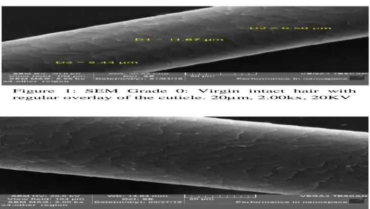
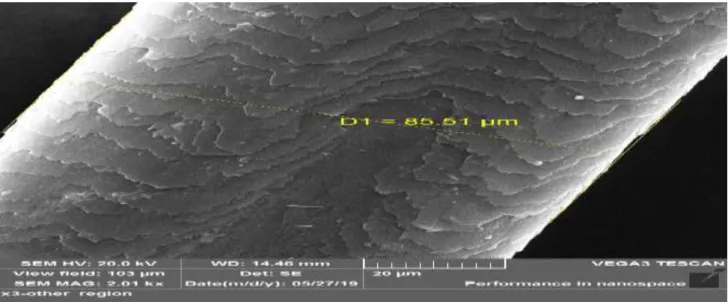
The damage to hair shaft under SEM was graded into five grades (based on the grading system developed by Kim et al [25].Grade 0-Virgin intact hair with regular overlay of the cuticle. Grade 1- Irregular overlay of the cuticle without cracks or holes. Grade 2 – Severe lift‑up of the cuticle with cracks or holes, but without exposure of the cortex. Grade 3 – Partial exposure of the cortex Grade 4-Complete exposure of the cortex.
Data Analysis and Statistics
Statistical analysis of the results was performed using the Excel statistical software 2016. All values were expressed as number, percent, range, minimum, maximum, means ± SD, and results were expressed as part per million (ppm) in analysis element concentration (U, Sr) that measured by ICP; OES, and to analyze degrees of hair damage, morphological and structural changes in human hair shaft under SEM. The statistical comparison of the results was carried out using t-Test: Two-Sample Assuming Equal Variances and Anova Single Factor: P ≤ 0.05) to evaluate differences between the radiation worker group and control group.
Results and Discussion
Grade of Hair Surface Damage by SEM
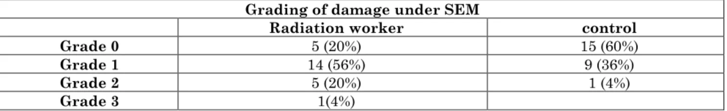
Table 1 shows the percentage of grading damage under SEM for both Radiation worker& control group. Of the study population (radiation worker), 5 (20%) had changes corresponding to (Grade 0), 14 (56%) had changes corresponding to (Grade 1), and 5 (20%) had changes corresponding to (Grade 2). Only 1 (4%) had intact hair (Grade 3) Figures 1‑4.Of the control samples 15 (60%) had (Grade 0) changes, whereas the remaining 9 (36%) showed intact hair on SEM (Grade 1) and only 1 (4%) had (Grade 2) Table 1.To comprehensively understand the degree of damage, we proposed a 3-stage evaluation scheme to describe the status of the hair’s surface, inner cuticle layers, and cortex. We ordered the grade of hair surface damage as follows: a lifting up of the cuticle edge as grade 0; an irregular overlay of cuticles without cracks or holes as grade 1; cracks or holes due to severe lifting up of cuticle layers as grade 2; exposure of cortical cells as grade 3; complete disappearance of cuticles as grade 4 [25].
Table 1: Show percentage of grading damage under SEM for both Radiation workers& control group.
Grading of damage under SEM
Radiation worker control
Grade 0 5 (20%) 15 (60%)
Grade 1 14 (56%) 9 (36%)
Grade 2 5 (20%) 1 (4%)
Grade 3 1(4%)
The cuticle is a chemically resistant area surrounding the cortex in animal hair fibers. It consists of flat overlapping cells (scales)
end) of the hair fiber. Each cuticle cell is approximately 0.5 mm high and approximately 45 to 60 mm long. The cuticle in human hair is generally 5-10 scales thick. Human hair cuticles usually contain smooth unbroken scale edges at the root end near the scalp. However, cuticle damage evidenced by broken scale edges that can usually be observed. Several centimeters away from the scalp is caused by weathering and mechanical damage from the effects of daily actions such as combing, brushing, and shampooing.
There have been efforts to describe the status of damaged hair objectively. According to the SEM findings, it has been suggested that stage 1 is characterized by intact, smooth scale edges and scale surfaces; stage 2 contains broken scale edges; in stage 3, the
scales have been partially removed; and in stage 4, the hair splits, indicating extensive cortical damage [26, 27]. Table 2 shows the Descriptive statistics of grade of hair damage, hair diameter, cell length under SEM.
A statistically significant difference was found between the radiation worker and control group with respect to the SEM scores with the extent of damage being more in the radiation worker group as compared to the control group (t-Test: Two-Sample Assuming Equal Variances, and Anova Single Factor: 0.001617, P ≤ 0.05). However, no statistically significant difference was seen in hair diameter and cell length between hair sample of radiation worker and control group (t-Test: Two-Sample Assuming Equal Variances and Anova Single Factor: 0.1385, P ≤ 0.05 of hair diameter, 0.7393 P ≤ 0.05 of cell length).
Table 2: Descriptive statistics of grade of hair damage, hair diameter, cell length under SEM Parameters Groups n Mean Standard
Deviation Standard Error P- value
Grading of hair damage under
SEM
Radiation
worker 25 1.08 0.759386 0.151877 0.00161 (S) control 25 0.44 0.583095 0.116619
Hair Diameter Radiation
worker 25 79.3805 16.76186 3.748066 0.1385 (NS) control 25 70.9795 18.32438 4.097456
Cell length Radiation
worker 25 7.884 2.064909 0.326491 0.7393 (NS) control 25 8.03625 2.012397 0.318188
S: Significant, NS: Non significant
Figure 1: SEM Grade 0: Virgin intact hair with
regular overlay of the cuticle. 20µm, 2.00kx, 20KV
Figure 2: SEM Grade 1: Irregular overlay of the cuticle without
Figure 3: SEM Grade 2: Severe lift‑up of the cuticle with cracks or holes but without exposure of the cortex. 50µm, 1.00kx, 20KV
Figure 4: SEM Grade 3: Partial exposure of the cortex. 20µm, 2.00kx, 20KV
The authors also noted that dermoscopy or light microscopy does not recognize hair damage due to chemical changes. SEM is a better modality to demonstrate hair shaft damage. According to Miteva and Tosti, although dermoscopy is a useful tool in the diagnosis of hair shaft abnormalities in general, it may not be useful in all conditions associated with hair shaft changes [28]. In general, SEM is considered a powerful complementary tool in the study of hair shaft abnormalities [29].
Measurement of Concentration of Trace Element by ICP-OES
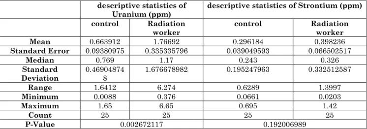
Table 3 shows the descriptive statistics, p-value of concentration of uranium and strontium (ppm) in hair of radiation worker and control group. Of the study, the average ± standard deviation of the former element of
uranium concentration in radiation worker (1.76692 ± 1.676678982) higher than control group (0.663912 ± 0.469048748).Also the average ± standard deviation of latter element of strontium concentration in radiation worker (0.398236± 0.332512587) higher than control group (0.296184± 0.195247963).A statistically significant difference was found between the radiation worker and control group with respect to the uranium concentration in (ppm) more in the radiation worker group as compared to the control group (t-Test: Two-Sample Assuming Equal Variances, 0.002672117, P ≤ 0.05).
Many studies were carried out to calculate the concentration of uranium and strontium in human hair. Puchyr, R. F. et al. have carried out the study on health care practitioners, the result that revealed of concentration of uranium and strontium is minimum level (0.253 ppm) and maximum level (0.503 ppm) respectively [4]. Another study, Wufuer, R. et al, carried out show significantly higher for people living in coal mining area than unexposed subjects in other areas, uranium concentrations in hair samples were 22.2-634.5 ng g-1 (mean: 156.2 ng g-1). Therefore, these results indicate that people living in coal mining area have been subjected to uranium exposure for long periods of time [30]. In order to make an assessment on an individual’s possible
exposure, knowledge of background uranium levels is indispensable. Hair considers common bio-indicators for uranium monitoring in humans [30, 32]. Gellein, K. et al. in his study on five individuals, three women and two men from range of age (28-35 year).The average value of strontium concentration is 5.01 µg/g compared with certified value 4.19 µg/g [31].Hair samples have several advantages over urine samples. Firstly, hair is stable and does not need special storage or handling. Besides, hair can reflect the total body intake over an extended period much longer than urine and feces. Therefore, hair is an excellent candidate for the biomonitoring of metals, particularly for the detection of trace elements [33].
Table 3: Descriptive statistics, p-value for concentration of Uranium and Strontium (ppm) in hair of radiation worker and control group
descriptive statistics of
Uranium (ppm) descriptive statistics of Strontium (ppm) control Radiation
worker control Radiation worker
Mean 0.663912 1.76692 0.296184 0.398236
Standard Error 0.09380975 0.335335796 0.039049593 0.066502517
Median 0.769 1.17 0.243 0.326
Standard
Deviation 0.469048748 1.676678982 0.195247963 0.332512587
Range 1.6412 6.274 0.6289 1.3997
Minimum 0.0088 0.376 0.0661 0.0203
Maximum 1.65 6.65 0.695 1.42
Count 25 25 25 25
P-Value 0.002672117 0.192006989
Conclusions
In conclusion, the obtained results occupationally exposed to low levels of ionizing radiation may be associated with increased deposits on the hair shaft surface; these findings may be consistent with those obtained in other studies, confirming that the
deficiency and efficiency of trace elements play a role in clinical disorders and the pathogenesis of many diseases. Furthermore, to prove that human hairs are useful in the studies pertaining to chronic body exposure and good biomarkers in clinical studies for radiation worker.
References
1. Sato I, Nakaki S, Murata K, Takeshita H, Mukai T (2010) Forensic hair analysis to identify animal species on a case of pet animal abuse. International journal of legal medicine, 124(3): 249-256.
2. Pozebon D, Scheffler GL, Dressler VL (2017) Elemental hair analysis: A review of procedures and applications. Analytica chimica acta, 992: 1-23.
3. Font L, Van der Peijl G, Van Wetten I, Vroon P, Van der Wagt B, Davies G (2012) Strontium and lead isotope ratios in human hair: investigating a potential tool
for determining recent human geographical movements. Journal of Analytical Atomic Spectrometry, 27(5): 719-732.
4. Puchyr RF, Bass DA, Gajewski R, Calvin M, Marquardt W, Urek K, Quig D (1998) Preparation of hair for measurement of elements by inductively coupled plasma-mass spectrometry (ICP-MS). Biological trace element research, 62(3): 167-182. 5. Carneiro MFH, Moresco MB, Chagas GR,
Brazil. Biological trace element research, 143(2): 815-824.
6. Zunic ZS, Tokonami S, Mishra S, Arae H, Kritsananuwat R, Sahoo SK (2012) Distribution of uranium and some selected trace metals in human scalp hair from Balkans. Radiation protection dosimetry, 152(1-3): 220-223.
7. Frazão SV, Saiki M (2007) Study on trace element determination in human head hair using neutron activation analysis. In 2007 International Nuclear Atlantic Conference-INAC.
8. Das S, Sarma K, Talukdar M, Rajkhowa J, Gautam C, Deka SSA (2018) Light and scanning electron microscopy aided with elementary analysis in characterization and identification of hair of Assam hill goat.
9. Rejah BK (2017) Specific activities of natural radionuclides and annual effective dose due to the intake of some types of children powdered milk available in Baghdad Markets. Baghdad Science Journal, 14(3): 619-624.
10. Czerny B, Krupka K, Ożarowski M, Seremak-Mrozikiewicz A (2014) Screening of trace elements in hair of the female population with different types of cancers in Wielkopolska region of Poland. The Scientific World Journal.
11. Momen AA, Khalid MAA, Elsheikh MAA, Ali DMH (2015) Trace elements in scalp hair and fingernails as biomarkers in clinical studies. Journal of Health Specialties, 3(1): 28.
12. Chojnacka K, Zielińska A, Michalak I, Górecki H (2010) The effect of dietary habits on mineral composition of human scalp hair. Environmental Toxicology and Pharmacology, 30(2): 188-194.
13. M'baku SB, Parr RM (1982) Inter laboratory study of trace and other elements in the IAEA powdered human hair reference material, HH-1. Journal of Radio analytical Chemistry, 69(1-2): 171-180.
14. Morishige Y, Kimura A (2008) Ionization interference in inductively coupled plasma-optical emission spectroscopy. Sci. Technical Review-English Edition, 66: 106. 15. Hou X, Amais RS, Jones BT, Donati GL
(2006) Inductively coupled plasma optical
emission spectrometry. Encyclopedia of Analytical Chemistry: Applications, Theory and Instrumentation, 1-25.
16. Scott RH, Fassel VA, Kniseley RN, Nixon DE (1974) Inductively coupled plasma-optical emission analytical spectrometry. Analytical Chemistry, 46(1): 75-80.
17. Devia DM, Rodriguez-Restrepo LV, Parra ER (2015) Methods employed in optical emission spectroscopy analysis: a review. Ingeniería y ciencia, 11(21): 239-267.
18. Dahlquist RL, Knoll JW (1978) Inductively coupled plasma-atomic emission spectrometry: analysis of biological materials and soils for major, trace, and ultra-trace elements. Applied Spectroscopy, 32(1): 1-30.
19. Charles B, Fredeen KJ (1997) Concepts, instrumentation and techniques in inductively coupled plasma optical emission spectrometry. Perkin Elmer Corporation.
20. Breton BC, McMullan D, Smith KC (Eds.) (2004) Sir Charles Oatley and the scanning electron microscope. Elsevier Academic Press.
21. Hafner B (2007) Scanning electron microscopy primer. Characterization Facility, University of Minnesota-Twin Cities, 1-29.
22. Akhtar K, Khan SA, Khan SB, Asiri AM (2018) Scanning Electron Microscopy: Principle and Applications in Nanomaterials Characterization. In Handbook of Materials Characterization 113-145. Springer, Cham.
23. Carter M, Shieh JC (2015) Guide to research techniques in neuroscience. Academic Press.
24. Ryabukhin YS (1976) Activation analysis of hair as an indicator of contamination of man by environmental trace element pollutants (No. IAEA-RL--50). International Atomic Energy Agency.
25. Kim YD, Jeon SY, Ji JH, Lee WS (2010) Development of a classification system for extrinsic hair damage: standard grading of electron microscopic findings of damaged hairs. The American Journal of Dermatopathology, 32(5): 432-438.
27. Hearle JWS (2000) A critical review of the structural mechanics of wool and hair fibres. International Journal of Biological Macromolecules, 27(2): 123-138.
28. Miteva, M., & Tosti, A. (2013) Dermatoscopy of hair shaft disorders. Journal of the American Academy of Dermatology, 68(3): 473-481.
29. Echeverría XP, Romero WA, Carreño NR, Zegpi MS, González SJ (2009) Hair shaft abnormalities. Pili bifurcati: A scanning electron microscopy analysis. Pediatric dermatology, 26(2): 169-170.
30. Wufuer R, Song W, Zhang D, Pan X, Gadd GM (2018) A survey of uranium levels in urine and hair of people living in a coal mining area in Yili, Xinjiang, China. Journal of environmental radioactivity, 189: 168-174.
31. Gellein K, Lierhagen S, Brevik PS, Teigen M, Kaur P, Singh T, Syversen T (2008) Trace element profiles in single strands of human hair determined by HR-ICP-MS. Biological trace element research, 123(1-3): 250-260.
32. Joksić AŠ, Katz SA (2014) Efficacy of hair analysis for monitoring exposure to uranium: A mini-review. Journal of Environmental Science and Health, Part A, 49(13): 1578-1587.