http://www.ijmedicine.com pISSN 2349-3925 | eISSN 2349-3933
Research Article
Clinical features and risk factors of pancytopenia: a study
in a tertiary care hospital
G. Pavan Kumar Reddy
1*, Kande V. Mallikarjuna Rao
2INTRODUCTION
Pancytopenia is one of the important hematological disorders, which is characterized by the decrease in all the three cellular elements of peripheral blood i.e. red blood corpuscles, white blood cells as well as platelets.1
The peripheral pancytopenia is normally a triad of findings which result from a number of diseases, of primary or secondary bone marrow disorders, the symptoms of which are usually attributed to anemia, leucopenia and thrombocytopenia.2 The usual symptoms presented by the patient are fatigue and weakness due to
anemia, increased susceptibility to infections due to leucopenia and excessive bleeding due to thrombocytopenia.3
The various disorders that result in pancytopenia vary according to the geographical distribution and genetic mutations.4-6 This can be due to failure of production of hematopoietic progenitors in bone marrow, malignant cell infiltration, antibody-mediated bone marrow suppression, ineffective hematopoiesis and dysplasia, chemotherapy, radiotherapy, or peripheral sequestration of blood cells in overactive reticulo endothelial system. 1,7-10
ABSTRACT
Background: Pancytopenia is one of the important hematological disorders seen in regular clinical practice, which is characterized by the decrease in all the three cellular elements of peripheral blood i.e. red blood corpuscles, white blood cells as well as platelets. The prevalence and risk factors are different in different geographical areas and hence this study was undertaken to identify the prevalence of pancytopenia in our area and analyse the risk factors.
Methods: 42 patients with pancytopenia, of all age groups and both the sexes were included in the study. A detailed demographic and clinical history was taken from the patients. All of them were subjected to thorough physical examination; blood tests were collected for hematological study. Bone marrow aspirate was also taken form these patients.
Results: Out of 42 patients, 54.8% patients were males and 45.3% were females, with most of them below 40 years of age. The most major cause of pancytopenia was megaloblastic anaemia comprising of 38.1% of the cases, followed by aplastic anaemia with 26.2%. The other minor contributors were leukaemia, malaria, malignancy, septicemia among others. Pallor, dyspnoea, fever were the most common symptoms observed.
Conclusions: The study shows that there is a great incidence of pancytopenia in younger and middle age group rather than in the elderly. Detailed hematological examination including bone marrow aspiration should be done in these patients to understand and diagnose this disease early so that proper intervention and management of these patients can be done.
Keywords: Pancytopenia, Hematological examination, Megaloblastic anemia, Aplastic anemia 1Department of Medicine, Mallareddy Medical College for women, Hyderabad, India
2Department of Medicine, Mallareddy Institute of Medical Sciences, Hyderabad, India
Received: 11 January 2016 Accepted: 27 January 2016
*Correspondence:
Dr. G. Pavan Kumar Reddy,
E-mail: drpavankumar_reddy@yahoo.co.in
Copyright: © the author(s), publisher and licensee Medip Academy. This is an open-access article distributed under the terms of the Creative Commons Attribution Non-Commercial License, which permits unrestricted non-commercial use, distribution, and reproduction in any medium, provided the original work is properly cited.
The most effective form of diagnosis is by complete blood picture and peripheral blood count of the patients including reticulocytes. Bone marrow studies also have been found to be effective.12 Various studies have been performed to identify the cause of the condition and has been found to be highly varied. In a study in North India, the most common cause has been found to be the most common cause while in Maharashtra it was hypersplenism.6,12,13 Aplastic Anemia was found to be the chief cause in Nepal14 while it was aplastic anemia along with malaria and Leishmania in Bangladesh.15
As the prevalence and risk factors were different in different geographical areas, we had undertaken this study to identify the prevalence of pancytopenia in our area and analyze the risk factors most prevalent here.
METHODS
This prospective study was conducted in the Department of Medicine, at Deccan College of Medical sciences. 42 patients with pancytopenia, of all age groups and both the sexes were included in the study. Patients under chemotherapy or radiotherapy, those diagnosed of malignancy, aplastic anemia, chronic liver diseases or with other genetic causes of pancytopenia were excluded from the study.
A detailed demographic and clinical history was taken from the patients. All of them were subjected to thorough physical examination. Symptoms of pancytopenia such as palpitations, fatigue, fever, shortness of breath were noted. X-rays of chest and ultrasound of the abdomen were taken for identifying the conditions of the internal organs.
After taking an informed consent, blood samples were collected in the EDTA tubes for complete blood picture, in plain tubes for biochemical tests and peripheral blood smears were done for all the patients.
The peripheral blood smear was stained by Leishmann Stain. The EDTA blood sample was used for estimation of the complete blood count, including hemoglobin and platelet levels. The biochemical tests performed were fasting blood glucose levels, Liver function tests, renal function tests, iron and ferritin, Vitamin B12 and folate levels.
Bone marrow aspiration was done and stained with Leishmann’s stain and trephine biopsy specimens were processed and paraffin wax blocks were made. They sections made from these blocks were made into thin smears and stained with hematoxylin and eosin stains.
RESULTS
Out of 42 patients, 23(54.8%) patients were males and 19 (45.3%) were females (Figure 1).
The patients were catagorised according to their ages. 31% of the patients belonged to 11-20 years age group followed by 19% in the 31-40 age group. The extreme age groups i.e. <10 and >71 showed only 1 case each.
Figure 1: Sex wise distribution of pancytopenia.
Figure 2: Age wise distribution of pancytopenia.
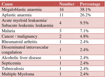
The most common cause of pancytopenia was megaloblastic anemia comprising of 38.1% of the cases. Aplastic anemia with 26.2% of the cases was the other major contributor. The other minor causes were leukemia, malaria, malignancy septicemia etc (Table 1).
Table 1:Distribution of causes of pancytopenia.
Cause Number Percentage
Megaloblastic anaemia 16 38.1%
Aplastic anaemia 11 26.2%
Acute myeloid leukaemia/
Subacute leukemic leukaemia 4 9.5%
Malaria 3 7.1%
Cancer / malignancy 2 4.8%
Rheumatoid arthritis 1 2.4%
Disseminated intravascular
coagulation 1 2.4%
Alcoholic liver disease 1 2.4%
Septicemia 1 2.4%
Tuberculosis 1 2.4%
Multiple Myeloma 1 2.4%
Males
Females
0 2 4 6 8 10 12 14
1 to 10
11 to 20
21 to 30
31 to 40
41 to 50
51 to 60
61 to 70
>71 1
13
7 8
6 4
Pallor was seen in most of the patients (64.3%) followed by dyspnea in nearly 60% of the patients. Many of the patients complained of pain in legs and abdomen and fever was seen in 45% of the cases (Table 2).
Table 2:Clinical symptoms.
Symptoms Number Percentage
Pallor 27 64.3%
Dyspnoea 25 59.5%
Pain in legs 20 47.6
Fever 19 45.2%
Hepatomegaly 13 31%
Splenomegaly 12 28.6%
Weakness 11 26.2%
Abdominal Pain 11 26.2%
Icterus 9 21.4%
Oedema 4 9.5%
Bleeding 3 7.1%
Weight loss 2 4.8%
Majority of the patients had a hemoglobin count of less than 5 g/dl. All of the 3 cases of malaria came under this category along woth some of the aplastic anemia cases. Most of the aplastic anemia patients had hemoglobin levels between 5.1-8.3 patients had total leucocyte count below 1000 while 17 of them had above 2500. Most of the patients had a platelet count below 50000. All the 3 cases of malria had very low platelet count (below 50000) (Table 3).
Table 3:Hematological presentation.
Parameters Category Number Percentage
Hb g/dl
1.5 – 5.0 17 40.5%
5.1-8.0 14 33.3%
>8.1 9 21.4%
TLC cumm
490 – 1000 3 7.1
1001-2500 22 52.4
>2501 17 40.5
PLT cumm
5000-50000 22 52.4
51000-80000 9 21.4
>81000 11 26.2
Figure 3: Bone marrow examination.
Bone marrow examination was done for all the patients. Of them, 47.6% of the patients had hypo cellular marrow. Hyper cellular and normocellular marrow were seen in 21.4% and 14.3% of the cases respectively (Figure 3).
DISCUSSION
Pancytopenia by its own is not a disease but a manifestation of different diseases that lead to reduction of the cellular components resulting in anemia, thrombocytopenia and leucopenia in the peripheral blood. It is a condition that is very commonly observed in clinical practice, nevertheless there are very few studies performed to identify the etiology of this pancytopenia.12 Most studies done are based on one particular etiology and broad spectrum studies are very few.
Albeit invasive, bone marrow examination is a very simple and safe procedure causing only moderate discomfort to the patients with very little or no bleeding. It is one of the routinely performed procedures in the clinical practice. It is mainly performed to evaluate the unexplained cytopenias and other malignant condition like leukemia.1
In our study, there was a predominance of males over females with pancytopenia, which was in accordance to a similar study by Prasad et al, who also observed a slight preponderance of males over females,16 Dasgupta et al17 and Das Makheja et al18 who observed the male: female ration to be 1.7:1 and 1.38:1 respectively. In contrast, a female preponderance was observed in studies by Agarwal et al, Kumar et al.19,20
69% of the patients in our study were below the age of 40 years with 11- 20 years age group being most affected. The same was corroborated by Prasad et al who observed 74% of the cases to be below 40 years of age16 and Khodke et al who observed 60% of patients to be below 40 years.21 In studies by Tilak et al,4 Khunger et al6 and Jha et al,22 the common age group affected was 1-30 years.
The most common cause for pancytopenia in our study was found to be megaloblastic anemia with 38.1% of the patients affected, followed by aplastic anemia in 26.2% of the cases. Leukemia and malaria contributed to about 9.5% and 7.1% respectively.
In a study by Yadav et al, 35.84% of the patients were observed to have megaloblastic anemia followed by 11.32% septicemia as causative agents for pancytopemia. Alcoholic and nonalcoholic liver diseases constituted 9.43% each. In our study, septicemia ad liver diseases constituted only 2.4% each.1
A study by Naseem et al based on bicytopenic and pancytopenic children observed acute leukemia to be the most common etiology in bicytopenic and aplastic anemia in pancytopenic children.23
20
9 6
7 Hypocellular
Hypercellular
Normocellular
The incidence of pancytopenia varies from 10-52.7%.4,24 In our study aplastic anemia was the second most common cause with 26.2% incidence. 14.285 were reported by Agarwal et al,19 Khodke et al,21 Khunger et al.6
Malaria was found to the most common cause in a study by Agarwal et al,19 Tareen et al7 and Gamal et al.9 Arya et al reported the lead cause of death among patients with pancytopenia was malaria.25 This high incidence was attributed to the endemic nature of malaria and the lack of sanitation and hygiene.
Among the studies performed around the world, various etiologies of pancytopenia were reported. In a study in Zimbabwe, megaloblastic anemia was the most common cause followed by aplastic anemia and acute leukemia.26 In another study in France, malignant myeloid disorders were found to be the most common cause accounting to about 42% of the total patients with pancytopenia and various malignant disorders accounted to about 18%.27 Aplastic anemia was seen in 10% of their cases only. Hypoplastic bone marrow was seen in 29% of the cases in a study in Nepal followed by megaloblastic anemia in 23.6% of the patients.22
We had observed in our study, pallor to be the commonest presentation by 64.3% of the patients, followed by dyspnea in 59.5%, fever in 45.2%, pain in legs in 47%. Fever and progressive pallor was observed in 81.4% patients in a study by Gupta et al.28 These were followed by bleeding manifestations. Megaloblastic anemia was observed in only around 6% of the children. Yet in another cohort study by Gayatri et al, general weakness and fever were the most common symptoms.12
CONCLUSIONS
Pancytopenia is a common hematological condition seen often in clinical practice and should be suspected in patients with unexplained anemia, prolonged fever, pallor, and tendency to bleed. The study shows that there is a great incidence of pancytopenia in younger and middle age group rather than in the elderly. Detailed hematological examination including bone marrow aspiration should be done in these patients to understand and diagnose this disease early so that proper intervention and management of these patients can be done.
Funding: No funding sources Conflict of interest: None declared
Ethical approval: The study was approved by the institutional ethics committee
REFERENCES
1. Yadav BS, Varma A, Kiyawat P. Clinical profile of pancytopenia: a tertiary care experience. Intern J Bioassays. 2015;4(01):3673-7.
2. Guinan EC, Shimamura A. Wintrobe's Clinical Hematology. In: Greer JP, Foerster J, Lukens JN, Rodgers GM, Paraskevas F, Glader B, editors. Acquired and inherited aplastic anaemia syndromes. 11th ed. Philadelphia: Lippincott Williams and Wilkins; 2004:1397-419.
3. Ujjan DI, Shaikh AI, Khokar AN, Memon AR, Farooq M. Frequency of causes of pancytopenia in patients admitted at Isra University Hospital Hyderabad. Pak J of Med Health Sci. 2010;4(4):416-8.
4. Tilak V, Jain R. Pancytopenia- a Clinico-hematologic analysis of 77 cases. Indian J Pathol. 1999;42(4):399-404.
5. Nanda A, Basu S, Marwaha N. Bone marrow trephine biopsy as an adjunct to bone marrow aspiration. JAPI. 2002;50:893-5.
6. Khunger JM, Arculselvi S, Sharma U, Ranga S, Talib VH. Pancytopenia- a clinico- haematological study of 200 cases. Indian J Pathol Microbiol. 2002;45(3):375-9.
7. Tareen SM, Bajwa MA, Tariq MM, Babar S, Tareen AM. Pancytopenia in two national ethnic groups of Baluchistan. J Ayub Med Coll Abottabad. 2011;23(2):82-6.
8. Gruchy D. Pancytopenia; aplastic anaemia. In: Firkin F, Chesterman C, Pennigton D, Rush B, editors. De Gruchy’s Clinical Hematology in Medical Practice. 5th ed. Oxford: Blackwell; 1989:119-36.
9. Gamal AH, Safa AR. Patterns of pancytopenia in Yemen. Turk J Hematol. 2008;25:71-4.
10. Mussarrat N, Fazl R. The incidence of underlying pathology in pancytopenia - an experience of 89 cases. J postgrad Instit. 2004;18:76-9.
11. Hayat AS, Khan AH, Baloch GH, Shaikh N. Pancytopenia; study for clinical features and etiological pattern of at tertiary care settings in Abbottabad. Professional Med J. 2014;21(1):060-5. 12. Gayathri BN, Rao KS. Pancytopenia: A clinico
hematological study. Physicians Lab J. 2011;3:15-20.
13. Jain A, Naniwadekar M. An etiological reappraisal of pancytopenia - Largest series reported to date from a single tertiary care teaching hospital. BMC Hematol. 2013;13:10.
14. Pathak R, Jha A, Sayami G. Evaluation of bone marrow in patients with pancytopenia. J Nepal Med Assoc. 2012;2:265-71.
15. Hossain MA, Akond AK, Chowdhary MK, Sikder AM, Rashid MA. Pancytopenia- a study of 50 cases. Bangladesh Journal Pathol. 1992;7:9-12.
16. Prasad BH, Sarode S, Kadam DB. Clinical profile of pancytopenia in adults. Int J Sc Res. 2013; 2(7):355-7.
18. Das Makheja K, Kumar Maheshwari B, Arain S, Kumar S, Kumari S, Vikash. The common causes leading to pancytopenia in patients presenting to tertiary care hospital. Pak J Med Sci. 2013;29:1108-11.
19. Agarwal R , Bharat V, Gupta BK, Jain S, ansal R, Choudhary A, Tiwari G. Clinical and hematological profile of pancytopenia. Intern J Clin Biochem Res. 2015;2(1):48-53.
20. Kumar DB, Raghupathi AR. Clinico-hematologic analysis of pancytopenia: study in a tertiary care centre. Basic and Applied Pathol. 2012;5:19-21. 21. Khodke K, Marwah S, Buxi G, Yadav RB,
Chaturvedi NK. Bone marrow examination in case of pancytopenia. J Ind Aca Clin Med. 2001;2(1 & 2):55-9.
22. Jha A, Sayami G, Adhikari RC, Panta AD, Jha R. Bone marrow examination in cases of pancytopenia. J Nepal Med Assoc. 2008;47(169):12-7.
23. Naseem S, Varma N, Das R, Ahluwalia J, Sachdeva US, Marwaha RK. Pediatric patients with bicytopenia/pancytopenia: review of etiologies and clinic hematological profile at a tertiary centre. IJPM. 2011;54(1):75-80.
24. Kumar R, Kalra SP, Kumar H, Anand AC, Madan M. Pancytopenia - a six year study. JAPI. 2001;49:1079-81.
25. Arya TV, Prasad RN. Fatal pancytopenia in falciparum malaria. J Assoc Physicians India. 1989;37(7):469-70.
26. Savage DG, Allen RH, Gangaidzo IT, Levy LM, Gwanzura C, Moyo A et al. Pancytopenia in Zimbabwe. Am J Med Sci. 1999;317:22-32.
27. Imbert M, Scoazec JY, Mary JY, Jouzult H, Rochant H, Sultan C. Adult patients presenting with pancytopenia: a reappraisal of underlying pathology and diagnostic procedures in 213 cases. Hematol Pathol 1989;3:159-67.
28. Gupta V, Tripathi S, Tilak V, Bhatia BD. A study of clinico-haematological profiles of pancytopenia in children. Trop Doct. 2008;38:241-3.