TARGETING THE RAS-RAL EFFECTOR PATHWAY
FOR CANCER TREATMENT
Leanna R. Gentry
A dissertation submitted to the faculty at the University of North Carolina at Chapel Hill in
partial fulfillment of the requirements for the degree of Doctor of Philosophy in the
Department of Pharmacology.
Chapel Hill
2015
Approved by:
Adrienne Cox
Channing Der
Lee Graves
Klaus Hahn
iii
ABSTRACT
LEANNA R. GENTRY: Targeting the Ras-Ral effector pathway for cancer treatment (Under the direction of Channing J. Der)
The RAS oncogene is the most frequently mutated gene in human cancers, and this activated Ras oncoprotein has been shown to be required for both cancer initiation and maintenance. Great strides have been made in understanding Ras signaling in cancer since the discovery of its involvement in human cancers in 1982, with numerous Ras effector pathways and modes of Ras regulation having been identified as contributing to Ras-driven oncogenesis. However, there has been limited success in developing strategies for therapeutically targeting Ras-driven oncogenesis. One effort that has gained popularity in recent years is the inhibition of Ras effector signaling. The Ral (Ras-like) small GTPases, discovered shortly after Ras in an attempt to identify RAS-related genes, are activated downstream of Ras by Ral guanine nucleotide exchange factors (RalGEFs). The Ral family members have since emerged as critical regulators of key cellular processes and, importantly, have been characterized as playing a role in tumorigenesis and invasion of multiple cancer types. Interestingly, divergent roles for RalA and RalB are often observed in within a cancer. Due to the high affinity of Ral for GTP, which activates Ral upon binding, the Ral GTPase family cannot be targeted directly. Therefore, indirect inhibition of Ral must be considered for targeting Ral-dependent phenotypes in Ras-driven cancers. This could be achieved through inhibition of Ral association with the plasma membrane, which is thought to be required for its activation and subsequent signaling. Alternatively, downstream effectors of Ral with validated roles in cancer could be inhibited.
iv
activation, and function. The first and essential step of this process is prenylation by GGTase. Prenylation signals for further CAAX processing by the enzymes RCE1 and ICMT, which are under consideration as therapeutic targets. We determined that the modifications regulated by these enzymes have distinct roles and consequences for Ral GTPases. We found that both RalA and RalB require RCE1 for association with the plasma membrane, and that the absence of RCE1 caused a sustained activation of both RalA and RalB. In contrast, ICMT deficiency disrupted plasma membrane localization of RalB but not RalA, whereas RalA depended on ICMT for efficient localization to recycling endosomes. Furthermore, ICMT deficiency caused increased stability of RalB protein but not RalA. Lastly, we found that palmitoylation was critical for proper subcellular localization of RalB but not RalA. In summary, we identified isoform-specific consequences of CAAX modifications that could be contributing to the divergent localization and activities of the Ral proteins.
In order to address inhibiting Ral effectors, we sought to determine the effect of inhibiting TBK1, a kinase that is a validated effector of RalB, in pancreatic ductal adenocarcinoma, a disease characterized by greater than 90% of cases containing a K-Ras mutation. We found that a novel small molecule inhibitor of TBK1, while effective at inhibiting signaling, had a minimal effect on
v
ACKNOWLEDGEMENTS
I want to thank my mentor, Dr. Channing Der, for teaching me to be independent and allowing me to take the science in my own direction while keeping me focused. I am fortunate to have been a part of the Der lab, and thank my fellow lab members who made this experience memorable and enjoyable. In particular, I want to thank Nicole Baker for being a wonderful and supportive friend from the first day of graduate school. I am thankful for Dr. Adrienne Cox, who was always there with her encouragement and advice when it came to both science and life. I would also like to thank my committee members, Drs. Gary Johnson, Lee Graves, and Klaus Hahn, for their creative ideas and insightful questions that have helped me complete this dissertation. I especially thank my parents, Gary and Mamta Gentry, for always encouraging me to pursue my love of learning and for teaching me to be tough. Without my mom’s unwavering support and my dad’s daily
vi
TABLE OF CONTENTS
LIST OF FIGURES ... ix
LIST OF ABBREVIATIONS ... x
CHAPTER 1: INTRODUCTION ... 13
RAL GTPASE FAMILY ... 13
RAL PROTEIN STRUCTURE ... 14
RAL GEFS ... 17
RAL EFFECTORS ... 21
RALBP1 ... 23
SEC5 AND EXO84 SUBUNITS OF THE EXOCYST ... 24
OTHER EFFECTORS ... 25
POST-TRANSLATIONAL MODIFICATION AND REGULATION OF RAL FUNCTION ... 26
RAL CAAX MODIFICATIONS ... 26
PHOSPHORYLATION REGULATION OF SUBCELLULAR LOCALIZATION AND EFFECTOR INTERACTION ... 29
UBIQUITINATION ... 30
DIVERGENT ROLES OF RAL IN CANCER ... 30
BLADDER CARCINOMA ... 31
COLORECTAL CARCINOMA ... 32
vii
LUNG ADENOCARCINOMA ... 32
MALIGNANT PERIPHERAL NERVE SHEATH TUMORS. ... 33
MELANOMA ... 34
OVARIAN CARCINOMA ... 34
PANCREATIC DUCTAL ADENOCARCINOMA ... 35
PROSTATE CARCINOMA ... 36
SQUAMOUS CELL CARCINOMA ... 36
CONCLUSIONS AND FUTURE PROSPECTS ... 36
CHAPTER 2: DIVERGENT ROLES OF CAAX MOTIF-SIGNALED
POSTTRANSLATIONAL MODIFICATIONS IN THE REGULATION AND
SUBCELLULAR LOCALIZATION OF RAL GTPASES
1... 38
BACKGROUND ... 38
INTRODUCTION ... 40
EXPERIMENTAL PROCEDURES ... 43
RESULTS ... 45
RALA AND RALB SHOW DISTINCT REQUIREMENTS FOR RCE1- AND ICMT-MEDIATED POSTTRANSLATIONAL MODIFICATIONS FOR THEIR SUBCELLULAR LOCALIZATION AND PLASMA MEMBRANE ASSOCIATION ... 45
ICMT REGULATES RALA LOCALIZATION TO RECYCLING ENDOSOMES ... 50
RCE1 REGULATES RAL ACTIVITY ... 52
ICMT REGULATES RALB STABILITY ... 54
DUAL LIPID MODIFICATION OF THE RAL CAAX MOTIF AFFECTS RALB SUBCELLULAR LOCALIZATION ... 56
DISCUSSION ... 58
CHAPTER 3: TARGETING TBK1 IN
KRAS
-MUTANT ... 62
viii
BACKGROUND ... 62
INTRODUCTION ... 64
EXPERIMENTAL PROCEDURES ... 66
RESULTS ... 67
LSN3090279 IS A TBK1 INHIBITOR ... 67
LSN3090279 HAS A MINIMAL EFFECT ON CANCER CELL PROLIFERATION IN VITRO AND IN VIVO ... 69
PDAC CELL LINES SHOW VARYING SENSITIVITY TO PHARMACOLOGIC TBK1 INHIBITION ... 70
COMBINED TBK1 AND ERK INHIBITION SYNERGISTICALLY DECREASES PDAC CELL PROLIFERATION... 76
DISCUSSION ... 77
ix
LIST OF FIGURES
Figure 1-1. Evolutionary conservation of Ral small GTPases. ... 15
Figure 1-2, A and B. Regulators of the Ral GDP-GTP cycle. ... 16
Figure 1-2, C and D. Regulators of the Ral GDP-GTP cycle. ... 20
Figure 1-3. Ral effectors and effector functions. ... 22
Figure 1-4. Regulation of Ral subcellular localization and membrane association... 28
Figure 2-1. Subcellular localization of Ral family proteins is differentially dependent on RCE1 and ICMT processing. ... 47
Figure 2-2. Hypervariable domain dictates Ral ICMT dependency. ... 49
Figure 2-3. RalA localization on recycling endosomes is impaired by loss of ICMT. ... 51
Figure 2-4. RCE1 deficiency increases activated Ral-GTP formed upon EGF stimulation. ... 53
Figure 2-5. ICMT deficiency increases RalB protein stability. ... 55
Figure 2-6. CA1AX palmitoylation dictates RalB subcellular localization. ... 57
Figure 3-1. LSN3090729 inhibits TBK1 ... 68
Figure 3-2. LSN3090729 has KRas-independent effect on cancer cell growth. ... 69
Figure 3-3. PDAC cells are resistant to LSN3090279. ... 71
Figure 3-4. TBK1 depletion does not decrease colony formation. ... 72
Figure 3-5. LSN3090729 disrupts autophagy in PDAC cells. ... 74
Figure 3-6. LSN3090729 minimally affects KRas-mutant tumor xenografts. ... 75
x
LIST OF ABBREVIATIONS
Akt Protein kinase B
Bcl-XL B-cell lymphoma extra large
CAAX Cysteine-aliphatic-aliphatic-terminal amino acid
CHX Cycloheximide
EGF Epidermal growth factor
EH Eps homology
ER Endoplasmic reticulum
ERK1/2 Mitogen activated protein kinase kinase kinase 1/2
Exo84 Exocyst complex component 8
FTase Farnesyl transferase
FTI Farnesyl transferase inhibitor
GAP GTPase activating protein
GDP Guanosine diphosphate
GDS Guanine nucleotide dissociation stimulator
GEF Guanine nucleotide exchange factor
GFP Green fluorescent protein
xi GGTI Geranylgeranyl transferase inhibitor
GTP Guanosine triphosphate
GTPase Guanosine triphosphatase
HVR Hypervariable region
ICMT Isoprenylcysteine carboxylmethyltransferase
IRF3 Interferon regulatory factor 3
LPS Lipopolysaccharide
mCh Red mCherry fluorescent protein
MEF Mouse embryonic fibroblast
MEK Mitogen-activated protein kinase kinase
mTOR Mammalian target of rapamycin
NF-κB Nuclear factor kappa-light-chain-enhancer of activated B cells
PARP Poly (ADP-ribose) polymerase
PDAC Pancreatic ductal adenocarcinoma
PH Pleckstrin homology domain
PI3K Phosphoinositide 3-kinase
Rab Ras-like proteins in brain
xii Ral Ras-like protein
RalBP1 Ral binding protein 1
Ras Rat sarcoma viral oncogene homolog
RBD Ral-binding domain
RCE1 Ras converting endopeptidase 1
REM Ras exchanger motif
Rgl RalGDS-like
Rho Ras homologous protein
RhoGDI Rho GDP dissociation inhibitor
Sec5 Exocyst complex component 2
TBK1 Tank binding kinase 1
TLR Toll-like receptor
TSC Tuberosclerosis complex
WT Wild-type
13
Chapter 1: Introduction
1Ral GTPase family. Identified initially as Ras-like (Ral) proteins, the Ral small GTPases are members of the Ras branch of the Ras superfamily of small GTPases [1]. RALA was identified initially using oligonucleotide probes to identify RAS-related genes in a cDNA library established from immortalized simian B-lymphocytes [2]. Three years later, using the simian RALA cDNA as a probe, human RALA and a related RALB gene were identified from a human pheochromocytoma cDNA library [3]. Subsequently, single RAL orthologs were identified in C. elegans (RAL-1) [4] and Drosophila (RalA) [5] (Fig. 1-1). Interestingly, although there are well-conserved RAS orthologs in yeast, no RAL orthologs are present in S. cerevisiae or S. pombe.
The three human RAS genes (HRAS, KRAS and NRAS) comprise one of the most frequently mutated gene families in human cancers [6]. Consequently, they have been the subject of intense research scrutiny and cancer drug discovery. Initially, the discovery of Ral proteins simply added to a rapidly growing roster of proteins that now comprise a large superfamily of >150 Ras-related small GTPases [1]. However, with discoveries that Ral GTPases are key regulators of vesicular trafficking and are effectors of Ras oncoprotein-driven growth transformation, Ral proteins stepped into the spotlight in 2003 to bask in their “15 minutes of fame” [7]. Since those initial findings, more discoveries on the role of Ral in normal and cancer cell physiology have ensured that their “fame” will last considerably more than 15
1 This chapter is adapted from a previously published work: Gentry LR, Martin TD, Reiner DJ, Der CJ. “Ral small
14
minutes. In this review, we summarize our current knowledge on Ral GTPases and we highlight recent findings in Ral function.
Ral protein structure. The highly related human RalA and RalB isoforms share 82% overall amino acid sequence identity (Fig. 1-1A) and are members of the Ras branch of the Ras superfamily (Fig. 1-1B). They share 46-51% sequence identity and domain architecture with Ras proteins [8]. However, Ral proteins contain an N-terminal 11 amino acid extension not found in Ras, accounting for the 11 residue shift in numbering compared with Ras residue numbering (Fig. 1-1C). This is followed by the G domain, involved in GTP binding and hydrolysis, and the C-terminal membrane targeting sequence. The majority of sequence divergence occurs within the C-terminal hypervariable regions (50% shared identity) (Fig. 1-1C).
Like Ras, Ral proteins cycle between inactive GDP-bound and active GTP-bound states (Fig. 1-2A). RalA and RalB share complete sequence identity in the switch I (SI) and II (SII) sequences that change conformation during GDP-GTP cycling [8] (Fig. 1-2B). As described below, SI and SII are involved in recognition by both regulators and effectors. The conservation of SI and SII sequences in Drosophila and C. elegans Ral proteins support their interaction with conserved regulators and effectors.
15
Figure 1-1. Evolutionary conservation of Ral small GTPases. A. Human and invertebrate Ral orthologs exhibit strong sequence identity. The RalA and RalB isoforms are found in all vertebrate species. There is one Ral ortholog in C. elegans (Ce) and D. melanogaster (Dm). Overall sequence identity was determined by CLUSTALW multiple sequence alignment. B. Ral GTPases are members of the Ras branch of the Ras superfamily. Shown here is a comparison with the four Ras proteins and representative members of the Ras family. The dendrogram was generated by CLUSTALW multiple sequence alignment. C. Dendrogram showing sequence relationship of human and invertebrate Ral proteins. D. Ral domain structure. Human RalA and RalB G domains (12–176) shares 88% sequence identity and contain the SI and SII domains that change in conformation during GDP–GTP cycling and are involved in interaction with regulators and effectors. The switch regions are conserved between human Ral proteins and Drosophila Ral and differ by a single residue in each switch in C. elegans Ral (identical residues indicated in blue text). The hypervariable (HV) C-terminus (50% identity) consists of the membrane targeting region and contains key post-translational phosphorylation sites that regulate Ral subcellular localization and effector interaction. Multiple sequence alignment was done by ClustalW analyses and domain topology by SMART analyses. Numbers correspond to the human Ral amino acid sequences
16
17
Ral GEFs. The first RalGEF identified, Ral guanine nucleotide dissociation stimulator (RalGDS) (Fig. 1-2B), was found by yeast two-hybrid screens performed in the early 1990s to identify Ras effectors [9-11]. RalGDS was found to catalyze nucleotide exchange on both RalA and RalB but not on other small GTPases including members of the Ras, Rho, and Rab families. Subsequent yeast two-hybrid library screening studies using H-Ras, R-Ras, TC21/R-Ras2, and Rit as baits identified three additional RalGEF proteins that were named Rgl (RalGDS-like), Rgl2/Rlf, and Rgl3 [12-14] [15]. These RalGEFs contain a common domain architecture including an N-terminal Ras exchanger motif (REM) domain followed by a CDC25 homology domain (RasGEF) and a C-terminal Ras-association (RA) domain (Fig. 1-2A) [16]. The CDC25 homology domain shares sequence identity with the catalytic domains of RasGEFs [17]. In addition to the three Ras isoforms, other Ras family small GTPases can also bind and activate the RA domain-containing RalGEFs [18].
RalGPS1 and RalGPS2 (Ral GEF with PH domain and SH3-binding motif) comprise a second distinct family of RalGEFs [19-21] (Fig. 1-2A). These two related proteins (63% identity) contain an N-terminal CDC25 homology RasGEF but lack a REM and RA domain. Instead, they contain a C-N-terminal pleckstrin homology (PH) domain. Additionally, they possess is a central proline-rich sequence with PxxP motifs recognized by Src homology 3 (SH3) domain-containing proteins.
The absence of an RA domain uncouples these RalGEFs from direct association with Ras family small GTPases. Instead, the PH domain has been shown to be sufficient for membrane targeting and necessary for Ral activation [19]. The regulation of these RalGEFs is poorly understood, but some evidence suggests that RalGPS2 plays a role in regulating the actin cytoskeleton [21]. Interestingly, members of both RalGEF subclasses have been implicated in cytokinesis [22].
18
domain (Fig. 1-2B). Furthermore, while the other RalGEFs described above are highly selective activators of Ral, RGR has also been described to activate other Ras family small GTPases [24].
RalGAPs. Although the existence of RalGAPs was first reported in 1991 [25], only recently has the molecular identification of RalGAPs been achieved (Fig. 1-2C). Work done by Feig and colleagues in the early 1990s detected and characterized RalGAP activity in brain and testes cytosolic extracts, and the putative RalGAP activity was distinct in size from Ras or Rho GAPs [25]. Subsequently, using a GTPase-deficient, persistently GTP-bound mutant of RalA for affinity chromatography, two distinct RalGAP complexes were identified in brain cytosol [26]. Each heterodimeric complex consists of a shared regulatory RalGAPβ subunit and one of two related catalytic RalGAPα1 and α2 subunits (53% overall sequence identity) (Figs. 1-2A and 1-2C). Independently, Saltiel and colleagues used a similar RalA affinity purification approach and identified RalGAPα2 (RGC2) and RalGAPβ (RGC1) as components of a Ral-selective GAP [27].
19
elegans (HGAP-1) and Drosophila (CG5521) share 37-39% and 58-59% sequence identity, respectively, with the GAP catalytic domains of their human counterparts.
20
Figure 1-2, C and D. Regulators of the Ral GDP-GTP cycle. C. The RalGAPs are heterodimeric complexes formed by either a RalGAPα1 or RalGAPα2 catalytic subunit with the regulatory RalGAPβ subunit. The RalGAPβ subunit serves to regulate the catalytic activity of the RalGAPα subunits, similar to TSC1 regulation of TSC2. Percentages indicate sequence identity with the RalGAPα1 catalytic domain. Orthologs of the human RalGAPα and RalGAPβ subunits are present in C. elegans and Drosophila. Multiple sequence alignment and sequence identity
21
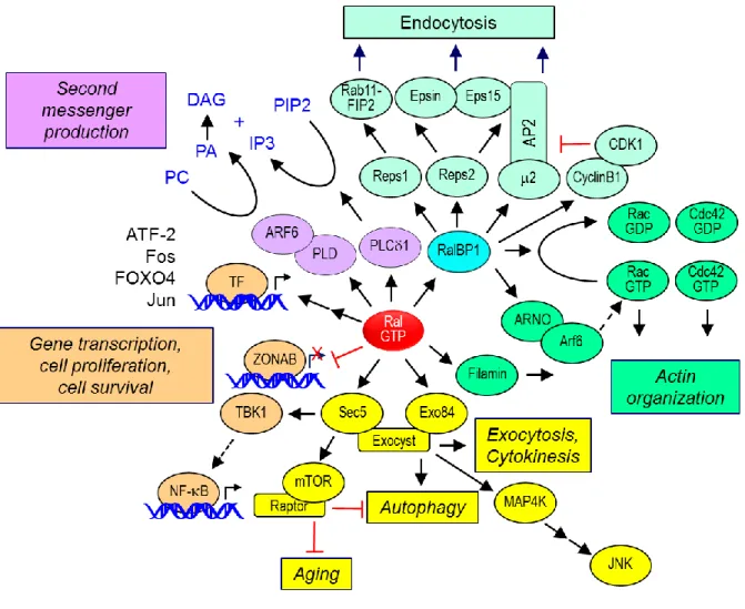
Ral effectors. Like Ras and other small GTPases, Ral interacts with a number of effector proteins when bound to GTP (Fig. 1-3). However, unlike with Ras, the Ral binding domains (RBD) lack primary sequence identity. The best characterized Ral effectors are RalBP1/RLIP76 and the Sec5 and Exo84 subunits of the octameric exocyst complex. The evolutionarily conserved exocyst complex mediates the tethering of post-Golgi secretory vesicles to the plasma membrane prior to exocytic fusion [33]. The exocyst subunits may also exist as monomers or subcomplexes, and can possess non-exocyst functions. Ral interaction with each subunit occurs in distinct subcellular locations, interacting with Sec5 at the plasma membrane and with Exo84 with intracellular vesicles [34, 35]. Although RalA and RalB can interact with the same set of effectors in vitro, as described below, the distinct biological functions of RalA and RalB are mediated by differences in subcellular localization, leading to their interaction with distinct subsets of effectors.
22
23
RalBP1. The first Ral effector described, RalBP1 (Ral binding protein 1; also called RLIP76 or
RIP1), was identified in screens for proteins that bound preferentially to activated RalA [40-42]. RalBP1 orthologs are found in Drosophila and C. elegans. RalBP1 contains a RhoGAP catalytic domain that has activity for the Cdc42 and Rac small GTPases, members of the Rho branch of the Ras superfamily [1]. Cdc42 stimulates filopodia formation whereas Rac stimulates lamellipodia formation. Thus, RalBP1 provides a link between Ral and modulation of the actin cytoskeleton changes that drive these cellular activities [40].
In additional to its RhoGAP domain, RalBP1 has additional functions (Fig. 1-3). Two ATP binding motifs have been identified in RalBP1 and shown to be important for transport function involving glutathione conjugates of electrophilic compounds [43, 44]. This transport function may facilitate the cellular export of chemotherapeutic drugs and radiation-induced oxidative damage byproducts [45]. RalBP1 overexpression has been found in a spectrum of human cancers, and suppression of RalBP1 expression can impair tumorigenic growth in vivo [46]. However, phenotypes attributed to RalBP1 do not necessarily implicate their role in Ral signaling [47].
RalBP1 also functions as a scaffold and interacts with a spectrum of functionally distinct proteins that regulate endocytosis and signal transduction (Fig. 1-3). The AP2 adaptor complex, a regulator of clathrin-mediated endocytosis from the plasma membrane, associates with the N-terminal region of RalBP1 [48]. The Eps homology (EH) domain-containing proteins Reps1 and Reps2 (POB1) were
identified as proteins that interacted with the C-terminus of RalBP1 [49, 50]. These proteins are known to be important for receptor tyrosine kinase-regulated endocytosis, with Reps1 interacting with Rab11-FIP2 and Reps2 binding Epsin and Eps15 [51, 52].
24
RalBP1 has been implicated as a key effector for several Ral-driven processes. In these studies, the typical approach has been the utilization of mutants of Ral that are selectively impaired in effector interaction. The D49N substitution impairs RalBP1 but not Sec5 or Exo84 effector binding, whereas the D49E mutation has the opposite consequence [40, 54, 55]. For example, shRNA silencing analyses determined that RalB but not RalA was required for invadopodia formation in pancreatic cancer cell lines [56]. RalB D49E but not D49N could rescue loss of endogenous RalB and restore invadopodia formation, indicating that RalBP1 was a critical effector for this RalB activity. This RalBP1 function was GAP-independent but abolished by mutation of the ATP binding motifs [56].
RalA was shown to utilize RalBP1 to regulate mitochondrial fission at mitosis [57]. Mitochondria exist as dynamic interconnected networks that are maintained through a balance of fusion and fission. Fission facilitates equal distribution of mitochondria to daughter cells during mitosis. Fission is controlled by the GTPase Drp1 on the outer mitochondrial membrane. RalA was found to recruit RalBP1 to
mitochondria, where RalBP1 acts as a scaffold to facilitate cyclin B/Cdk1 phosphorylation of Drp1 to promote mitochondrial fission. Suppression of either RalA or RalBP1 expression caused a loss of mitochondrial fission at mitosis.
Recently, RalBP1 was shown to be necessary and sufficient for RalA-driven mislocalization of the cyclin-dependent kinase inhibitor p27 KIP1, leading to inhibition of TGF-β–mediated growth arrest in epithelial cells [58]. This function appeared to require an intact RhoGAP domain.
Sec5 and Exo84 subunits of the exocyst. The best-characterized Ral effectors are two components
25
interacting with Sec5 and Exo84 [55, 60]. Ral interaction with Sec5 may also regulate exocyst-independent functions.
Recent evidence suggests that Ral engages exocyst subunits to perform a variety of cellular processes independent of their roles in exocytosis. White and colleagues found that the association of RalB with Sec5 is critical in the innate immune response [61]. RalB binding to Sec5 leads to an
interaction of Sec5 with TBK1, a protein kinase known to regulate NF-B signaling. Intriguingly, TBK1 has recently been identified in siRNA screens as a synthetic lethal partner of activated K-Ras [62], although a subsequent study failed to support this relationship [63]. Recently, a mechanism where the integrin αvβ3 recruited a K-Ras-RalB complex to the plasma membrane to activate TBK1 and NF-B signaling was identified (Fig. 1-3) [64]. This signaling mechanism regulated tumor initiation and growth. Chapter 3 contains a study of TBK1 inhibition in Ras-driven pancreatic ductal adenocarcinoma (PDAC).
The association of RalB with the exocyst has also been shown to regulate macroautophagy [34]. When cells are grown in nutrient-rich conditions, RalB engages Sec5. Upon nutrient starvation, RalB then engages Exo84 and the exocyst, leading to an upregulation of autophagosome formation. This process is mediated through the assembly of the ULK1 serine/threonine kinase and Beclin1-VPS34 complexes on the exocyst. Autophagy has emerged as a key component of Ras-driven transformation in a variety of cell types, perhaps highlighting an underlying importance of Ras-RalGEF signaling in tumor cell autophagy.
Other effectors. One lesser-characterized Ral effector is phospholipase D1 (PLD1) [65, 66].
However, unlike other effectors, the association with Ral is not GTP-dependent and instead the
association is with the N-terminal 11 amino acid extension (Fig. 1-1D). PLD1 is best known for its role in converting phosphotidylcholine to phosphatidic acid and choline in response to G-protein coupled
26
promote proper p27 localization, thus allowing for proper TGF-β signaling [58]. The interaction of both RalA and RalB with PLD1 has been shown to be critical for HeLa cell cytokinesis [22].
Filamin is an important component of the actin cytoskeleton and is involved in actin crosslinking and lamellipodia formation. The association of RalA with filamin was found to be important for filopodia formation in Swiss-3T3 cells [68]. Additionally, RalA did not induce filopodia in a human melanoma cell line that lacks expression of filamin.
Lastly, active RalA has been shown to engage the transcription factor ZONAB (zonula occludens 1-associated nucleic acid binding protein) in a cell density dependent manner in MDCK cells [69]. At high cell densities, RalA engages ZONAB, unlocking the transcription of ZONAB targets, but it is unclear which genes are turned on [69]. While a direct role for Ral association with these lesser-studied effectors has not been found in Ral-driven cancers, their important roles in mitosis, motility, and gene regulation make them intriguing targets as Ral studies progress.
Post-translational modification and regulation of Ral function. RalA and RalB exhibit the most significant sequence divergence in their C-terminal membrane targeting sequences (50% identity) (Fig. 1-1C). This sequence divergence results in their distinct subcellular localization that contributes to the functional differences described for RalA and RalB by regulating effector utilization in vivo [56, 70-73]. Both isoforms can be found at the plasma membrane as well as in endomembranes, with cell type
differences seen. In this section we summarize the role of posttranslational modifications that regulate Ral subcellular localization.
Ral CAAX modifications. Like the majority of Ras family small GTPases, RalA and RalB
27
prenyltransferase specificity [74]. When X = S, A, Q, and M, the protein is preferentially recognized by farnesyltransferase (FTase)-catalyzed addition of a C15 farnesyl isoprenoid lipid; when X = L or I, it signals for geranylgeranyltransferase type I (GGTase-I)-catalyzed addition of a C20 geranylgeranyl isoprenoid.
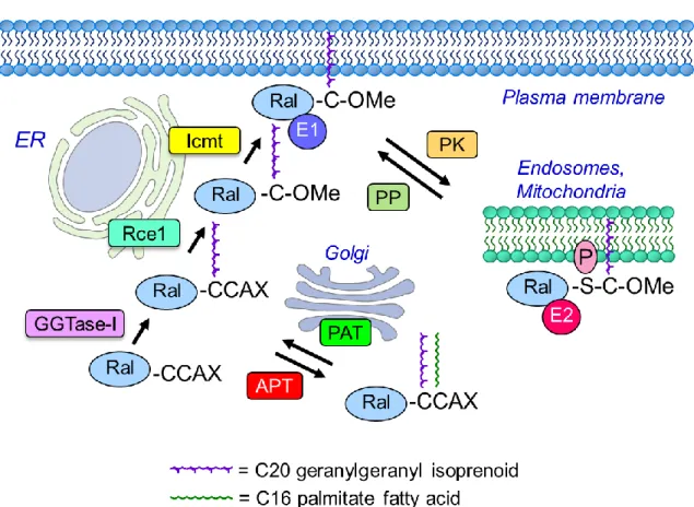
For Ral, the initial step is catalyzed by covalent addition of geranylgeranyl to the cysteine residue of the CAAX motif by cytosolic GGTase-I [75]. This is followed by endoproteolytic removal of the AAX residues, catalyzed by endoplasmic reticulum-associated Ras converting enzyme 1 (RCE1), and
subsequent carboxylmethylation of the now terminal prenylated cysteine residue, catalyzed by isoprenylcysteine carboxyl methyltransferase (ICMT).
The CAAX-signaled modifications are critical for both RalA and RalB function. Mutation of the cysteine residue to prevent all CAAX-signaled modifications disrupts Ral membrane association and function [75]. Similarly, treatment with a pharmacologic inhibitor of GGTase-I also disrupted Ral membrane association and signaling. Since inhibition of the GGTase-I modification prevents all
subsequent modifications, the role of the Rce1 and ICMT catalyzed modifications in Ral function remain to be addressed.
Recently, the Ral CAAX motifs were identified as members of a distinct subset of CA1A2X
motifs where the A1 residue is a second cysteine residue (CCAX). CCAX motifs can undergo an
alternative modification pathway (Fig. 1-4). As shown for a Rho family small GTPase (Cdc42), this motif can signal for dual lipid modification: prenylation followed by covalent addition of a palmitate fatty acid [76]. For Cdc42, after the initial GGTase-I catalyzed prenylation step, a Golgi-associated protein
28
this palmitate modification on Ral subcellular localization and membrane association, and function, have not been determined previously. Chapter 2 explores the consequence of RCE1 and ICMT processing as well as palmitoylation of the Ral CAAX motif on Ral localization and function.
29
Phosphorylation regulation of subcellular localization and effector interaction. An emerging
theme in the regulation of small GTPases is reversible post-translational modifications that dynamically regulate subcellular localization, thereby influencing effector interaction and biological activity [77]. In particular, recent studies have highlighted protein kinase-mediated phosphorylation of small GTPases in their C-terminal membrane-targeting regions. For example, K-Ras4B phosphorylation by protein kinase C (PKC) on S181 in its C-terminal membrane targeting sequence altered K-Ras4B subcellular
localization [78]. Nonphosphorylated K-Ras4B was plasma membrane associated, whereas S181 phosphorylation K-Ras4B caused translocation to mitochondrial and endoplasmic reticulum (ER)
membranes. S181 is positioned within a polybasic amino acid stretch in K-Ras4B that serves as a second signal that together with the CAAX modifications promote full plasma membrane association. The negative charge caused by phosphorylation reduces the positive charge of the polybasic stretch. The ER-associated K-Ras4B then ER-associated with inositol trisphosphate receptors (InsP3) on the ER in a Bcl-xL-dependent fashion, blocking the ability of Bcl-xL to potentiate the InsP3 regulated flux of calcium from ER to mitochondria that is required for respiration, inhibition of autophagy, and cell survival [79].
The Ral proteins are also regulated by similar mechanisms, with distinct protein kinases
30
Studies by our lab and others have found that RalB is similarly regulated by PKCα
phosphorylation of S198 in the C-terminal membrane targeting sequence [72, 81]. In one study, it was found that S198 phosphorylation caused RalB translocation from the plasma membrane to endocytic vesicles [72]. Associated with this change in subcellular localization was a switch in effector utilization. Whereas unphosphorylated RalA preferentially associated with Sec5, S198 phosphorylation caused preferential association with RalBP1. Phosphorylation of RalB S198 was necessary for proper exocytic vesicle trafficking and fusion at the plasma membrane, with delivery of surface alpha-5 integrin being regulated by dynamic RalB phosphorylation. Independently, Theodorescu and colleagues found that phosphorylation of RalB S198 was critical in regulating the ability of RalB to promote the metastatic growth of bladder cancer cells in a nude mouse model [81].
Ubiquitination. In the past few years, regulation of small GTPases by ubiquitination has gained
recognition [77]. For example, monoubiquitination of K-Ras on K147 reduces GAP sensitivity, thus allowing K-Ras to remain active and signaling in the absence of upstream input [83]. Ubiquitination of the Ral proteins has also been shown to influence their activity and function. Regulation of the
ubiquitination of RalA modulated RalA activity as well as lipid raft exposure [84]. Furthermore, ubiquitination of RalB promoted binding to Sec5 to regulate innate immunity, whereas deubiquitination allowed for binding to Exo84 and subsequent induction of autophagy [85].
31
human cancer was observed, suggesting species differences in the effectors that are important in Ras oncogene function [88].
That Ral GTPases serve critical roles in human cancer cell growth gained greater traction when White and colleagues found that RalB was critical for tumor but not normal cells for survival, while RalA was necessary for the anchorage-independent growth of cancer cells [89]. Importantly, this also marked the first time RalA and RalB were found to have non-overlapping functions. Since these key studies, a major theme of Ral proteins is their significant and often divergent roles in numerous cancer types. In the following section we review some of the key findings made with regards to the role of the two Ral isoforms as drivers in different human cancers. Since the RA domain-containing RalGEFs can be activated by other Ras family small GTPases, as well as by non-Ras mechanisms, and since some RalGEFs are regulated by non-Ras mechanisms, an involvement of Ral in cancers where RAS mutations are not common is not surprising.
Bladder carcinoma. Evaluation of a panel of human bladder cancer cell lines found preferentially
increased levels of activated RalA and RalB in RAS-mutant [90] or invasive cell lines [91]. Using RNAi or ectopic expression of activated Ral mutants, Theodorescu and colleagues found that RalA and RalB played antagonistic roles in the migratory activity of the KRAS-mutant UM-UC-3 bladder cancer cell line, with RalA suppressing and RalB enhancing motility [92].
32
Colorectal carcinoma. Oncogenic KRAS and NRAS mutations occur in 45% and 8%, respectively, of
colorectal cancer (CRC) tumors. Ral signaling has been shown to be a critical regulator of the anchorage-independent growth properties of CRC tumor cells [93]. Martin et al found that RNAi-mediated
suppression of RalA resulted in a decrease in soft agar colony growth while loss of RalB had the opposite effect, leading to an enhancement of anchorage-independent growth. They found that RalA and RalB modulated this phenotype by utilizing both common and distinct effector proteins. Using Ral effector binding mutants that are selectively uncoupled from Exo84, Sec5, or RalBP1, they showed that RalA required Exo84 and RalBP1 binding to promote the anchorage-independent growth of CRC cells. Conversely, RalB required Sec5 and RalBP1 to suppress soft agar colony formation. Intriguingly, loss of one Ral isoform was found to increase the activation of the other isoform suggesting compensatory crosstalk between RalA and RalB. What specifically mediates this crosstalk between RalA and RalB is unknown, but it could be through either enhanced RalGEF accessibility for the remaining Ral protein or a downregulation of RalGAP activity upon single Ral isoform depletion. Depletion of RalB has also been shown to cause apoptosis in colorectal cancer cells [61].
Hepatocellular carcinoma. RAS mutations are rare (>2%) in hepatocellular carcinoma (HCC). RalA
was found to be significantly overactivated in hepatocellular carcinoma (HCC) cells and tissues compared to nonmalignant samples. Suppression of RalA expression caused a significant decrease in the viability and invasiveness of HCC cells. A role for RalB was not addressed. Finally, in a transgenic mouse model for HCC (farnesoid X receptor–deficiency induced) elevated RalA-GTP was detected in the liver tumors [94].
Lung adenocarcinoma. KRAS mutations are found in 30% of lung adenocarcinomas and several
33
adenocarcinoma cell line reduced the proliferation and invasion in vitro. In a second more comprehensive study, immunohistochemistry analyses of non-small cell lung cancers (NSCLC), it was found that high RalA and RalB protein expression was associated with poor survival. The levels of activated RalA but not RalB were higher in KRAS-mutant NSCLC cell lines [96]. Depletion of RALA or RALB or both reduced anchorage-dependent and –independent growth for either KRAS mutant and wild type cell lines.
Depletion of RALA, RALB, or both also impaired the tumorigenic growth of KRAS-mutant NSCLC cells. Interesting, very limited analyses in this and another study suggested mutation-selective involvement of Ral in NSCLC, where KRAS G12C mutant NSCLC cell lines showed greater activation and/or
dependence on Ral for growth [97].
In contrast to human lung tumor cell line studies, RalA and RalB were found to exhibit redundant functions when assessed in mouse development and in a Kras G12D-driven mouse model of lung
adenocarcinoma [73]. Ralb deficient mice were viable with no overt phenotype whereas a Rala deficiency caused embryonic lethality that was further exacerbated by a combined Ralb deficiency. Neither a Rala nor a Ralb deficiency impaired Kras-driven lung tumor development. However, a combined loss of both Rala and Ralb significantly reduced lung tumor development. These results suggest redundancy in RalA and RalB function for tumor development. One possible explanation for this different conclusion may be that the human lung tumor cell line studies addressed the role of Ral in tumor maintenance whereas the mouse study addressed the role of Ral in tumor initiation and progression.
Malignant peripheral nerve sheath tumors. Loss of the NF1 RasGAP, rather than RAS mutational
34
human MPNST cell lines and tissue, and restoration of NF1 GAP activity reduced RalA activity, indicating that this was associated with Ras activation.
Melanoma. RAS mutations, predominantly NRAS, occur in 28% of skin cutaneous melanomas. With
BRAF mutations seen in 60% of melanomas in a non-overlapping frequency with RAS mutations,
activation of the canonical Raf-MEK-ERK mitogen-activated protein kinase (MAPK) pathway alone may seem to be sufficient for Ras-driven melanoma growth. However, analysis of Ral activation in a panel of human melanoma cells showed a consistently high level of total and activated RalA, but not RalB, activation that was independent of NRAS or BRAF mutation status [100]. Additionally, RalA and to a lesser degree RalB are necessary for the tumorigenic growth of melanomas, also regardless of BRAF and NRAS mutation status.
Studies using tumor suppressor Arf-deficient immortalized mouse melanocytes to investigate the contributions of Ras downstream signaling to melanomagenesis also indicated a role for Ral signaling [101]. Ectopic expression of the RalGEF Rgl2 engineered to contain a membrane localization sequence (to mimic Ras activation of RalGEF) was sufficient to promote the anchorage-independent growth and Matrigel invasion of these melanocytes similar to that caused by oncogenic N-Ras. Surprisingly, in contrast, activated BRAF V600E did not enhance proliferation or invasion. Finally, ectopic expression of a dominant negative mutant of RalB that blocks RalGEF function partially impaired the growth of NRAS-transformed melanocytes. Thus, together with the findings of Zipfel et al. [100], Ral GTPases can act as drivers of melanoma cancer growth in both RAS wild type and mutant cancer cells.
Ovarian carcinoma. One study has now revealed that Ral signaling has a role in ovarian cancer.
35
Pancreatic ductal adenocarcinoma. A significant requirement for activated Ral signaling in
pancreatic adenocarcinoma (PDAC) cell line tumorigenic and invasive growth has been established. Human PDAC has a high frequency or activating KRAS mutations and Ral activation is seen in both human tissue samples and tumor cell lines [103-105]. Interestingly, activation of RalA was found at a higher frequency than the activation of either ERK or AKT in PDAC cells, suggesting a critical role for the RalGEF-Ral pathway downstream of oncogenic K-Ras.
Depletion of RalA and RalB via RNAi has elucidated roles for RalA in anchorage-independent and tumorigenic growth and RalB in invasive and metastatic growth of PDAC cells [105]. PDAC cells with stable RNAi depletion of RalA results in reduced subcutaneous tumor formation upon injection into immune compromised mice. These same cells expressing RalB RNAi do not form lung metastases post-injection into the tail-vein of nude mice. In addition to playing a role in tumor initiation, RalA has also been shown to be necessary for PDAC tumor maintenance. The use of inducible RNAi to stably deplete RalA from established primary tumors resulted in regression of the tumor, indicating a necessity for persistent RalA signaling in established PDAC tumors.
There is also recent evidence that active K-Ras signaling to RalB but not RalA plays a critical role in the formation of invadopodia in PDAC cells [56]. Invadopodia are actin-rich membrane protrusions that are known to be involved in the secretion of matrix metalloproteases (MMP) during tumor cell invasion. RalB requires the ability to interact with RalBP1 to mediate this process and RalBP1 itself is necessary for the formation of invadopodia in PDAC cells. Surprisingly, the RhoGAP activity of RalBP1 is not necessary for invadopodia formation while the ATPase activity is required. Why the ATPase activity is necessary for RalBP1 to mediate invadopodia formation is unclear.
36
and RalB activation. Interestingly, expression of constitutively active RalA could not rescue soft agar growth after the loss of Rgl2 indicating that Rgl2 may have non-Ral regulatory functions or that the RalA interaction with Rgl2 is critically important for the regulation of anchorage-independent growth. Rgl2 was found to be co-localized with RalB but not RalA at the leading edge of migrating CFPac-1 PDAC cells. Loss of Rgl2 results in a loss of RalB from the leading edge, perhaps giving insight into how the migratory and invasive activity of PDAC cells relies on Rgl2/RalB signaling.
Prostate carcinoma. Increased RalA-GTP levels were observed in the RAS wild type human prostate
carcinoma cell line PC3. Suppression of RalA did not impair tumor formation but did abolish bone metastasis [107]. In contrast, suppression of RalB expression did not impair metastasis.
Squamous cell carcinoma. Squamous cell carcinoma is the second most common type of skin
cancer. Using an in vitro model of Ras-induced human squamous cell carcinoma (SCC) of the skin, it was found that RalA suppressed rather than promoted progression [108]. Suppression of RalA but not RalB stimulated the progression of HRAS-transformed human keratinocytes to a more invasive state.
In contrast to the in vitro observations, different roles for Ral were observed in a mouse model of carcinogen-induced SCC [73]. Single application of the mutagen DMBA, followed by repeated
applications of phorbol ester 12-O-tetradecanoylphorbol 13-acetate (TPA) causes Hras mutation and the development of benign papillomas, with a subset progressing to SCC. Neither a Rala nor a Ralb
deficiency impaired papilloma development. As described above for Kras-driven lung adenocarcinoma formation, only combined loss of both Rala and Ralb significantly reduced papilloma development. Genetic ablation of Ralgds in this same carcinogenesis model also significantly reduced tumor incidence, size, and progression [109].
37
38
Chapter 2: Divergent Roles of CAAX Motif-Signaled Posttranslational
Modifications in the Regulation and Subcellular Localization of Ral GTPases
1Background
As mentioned in Chapter 1, the Ral proteins, like Ras, contain a C-terminal CAAX motif that undergoes prenylation followed by cleavage by RCE1 and carboxylmethylation by ICMT (Fig. 1-4). These processing enzymes are being considered as therapeutic targets in the treatment of cancer since their activities are thought to be necessary for proper subcellular localization and function of CAAX-containing proteins. While their usefulness as therapeutic targets is still not fully understood, progress has been made in further elucidating the roles of RCE1 and ICMT, which will aid in determining how to implement inhibitors against these processing enzymes for the treatment of cancer.
RCE1 was initially discovered as a Ras processing protein in S. cerevisiae, where RCE1 disruption was shown to cause Ras mislocalization and signaling [110]. RCE1 is an integral membrane protein of the ER [111]. It is the only enzyme with this particular cleavage activity, and was found to cleave most CAAX sequences efficiently with few exceptions [112-114]. Since targeting Ras has long been of interest in the field of cancer therapeutics, many studies focused on the role of RCE1 catalytic activity in Ras-dependent processes. It has been shown that RCE1 can process all Ras isoforms, and that deletion of RCE1 reduced Ras-driven transformation of mouse embryo fibroblasts [115, 116]. Although there have been numerous discoveries about RCE1 biology, progress toward RCE1 inhibitors has been
1
Adapted from previously published work: Gentry LR, Nishimura A, Cox AD, Martin TD, Tsygankov D, Motohiro N, Elston TC, and Der CJ. “Divergent roles of CAAX motif-signaled posttranslational
39
limited. The first documented RCE1 inhibitors were AOMKs ((acyloxy)methyl ketones), which were shown to block K-Ras4B cleavage [117, 118]. Another study showed compounds identified in an in vitro screen for small molecule inhibitors of RCE1 that disrupted Ras subcellular localization [119, 120]. However, these inhibitors lack sufficient potency and selectivity and have not been implemented in cancer cell assays. In the absence of useful pharmacologic inhibitors, the assessment of their roles in cancer growth remains unknown and underexplored. Thus, Rce1-/- MEFs remain one of the best options for studying the effects of RCE1 loss.
ICMT was initially discovered in S. cerevisiae for its ability to methylate a-factor mating pheromone, and it was quickly shown that ICMT methylates Ras as well [121, 122]. ICMT is also localized at the ER [123]. One study found that ICMT was required for Ras-driven myeloproliferative disease. In contrast, a second study demonstrated its absence resulted in exacerbation of K-Ras-driven pancreatic cancer growth. These opposing observations highlight the fact that our understanding of whether ICMT is a useful therapeutic target in Ras-driven cancer remains to be clarified [124, 125]. In contrast to RCE1, there has been better progress in developing and characterizing ICMT inhibitors in cancer models. Many compounds have been found to inhibit ICMT, including methotrexate and lysine nitrilotriacetic acid [126-129]. One such small molecule, cysmethynil, has been shown to decrease both anchorage-dependent and –independent growth and cause mislocalization and impaired signaling of Ras in colorectal cancer cells [130]. However, this inhibitor has off-target activities that complicate
interpretation from studies utilizing this inhibitor to assess ICMT as a therapeutic target. In the absence of useful inhibitors, cell models with genetic loss of ICMT function remain the best option. Icmt-/- MEFs have been used widely as the cell system most suited to study the impact of ICMT loss on protein
function.
40
results in a dual prenyl and palmitoyl form of the CCAX-containing protein that then does not undergo subsequent RCE1- and ICMT-mediated processing steps. Palmitoylation at this site of the brain-specific isoform of the Rho family protein Cdc42 was shown to prevent RhoGDIα binding in vitro and promote stable Cdc42 membrane association in cells, as opposed to its fully processed form that was instead found in the cytosol in addition to membrane fractions [76]. Furthermore, Cdc42 that was alternatively
palmitoylated displayed greater serum response element (SRE) activity than the canonically processed Cdc42, suggesting the palmitoyl from has a greater ability to stimulate downstream signaling. containing RalA, RalB, and PRL-3 were shown to be palmitoylated at this site, and other CCAX-containing proteins that undergo prenylation may also be subject to palmitoylation of the motif, but the consequences of this newly-identified processing pathway on Ral GTPase function has not been studied. Unlike Cdc42, Ral GTPases lack a functional counterpart to RhoGDI. Therefore, the consequences of palmitoylation on Ral function are not known.
Interestingly, although RCE1 and ICMT expression has been shown to be necessary for certain Ras-driven cancers, depletion of either processing enzyme does not fully mislocalize Ras, and it is likely that other RCE1- and ICMT-dependent proteins downstream of Ras are at least partially responsible for mediating these anti-proliferative effects. It is speculated that there may be as many as 400 mammalian CAAX-terminating proteins [131]. Also, while inhibition of palmitoylation is being considered as a therapeutic route in Ras driven cancers given recent findings that palmitoylated KRas4A, as well as N-Ras and H-N-Ras, are involved in colorectal and other cancers, the consequence of this modification on other small GTPases is not understood. In Chapter 2, I explore the effect of RCE1 and ICMT loss as well as the consequence of palmitoylation on the function of CCAX-containing Ral proteins.
Introduction
41
switches and cycle between inactive GDP- and active GTP-bound states. The cycle is catalyzed by Ral-selective guanine nucleotide exchange factors (RalGEFs) and GTPase-activating proteins (RalGAPs), which accelerate slow intrinsic exchange and GTPase activities, respectively [106]. Ral GTPases are activated downstream of Ras oncoproteins. Activated Ras-GTP binds to and activates RalGEFs (e.g., RalGDS), stimulating formation of Ral-GTP. Active Ral-GTP binds preferentially to a spectrum of functionally diverse downstream effectors to regulate a diversity of cellular processes that include actin cytoskeletal organization, endocytosis and exocytosis, and mitochondrial function [57, 70, 135, 136] as well as cell proliferation, survival and autophagy [31, 34]. The best validated effectors of Ral are the Sec5 and Exo84 subunits of the exocyst complex and RalBP1/RLIP76, a GAP for Rho family small GTPases [132-134].
RalA and RalB share significant overall sequence, structural, and biochemical identity [134]. Their amino-terminal G-domains (amino acids 12-176) involved in GTP binding and hydrolysis, RalGEF and RalGAP regulation and effector binding share 88% sequence identity. Remarkably, despite their regulation by shared GEFs and GAPs and interaction with shared effectors in vitro, RalA and RalB display divergent biological roles in normal and neoplastic cell growth. RalB was found to be specifically required for survival of tumor but not normal cells [89]. In contrast, RalA was dispensable for survival, but was required for anchorage-independent proliferation. Differences in Ral isoform function have also been observed in various cancer types. Distinct aspects of cell migration were differentially regulated by RalA versus RalB in bladder cancer cells [92]. RalA was essential for tumorigenic growth of pancreatic cancer cells, whereas RalB was required for invasive and metastatic growth [105]. In contrast, RalA promoted whereas RalB antagonized anchorage-independent growth of colorectal carcinoma cells [93].
42
the cysteine residue of the CAAX motif [131]. The X residue determines protein prenyltransferase specificity [74]. RalA and RalB proteins, both of which terminate in leucine, are modified by addition of a geranylgeranyl lipid, catalyzed by cytosolic geranylgeranyltransferase-I (GGTaseI) [75, 137]. Subsequent cleavage of the –AAX tripeptide by endoplasmic reticulum-associated Ras converting CAAX endopeptidase 1 (RCE1) leaves a lipid-modified terminal cysteine. This cysteine is then methylated by endoplasmic reticulum-associated isoprenylcysteine carboxylmethyltransferase (ICMT) [138].
The RalA and RalB functional differences identified to date have been attributed to the carboxyl-terminal sequences immediately adjacent to their CAAX motifs [70, 104]. Termed the hypervariable region (HVR; amino acids 177-202), this sequence exhibits the greatest divergence between RalA and RalB (44% identity) and confers secondary membrane-targeting specificity. In particular, the RalA and RalB carboxyl termini possess phosphorylation sites recognized by distinct protein kinases, where phosphorylation causes translocation from the plasma membrane to endomembranes and a change in effector interaction [71, 72, 80-82]. However, posttranslational modifications at other sites may also play an important role in Ral isoform functions.
A recent study of the brain-specific isoform of the Rho family small GTPase Cdc42 concluded that proteins terminating in a CA1A2X motif in which the A1 residue is a cysteine (CA1A2X = CCAX) can
undergo an alternative processing pathway [76]. While a majority of Cdc42 undergoes the conventional post-prenyl modification pathway involving RCE1 and ICMT, a subset (5-20%) does not undergo RCE1-catalyzed removal of the AAX residues, and instead the A1 cysteine is covalently modified by
43
CA1A2X motifs [76], the impact of dual lipid modification of the CAAX motif on Ral subcellular
localization was not determined.
Additionally, the contributions of the CAAX-signaled RCE1 and ICMT modifications to RalA and RalB subcellular localization and membrane association have not been addressed. We therefore determined the consequences of RCE1 or ICMT deficiency on these properties of each Ral isoform. We found that both RalA and RalB required RCE1 for efficient plasma membrane targeting, whereas only RalB required ICMT for this association. Interestingly, while RalA could still concentrate at the plasma membrane in the absence of ICMT, it depended on ICMT for localization to recycling endosomes. We also found that RCE1 deficiency increased steady-state activity of both RalA and RalB, whereas ICMT deficiency increased steady-state levels of RalB but not RalA. Finally, we determined that CA1AX
palmitoylation was essential for plasma membrane association of RalB, but not of RalA. Pharmacologic inhibitors of RCE1 and ICMT are being considered as anti-Ras therapeutic strategies [139]. Since Ral GTPases are key drivers of Ras-dependent cancer growth, it will be critical to understand the complex and differential consequences of RCE1 and ICMT inhibition for Ral function.
Experimental Procedures
Plasmids and Cell Lines – cDNAs encoding green fluorescent protein (GFP)-tagged human RalA was
described previously [71] and RalB was generated for this study as described for RalA. Site-directed mutagenesis was done to generate cysteine-to-serine missense mutants of the A1 cysteine residue of the
Ral CCI/LL CA1A2X motif, which were then subcloned into pEGFP-C3. The pEGFP-RalA/B chimeric
44
(Rce1-/-) or ICMT (Icmt-/-) were provided by Dr. Steven Young (UCLA) and cultured as described previously [112, 140].
Transfection, Transferrin Labeling, and Microscopy – Cells were plated, transfected, and imaged as described previously [141]. Lipofectamine Plus was used for DNA transfections. Three h after transfection, cells were washed and cultured in phenol-red free Dulbecco’s Modified Eagle’s Medium (DMEM) supplemented with 10% fetal calf serum. Cells were labeled with transferrin following the manufacturer’s protocol (Life Technologies). Cells were imaged in 35-mm culture dishes containing a 15 mm coverslip on the bottom made of number 1.5 glass (MatTek). Cells were examined with an inverted laser-scanning confocal microscope (Zeiss 710 LSM) using an oil immersion 63X objective with a 1.4 numerical aperture. Images of GFP expression were captured by scanning with a 488 nm line of a solid-state laser using a BP 490-555 emission filter. Images of mCh and red fluorescent transferrin probes were captured by scanning with a 555 nm solid-state laser with the variable secondary dichroic set to 580 nm. Images were processed with ImageJ software, and thresholded Manders coefficients using were calculated using the colocalization threshold plugin from the Wright Cell Imaging Facility.
GST Pulldown Assays – Endogenous activated RalA- and RalB-GTP were assayed as we described previously, by pulldown analyses using GST-Sec5-RBD (glutathione S-transferase fusion of the Ral-GTP binding domain of Sec5) [104]. After SDS-PAGE and immunoblotting with the RalA- (BD Biosciences) or RalB- (Millipore) specific antibodies, the amount of active Ral-GTP was normalized to total Ral protein levels in total cellular lysates. Western blot analyses with anti--actin (Sigma Aldrich) or anti-GAPDH (Sigma Aldrich) were done to verify equivalent loading of total cellular protein.
Detection of Protein Palmitoylation - Palmitoylated proteins were purified using acyl-resin assisted capture (acyl-RAC). Cells were lysed with RIPA buffer (20 mM Hepes-NaOH pH 7.4, 100 mM NaCl, 3 mM MgCl2, 1% Triton X-100, 0.5% deoxycholate, and 0.1% SDS) supplemented with protease inhibitor.
45
buffer (100 mM Hepe-NaCl pH 7.4, 1 mM EDTA, and 1% SDS). Samples were mixed with thiopropyl-Sepharose in the presence of 250 mM hydroxylamine to cleave thioester linkages. As a negative control, the same volume of NaCl was added instead of hydroxylamine. After 3 h rotation at room temperature, the resins were washed four times with binding buffer, and samples were eluted in buffer containing 50 mM Tris-HCl pH 6.8, 50 mM DTT, 10% glycerol, and 1% SDS. After immunoblotting with the RalA- and RalB- specific antibodies, the palmitoylation stoichiometry was calculated by a ratio of the precipitate intensity to the input intensity.
Quantification of Fluorescence – The statistics of fluorescence intensity were extracted from images of individual cells as a function of the distance from the cell edge. Cell masks (binary images) were obtained by thresholding with the MovThresh module of the CellGeo package [142]. Each cell mask was then morphologically eroded using the MATLAB function “imerode” with a disk of radius 1 to 50 pixels as a structural element. At each step of the morphological erosion, the boundary was traced and the intensity of the corresponding boundary pixels was determined from the original image. This allowed the mean, median, and standard deviation to be calculated for each cell as function of the distance 1 to 50 pixels from the cell edge. Finally, the intensity profiles for individual cells were normalized to their peak (max) values, and then averaged over all cells in a given genotype. These four processing steps constitute a single MATLAB code for batch processing.
Results
RalA and RalB show distinct requirements for RCE1- and ICMT-mediated posttranslational
modifications for their subcellular localization and plasma membrane association - We first addressed
46
47
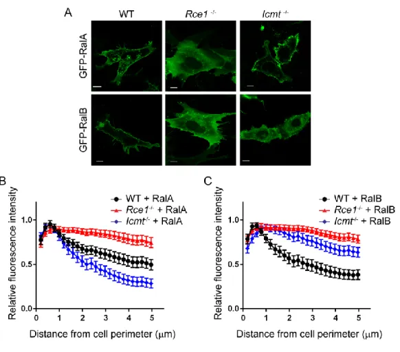
Figure 2-1. Subcellular localization of Ral family proteins is differentially dependent on RCE1 and ICMT processing. Wild-type (WT), Rce1-/-, and Icmt-/- MEFs were transiently transfected with expression vectors encoding EGFP-tagged RalA or RalB. A, live cells were visualized using confocal microscopy. Images shown are representative of three independent experiments, each of which examined >40 cells. Images were analyzed using our
MATLAB image processing script for B, EGFP-RalA distribution and C, EGFP-RalB distribution in WT, Rce1-/-,
48
Since RCE1 deficiency also prevents the subsequent ICMT modification, we next determined the consequences of ICMT deficiency alone. Although the plasma membrane targeting of both Ral isoforms depended similarly on RCE1, surprisingly, we found a differential requirement for ICMT in regulating RalA versus RalB plasma membrane targeting. In Icmt-/- MEFs, we observed that RalA remained concentrated at the plasma membrane, similar to its localization in WT MEFs. Thus, the significant disruption of RalA plasma membrane association in Rce1-/- cells was caused by the absence of AAX proteolysis. In striking contrast, RalB association with the plasma membrane was reduced significantly in Icmt-/- MEFs, instead showing increased diffuse localization in the cytosol. Thus, plasma membrane association of RalB displays a greater requirement for ICMT compared to that of RalA.
49
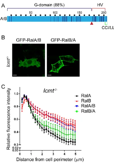
consequences of ICMT deficiency seen with RalA and RalB are due to sequence divergence in the HVR rather than in the G-domain.
Figure 2-2.Hypervariable domain dictates Ral ICMT dependency. A, Chimeric Ral proteins were constructed by a reciprocal switch of the carboxyl-terminal residues 176-206 (red arrow) comprised of the hypervariable domain and the CAAX motif. RalA/B is comprised of the RalA G-domain (residues 1-176) followed by RalB residues 177-206, whereas RalB/A is comprised of the RalB G-domain (residues 1-176) followed by RalA residues 177-206. WT,
Rce1-/- and Icmt-/- MEF cells were transiently transfected with expression constructs encoding EGFP-tagged Ral
chimeras RalA/B or RalB/A. B, live cells were visualized by confocal microscopy. Images shown are representative
of three independent experiments, each of which examined >40 cells. C, Images from B were analyzed using our
50
Icmt regulates RalA localization to recycling endosomes - Although ICMT deficiency did not
noticeably impact RalA plasma membrane targeting, we did observe a decreased level of endomembrane-associated RalA in Icmt-/- MEFs (Fig. 2-1A). Therefore, we used our MATLAB image processing script to quantify the spatial distribution of GFP-RalA. Our analysis revealed a decreased concentration of RalA closer to the nucleus in Icmt-/- MEFs. In contrast, RalB association with internal organelles was not impacted significantly by ICMT deficiency (Fig. 2-1B). Thus, RalA and RalB association with endomembranes exhibited different requirements for ICMT.
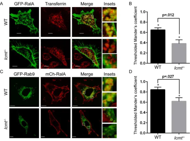
Since RalA is known to localize to recycling endosomes in various cell types [70, 136], we speculated that the RalA endomembrane localization that was disrupted in the absence of ICMT included this compartment. To address this possibility, we detected recycling endosomes by live cell microscopy using a fluorescent transferrin probe, and measured the colocalization of GFP-RalA with transferrin-loaded endosomes. Image analysis revealed a significant decrease in colocalization of RalA with transferrin in Icmt-/- MEFs compared to WT MEFs, indicating that RalA localization to recycling endosomes was impaired in the absence of ICMT (Figs. 2-3A and 2-3B).
51
Figure 2-3.RalA localization on recycling endosomes is impaired by loss of ICMT. A, WT and Icmt-/- MEF cells were transiently transfected with an expression construct encoding EGFP-RalA and incubated with pHrodo red transferrin conjugate for 30 min before visualization by confocal microscopy. The images were overlapped (Merge)
to indicate the degree of colocalization on endosomes in WT cells compared with Icmt-/-. B, Twenty-five images per
condition were quantified. Values shown are means of Manders coefficients using thresholds ± S.E., and an
unpaired t test was used to determine significance (p<0.05). C, WT and Icmt-/- MEF cells were transiently
52
RCE1 regulates Ral activity – Since we previously observed that phosphorylation-mediated
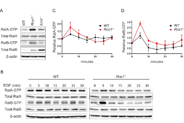
translocation of RalA or RalB from the plasma membrane to endosomes was associated with an increase in active GTP-bound protein [71, 72], we speculated that the altered subcellular localization of RalA and RalB seen in RCE1- or ICMT-deficient cells might also be accompanied by altered regulation of their GDP-GTP cycle and consequently of their activation state. To address this possibility, we first performed pulldown analyses to determine the level of activated, GTP-bound endogenous RalA and RalB in WT, Rce1-/- and Icmt-/- MEFs. When normalized to total protein, we found that the steady-state levels of both endogenous RalA-GTP and RalB-GTP were elevated strikingly in Rce1-/- MEFs compared to WT MEFs (Fig. 2-4A). In contrast, there was limited increase in RalA-GTP and no increase in RalB-GTP levels in Icmt-/- MEFs, although RalB protein levels were markedly higher in these cells compared to WT MEFs.
53
mislocalization that we observed in Rce1-/- MEFs is more likely to alter their regulation by one or more of the 6 RalGEFs than by RalGAPs [134].
Figure 2-4.RCE1 deficiency increases activated Ral-GTP formed upon EGF stimulation. A, Endogenous levels
of RalA-GTP and RalB-GTP were compared in WT, Rce1-/- and Icmt-/- MEFs by a pulldown assay using GST-Sec5
RBD. Western blot analyses were done with anti-RalA or anti-Ral antibody to determine levels of Ral-GTP and total
Ral proteins. -actin was used to verify equivalent total protein loading. B, WT and Rce1-/- MEFs were
serum-starved then stimulated with EGF. Cells were lysed at the indicated time points, followed by western blot analyses
as described in panel A. C, RalA-GTP and D, RalB-GTP expression levels were quantified by densitometry of
54
Icmt regulates RalB stability – As described above, we observed that levels of endogenous RalB but
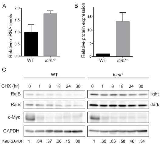
not RalA proteins were stably elevated in Icmt-/- MEFs compared to WT MEFs (Fig. 2-4A). Previous studies demonstrated that RhoA protein was destabilized upon pharmacologic inhibition or genetic loss of ICMT, whereas Ras protein was stabilized [145, 146]. While we did find that the steady-state transcript levels of RalB were slightly greater in Icmt-/- MEFs compared to WT MEFs (Fig. 2-5A), this small change likely was an insufficient basis for the large difference in abundance of RalB protein in the two cell types (Fig. 2-5B). We therefore hypothesized that the increases in RalB protein in Icmt-/- MEFs were due to altered regulation of RalB stability in the absence of ICMT.
55
Figure 2-5. ICMT deficiency increases RalB protein stability. A, RalB transcript levels were determined by
TaqMan qPCR analysis and normalized to GABPB1 in a triplicate set of assays ± SE. Data displayed as relative to
WT MEFs. B, Endogenous RalB protein levels were quantified by densitometry of western blot and normalized to
total protein. Data displayed as relative to WT MEFs. Experiment performed in triplicate. C, WT and Icmt-/- MEFs
56
Dual lipid modification of the Ral CAAX motif affects RalB subcellular localization –In addition to
the conventional modification pathway involving RCE1 and ICMT, RalA and RalB can also undergo an alternative pathway, whereby the newly GGTaseI-modified protein does not undergo subsequent RCE1-mediated proteolytic removal of the A1A2X residues but instead undergoes palmitoylation of the cysteine
residue at the A1 position [76]. While the mechanism that regulates which pathway will be taken has not
been elucidated, it was reported that the level of palmitoylation of RalA was increased in RCE1-deficient MEFs. Thus, impairment of one pathway can then favor a shift to the other pathway. We hypothesized that the altered subcellular localization of RalA and RalB that we observed in Rce1-/- MEFs (Fig. 2-1) might reflect not only loss of the RCE1- and ICMT-catalyzed modifications but additionally reflect alterations in dual lipidation by geranylgeranylation and palmitoylation of the CAAX motif.
To determine a role for this alternative dual lipid modification pathway in regulating RalA and RalB localization, we first generated the same palmitoylation-deficient Ral mutants utilized by Nishimura and Linder in defining the pathway [76]. To do so, we introduced a C to S amino acid substitution at the A1
position of each Ral isoform (C204S), designated RalA-CSIL and RalB-CSLL. This substitution ablates palmitoylation but not geranylgeranylation of the CAAX motif [76]. Although we did not observe a significant alteration in RalA-CSIL subcellular localization compared to WT RalA, we did find reduced