THE IDENTIFICATION OF BIOMARKERS THAT PREDICT IMPENDING HEART FAILURE PRESERVED EJECTION FRACTION (HFpEF)
Carolyn L. Lekavich
A dissertation submitted to the faculty of the University of North Carolina at Chapel Hill in partial fulfillment of the requirements for the degree of Doctor of Philosophy in the School of Nursing
Chapel Hill 2015
Approved by:
Debra J. Barksdale
Jamie Crandell
Virginia Neelon
Eric J. Velazquez
iii ABSTRACT
CAROLYN L. LEKAVICH: The Identification of Biomarkers that Predict Impending Heart Failure Preserved Ejection Fraction (HFpEF)
(Under the direction of Dr. Debra J. Barksdale)
The overall purpose of this investigation was to utilize pathophysiologic,
methodologic and empirical approaches to address gaps in our understanding about the
identification of biomarkers that predict impending HFpEF. To guide this exploration, three
papers were developed that outline the current scope of the problem, define strategies to
contribute to the current science, analyze the patient population, report the results of one
iv
ACKNOWLEDGEMENTS
The privilege of pursuing my PhD over the last four and a half years has brought
many incredible gifts into my life. I have deep appreciation for many family members,
friends, and colleagues and for the experiences that this journey enabled.
Throughout this process I consistently relied on my spiritual practice and the primary
themes of kindness and respect.
To my mother, Florence, for her unwavering support and love regardless of the
situation or time. I know that Dad would have loved to have shared this time with us. To my
daughter Venia who teaches me every day about what matters most in life. To my brothers
Ken, Tom, late brother, Barry, and my sister in law, Mary, thank you for your care and
willingness to ‘always be there’. To my godmother, MaryAnn, who is a mentor and inspired
me to become a nurse.
For my friends who supported me through the entire process, I’d like to thank Sandee,
Ellen, Judie and Christine who helped me stay focused and grounded. To my colleagues at
work, especially Midge Bowers, Allison Dimsdale and Karol Harshaw-Ellis, thank you for
your ongoing support, inspiration and example of professionalism and caring.
So many components of the research process had to come together to support the
completion of this dissertation. I would especially like to acknowledge Mike Foster, RCW,
v
RDCS. And to Dr. Eric Velazquez, who has consistently been a mentor and advocate of my
clinical and research aspirations over the last 16 years.
To my dissertation committee, I will always be appreciative of your guidance and
expertise. A sincere thank you to Drs. Debra Barksdale, Eric Velazquez, Jamie Crandell,
Virginia Neelon and Jia-Rong Wu, I am appreciative of your wisdom, insights, and high
ethical and academic standards. A special note of gratitude to Dr. Barksdale, as my
vi
TABLE OF CONTENTS
LIST OF TABLES ... ix
Chapter I. INTRODUCTION ...1
Statement of the Problem ...2
Purpose ...2
Background and Significance ...3
Research Proposal ...9
Manuscript Organization ...10
II. PAPER 1: “HEART FAILURE PRESERVED EJECTION FRACTION (HFpEF) STATE OF THE SCIENCE: AN INTEGRATED AND STRATEGIC REVIEW” ...12
Overview ...12
Background ...14
Physiology...16
Biomarkers ...18
Guidelines ...23
Treatment ...25
Population ...29
vii
III. PAPER 2: “COMPARING NOVEL BIOMARKERS ASSOCIATED WITH HEART FAILURE PRESERVED EJECTION FRACTION (HFpEF): A
MATCHED CASE-CONTROL ANALYSIS” ...32
Overview ...32
Background ...34
Methods...36
Sample and Setting ...37
Results ...41
Discussion ...44
Limitations ...45
Conclusion ...46
IV. PAPER 3: “INCIDENT HEART FAILURE PRESERVED EJECTION FRACTION (HFpEF): RECOGNIZING KEY PATIENT ATTRIBUTES” ...47
Overview ...47
Background ...49
Methods...52
Results ...55
Discussion ...58
Limitations ...60
Conclusion ...61
V. DISCUSSIONS AND CONCLUSIONS ...62
Summary ...62
Implications for Nursing Research ...64
Implications for Nursing Practice ...65
viii
APPENDICES ...68
ix
LIST OF TABLES
Tables
3.1 Comparison of Biomarkers for the Matched Case-Control ...43
3.2 Conditional Logistic Regression Results ...44
4.1 Characteristics of Incident HFpEF ...56
4.2 Age of Incident HFpEF at Diagnosis by Race ...56
4.3 Physiologic Markers of Incident HFpEF ...57
1 CHAPTER 1 INTRODUCTION
Heart failure (HF) is a clinical syndrome defined by characteristic symptoms and
physical findings resulting from structural or functional impairment of left ventricular (LV)
filling or ejection in which the heart is unable to pump enough blood to meet the metabolic
needs of the body (Borlaug & Paulus, 2011; Redfield et al., 2003; Yancy et al., 2013). Based
on HF guidelines published in 2013 by the American College of Cardiology Foundation
(ACCF)/American Heart Association (AHA), HF can result from disorders of the great
vessels, myocardium, endocardium, pericardium, heart valves and from metabolic
abnormalities, however, most commonly HF is a disease of the left ventricle (LV) (Yancy et
al., 2013).
The pathophysiological understanding of HF has changed notably over the last 25
years (Little & Zile, 2012). Previously, HF was typically associated with ischemia due to
coronary artery disease that resulted in LV systolic dysfunction “pump failure” with reduced
ejection fraction (EF). With emerging evidence that symptoms of HF could be associated
with a wide range of LV function ranging from severe dilatation and reduced EF to preserved
EF suggested that HF was not just a condition of systolic dysfunction (Little & Zile, 2012;
Yancy et al., 2013). Terminology evolved to include HF syndromes with EF >50%
described as diastolic heart failure (DHF) and as the pathophysiological mechanisms of HF
2
and heart failure reduced ejection fraction (HFrEF) (Little & Zile, 2012). The extent to
which HFpEF and HFrEF overlap versus represent distinct phenotypes is controversial
(Owan et al., 2006). A more recent paradigm suggests that HFPEF is a pro-inflammatory
state driven by multiple co-morbidities (Paulus & Tschope, 2013).
To more precisely describe this syndrome, the term HF is not synonymous with terms
such as cardiomyopathy or LV systolic or diastolic dysfunction; instead these terms describe
possible structural or physiological states that contribute to HF (Yancy et al., 2013). To be
comprehensive in describing the scope of this syndrome a list of conditions that may
contribute to the development of HF include: a) familial causes (found through genotyping),
b) metabolic causes; obesity (excessive adipose tissue causing an increase in circulating
blood volume), diabetic (considered a risk factor for HF), thyroid disease (hyperthyroidism
associated with sinus tachycardia or hypothyroidism associated with bradycardia and
decreased ventricular filling), c) toxic causes; alcohol (causes biventricular dysfunction and
dilation), cocaine (dilatation), cancer therapies/chemotherapy (anthracyclines, Herceptin,
cyclophosphadmide, taxoids, mitomycin-C, 5-fluirouracil and interferons, d) other toxins
causes; ephedra, anabolic steroids, chloroquine, clozapine, amphetamine, methyphenidate
and catecholamines and nutritional deficiencies such as anorexia, AIDS, pregnancy (thiamine
deficiency related) definciency in l-carnitiine, e) tachycardia induced causes; (duration and
rate of the increased heart rate) such as atrial fibrillation with rapid ventricular rate or
supraventricular tachycardia, f) inflammatory/infectious causes; postvirus, medications,
systemic diseases such as systemic lupus erythematosus, HIV cardiomyopathy, post partum
cardiomyopathy, giant cell, refractory ventricular arrthymias, Chagas (biventricular
3
thrombi), g) inflammatory/noninfectious causes; allergy/hypersensitivity (peripheral
eosinophilia-drug induced such as Amphtericin B, streptomycin, phentyoin, isoniazid,
tetanus toxoid, hydrocholorthiazaide, dobutamine, chlorthalidone), rheumatologic/connective
tissue (pericarditis, pericardial effusion atrioventricular heart block), scleroderma, peripartum
cardiomyopathy (last trimester of pregnancy, risk factors maternal age, mulitparity, African
descent- focus on hemodynamic and immunologic causes), iron overload (hemochromatosis,
beta thalassemia major), amyloidosis (deposition of insoluble proteins as fibrils in the heart,
3-4% of African Americans carry an amyloidogenic allele of the human serum protein
transthyretic which appears to increase cardiac amyloid deposition), sarcoidosis (cardiac
sarcoid may affect as many as 25% of patients with sarcoid), stress (Takotsubo) (acute
reversible LV dysfunction in the absence of significant CAD, triggered by acute emotional or
physical stress with a distinctive pattern of apical ballooning that most often affects
postmenopausal women with similar presentation to acute coronary syndrome) (Yancy et al.,
2013).
Physiology of Diastolic and Systolic Function
Diastole starts when the aortic valve closes and includes LV pressure fall, rapid
filling, diastasis and atrial contraction (Brutsaert, Sys, & Gillebert, 1993). Key aspects of
diastolic function are: a) myocardial relaxation and passive LV filling, properties modulated
by myocardial tone, b) myocardial relaxation determined by load, and c) myocardial stiffness
determined by the myocardial cell and interstitial matrix (Wang & Nagueh, 2009).
Systole starts when the mitral valve closes and lasts until aortic valve closes (Otto,
2004). In terms of ventricular pressure changes, systole begins when LV diastolic pressure
4
increases, exceeds aortic pressure and the aortic valve opens, ejection occurs, LV volume
drops and the aortic valve closes (Otto, 2004). LV systolic function is best described by
contraction and is affected by heart rate, preload and afterload (Otto, 2004).
Optimal performance of the LV is dependent upon the ability of heart to cycle two
states; a) a compliant left ventricle that allows the LV to fill during diastole from low left
atrial pressures, and b) a firm LV chamber in systole that ejects the stroke volume (volume of
blood pumped from one ventricle of the heart with each beat) at arterial pressures (Wang &
Nagueh, 2009). In addition, the stroke volume (SV) must accommodate to meet the
metabolic needs of the body, as with exercise without much increase in left atrial (LA)
pressure (Brutsaert et al., 1993).
The primary measurements of systolic function are measures of contractility that
include EF, cardiac output (CO) and SV (Otto, 2004). Global cardiac function is most
commonly assessed with echo by measuring the EF (Otto, 2004). EF is directly calculated
from ventricular volumes and is load dependent, rather than a true measure of cardiac
contractility (Braunwald & Zipes, 2001). Although EF may not consistently be a valid or
reliable estimate of true myocardial contractility, it is the most commonly used method for
assessing LV function (Feigenbaum, Armstrong, & Ryan, 2005). As a result, myocardial EF
is used as an important classification in distinguishing patient demographics, co-morbid
conditions, prognosis and treatment (Yancy et al., 2013). It is recognized that most HF
patients may have varying degrees of systolic and diastolic LV abnormalities not reflected in
the EF (Borlaug & Paulus, 2011). Although EF is maintained in HFpEF, myocardial systolic
function and LV systolic elastance, (Ees) is not normal (Gladden, Linke, & Redfield, 2014).
5
contractility (systole) suggesting a reduction in myocardial contractility (Borlaug & Paulus,
2011). This is an important pathophysiological finding given that reduced myocardial
contractility has been associated with increased mortality (Borlaug, Lam, Roger, Rodeheffer,
& Redfield, 2009). As a measure of systolic function, contractility is more accurately
represented by measures that reflect the interaction of ventricular and arterial elastance
(Borlaug & Paulus, 2011).
Diastolic and Systolic Dysfunction
The two main mechanisms involved in diastolic dysfunction (DD) are abnormal
relaxation and passive stiffness (decreased compliance of the LV) that manifests as
prolonged isovolumetric relaxation, slow LV filling and increased diastolic stiffness as the
cause (Braunwald & Zipes, 2001; Libby, 2008). Cardiac relaxation is dependent upon
calcium (Ca2+) reuptake and elastic recoil related directly to the sarcoplasmic reticulum Ca2
ATPase pump (Gladden et al., 2014). Elevated filling pressures are the main physiological
consequence of diastolic dysfunction (Brutsaert et al., 1993). As an important early finding,
LV pressures and relaxation can be normal at rest, exercise testing can reveal impaired LV
relaxation at elevated heart rates (measured by elevated LV mean and LV end diastolic
pressure LVEDP) (Hay, Rich, Ferber, Burkhoff, & Maurer, 2005)
In previous studies evaluating potential causes of HFpEF, DD was considered a
primary antecedent to HFpEF (Hanrath, Mathey, Siegert, & Bleifeld, 1980). HFpEF was
commonly thought to represent a process of maladaptive age and HTN related remodeling
which resulted in DD (Dunlay, Roger, Weston, Jiang, & Redfield, 2012). However, with the
progression of Doppler echo imaging and new science describing physiology, evidence of
6
& Paulus, 2011). Other factors with important mechanisms underlying HFpEF include
resting and exercise systolic dysfunction, impaired ventricular-arterial coupling, chronotropic
incompetence (Borlaug, Olson, et al., 2010; Dunlay et al., 2012), endothelial dysfunction,
microvascular CAD (Borlaug & Paulus, 2011) and pulmonary arterial hypertension (Lam et
al., 2009).
HFpEF is typically characterized by EF>50% (Borlaug & Paulus, 2011). Four sets of
diagnostic criteria for the diagnosis of HFpEF have so far been published from ("How to
diagnose diastolic heart failure. European Study Group on Diastolic Heart Failure," 1998;
Paulus et al., 2007; Vasan & Levy, 2000; Yturralde & Gaasch, 2005). All of the guidelines
require the presence of signs or symptoms of HF, evidence of normal systolic LV function,
and evidence of diastolic dysfunction or surrogate markers that include LV hypertrophy, LA
enlargement, atrial fibrillation or elevated BNP levels (Paulus et al., 2007). None of the
guidelines have been validated (Borlaug & Paulus, 2011) and consistent use in the literature
is lacking. The first set came from the Working Group on Myocardial Function of the
European Society of Cardiology ("How to diagnose diastolic heart failure. European Study
Group on Diastolic Heart Failure," 1998). The second set came from the National Heart
Lung Blood Institute Framingham Heart Study in which invasive evidence of diastolic
dysfunction was required (Vasan & Levy, 2000). The third set came from the Lahey Clinic
where a scoring system was designed along with surrogate markers for DD by LVH and LAE
(Yturralde & Gaasch, 2005). The last set of guidelines came from the ESC requiring signs
and symptoms of HF, LVEF >50%, evidence of DD (elevated left ventricular end diastolic
pressure (LVEDP)>16mmHg, pulmonary capillary wedge pressure>12mmHg, E/E’>15,
7
LA size>40mL/m2, or LV mass>149g/m2-men or >122g/m2-women (Paulus et al., 2007).
In addition the ACCF/ACC 2013 HF Guidelines for the Management of Heart Failure
published a chart with HFpEF and HFrEF definitions (Yancy et al., 2013); however, a
specific endorsement of HFpEF diagnostic criteria was not mentioned. Lastly, no
improvements in early detection or therapeutic intervention for HFpEF have been established
within the last two decades (Owan et al., 2006).
As a measure of systolic function, contractility is more accurately represented by
measures that reflect the interaction of ventricular and arterial elastance (Borlaug & Paulus,
2011). The impact of ventricular-arterial (vascular) coupling (measured by the ratio of
effective arterial elastance (Ea) to LV end systolic elastance (Ees) may play a key role in
understanding the pathogenesis of HFpEF (Borlaug & Redfield, 2011).
Diastolic and Systolic Dysfunction
The two main mechanisms involved in diastolic dysfunction (DD) are abnormal
relaxation and passive stiffness (decreased compliance of the LV) that manifests as
prolonged isovolumetric relaxation, slow LV filling and increased diastolic stiffness as the
cause (Braunwald & Zipes, 2001; Libby, 2008). Cardiac relaxation is dependent upon
calcium (Ca2+) reuptake and elastic recoil related directly to the sarcoplasmic reticulum Ca2
ATPase pump (Gladden et al., 2014). Elevated filling pressures are the main physiological
consequence of diastolic dysfunction (Brutsaert et al., 1993). As an important early finding,
LV pressures and relaxation can be normal at rest, exercise testing can reveal impaired LV
relaxation at elevated heart rates (measured by elevated LV mean and LV end diastolic
8
In previous studies evaluating potential causes of HFpEF, DD was considered a
primary antecedent to HFpEF (Hanrath et al., 1980). HFpEF was commonly thought to
represent a process of maladaptive age and HTN related remodeling which resulted in DD
(Dunlay et al., 2012). However, with the progression of Doppler echo imaging and new
science describing physiology, evidence of DD was found to be one of several factors that
impact the development of HFpEF (Borlaug & Paulus, 2011). Other factors with important
mechanisms underlying HFpEF include the inflammatory effect of multiple co-morbidities,
resting and exercise systolic dysfunction, impaired ventricular-arterial coupling, chronotropic
incompetence (Borlaug, Olson, et al., 2010; Dunlay et al., 2012) endothelial dysfunction,
microvascular CAD (Borlaug & Paulus, 2011) and pulmonary arterial hypertension (Lam et
al., 2009).
HFpEF is typically characterized by EF>50% (Borlaug & Paulus, 2011). Four sets of
diagnostic criteria for the diagnosis of HFpEF have so far been published from ("How to
diagnose diastolic heart failure. European Study Group on Diastolic Heart Failure," 1998;
Paulus et al., 2007; Vasan & Levy, 2000; Yturralde & Gaasch, 2005). All of the guidelines
require the presence of signs or symptoms of HF, evidence of normal systolic LV function,
evidence of diastolic dysfunction or surrogate markers that include LV hypertrophy, LA
enlargement, atrial fibrillation or elevated BNP levels (Paulus et al., 2007). None of the
guidelines have been validated (Borlaug & Paulus, 2011) and consistent use in the literature
is lacking. The first set came from the Working Group on Myocardial Function of the
European Society of Cardiology ("How to diagnose diastolic heart failure. European Study
Group on Diastolic Heart Failure," 1998). The second set came from the National Heart
9
dysfunction was required (Vasan & Levy, 2000). The third set came from the Lahey Clinic
where a scoring system was designed along with surrogate markers for DD by LVH and LAE
(Yturralde & Gaasch, 2005). The last set of guidelines came from the ESC requiring signs
and symptoms of HF, LVEF >50%, evidence of DD (elevated left ventricular end diastolic
pressure (LVEDP)>16mmHg, pulmonary capillary wedge pressure>12mmHg, E/E’>15,
mitral flow velocity Doppler signal showing E/A ratio<0.5+deceleration time (DT) >280ms,
LA size>40mL/m2, or LV mass>149g/m2-men or >122g/m2-women (Paulus et al., 2007).
In addition the ACCF/ACC 2013 HF Guidelines for the Management of Heart Failure
published a chart with HFpEF and HFrEF definitions (Yancy et al., 2013); however, a
specific endorsement of HFpEF diagnostic criteria was not mentioned. Lastly, no
improvements in early detection or therapeutic intervention for HFpEF have been established
within the last two decades (Owan et al., 2006).
Substantial evidence supports the enormity of the HFpEF as a major public health
problem requiring focused inquiry. HFpEF is without treatment, lacks consistent diagnostic
criteria, is without defined early markers and there is controversy in defining those most
affected (Gladden et al., 2014; Yancy et al., 2013). To build on the current science of HFpEF
the following study plan outlines a research guide to address the gaps outlined in the current
literature. The study design was an observational retrospective secondary data analysis that
included: a) retrospective matched case-control analysis and b) an analysis of the case group.
Research plan
The dissertation was organized into three publishable manuscripts. The first
manuscript (Chapter 2) presents an integrated and strategic review of the current literature on
10
HFpEF, demonstrate gaps in the literature and suggest new approaches to study the syndrome
of HFpEF. This paper includes a description of patient characteristics, pathophysiology,
current role and definitions of echocardiography and physiological markers, diagnostic
criteria, treatment approaches and outcomes.
The second manuscript (Chapter 3) presents the original data based findings from a
retrospective matched case-control study. This paper presents the results of this proposed
study that analyzes the differences between the case group (patients discharged with incident
HFpEF) and the control group (no prior history of HFpEF or HFrEF), matched by age, race
and sex, on echocardiographic and physiologic markers.
The third manuscript (Chapter 4) presents an analysis of the incident HFpEF patient
population. This paper presents key details about race, age at diagnosis and time to death as
it relates to incident HFpEF.
The final chapter consists of an overall discussion and conclusion. The purpose of
this chapter is to integrate findings from each of the three publishable manuscripts that
synthesize the current literature, study methodology and results and implications for research
and practice. In summary, this dissertation report includes background and significance, a
review of the literature, results of an empirical study, an analysis of the incident HFpEF
patient population and a synthesis and discussion of the three papers.
Study aims
The overall purpose of this dissertation is to identify biomarkers that predict
impending HFpEF but precedes the actual onset of HFpEF. This study addressed the
following hypotheses: 1) that subjects with HFpEF demonstrate differences in biomarkers
11
age/race/sex function as moderators that impact the extent to which biomarkers predict
HFpEF, and 3) case group analysis of the incident HFpEF group demonstrate differences in
key attributes that define this patient population. The impact of this study contributes to the
current literature by enabling early recognition of potential biomarkers that precede the
development of HFpEF, use of diagnostic criteria to consistently identify the patient
population and provides new perspectives on the populations most affected, thus making a
substantial contribution to the current science of HFpEF.
12 CHAPTER 2
PAPER 1: HEART FAILURE PRESERVED EJECTION FRACTION (HFpEF): AN INTEGRATED AND STRATEGIC REVIEW
Overview Background
In the United States (U.S.), 5.1 million Americans >20 years have heart failure (HF) and
heart failure preserved ejection fraction (HFpEF) accounts for at least 50% of all hospital
admissions for HF. HFpEF has no single guideline for diagnosis or treatment, the patient
population that is heterogeneously and inconsistently described and longitudinal studies are
lacking.
Objective
The primary aims of this manuscript were to present an integrated review of the current state
of the science on heart failure preserved ejection fraction (HFpEF), demonstrate gaps in the
literature and provide the rationale for the design and implementation of future research to
yield insight into the syndrome of HFpEF.
Methods
The scientific literature was comprehensively reviewed on HFpEF pathophysiology, patient
characteristics, diagnostic criteria, echocardiography biomarkers, treatment approaches and
outcomes. Discrepancies in patient characteristics, diagnostic criteria, study methods, and
13 Discussion
This review indicates that no single test or guideline exists for diagnosis or treatment for
HFpEF; heterogeneity of the population is complicated by multiple comorbidities that factor
into onset, race and age are likely important factors and the absence of longitudinal studies
that identify early markers of impending HFpEF. Studies designed and powered to include
race and age stratification, concisely defined biomarkers and consistent use of defined
14
PAPER 1: HEART FAILURE PRESERVED EJECTION FRACTION (HFpEF): AN INTEGRATED AND STRATEGIC REVIEW
Objective
This manuscript presents an integrated review of the current state of the science on
heart failure preserved ejection fraction (HFpEF), demonstrates gaps in the literature and
provides the rationale for the design of future research. A description of current theories of
pathophysiology and associated biomarkers, limitations of the current diagnosis and
treatment guidelines and inconsistencies in defining the patient populations most affected by
incident HFpEF are key areas of focus.
Background
Heart failure preserved ejection fraction (HFpEF) is a clinical syndrome resulting
from increased resistance in the filling of the left ventricle (LV) leading to symptoms of
congestion (Yancy et al., 2013) although the exact cause continues to be unknown and the
identification of markers that predict HFpEF risk have not been proven (Owan et al., 2006;
Wong et al., 2013). Data from the National Health and Nutrition Examination Survey
(NHANES) suggests an estimated 5.1 million Americans >20 years have HF, HFpEF
accounts for at least 50% of all HF hospital admissions and forecasts a 46% increase in
HFpEF prevalence by 2030 (AlJaroudi et al., 2012; Heidenreich, Albert, Allen, Bluemke, &
Trogdon, 2013). However, biomarkers that enable prevention, diagnostic and treatment
guidelines and population specific characteristics are not evident in the literature (Gladden et
al., 2014; Wong et al., 2013; Yancy et al., 2013).
The pathophysiologic understanding of HF has changed notably over the last 25 years
15
fraction (EF) >50% described as diastolic heart failure (DHF) and as the pathophysiological
mechanisms of HF became clearer the terms were changed to HFpEF and heart failure
reduced ejection fraction (HFrEF) which are currently used (Little & Zile, 2012). Although
the understanding of HFpEF pathophysiology has progressed, definitive research on
population specific pathophysiology, consistent use of biomarkers and guidelines for
diagnosis and treatment are not yet established.
Pathophysiology and Biomarkers of HFpEF
Because HFpEF is a complex syndrome an integrated approach best explains the
phenomenon (Gladden et al., 2014). Multiple aspects of pathophysiology are involved in the
progression from asymptomatic dysfunction to incident HFpEF and include: systemic
inflammation, LV hypertrophy, slow LV relaxation, LV diastolic stiffness, decreased LV
systolic performance, left atrial remodeling, peripheral vascular resistance, impaired
epithelial function, increased pulmonary arterial and venous resistance, neurohormonal
activation and ventricular-arterial coupling (Gladden et al., 2014). Borlaug and Kass (2011)
described one possible physiological theory of the progressive maladaptive process in the
context of ventricular-arterial coupling, defined as LV systolic elastance (Ees) and arterial
elastance (Ea) (Borlaug & Kass, 2011). With vascular dysfunction, acute afterload elevation
combined with ventricular-arterial stiffening contributes to increases in blood pressure
(Borlaug et al., 2011). This mechanism then impairs diastolic LV relaxation leading to
further increases in blood pressure contributing to dramatic increases in LV filling pressures
during stress thus the adaptive physiologic response evolves to a pathologic response such as
16
Paulus and colleagues (2013) describe HFpEF from the perspective of the
pro-inflammatory effect of multiple co-morbidities such as obesity, diabetes, hypertension and
chronic kidney disease that factor into the onset of HFpEF (Paulus & Tschope, 2013). This
conceptual paradigm shifts emphasis from LV overload excess to including coronary
microvascular inflammation (Paulus & Tschope, 2013). From this perspective, comorbidities
and plasma markers of inflammation should be factored into diagnostic algorithms for
HFpEF (Paulus & Tschope, 2013).
Hypertension (HTN) or elevated blood pressure is a well-documented antecedent to
the development of HF and as many as 90% of HF cases are preceded by the diagnosis of
HTN (Pickering, 2004). In the setting of elevated blood pressure, LV and arterial stiffening
are abnormally elevated in patients with HFpEF (Borlaug & Kass, 2011; Borlaug & Paulus,
2011). One application of ventricular/arterial coupling (Ees/Ea) physiology suggests that
HTN is antecedent to the development of HFpEF (Antonini-Canterin et al., 2013). In the
Efficacy in Diastolic Dysfunction (EXCEED) trial, 527 patients with early stage HTN, after
24-48 weeks of antihypertensive therapy, there was evidence of an increase in the effective
Ea/Ees ratio (r=-0.25, p<0.01) (Lam et al., 2013). The impact of antihypertensive treatment
on patients with HTN and diastolic dysfunction (DD) on the Ea/Ees suggests that blood
pressure control is one method to prevent the progression of hypertensive heart disease to
decompensated HFpEF, even in the early stages of disease (Lam et al., 2013).
Physiology of Diastolic and Systolic Function
Diastole starts when the aortic valve closes and includes LV pressure fall, rapid
filling, diastasis and atrial contraction (Brutsaert et al., 1993). Key aspects of diastolic
17
myocardial tone, b) myocardial relaxation determined by load, and c) myocardial stiffness
determined by the myocardial cell and interstitial matrix (Nagueh et al., 2009).
Systole starts when the mitral valve closes and lasts until aortic valve closes (Otto,
2004). In terms of ventricular pressure changes, systole begins when LV diastolic pressure
exceeds left atrial pressure, resulting in closure of the mitral valve, ventricular pressure
increases, exceeds aortic pressure and the aortic valve opens, ejection occurs, LV volume
drops and the aortic valve closes (Otto, 2004). LV systolic function is best described by
contraction and is affected by heart rate, preload and afterload (Otto, 2004).
Optimal performance of the LV is dependent upon the ability of heart to cycle two
states; a) a compliant left ventricle that allows the LV to fill during diastole from low left
atrial pressures, and b) a firm LV chamber in systole that ejects the stroke volume (volume of
blood pumped from one ventricle of the heart with each beat) at arterial pressures (Nagueh et
al., 2009). In addition, the stroke volume (SV) must accommodate to meet the metabolic
needs of the body, as with exercise without much increase in left atrial (LA) pressure
(Brutsaert et al., 1993).
The 2-D (dimensional), M (motion)-mode Doppler echocardiogram (echo) is the most
commonly used imaging technique to evaluate diastolic and systolic function in addition to
evaluating functional abnormalities of the heart muscle, valves and pericardium (Nagueh et
al., 2009; Yancy et al., 2013). In describing physiology the term biomarker will be used as
defined by the National Institute of Health (NIH) as an objective functional or physiologic
measure of biological, pathological or therapeutic interventions (2001).
18
The two main mechanisms involved in diastolic dysfunction (DD) are abnormal
relaxation and passive stiffness (decreased compliance of the LV) that manifests as
prolonged isovolumetric relaxation, slow LV filling and increased diastolic stiffness
(Braunwald & Zipes, 2001; Libby, 2008). Cardiac relaxation is dependent upon calcium
(Ca2+) reuptake and elastic recoil related directly to the sarcoplasmic reticulum Ca2 ATPase
pump (Gladden et al., 2014). The primary biomarkers of diastolic function are mitral inflow
that include peak early filling (E-wave) and late diastolic filling (A-wave) velocities, the E/A
ratio reflects grade of diastolic dysfunction (DD), (Nagueh et al., 2009). Different DD grades
suggest varying levels of dysfunction and are reflected by the following parameters (Nagueh
et al., 2009) Please see Appendix A. for a complete list of all abbreviations.
Based on diagnostic guidelines from the European Society of Cardiology (ESC), in
addition to clinical symptoms, early mitral valve annular velocity (E’), and the ratio of E
(early ventricular filling)/e’(early mitral valve annular velocity) are important in estimating
LV filling pressures (Nagueh et al., 2009). Evidence of E/e’>15 is diagnostic of LV DD and
E/e’<8 is diagnostic of absence of HFpEF and E/E’ ranges from 8-15 are suggestive of LV
DD that require further evaluation with biomarkers such as E/A ratio or brain natriuretic
peptide BNP (Paulus et al., 2007).
In previous studies evaluating potential causes of HFpEF, DD (diastolic dysfunction
grade 1, 2 or 3/4) was considered a primary antecedent to HFpEF (Hanrath et al., 1980).
HFpEF was commonly thought to represent a process of maladaptive age and HTN related
remodeling which resulted in DD (Dunlay et al., 2012). However, with the progression of
Doppler echo imaging and new science describing physiology, evidence of DD was found to
19
Reasons to consider an integrated physiology approach to HFpEF include the multiple other
factors with important mechanisms underlying HFpEF, such as: stiff cardiomyocytes and
fibrosis, resting and exercise systolic dysfunction, impaired ventricular-arterial coupling,
chronotropic incompetence (Borlaug, Olson, et al., 2010; Dunlay et al., 2012) endothelial
dysfunction, microvascular CAD (Borlaug & Paulus, 2011) and pulmonary arterial
hypertension (Lam et al., 2009). Therefore DD likely plays a key role in the cause of
symptoms in HFpEF however, DD is not necessarily required to produce HFpEF (Redfield et
al., 2003).
The primary measurements of systolic function are measures of contractility that
include EF, cardiac output (CO) and stroke volume (SV) (Otto, 2004). Global cardiac
function is most commonly assessed with echo by measuring the EF (Otto, 2004). EF is
directly calculated from ventricular volumes and is load dependent, rather than a true
measure of cardiac contractility (Braunwald & Zipes, 2001). Although EF may not
consistently be a valid or reliable estimate of true myocardial contractility, it is the most
commonly used method for assessing LV function (Feigenbaum et al., 2005). As a result,
myocardial EF is used as an important classification in distinguishing patient demographics,
co-morbid conditions, prognosis and treatment (Yancy et al., 2013). It is recognized that
most HF patients may have varying degrees of systolic and diastolic LV abnormalities not
reflected in the EF (Borlaug & Paulus, 2011).
Evidence of decreased exercise reserve is likely the first sign of early HFpEF
(Gladden et al., 2014). Both systolic and diastolic reserve are impaired with HFpEF and
exercise may reveal the mild defects (Borlaug & Paulus, 2011). One theory that explains the
20
increases, wide pressure swings occur that interfere with flow and worsen pressure (Borlaug
& Kass, 2011) The increase in pressure results in greater work requiring greater oxygen
consumption, this increase in work-load results in impaired reserve, labile BP, decreased
coronary flow and increased diastolic filling pressures (Borlaug & Kass, 2011). In addition,
exertional intolerance is common with HFpEF due to elevated filling pressures reflected in
elevated E/E’ (Gladden et al., 2014). Many patients have exercise intolerance without
evidence of volume overload (Borlaug & Paulus, 2011). Pulmonary artery pressures
correlate closely with left heart filling pressures in early stage HFpEF (Borlaug, Nishimura,
Sorajja, Lam, & Redfield, 2010), therefore tracking pulmonary pressures (PCWP>12mmHg
and LVEDP>16mmHg) with echo may be an optimum screening tool in detecting early
HFpEF in patients with normal EF and exertional dyspnea (Borlaug & Paulus, 2011).
As a measure of systolic function, contractility is more accurately represented by
ventricular-arterial coupling versus ejection fraction alone suggesting that EF does not reflect
all aspects of LV contractility (Borlaug & Paulus, 2011). The impact of ventricular-arterial
coupling, (Ees) to effective arterial elastance (Ea) likely has a key role in understanding the
pathogenesis of HFpEF (Borlaug & Redfield, 2011). In the context of ventricular-arterial
coupling and vascular dysfunction, acute afterload elevation combined with
ventricular-arterial stiffening contributes to increases in blood pressure (Borlaug & Paulus, 2011). In the
setting of HFpEF, systemic vasorelaxation during exercise is diminished, impairing blood
flow to skeletal muscle (Borlaug, Olson, et al., 2010; Borlaug & Paulus, 2011). Patients with
HFpEF have systolic-ventricular and arterial stiffening beyond that associated with aging
and/or HTN and is attributed to stress (Kawaguchi, Hay, Fetics, & Kass, 2003). Elevated
21
raising diastolic pressures (Kawaguchi et al., 2003), although EF is maintained (Gladden et
al., 2014). Lam and colleagues (2013) demonstrate that blood pressure control decreases
measures of arterial stiffness and ventricular arterial coupling and acts to prevent HFpEF,
however, once HFpEF occurs, antihypertensive medications have not significantly altered
HFpEF death outcomes (Gladden et al., 2014).
The single beat method of measuring ventricular elastance (Ees) has been validated
against invasive hemodynamic measurement of LV performance (Borlaug & Kass, 2009;
Borlaug et al., 2009; Chen et al., 2001; Ky et al., 2013). Both measures; Ees and Ea are
expressed in mmHg/ml and therefore comparable mathematically in a ratio. Normal Ea and
Ees values in resting subjects are 2.2 ± 0.8 mmHg/ml and 2.3 ± 1.0 mmHg/ml, respectively,
and when the Ea/Ees ratio is 0.5-1.0, this suggests optimal function, but when the ratio is
>1.0, this suggests that the LV becomes progressively less efficient (Antonini-Canterin et al.,
2013; Borlaug et al., 2009).
The following is the single beat formulae used to calculate Ees(sb) (Chen et al.,
2001):
Ees=(DBP − (E nd(est) × SBP × 0.9))/E nd(est) × SV - with the E nd(est)=estimated normalized
ventricular elastance at the onset of ejection, SV=Doppler derived stroke volume (Chen et al.,
2001).
The following formulae is used to calculate arterial afterload (Ea):
Ea=0.9 x ESP (brachial systolic pressure (mmHg)/SV with SV=Doppler derived stroke
volume (Borlaug & Kass, 2011).
One of the main applications is the Ea/Ees physiology is the application to HTN as a
22
Ees/Ea are abnormally elevated in patients with HFpEF (Borlaug & Paulus, 2011). Also in
the Efficacy in Diastolic Dysfunction (EXCEED) trial, the impact of antihypertensive
treatment on patients with HTN and DD on the Ees/Ea ratio suggested that blood pressure
control may be one way to prevent progression of hypertensive heart disease to
decompensated HFpEF, even in the early stages of disease (Lam et al., 2013). In addition,
studies have confirmed that Ees is increased in the setting of impaired myocardial
contractility (systole) suggesting a reduction in myocardial contractility (Borlaug & Paulus,
2011). This is an important pathophysiological finding given that reduced myocardial
contractility has been associated with increased mortality (Borlaug et al., 2009).
Also measured by echo, another marker of LV contractility is strain rate imaging.
Specifically global longitudinal strain (GLS) has been shown to have diagnostic and
predictive morbidity/mortality benefit in studying HFpEF superior to measurement of EF.
GLS has been closely coupled with evaluating diastolic function, acute decompensated
HFpEF, acute MI and evaluating reduced exercise capacity in HFpEF patients (Ersboll et al.,
2013; Hasselberg et al., 2015; Yoon et al., 2014).
In addition to echo, cardiac magnetic resonance imaging (cMRI) has demonstrated
utility in detecting functional abnormalities for both diastolic and systolic dysfunction and
especially myocardial fibrosis (Borlaug & Paulus, 2011; Mewton, Liu, Croisille, Bluemke, &
Lima, 2011). However the finding of fibrosis on cMRI is not an early finding of HFpEF and
may not be specific to HFpEF (Desai et al., 2014).
Neurohormonal activation has not been extensively studied in HFpEF but limited
evaluation suggests activation of the adrenergic and renin-angiotensin-aldosterone system
23
2012). Given that HTN, specifically systolic HTN, is present in most patients with HFpEF it
strengthens the hypothesis that activation of the RAAS is a unifying concept in the genesis of
HFpEF (Gladden et al., 2014; Steinberg et al., 2012). Given the high associated comorbidity
of chronic kidney disease associated with HFpEF, this further supports that multiple systems
are involves in the genesis of this syndrome (Paulus & Tschope, 2013).
Cardiac arrhythmia, specifically atrial fibrillation associated with left atrial
remodeling may be a marker for HFpEF. The left atrium is typically enlarged with HFpEF
and the degree of enlargement may be a rough estimate of the chronicity of HFpEF
(Melenovsky et al., 2007). Structural remodeling of the LA leads to electrical remodeling
which predisposes HFpEF patients to atrial fibrillation (AF) (70% of patients with HFpEF
have AF) (Zakeri, Chamberlain, Roger, & Redfield, 2013). It is unclear if AF is a marker of
more advanced LV dysfunction or contributor to the pathophysiology (loss of
atrial-ventricular synchrony, irregular heart rate, impaired LA compliance) associated with HFpEF
(Gladden et al., 2014).
Guidelines for HFpEF Diagnosis
HFpEF is typically characterized by EF>50% (Borlaug & Paulus, 2011). Four sets of
diagnostic criteria for the diagnosis of HFpEF have so far been published, including the
ACCF/ACC HF Guidelines for the Management of Heart Failure ("How to diagnose diastolic
heart failure. European Study Group on Diastolic Heart Failure," 1998; Paulus et al., 2007;
Vasan & Levy, 2000; Yturralde & Gaasch, 2005). All of the HFpEF guidelines require the
presence of signs or symptoms of HF, evidence of normal systolic LV function, evidence of
diastolic dysfunction or surrogate markers that include LV hypertrophy, LA enlargement,
24
been validated (Borlaug & Paulus, 2011). The first set came from the Working Group on
Myocardial Function of the European Society of Cardiology ("How to diagnose diastolic
heart failure. European Study Group on Diastolic Heart Failure," 1998). The second set
came from the National Heart Lung Blood Institute Framingham Heart Study in which
invasive evidence of diastolic dysfunction was required (Vasan & Levy, 2000). The third set
came from the Lahey Clinic where a scoring system was designed along with surrogate
markers for DD by LVH and left atrial enlargement (Yturralde & Gaasch, 2005). The fourth
set of guidelines was initially published in 1998 by the European Society of Cardiology
(ESC) and then was revised in 2007 including signs and symptoms of HF, LVEF >50%,
evidence of DD (elevated left ventricular end diastolic pressure (LVEDP)>16mmHg,
pulmonary capillary wedge pressure>12mmHg, E/E’>15, mitral flow velocity Doppler signal
showing E/A ratio<0.5+deceleration time (DT) >280ms, LA size>40mL/m2, or LV
mass>149g/m2-men or >122g/m2-women (Paulus et al., 2007). The ACCF/ACC 2013 HF
Guidelines for the Management of Heart Failure published a chart (Table 1) with HFpEF
definitions (Yancy et al., 2013), however a specific endorsement of HFpEF diagnostic
criteria was not suggested.
The most commonly referenced diagnostic HF criterion is the Framingham criteria.
The Framingham criteria was published by McKey et al., in 1971 to describe symptoms
suggestive of congestive heart failure and requires that major and minor clinical symptoms
be present such as paroxysmal nocturnal dyspnea, neck vein distention or acute pulmonary
edema (there are 16 possible symptoms (McKee, Castelli, McNamara, & Kannel, 1971).
25
biomarkers are not incorporated into the Framingham criteria and the criteria is not specific
to HFpEF.
HFpEF Treatment
Pharmacologic trials including ace inhibitors, ARBs, β-blockers, statins and
aldosterone antagonists and the consideration of electrophysiology trials have all been
evaluated in the treatment of HFpEF. Pharmacologic treatments for HFpEF typically
manage symptoms and have otherwise been ineffective in showing benefit (Yancy et al.,
2013) with the exceptions of one trial evaluating statins and a post hoc analysis of a second
trial that showed benefit with aldosterone-antagonist (Fukuta, Sane, Brucks, & Little, 2005;
Pfeffer et al., 2015). Details of each treatment option for HFpEF are detailed in the
following summary.
Ace1-inhibitors/ARBs. The effect of ace inhibitors and ARBs on HF is not
completely understood and likely has mechanistic properties that include more than
decreasing preload and afterload (Haywood et al., 1997). The perindopril in elderly people
with chronic heart failure (PEP-CHF) trial was the first prospective randomized trial that
evaluated ace inhibitors in HFpEF patients (Cleland et al., 2006). The study evaluated 850
patients with EF>40%, and >70 years of age on perindopril 4mg daily (Cleland et al., 2006).
The trial was insufficiently powered to meet the primary endpoint of morbidity and mortality
but patients on treatment reported improved symptoms, exercise capacity (p=0.011) and
fewer hospitalizations (p=0.033) in the first year on therapy (Cleland et al., 2006). In the
Candesartan in Heart failure: Assessment of Reduction in Mortality and Morbidity
(CHARM)-Preserved Trial, an ARB was evaluated in HFpEF patients on the effect on
26
EF>40% Candesartan 32mg daily, and reduced hospitalizations (p=0.017) but showed no
difference in mortality (p=0.118) (Yusuf et al., 2003). In the Irbesartan in Heart Failure with
Preserved Ejection Fraction Study (I-PRESERVE) trial an ARB was evaluated in HFpEF
patients on the effect on mortality, hospitalization and quality of life (Massie et al., 2008).
This study evaluated 4128 patients with EF>45% and >60 years of age with findings that that
irbesartan did not improve outcomes in patients with HFpEF (Massie et al., 2008). With
HFpEF, both LV and arterial elastance are increased suggesting the LV and arterial system
are functioning at maximum capacity (stretched) which explains why arterial vasodilatation
(ace inhibitors) work with HFrEF but not with HFpEF (Borlaug & Paulus, 2011;
Schwartzenberg et al., 2012).
Beta-blockers. Traditionally β-blockers have been recommended for the treatment of
HFpEF for the negative chronotropic (decrease heart rate) effect to increase the diastolic
filling period (Borlaug & Paulus, 2011). However studies have shown that chronotropic
incompetence is highly prevalent in HFpEF and use of β-blockers in the absence of
tachycardia decreases exercise capacity (Borlaug, Nishimura, et al., 2010). In the Organized
Program to Initiate Lifesaving Treatment in Hospitalized Patients with Heart Failure
(OPTIMIZE-HF) registry, the use of β-blockers was evaluated in HFpEF and HFrEF patients
(Hernandez et al., 2009). The study evaluated 7154 hospitalized patients, >72years of age,
with HF and divided EF <30%, 30-39%, 40-49% and >50% and showed that β-blockers had
no effect on 1 year mortality or hospitalization rates in HFpEF patients but significantly
improved both endpoints in the HFrEF group (Hernandez et al., 2009).
Statins. The effect of statin therapy (Hydroxymethylglutaryl coenzyme A – (HMG
27
remodeling (Indolfi et al., 2002), sympathetic nervous system activation (Pliquett, Cornish,
Peuler, & Zucker, 2003), inflammation (Rauchhaus, Coats, & Anker, 2000) and endothelial
function (Bates, Ruggeroli, Goldman, & Gaballa, 2002). Fukuta and colleagues studied the
mortality effect of statin therapy on 185 HFpEF patients with EF>50%, on statin
(atorvastatin, mean dose 22mg, simvastatin mean dose 37mg, pravastatin mean dose 30mg,
fluvastatin mean dose 80mg daily) and showed a statistically significant (p=0.013) reduction
in mortality in patients with HFpEF on mortality (Fukuta et al., 2005). The cause remains
unclear but may be related to microvascular myocardial ischemia (50% of HFpEF patients
have a diagnosis of CAD) (Chattopadhyay et al., 2010) (Mohammed et al., 2014), impaired
β-adrenergic signaling (Phan et al., 2009), or abnormal calcium handling (Liu et al., 1993).
Observational studies have demonstrated better outcomes in HFpEF patients treated with
statins however it is unclear if this is due to the effect on CAD progression or decrease in
endothelial inflammation (Paulus & Tschope, 2013). To date studies have not been
conducted to evaluate microvascular chronic ischemia as a cause of HFpEF (Hoenig,
Bianchi, Rosenzweig, & Sellke, 2008).
Aldosterone-antagonists. Aldosterone and the pathogenesis of vascular stiffening and
endothelial dysfunction has been studied in both HFpEF and HFrEF (Weber, 2001). In the
Treatment of Preserved Cardiac Function in Heart Failure with an Aldosterone Antagonist
(TOPCAT) trial, spironolactone titrated up to 45mg daily was studied in HFpEF patients for
the effect on mortality and hospitalization rate (Pitt et al., 2014). This international study
conducted in 6 countries that included Russia and Georgia, United States, Canada, Argentina
and Brazil evaluated 3445 patients with EF>45% and >50 years of age, randomized to either
28
results did not suggest statistical significance in mortality benefit (p=0.14) but did show
benefit in reducing hospitalizations (p=0.04), (Pitt et al., 2014). However, post hoc analysis
of TOPCAT suggested regional variation in the Americas with statistically significant benefit
in both mortality and hospitalization with a hazard ratio for treatment with spironolactone
with respect to cardiovascular death 0.74 (95% CI, 0.57–0.97), and hazard ratio for heart
failure hospitalization with spironolactone treatment was 0.82 (95% CI, 0.67–0.99) (Pfeffer
et al., 2015). Inconsistencies in HFpEF diagnostic criteria in Russia and Georgia have been
referenced as the cause of mortality insignificance with the initial reporting of TOPCAT
(Pfeffer et al., 2015).
Calcium Channel Blockers. Experimental investigations with a selective calcium
channel inhibitor ivabradine have demonstrated benefit with short term treatment (Kosmala et al., 2013). Tested in prospective trial of 30 patients in the placebo group and 30 patients
treated twice daily for 7 days with ivabradine. The treatment group showed improved
exercise capacity (4.2 ± 1.8 METs vs. 5.7 ± 1.9 METs, p = 0.001) and peak oxygen uptake
(14.0 ± 6.1 ml/min/kg vs. 17.0 ± 3.3 ml/min/kg, p = 0.001). This benefit may be explained
by improved diastolic filling as a result of the rate slowing effect and improved myocardial
relaxation (Kosmala et al., 2013). To date, evidence is lacking to support longer term
treatment with ivabradine.
Electrophysiology. Although chronotropic incompetence is found in patients with
incident HFpEF, a therapeutic indication for cardiac pacing in HFpEF patients has not been
established (Borlaug & Paulus, 2011). Atrial fibrillation is common with HFpEF but
treatment is focused on AF management not the underlying pathophysiology (Zakeri et al.,
29
Exercise. Exercise training in HFpEF has shown benefit in improving exercise
tolerance and managing obesity (Taylor et al., 2012). IN the HF-Action trial, O’Connor and
colleagues demonstrated that exercise had a modest reduction for both all-cause mortality
and re-hospitalization but was focused on HFrEF patients (O'Connor et al., 2009). In a
meta-analysis of exercise training in HFpEF, findings suggest that exercise training was associated
with a fold improvement in cardiovascular fitness 2.72 (CI 1.79-3.65), and 4 fold increase in
quality of life, 3.97 (CI -7.21, -0.72) and non-significant changes in LV systolic or diastolic
function, 1.26 (CI -0.13, 2.66), 0.08 (CI -0.01-0.16) (Pandey et al., 2015). Interestingly, this
study used EF and diastolic parameters (such as diastolic grade) as the primary measures of
systolic and diastolic function.
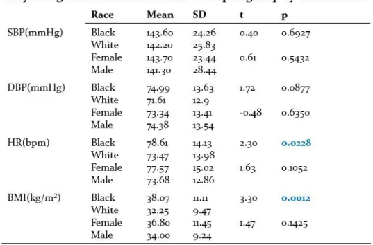
Population Stratification: Age Race and Sex as Key Attributes
Controversy exists in the scientific literature on the description of patient populations
most commonly affected by incident HFpEF. The ACCF/AHA guidelines suggest that
White women, >65 years of age with a history of HTN are most commonly affected by
HFpEF (Crowder, Irons, Meyerrose, & Seifert, 2010; Yancy et al., 2013). However, studies
with a racially diverse sample that included subjects <65 years of age, indicate Black women
are most commonly affected by incident HFpEF (Desai et al., 2013; Wong et al., 2013).
Given that at least 90% of HFpEF cases are preceded by a diagnosis of HTN (Pickering,
2004) and that Black patients had twice the incidence of new HTN compared to White
patients over a 10 year period (Egan, Zhao, & Axon, 2010; Ford & Cooper, 1991; Gillum,
1991) more definitive exploration of race and age in future HFpEF is indicated.
In a trial evaluating the relationship of race to HFpEF incidence and outcomes,
30
within the U.S. were suggested that Blacks had a 48% higher risk of hospitalization
compared to Whites and Asians and with patients <65 years of age and that incident HF
admission occurred more commonly in Blacks compared to Whites and Asians (Gurwitz et
al., 2014). Also the prevalence of HF among different ethnic and racial groups is expected to
increase substantially (Al-Dubai, Alshagga, Rampal, & Sulaiman, 2012) with the highest
prevalence to be among Black patients increasing by 29% between 2012 and 2030, from
2.8% to 3.6% of the Black population (Heidenreich et al., 2013), followed by Hispanic,
White, and Chinese (Bild et al., 2005). In addition to alarming trends in incidence and
prevalence, mortality data suggest a 34% higher five year mortality rate for Blacks with
HFpEF compared to White patients (East, Peterson, Shaw, Gattis, & O'Connor, 2004)
Conclusions
HFpEF is a complex clinical syndrome with increasingly significant health and fiscal
implications (Gladden et al., 2014). Multiple co-morbidities and the associated
pro-inflammatory state contribute to the onset of HFPEF (Paulus & Tschope, 2013). Although
this syndrome is not limited to a problem of LV contractility, the use of Ea and Ees to reflect
arterial-ventricular coupling have demonstrated physiologic utility in studying the
phenomenon of HFpEF (Borlaug & Paulus, 2011). In addition to the complexity of
pathophysiology, diagnostic guidelines for HFpEF are not consistently used. In the
TOPCAT trial the study initially reported non-significant mortality findings; however, post
hoc analysis stated ‘regional variation’ in the Americas and re-analysis did show mortality
benefit (Pitt et al., 2014). In TOPCAT the diagnostic criteria used for inclusion was the
Framingham HF criteria which is not specific to HFpEF and does not include biomarkers in
31
evolving and the exact cause remains unclear, without consistent use of current diagnostic
criteria in studying HFpEF, more confusion in defining this heterogeneous population is
likely to result.
This review outlines the pathophysiology of current and novel biomarkers and
consideration of the pro-inflammatory effect of multiple co-morbidities, demonstrates the
infrequent use of diagnostic criteria and emphasizes the importance of implementing a
diverse study sample. Incorporating each component (diagnosis, biomarkers,
co-morbidities, representative population) into comparisons between adults with and without
HFpEF over time would greatly contribute to our current scientific knowledge base. In
summary, consistent use of HFPEF diagnostic criteria with standardized measures of
32 CHAPTER 3
PAPER 2: COMPARING NOVEL BIOMARKERS ASSOCIATED WITH HEART FAILURE PRESERVED EJECTION FRACTION (HFpEF): A MATCHED
CASE-CONTROL ANALYSIS
Overview Objectives:
This study was designed to detect differences in biomarkers associated with incident heart
failure preserved ejection fraction (HFpEF) when comparing matched case-control groups.
Background:
Evidence continues to demonstrate increasing prevalence, cost and mortality implications of
HFpEF. Early identification of biomarkers associated with incident HFpEF would contribute
greatly to the current science.
Methods:
A study cohort of 310 patients, that included case (incident HFpEF patients, n=155) and
matched control (patients with no prior HF, n=155) groups were retrospectively identified.
Matching criteria included race, sex, age (within 3 years) and previously acquired
echocardiogram biomarkers (within 1year). Physiologic and echocardiogram biomarkers
were collected from previously acquired 2-D (dimensional) M-mode Doppler
echocardiograms. Echo images were re-analyzed from previously obtained echo to calculate
measures factored into calculating ventricular-arterial coupling. To estimate ventricular
33
conditional logistic regression and controlling for covariates, models were fit to detect
differences in HFpEF biomarkers between the matched case-control groups.
Results:
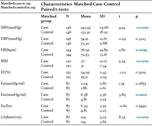
Statistically significant differences in biomarkers that reflect ventricular elastance (Ees)
(p=0.0030) and left atrial diameter (LAdiam) (p=0.0002) were detected when comparing the
case and control groups. Conditional logistic regression analyses suggested a 30% higher
odds of converting to the case group with every 1 unit increase in Ees, OR 1.315 (1.097,
1.575) and a 4.57 times higher odds of being in the case group for every 1 unit increase in
LAdiam, OR 4.565 (2.038, 10.223).
Conclusions
Ees and LAdiam may have a role in tracking physiology as it relates to HFpEF. This study
demonstrates that it is feasible to calculate both the Ees and LAdiam from routinely obtained
echo images without increasing cost or risk. Prospective studies are indicated that explore
34
PAPER 2: COMPARING NOVEL BIOMARKERS ASSOCIATED WITH HEART FAILURE PRESERVED EJECTION FRACTION (HFpEF): A MATCHED
CASE-CONTROL ANALYSIS
Objective
This manuscript presents the findings of a matched case-control study. The case
group consisted of patients previously discharged with incident (first case) heart failure
preserved ejection fraction (HFpEF). The control group consisted of patients with no prior
history of heart failure (HF). Groups were matched based on age, race, sex and date of
echocardiogram. This paper includes an analysis of differences within the matched cohorts
comparing physiologic and echocardiographic biomarkers.
Background
Heart failure preserved ejection fraction (HFpEF) accounts for at least 50% of all
hospital admissions for heart failure (HF) in the United States (U.S.) yet measures that
predict risk and onset of HFpEF are lacking (Gladden et al., 2014; Heidenreich et al., 2013).
HFpEF is a heterogeneous syndrome likely affected by multiple comorbidities with
inflammatory precursors (Lam et al., 2011; Paulus & Tschope, 2013). Clinical diagnosis is
typically based on patient history and physical examination; however, epidemiologic studies
suggest that some patients are asymptomatic but have evidence of abnormal biomarkers such
as diastolic dysfunction (Owan & Redfield, 2005; Yancy et al., 2013). The relative risk
associated with specific biomarkers (Ky et al., 2013) and the impact of age, gender and race
have not been established (Shah, 2012). Therefore, studies that include samples stratified by
age, race and sex that identify biomarkers that predict impending HFpEF may help identify
35
Previous studies predicting risk or outcomes in HFpEF patients, most of which
compared HFpEF and HFrEF groups to control groups and some of which matched on age
and gender, suggested utility in echo biomarkers that include measurement of left ventricular
hypertrophy (LVH), estimates of left ventricular filling (E/e’), left atrial (LA) size, global
longitudinal strain (GLS), arterial elastance (Ea), ventricular elastance (Ees) and
ventricular-arterial coupling (Borlaug et al., 2009; Mohammed et al., 2012; Shuai et al., 2011; Stampehl
et al., 2015). However, none of the studies included race in the matched groups. In addition,
the pathophysiology of HFpEF is complex reflected by LV hypertrophy, pro-inflammatory
markers, slow LV relaxation, LV diastolic stiffness, decreased LV systolic performance, left
atrial remodeling, peripheral vascular resistance, impaired epithelial function, increased
pulmonary arterial and venous resistance, neurohormonal activation and mismatched
ventricular-arterial coupling (Gladden et al., 2014; Paulus & Tschope, 2013).
LV ejection fraction (EF) is an important volumetric measure and predictor of
outcome, however, EF does not incorporate load, contractility and the interaction of
ventricular-arterial stiffening (Borlaug et al., 2009). Non-invasive measures have been
established such as LV systolic elastance (Ees)(mmHg/ml) which incorporates measures of
LV end-diastolic volume (EDV), LV size and cardiac remodeling and the end-systolic
pressure volume relationship (ESPVR) which is a load-dependent measure of contractile
function (Ky et al., 2013). Arterial elastance (Ea)(mmHg/ml) is a measure that incorporates
end-systolic pressure (ESP) and end-diastolic volume to incorporate stroke volume (SV),
therefore Ea=ESP/SV (Ky et al., 2013). The net interaction of the ventricular and arterial
systems is measured by Ea/Ees which significantly impacts cardiac performance (Borlaug &
36
Previous studies suggest that optimal ventricular-arterial functioning is reflected in an
Ea/Ees ratio of 0.5-1.0 which is maintained with normal aging but reportedly declines in
women because of ventricular stiffening increases out of proportion to vascular load (Borlaug
et al., 2009; Kawaguchi et al., 2003). However, studies that evaluate Ea, Ees and Ea/Ees and
incorporate a diverse range of age, race and gender into the study design have not been
completed to date. Ea (arterial elastance) and Ees (ventricular elastance) are measures that
reflect the interaction of ventricular-arterial coupling. Therefore in order to better understand
the phenotype of HFpEF, collecting data on Ea, Ees in addition to stratifying the study group
by age, race and sex are greatly needed. For a complete list of all abbreviations used in this
study, please refer to Appendix A.
Methods
Design
The study was observational with a retrospective matched case-control design. The
procedure of case-control group selection began with the consideration of key
methodological principles in control group selection (Wacholder, Silverman, McLaughlin, &
Mandel, 1992a). This framework includes three principles to maximize comparability
between case-control. The concepts include compariability of: a) study base, b)
deconfounding, and c) comparative accuracy (Wacholder, McLaughlin, Silverman, &
Mandel, 1992). To control the study base, for both the case and control groups, the study
samples only included residents of Durham County, North Carolina. Given that the selected
academic institution is a regional, national and international referral instituion, selecting only
residents of Durham County helped control; a) socioeconomic, b) racial and c) access to
medical care variables, which preserved both the heterogeneity and the homogeniety of the
37
(dimensional) M (motion)-mode Doppler echocardiograms, ordered within the health system,
read at a core laboratory that is certified, regulated and adhered to standards set by the
American Society of Echocardiography (ASE) (Wang & Nagueh, 2009) on patients that met
the inclusion and exclusion criteria, were eligible for this study.
The case group only included patients with an incident (first) HFpEF hospital
admission and the control group only included patients with no history of incident HFpEF (or
any type of HF) hospital admission. Matching on the variables age, race and sex also helped
reduce the effects of confounding variables (Wacholder, Silverman, McLaughlin, & Mandel,
1992b).
To maximize comparable accuracy, this study included data extracted from
previously obtained (derived) data with the exception of echo image measurements for
ventricular-arterial coupling. For the coupling variables, two nationally certified echo
sonographers obtained measures of the LV outflow tract diameter at the pulse wave Doppler
signal and measures of the flow onset time and end time from the LVOT waveform. For
both the case and control groups, all echos had previously been ordered, interpreted and
reported based on standards created by the ASE (Wang & Nagueh, 2009) and thus favorably
contributed to the validity of group to group comparisons.
Sample and Setting
Based on power analysis, the number of matched pairs required for this study was
n=90 or a total of 180 subjects. Patients in both the case and control groups were required to
be 18 years of age or older. All study data were extracted from pre-existing electronic
sources that included a storage data base, medical record, echocardiogram database and