Cochrane
Database of Systematic Reviews
Perioperative antibiotics for prevention of acute
endophthalmitis after cataract surgery (Review)
Gower EW, Lindsley K, Tulenko SE, Nanji AA, Leyngold I, McDonnell PJ
Gower EW, Lindsley K, Tulenko SE, Nanji AA, Leyngold I, McDonnell PJ.
Perioperative antibiotics for prevention of acute endophthalmitis after cataract surgery. Cochrane Database of Systematic Reviews2017, Issue 2. Art. No.: CD006364.
DOI: 10.1002/14651858.CD006364.pub3.
T A B L E O F C O N T E N T S
1 HEADER . . . .
1 ABSTRACT . . . .
2 PLAIN LANGUAGE SUMMARY . . . .
4 SUMMARY OF FINDINGS FOR THE MAIN COMPARISON . . . .
8 BACKGROUND . . . .
9 OBJECTIVES . . . .
9 METHODS . . . .
11 RESULTS . . . .
Figure 1. . . 12 Figure 2. . . 14 17 DISCUSSION . . . .
19 AUTHORS’ CONCLUSIONS . . . .
20 ACKNOWLEDGEMENTS . . . .
20 REFERENCES . . . .
24 CHARACTERISTICS OF STUDIES . . . .
36 DATA AND ANALYSES . . . .
36 ADDITIONAL TABLES . . . .
36 APPENDICES . . . .
40 WHAT’S NEW . . . .
41 HISTORY . . . .
41 CONTRIBUTIONS OF AUTHORS . . . .
42 DECLARATIONS OF INTEREST . . . .
42 SOURCES OF SUPPORT . . . .
42 DIFFERENCES BETWEEN PROTOCOL AND REVIEW . . . .
[Intervention Review]
Perioperative antibiotics for prevention of acute
endophthalmitis after cataract surgery
Emily W Gower1, Kristina Lindsley2, Samantha E Tulenko1, Afshan A Nanji3, Ilya Leyngold4, Peter J McDonnell5
1University of North Carolina, Gillings School of Global Public Health, Chapel Hill, North Carolina, USA.2Department of
Epi-demiology, Johns Hopkins Bloomberg School of Public Health, Baltimore, Maryland, USA.3Casey Eye Institute, Oregon Health & Science University, Portland, Oregon, USA.4Division of Oculofacial Plastic and Reconstructive Surgery, Duke University Hospital Department of Ophthalmology, Durham, North Carolina, USA.5Wilmer Eye Institute, Johns Hopkins University School of Medicine, Baltimore, Maryland, USA
Contact address: Emily W Gower, University of North Carolina, Gillings School of Global Public Health, 135 Dauer Drive, 2102A McGavran Greenberg, CB#7435, Chapel Hill, North Carolina, 27599, [email protected].
Editorial group:Cochrane Eyes and Vision Group.
Publication status and date:New search for studies and content updated (no change to conclusions), published in Issue 2, 2017. Review content assessed as up-to-date: 6 December 2016.
Citation: Gower EW, Lindsley K, Tulenko SE, Nanji AA, Leyngold I, McDonnell PJ. Perioperative antibiotics for prevention of acute endophthalmitis after cataract surgery.Cochrane Database of Systematic Reviews2017, Issue 2. Art. No.: CD006364. DOI: 10.1002/14651858.CD006364.pub3.
Copyright © 2017 The Cochrane Collaboration. Published by John Wiley & Sons, Ltd.
A B S T R A C T
Background
Endophthalmitis is a severe inflammation of the anterior or posterior (or both) chambers of the eye that may be sterile or associated with infection. It is a potentially vision-threatening complication of cataract surgery. Prophylactic measures for endophthalmitis are targeted against various sources of infection.
Objectives
To evaluate the effects of perioperative antibiotic prophylaxis for endophthalmitis following cataract surgery compared with no pro-phylaxis or other form of propro-phylaxis.
Search methods
We searched CENTRAL (which contains the Cochrane Eyes and Vision Trials Register) (2016, Issue 12), Ovid MEDLINE, Epub Ahead of Print, In-Process & Other Non-Indexed Citations, Ovid MEDLINE Daily (January 1946 to December 2016), Embase (January 1980 to December 2016), Latin American and Caribbean Health Sciences Literature Database (LILACS) (1982 to December 2016),the ISRCTN registry (www.isrctn.com/editAdvancedSearch), ClinicalTrials.gov (www.clinicaltrials.gov), and the World Health Organization (WHO) International Clinical Trials Registry Platform (ICTRP) (www.who.int/ictrp/search/en). We used no date or language restrictions in the electronic searches for trials. We last searched the electronic databases on 6 December 2016. We also searched for additional studies that cited any included trials using the Science Citation Index.
Selection criteria
Data collection and analysis
Two review authors independently reviewed abstracts and full-text articles for eligibility, assessed the risk of bias for each included study, and abstracted data.
Main results
Five studies met the inclusion criteria for this review, including 101,005 adults and 132 endophthalmitis cases. While the sample size was very large, the heterogeneity of the study designs and modes of antibiotic delivery made it impossible to conduct a formal meta-analysis. Interventions investigated included the utility of adding vancomycin and gentamycin to the irrigating solution compared with standard balanced saline solution irrigation alone, use of intracameral cefuroxime with or without topical levofloxacin perioperatively, periocular penicillin injections and topical chloramphenicol-sulfadimidine drops compared with topical antibiotics alone, and mode of antibiotic delivery (subconjunctival versus retrobulbar injections; fixed versus separate instillation of gatifloxacin and prednisolone). The risk of bias among studies was low to unclear due to information not being reported. We identified one ongoing study.
Two studies compared any antibiotic with no antibiotic. One study, which compared irrigation with antibiotics in balanced salt solution (BSS) versus BSS alone, was not sufficiently powered to detect differences in endophthalmitis between groups (very low-certainty evidence). One study found reduced risk of endophthalmitis when combining intracameral cefuroxime and topical levofloxacin (risk ratio (RR) 0.14, 95% confidence interval (CI) 0.03 to 0.63; 8106 participants; high-certainty evidence) or using intracameral cefuroxime alone (RR 0.21, CI 0.06 to 0.74; 8110 participants; high-certainty evidence) compared with placebo, and an uncertain effect when using topical levofloxacin alone compared with placebo (RR 0.72, CI 0.32 to 1.61; 8103 participants; moderate-certainty evidence).
Two studies found reduced risk of endophthalmitis when combining antibiotic injections during surgery and topical antibiotics compared with topical antibiotics alone (risk ratio (RR) 0.33, 95% confidence interval (CI) 0.12 to 0.92 (periocular penicillin and topical chloramphenicol-sulfadimidine; 6618 participants; moderate-certainty evidence); and RR 0.20, 95% CI 0.04 to 0.91 (intracameral cefuroxime and topical levofloxacin; 8101 participants; high-certainty evidence)).
One study, which compared fixed versus separate instillation of gatifloxacin and prednisolone, was not sufficiently powered to detect differences in endophthalmitis between groups (very low-certainty evidence). Another study found no evidence of a difference in endophthalmitis when comparing subconjunctival versus retrobulbar antibiotic injections (RR 0.85, 95% CI 0.55 to 1.32; 77,015 participants; moderate-certainty evidence).
Two studies reported any visual acuity outcome; one study, which compared fixed versus separate instillation of gatifloxacin and prednisolone, reported only that mean visual acuity was the same for both groups at 20 days postoperation. In the other study, the difference in the proportion of eyes with final visual acuity greater than 20/40 following endophthalmitis between groups receiving intracameral cefuroxime with or without topical levofloxacin compared with no intracameral cefuroxime was uncertain (RR 0.69, 95% CI 0.22 to 2.11; 29 participants; moderate-certainty evidence).
Only one study reported adverse events (1 of 129 eyes had pupillary membrane in front of the intraocular lens and 8 eyes showed posterior capsule opacity). No study reported outcomes related to quality of life or economic outcomes.
Authors’ conclusions
Multiple measures for preventing endophthalmitis following cataract surgery have been studied. High-certainty evidence shows that injection with cefuroxime with or without topical levofloxacin lowers the chance of endophthalmitis after surgery, and there is mod-erate-certainty evidence to suggest that using antibiotic eye drops in addition to antibiotic injection probably lowers the chance of endophthalmitis compared with using injections or eye drops alone. Clinical trials with rare outcomes require very large sample sizes and are quite costly to conduct; thus, it is unlikely that many additional clinical trials will be conducted to evaluate currently available prophylaxis. Practitioners should rely on current evidence to make informed decisions regarding prophylaxis choices.
P L A I N L A N G U A G E S U M M A R Y
Antibiotics at the time of cataract surgery to prevent bacterial infection of the eye
What is the aim of this review?
Key messages
There is a very small chance of endophthalmitis after cataract surgery. Antibiotics injected into the eye during surgery lower this small chance of infection (high-certainty evidence). Antibiotic injection and antibiotic eye drops given together probably lower the chance of infection compared with using either injection alone or eye drops alone. Information on adverse effects was not provided in most studies.
What was studied in this review?
Endophthalmitis is a rare, but potentially serious, complication of cataract surgery that may lead to blindness. It is caused by bacteria that enter the eye during surgery or in the first few days after surgery. There are many ways to stop infection during and after surgery, such as using antibiotics at the time of surgery. There are several different types of antibiotic that can be used, and these may be used in different ways (either by injection into the eye, or infusion into the blood, or eye drops) or at different times (before, during, or after surgery).
What are the main results of the review?
Cochrane researchers found five relevant studies. Two studies were conducted in Pakistan, one study in several European countries, one study in Brazil, and one study in Turkey. These studies all looked at different treatments: one study compared four different treatments - antibiotic injection combined with antibiotic eye drops versus antibiotic injection alone versus antibiotic eye drops alone versus placebo eye drops; one study compared combined antibiotic injection and antibiotic eye drops versus antibiotic eye drops alone; one study compared combined antibiotics and steroids versus antibiotics and steroid given individually; one study compared two different locations for the antibiotic eye injection; one study compared adding antibiotics to the sterile fluid used during surgery versus not adding antibiotics to this fluid.
The review shows that:
• Antibiotic injection in the eye (cefuroxime) at the end of surgery lowers the chance of endophthalmitis after surgery (high-certainty evidence).
• Using antibiotic eye drops (either levofloxacin or chloramphenicol) in addition to antibiotic injection (either cefuroxime or penicillin) probably lowers the chance of endophthalmitis compared with using injections or eye drops alone (moderate certainty evidence).
• It is very uncertain whether adding antibiotic to the sterile irrigating fluid used during cataract surgery lowers the chance of endoph-thalmitis (very low-certainty evidence).
• It is very uncertain if using antibiotics and steroids individually or in combination makes a difference to the chance of developing endophthalmitis (very low-certainty evidence).
How up to date is this review?
S U M M A R Y O F F I N D I N G S F O R T H E M A I N C O M P A R I S O N [Explanation]
Perioperative antibiotics for prevention of endophthalmitis after cataract surgery
Population:participants undergoing cataract surgery
Settings:eye hospital or clinic
Outcome:risk of endophthalm itis af ter surgery
Perioperative prophylaxis versus no prophylaxis
Study ID No. eyes and
par-ticipants
Follow- up Comparison (intervention vs comparator)
Risk of endophthalmitis by study group RR (95% CI)
Treatment vs control
Certainty of the ev-idence
(GRADE)
Presumed cases* Proven cases* * Presumed cases* Proven cases* *
Sobaci 2003 644 eyes of 640
participants
6 weeks Treatment:
BSS with antibi-otics (vancom ycin 20 m g/ m L and gen-tam icin 8 m g/ m L)
Not reported 0/ 322 (0%) eyes Not reported 0.20 (0.01 to 4.15) ⊕
Very low1,2
Control: BSS-only irrigating inf usion f luid
Not reported 2/ 322 (0.62%) eyes
ESCRS 2007 16,603 eyes of 16,
603 participants
6 weeks Treatment 1:com -bined intracam eral
ce-f uroxim e and topi-cal levof loxacin
2/ 4052 (0.05%) eyes
1/ 4052 (0.02%) eyes
0.14 (0.03 to 0.63) 0.10 (0.01 to 0.78) ⊕⊕⊕⊕
High
Treatment 2: intra-cam eral cef urox-im e 0.9%
3/ 4056 (0.07%) eyes
2/ 4056 (0.05%) eyes
0.21 (0.06 to 0.74) 0.20 (0.04 to 0.91) ⊕⊕⊕⊕
Treatment 3: topi-cal levof loxacin 0. 5%
10/ 4049 (0.25%) eyes
7/ 4049 (0.17%) eyes
0.72 (0.32 to 1.61) 0.70 (0.27 to 1.84) ⊕⊕⊕
M oderate3
Control: placebo drops
14/ 4054 (0.35%) eyes
10/ 4054 (0.25%) eyes
Comparisons of combinations of antibiotics with specific antibiotics
Study ID No. eyes and
par-ticipants
Follow- up Interventions Risk of endophthalmitis by study group RR (95% CI)
Treatment 1 vs treatment 2
Certainty of the ev-idence
(GRADE)
Presumed cases* Proven cases* * Presumed cases* Proven cases* *
Christy 1979 6618 eyes of 6618
participants
1 week Treatment 1:com -bined prophylaxis (topical regim en + periocular peni-cillin at the tim e of surgery)
5/ 3309 (0.15%) eyes
Not reported 0.33 (0.12 to 0.92) Not reported ⊕⊕⊕
M oderate4
Treatment 2: topi-cal regim en alone (chloram phenicol-sulf adim idine)
15/ 3309 (0.45%) eyes
Not reported
ESCRS 2007 16,603 eyes of 16,
603 participants
6 weeks Treatment 1:com -bined intracam eral
ce-f uroxim e and topi-cal levof loxacin
2/ 4052 (0.05%) eyes
1/ 4052 (0.02%) eyes
Treatment 1 vs
treatment 2: 0.67 (0.11 to 3.99)
Treatment 1 vs
treatment 2: 0.50 (0.05 to 5.52)
⊕⊕⊕
M oderate3
Treatment 2: intra-cam eral cef urox-im e 0.9%
3/ 4056 (0.07%) eyes
2/ 4056 (0.05%) eyes
Treatment 2 vs
treatment 3: 0.30 (0.08 to 1.09)
Treatment 2 vs
treatment 3: 0.29 (0.06 to 1.37)
⊕⊕⊕
M oderate3
Treatment 3: topi-cal levof loxacin 0. 5%
10/ 4049 (0.25%) eyes
7/ 4049 (0.17%) eyes
Treatment 1 vs
treatment 3: 0.20 (0.04 to 0.91)
Treatment 1 vs
treatment 3: 0.14 (0.02 to 1.16)
⊕⊕⊕⊕
High
Mode of antibiotic delivery
Study ID No. eyes and
pa-tients
Follow- up Interventions Risk of endophthalmitis by study group RR (95% CI) M ode 1 vs mode 2
Certainty of the ev-idence
(GRADE)
Presumed cases* Proven cases* * Presumed cases* Proven cases* *
Christy 1986 77,015 eyes of 77,
015 participants
1 week M ode 1: Ante-rior sub-Tenon in-jections (subcon-junctival)
38/ 39,752 (0.10%) eyes
Not reported 0.85 (0.55 to 1.32) Not reported ⊕⊕⊕
M oderate4
M ode 2:
Poste-rior sub-Tenon in-jections (retrobul-bar)
42/ 37,263 (0.11%) eyes
Not reported
Cunha 2013 108 eyes of 108
participants
3 weeks Treatment 1:f ixed com
-bination of topical gatif loxacin 0.3% and prednisolone acetate 1%
0/ 47 (0%) eyes Not reported 0.43 (0.02 to 10. 34)
Not reported ⊕
Very low1,5
Treatment 2:
in-dividual instillation of topical gati-f loxacin 0.3% and prednisolone ac-etate 1%
1/ 61 (2%) eyes Not reported
GRADE Working Group grades of evidence
High- certainty:Further research is very unlikely to change our conf idence in the estim ate of ef f ect.
M oderate- certainty:Further research is likely to have an im portant im pact on our conf idence in the estim ate of ef f ect and m ay change the estim ate.
Low- certainty:Further research is very likely to have an im portant im pact on our conf idence in the estim ate of ef f ect and is likely to change the estim ate.
Very low- certainty:We are very uncertain about the estim ate. BSS: balanced salt solution; CI: conf idence interval; RR: risk ratio.
* Presum ed cases: includes both culture-proven and clinically diagnosed cases of postoperative endophthalm itis. * * Proven cases: cases conf irm ed by at least one of Gram stain, culture, or polym erase chain reaction (PCR)
1Downgraded f or im precision (-2) as the study did not enroll a suf f icient num ber of participants to detect dif f erences between
groups.
2Downgraded f or high risk of attrition bias (-1) as the study authors excluded participants at the tim e of surgery based on the
surgeon’s discretion (num ber excluded not reported).
3Downgraded f or im precision (-1) as the conf idence interval of the ef f ect estim ate between groups was wide.
4Downgraded f or indirectness (-1) as the study was conducted m ore than 30 years ago and the techniques f or cataract
surgery have since changed substantially.
5Downgraded f or high risk of attrition bias (-1) as the study authors excluded participants who did not return f or f ollow-up
(16% of study population).
B A C K G R O U N D
Description of the condition
Age-related cataract is a leading cause of reduced vision in both high-income and low-income countries (Friedman 2004; Resnikoff 2004). Surgery for cataract involves removal of the opaque lens and replacement with an intraocular lens (IOL). In the few cases where IOL implantation is not possible, contact lenses and glasses are valid options for the correction of the re-fractive error that results from being aphakic (without a lens). En-dophthalmitis is a potentially vision-threatening complication of cataract surgery. Endophthalmitis is a severe inflammation of the anterior or posterior (or both) chambers of the eye and may be sterile or associated with infection. It most commonly occurs as a complication of cataract surgery, but also may occur following other ocular procedures, trauma to the eye, metastatic systemic infections, and systemic inflammatory disorders.
Epidemiology
Reported endophthalmitis rates vary substantially, with some in-dividual centers reporting no endophthalmitis in a several-year period (Galvis 2014;Monica 2005), while others report rates as high as 1 in 200 or 300 surgeries (ESCRS 2007;Garcia-Arumi 2007). One systematic review that included studies from high-income and low-high-income countries indicated a decreasing inci-dence of endophthalmitis following cataract surgery until the early 1990s, followed by an increase in incidence (Taban 2005a). The pooled estimate for incidence of endophthalmitis was 1.09 per 1000 surgeries from 1963 to 1999 and 2.65 per 1000 surgeries from 2000 to 2003 (Taban 2005a). In addition, an analysis of US Medicare data reported a 40% increase in the adjusted risk of endophthalmitis comparing data from 1998 to 2001 against 1994 to 1997, with annual rates ranging from 1.79 to 2.47 cases per 1000 surgeries (West 2005). An analysis of Medicare fee-for-service cataract surgeries reported that rates declined to 1.32 per 1000 surgeries in 2003 and 1.11 per 1000 surgeries in 2004 (Keay 2012). Rates appear to have remained relatively consistent, with two more recent Medicare analyses showing rates of 1.2 per 1000 surgeries (Coleman 2015; Du 2014). Other national-level data have shown a decline, with Sweden’s reported rate dropping from 0.48 per 1000 surgeries in 2002 through 2004 to 0.29 per 1000 surgeries for 2005 through 2010 (Friling 2013), and Iran reports an overall rate of 0.02% (Jabbarvand 2016). Furthermore, India has recently reported a rate of 0.08% among patients not receiving intracameral antibiotics and 0.02% among those receiving intra-cameral antibiotics (Haripriya 2016).
Presentation and diagnosis
Endophthalmitis usually presents within a few days following cataract surgery, and 80% of cases present within six weeks.
Presenting features include decreased visual acuity (VA), pain, swelling and redness of the eyelids, redness of the conjunctiva, haziness of the cornea due to edema, and increased cellularity of fluid in the anterior chamber of the eye with or without hypopyon (pus). Signs of infection and inflammation of the retina and vitre-ous usually are observed during exam. Although endophthalmitis is a rare infection, it often results in significant long-term mor-bidity, even when treated appropriately. Approximately 50% of people do not regain vision of 20/40 or better despite treatment (Gower 2015; Lalwani 2008), and often nearly one-third have acuity worse than 20/200 following treatment (Gower 2015;Ng 2005;Sheng 2011).
Description of the intervention
Several factors are thought to contribute to the incidence of en-dophthalmitis following cataract surgery. One primary factor is the type of incision used for surgery (Lundstrom 2007;Taban 2005a; Taban 2005b). In addition, many research studies have focused on the role of antibiotics used prophylactically to target ocular surface flora. Examples of prophylactic measures include preoper-ative lash-trimming and irrigation of the lacrimal drainage system with antibiotics, antiseptic preparation of the operative site using agents such as povidone iodine, and preoperative, intraoperative, and postoperative administration of antibiotics. Perioperative an-tibiotics may be administered through parenteral, topical, or in-travitreal routes, using a variety of antibiotics. This review focuses only on perioperative antibiotic use as a prophylactic measure.
How the intervention might work
humor of the eye (i.e. the fluid in the anterior chamber). Some antibiotics administered orally achieve intraocular concentrations via systemic delivery, while other antibiotics do not effectively pen-etrate the eye. Intracameral antibiotic administration is the most direct route of delivery to the site of potential infection. Perioperative antibiotics eliminate etiologic organisms by either bacteriostatic or bactericidal mechanisms. Bacteriostatic agents ar-rest the growth and replication of bacteria found on the ocular surface, eyelids, or those already iatrogenically introduced into the aqueous humor. Thus, these drugs limit the spread of infection while the body’s immune system eliminates the nonproliferating pathogens. Bactericidal antibiotics kill the bacteria directly, de-creasing the total concentration of viable microorganisms. Bac-tericidal agents are more commonly used in ocular surgery as they can achieve more rapid destruction of invading bacteria. Fre-quently used perioperative antibiotics with bactericidal proper-ties include fluoroquinolones, vancomycin, aminoglycosides, and cephalosporins.
Why it is important to do this review
Cataract surgery is the most common operative procedure in the aged population. While endophthalmitis is relatively rare, the fre-quency of the procedure makes the absolute number of cases sig-nificant enough to be a public health problem. In 2003 to 2004, nearly 1.6 million cases of cataract surgery were performed annu-ally in the US Medicare fee-for-service population alone (Schein 2012), and an estimated 10 million procedures were performed worldwide annually in the 1990s (Foster 2001). Experts estimate that the annual target for cataract surgery should be above 30 mil-lion surgeries (Foster 2001). At that rate, and assuming an inci-dence of one case per 1000 surgeries, 30,000 cases of postcataract surgery endophthalmitis would occur annually, with about 10,000 leading to blindness in the operated eye. Visual recovery following acute postoperative endophthalmitis remains poor across differ-ent clinical settings, despite advances in treatmdiffer-ent (Lalitha 2005; Miller 2005;Ng 2005;Sheng 2011). The extensive use of surgery to provide better vision for people with cataracts across the world calls for adoption of evidence-based methods to prevent acute endophthalmitis. This systematic review update aims to identify the current evidence to facilitate the adoption of evidence-based practices for prophylaxis of acute endophthalmitis after cataract surgery.
O B J E C T I V E S
To evaluate the effects of perioperative antibiotic prophylaxis for endophthalmitis following cataract surgery compared with no pro-phylaxis or other form of propro-phylaxis.
M E T H O D S
Criteria for considering studies for this review
Types of studies
We included randomized controlled trials (RCTs). We employed no date or language restrictions.
Types of participants
We included trials enrolling adults undergoing cataract surgery with any procedure for lens opacities due to any origin.
Types of interventions
We included trials evaluating preoperative antibiotics, intraopera-tive (intracameral, subconjunctival, or systemic), or postoperaintraopera-tive antibiotic prophylaxis for acute endophthalmitis. Comparisons of interest included:
• any prophylaxis versus no prophylaxis;
• preoperative versus postoperative or intraoperative prophylaxis or combinations;
• specific antibiotics used in included trials; • mode of perioperative antibiotic delivery.
We excluded studies that evaluated antiseptic preoperative prepa-ration using agents such as povidone iodine. In addition, excluded studies that evaluated antibiotics for treating acute endophthalmi-tis after cataract surgery.
We excluded studies with less than one week of follow-up after surgery.
Types of outcome measures
Primary outcomes
• Endophthalmitis: both presumed and culture-proven endophthalmitis within six weeks after cataract surgery. Our primary analysis was based on six-week outcomes; however, we also evaluated data from weeks one to four.
Secondary outcomes
• Adverse effects: specific adverse effects of interest were postoperative bacterial keratitis, antibiotic resistance if
documented, allergy and anaphylaxis. We also summarized other adverse effects as reported in included trials.
• Quality of life measures. • Economic data
Search methods for identification of studies
Electronic searches
We searched CENTRAL (which contains the Cochrane Eyes and Vision Trials Register) (2016, Issue 12), Ovid MEDLINE, Epub Ahead of Print, In-Process & Other Non-Indexed Cita-tions, Ovid MEDLINE Daily (January 1946 to December 2016), Embase (January 1980 to December 2016), Latin American and Caribbean Health Sciences Literature Database (LILACS) (1982 to December 2016),the ISRCTN registry (www.isrctn.com/ editAdvancedSearch), ClinicalTrials.gov (www.clinicaltrials.gov), and the World Health Organization (WHO) International Clini-cal Trials Registry Platform (ICTRP) (www.who.int/ictrp/search/ en). We used no date or language restrictions in the electronic searches for trials. We last searched the electronic databases on 6 December 2016.
See: Appendices for details of search strategies for CENTRAL (Appendix 1), MEDLINE (Appendix 2), EMBASE (Appendix 3), LILACS (Appendix 4), the ISRCTN (Appendix 5), ClinicalTri-als.gov (Appendix 6) and the ICTRP (Appendix 7).
Searching other resources
We searched for additional studies that cited any included refer-ences using the Science Citation Index Expanded database (Web of Science).
Data collection and analysis
Selection of studies
Two review authors independently reviewed the titles and abstracts resulting from the literature searches according to the inclusion criteria. We classified abstracts as ’definitely exclude’, ’unsure’ or ’definitely include’. We obtained the full-text for articles in the ’unsure’ category and reassessed them for inclusion. A third re-view author resolved any disagreement between the two rere-view authors. Studies excluded after full-text review are listed in the Characteristics of excluded studiestable along with the reasons for exclusion.
Data extraction and management
We developed data extraction forms to collect data from the in-cluded studies. We tested the forms using a few studies prior to extracting data for all included studies. Two review authors inde-pendently extracted study characteristics, methods, and outcomes data, and assessed risk of bias for all included studies. The two review authors compared data extraction forms and resolved dis-crepancies between them by discussion. One review author en-tered the data into Review Manager 5 (RevMan 2014), and a sec-ond review author checked the entered data for accuracy.
Assessment of risk of bias in included studies
Two review authors independently assessed the included studies for risk of bias according to guidelines set out in Chapter 8 of the Cochrane Handbook for Systematic Reviews of Interventions(Higgins 2011), and a third review author resolved any discrepancies. For each domain related to systematic biases, we made judgments of ’low risk of bias’, ’unclear risk of bias’, or ’high risk of bias’ for each included study.
• Selection bias: adequate sequence generation and allocation concealment. Examples of adequate sequence generation included using computerized randomization or random number lists. Methods such as centralized randomization and
sequentially numbered, sealed, opaque envelopes provided adequate allocation concealment.
• Performance bias: masking of study participants and personnel. For studies in which masking was not done or not possible (e.g. surgeons administering subconjunctival versus retrobulbar injections), we considered whether the person knowing the treatment assignment could have influenced the treatment effects.
• Detection bias: masking of outcome assessors. • Attrition bias: incomplete outcome data. We assessed whether follow-up rates and reasons for losses to follow-up were similar in the comparison groups and whether all participants were analyzed in the group to which they were randomized.
• Reporting bias: selective outcome reporting. Studies that reported results for all study outcomes described in the methods section of the included papers were considered to have low risks of reporting bias.
• Other sources of bias: other potential sources of bias that were considered included, but were not limited to, funding source, study design, and imbalance in baseline characteristics.
Measures of treatment effect
For individual studies, we presented dichotomous outcomes as risk ratios (RR) with 95% confidence intervals (CI). We did not conduct meta-analyses as part of this review.
Unit of analysis issues
The unit of analysis was the individual (one eye per participant) in four studies (Christy 1979;Christy 1986;Cunha 2013;ESCRS 2007). In theSobaci 2003study, both eyes of 4/640 (less than 1%) participants were included in the analysis; for the remaining 636 participants, only one eye was included.
Dealing with missing data
In the event of missing or unclear data, we contacted the primary investigators for additional information. We allowed six weeks for a response; failing that, we used the information as available in identified reports. We analyzed outcome data using the available data, assuming data were missing at random. We did not perform missing data statistics as the proportion of missing data was low (less than 1% of included participants) in most studies.
Assessment of heterogeneity
We assessed clinical heterogeneity using qualitative information on trial methodology, participant characteristics, interventions com-pared, routes of administration of prophylactic measures, duration of follow-up, and losses to follow-up. We performed no statistical tests for heterogeneity.
Assessment of reporting biases
Typically, funnel plots are used to examine reporting biases when 10 or more studies contribute to a given outcome. In this review with only five included studies and no meta-analysis, funnel plots were not appropriate.
Data synthesis
Because of the small number and heterogeneity of the included studies, we described data for each study narratively.
Subgroup analysis and investigation of heterogeneity
We performed no subgroup analyses.
Sensitivity analysis
We conducted no sensitivity analyses, given the small number of included studies.
’Summary of findings’ table
We prepared a ’Summary of findings’ table including relative and absolute effects for the outcome of endophthalmitis for all com-parisons. We assessed the certainty of evidence for all outcomes in this review using the GRADE classification system (GRADEpro 2014).
R E S U L T S
Description of studies
Results of the search
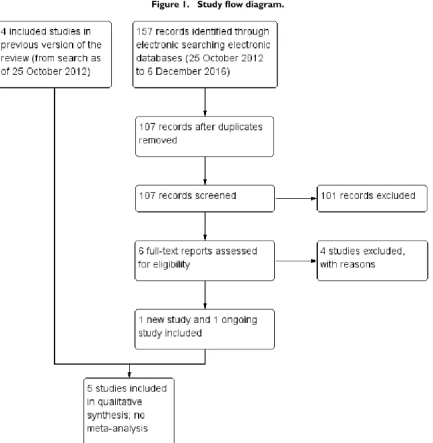
Electronic literature searches as of 25 October 2012 identified 491 potentially relevant titles and abstracts for this review (Gower 2013). After duplicate independent abstract review, 12 records were assessed at the full-text level, of which four were excluded and eight were included in the review. The eight records reported four studies. A review of references that cited the included studies and the reference lists of included studies identified one additional record that was excluded after full-text assessment.
Figure 1. Study flow diagram.
Included studies
We include five RCTs in this review (seeCharacteristics of included studies table). The studies enrolled 101,005 adults undergoing cataract surgery. The five studies varied widely in the approaches and prophylactic measures examined.
The first two studies were conducted at cataract surgery camps in northern Pakistan where intracapsular cataract extraction was performed, and participants were followed postoperatively for one week (Christy 1979;Christy 1986). Endophthalmitis diagnosis was made based on clinical signs. Although intracapsular cataract extraction is rarely performed in the 21st century, and hence the
Christy 1986compared subconjunctival versus retrobulbar injec-tion of antibiotics in 77,015 people/eyes. All participants received five applications of a sulfadimidine-chloramphenicol solution in the 20 hours before surgery. In both studies, participants were fol-lowed for one week after surgery and evaluated for endophthalmi-tis based on clinical signs.
The three more recent studies employed phacoemulsification. Sobaci 2003was conducted in Turkey and compared antibiotics (vancomycin and gentamycin) in balanced salt solution (BSS) ir-rigating infusion fluid with BSS-only irir-rigating infusion fluid in 644 eyes of 640 participants. All were treated with ofloxacin and diclofenac sodium four times on the day prior to surgery. Povi-done iodine was utilized for antisepsis at the time of surgery and a solution of ofloxacin, dexamethasone, and indomethacin was given postoperatively. Follow-up was for six weeks postoperation. Since the incidence of endophthalmitis following cataract surgery is low (the study authors ofSobaci 2003reported the rate of post-operative endophthalmitis at their institution was 0.109%) and because only 644 eyes were included in the study (with less than one eye expected to be affected), the study lacked sufficient power to detect valid differences between treatments.
ESCRS 2007conducted at multiple sites throughout Europe and Turkey, implemented a two-by-two factorial design to evaluate in-tracameral cefuroxime injected at the end of surgery and topical levofloxacin given immediately preoperatively (within one hour of surgery) and up to 15 minutes following surgery in 16,603 partic-ipants. In a factorial design studying two drugs or procedures that are expected to act independently, treatment arms were allocated such that both drugs could be evaluated alone and in combina-tion. InESCRS 2007, the two interventions studied were intra-cameral cefuroxime and topical levofloxacin. One group received only intracameral cefuroxime, one group received only topical lev-ofloxacin, one group received both intracameral cefuroxime and topical levofloxacin, and one group received neither intervention. Povidone iodine was used for antisepsis at the time of surgery and topical levofloxacin was given to all participants starting the morn-ing after surgery. Follow-up was for six weeks postoperation. InCunha 2013, all participants underwent phacoemulsification with IOL implantation. The use of a fixed combination of gat-ifloxacin 0.3% and prednisolone acetate 1% (i.e. both drugs in
a single bottle) was compared with the administration of gati-floxacin 0.3% alone and prednisolone acetate 1% alone. Partic-ipants instilled the drops beginning one day before the cataract surgery until 15 days postoperation. Although the study authors reported endophthalmitis as an adverse outcome, the study was not designed to assess differences in endophthalmitis rates between intervention groups. Further, with only 129 enrolled participants, the study did not have sufficient power to detect valid differences between treatment groups.
Excluded studies
We excluded nine studies overall: six were not RCTs and three did not evaluate the risk of endophthalmitis (seeCharacteristics of excluded studiestable).
Risk of bias in included studies
Allocation
Masking (performance bias and detection bias)
One study reported masking all study participants and person-nel by distributing identical bottles with masked labels (Cunha 2013).ESCRS 2007masked participants and clinicians by us-ing coded droppers with either active or placebo drops. Partici-pants and physicians were not masked for the injections since no sham injections were performed for those not receiving the in-tracameral cefuroxime injection. Participants and physicians who were present during the surgery were masked to the drops, and other clinical partners were masked to both drops and injections throughout the study. It was unclear whether the physicians who were present during the surgery or their clinical partners were as-sessing the outcomes for the study. One other study was reported to be masked, but details of who was masked and how masking was accomplished were not reported (Christy 1979). We assessed these three studies as having low risks of performance and detec-tion bias, since masking was reported and we would not expect the diagnosis of endophthalmitis to be affected if masking was bro-ken. The two remaining included studies did not report masking (Christy 1986;Sobaci 2003).
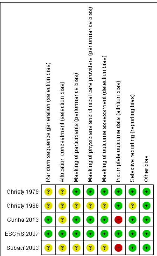
Incomplete outcome data
Risk of bias due to incomplete outcome data was low in three studies and high in two studies (Figure 2). InSobaci 2003, judged as having a high risk of bias, eyes of participants for which the surgical procedure was modified according to physician discretion during surgery were excluded from the study. Reasons for modi-fying the protocol included administering subconjunctival antibi-otics and adding a suture. The number of excluded participants was not reported. InCunha 2013, more than 15% of participants were not included in the analyses, most due to missing the follow-up visit.Christy 1979andChristy 1986reported no exclusions or losses to follow-up; however, the study authors noted that data were limited to early postoperative infections occurring one week after surgery, since most participants lived too far away for follow-up visits once discharged. The studies undertook no bacteriologic confirmation of infection. AlthoughESCRS 2007reported fol-lowing intent-to-treat analyses, 324 (2%) participants who were lost to follow-up and 68 (0.4%) participants who did not undergo the planned surgery or withdrew consent (timing of withdrawal not specified) were excluded from the analyses.
Selective reporting
Risk of selective reporting bias in these studies was low. All studies employed commonly used methods for reporting endophthalmitis cases. Two studies reported results for suspected cases without bacteriological confirmation (Christy 1979;Christy 1986).Sobaci
2003reported results for bacteriologically confirmed cases, and ESCRS 2007reported results for all suspected cases as well as for bacteriologically confirmed cases.Cunha 2013did not report how endophthalmitis was diagnosed.
Christy 1986used two types of antibiotics for injections, deter-mined by the surgeon doing the operation. The study authors re-ported that infection rates were similar between the two types of antibiotics and the two surgeons, but did not report infection rates for treatment groups (anterior versus posterior injections) sepa-rately by type of antibiotic.
Other potential sources of bias
We did not identify other potential sources of bias for the included studies.
Effects of interventions
See: Summary of findings for the main comparison Perioperative antibiotics for prevention of endophthalmitis after cataract surgery
The results of the five studies are described individually below. Interventions differed between them. Given the heterogeneity of study designs and modes of antibiotic delivery, we decided against conducting meta-analyses. We describe outcome data and present a summary of postoperative endophthalmitis for all comparisons inSummary of findings for the main comparison.
The primary outcome for four studies was postoperative en-dophthalmitis following cataract surgery; the fifth study investi-gated prophylaxis and control of inflammation following cataract surgery with endophthalmitis reported as an adverse outcome. The two earliest studies relied on clinical diagnosis of endophthalmitis (Christy 1979;Christy 1986).Sobaci 2003reported results for bacteriologically confirmed cases only,ESCRS 2007reported re-sults for all suspected cases as well as the subset of bacteriologically confirmed cases, andCunha 2013did not report how endoph-thalmitis was defined.
Perioperative prophylaxis versus no prophylaxis
Irrigation with antibiotics in balanced salt solution versus balanced salt solution alone
4.15). We assessed the certainty of evidence for this outcome as very low, downgrading for imprecision of the effect estimate and high risk of attrition bias in the study.
Intracameral with or without topical antibiotics versus no antibiotics
InESCRS 2007, the risk of clinically diagnosed (presumed) post-operative endophthalmitis at six weeks was significantly reduced for eyes that received intracameral cefuroxime injections, with or without topical levofloxacin, compared with no prophylaxis (nei-ther injection nor topical levofloxacin) (RR 0.14, 95% CI 0.03 to 0.63 with topical levofloxacin; RR 0.21, 95% CI 0.06 to 0.74 without topical drops). There were similar results when analyz-ing culture-proven cases of postoperative endophthalmitis. We as-sessed the certainty of evidence for these outcomes as high, finding no reason to downgrade the assessment.
The effect of topical levofloxacin alone compared with no prophy-laxis to reduce the risk of postoperative endophthalmitis was less certain (RR 0.72, 95% CI 0.32 to 1.61 for presumed cases; RR 0.70, 95% CI 0.27 to 1.84 for culture-proven cases). We assessed the certainty of evidence for this outcome as moderate, downgrad-ing for imprecision.
Comparisons ofcombinations of antibiotics with specific antibiotics
Chloramphenicol-sulfadimidine drops with versus without periocular penicillin
In the Christy 1979 study of chloramphenicol-sulfadimidine drops with or without periocular penicillin injection, 5/3309 (0.15%) eyes that received combined prophylaxis (drops and in-jection at the time of surgery) had postoperative endophthalmitis at one week, compared with 15/3309 (0.45%) eyes that received the topical regimen alone (RR 0.33, 95% CI 0.12 to 0.92). We assessed the certainty of evidence for this outcome as moderate, downgrading for indirectness as the study was conducted in the mid-to-late 1970s and the techniques for cataract surgery have since changed substantially.
Intracameral and topical antibiotics versus either antibiotic alone
InESCRS 2007, a risk reduction was observed for eyes treated with combined intracameral cefuroxime and topical levofloxacin compared with eyes treated with topical levofloxacin alone for presumed cases of postoperative endophthalmitis (RR 0.20, 95% CI 0.04 to 0.91), but this difference was less precise for culture-proven cases (RR 0.14, 95% CI 0.02 to 1.16). We assessed the certainty of evidence for this outcome as high, finding no reason to downgrade the assessment.
When comparing combined intracameral cefuroxime and topical levofloxacin with eyes treated with intracameral cefuroxime alone, the difference of postoperative endophthalmitis was unclear (RR 0.67, 95% CI 0.11 to 3.99 for presumed cases; RR 0.50, 95% CI 0.05 to 5.52 for proven cases). We assessed the certainty of evidence for this outcome as moderate, downgrading for imprecision. Additionally, the head-to-head comparison of intracameral ce-furoxime alone compared with topical levofloxacin alone suggested that intracameral cefuroxime may perform better or as good as topical levofloxacin for preventing postoperative endophthalmitis (RR 0.30, 95% CI 0.08 to 1.09 for presumed cases; RR 0.29, 95% CI 0.06 to 1.37 for proven cases). We assessed the certainty of evidence for this outcome as moderate, downgrading for im-precision.
Mode of antibiotic delivery
Subconjunctival versus retrobulbar antibiotic injection
InChristy 1986, at one week after surgery, 38/39,752 (0.10%) eyes receiving subconjunctival injection had presumed postopera-tive endophthalmitis, compared with 42/37,263 (0.11%) eyes re-ceiving retrobulbar antibiotic injection. The risk of postoperative endophthalmitis was similar between groups (RR 0.85, 95% CI 0.55 to 1.32). We assessed the certainty of evidence for this out-come as moderate, downgrading for indirectness as the techniques of the cataract surgery used in the study were different compared with current cataract surgery methods.
Fixed combination versus individual instillation of topical antibiotic and corticosteroid
Cunha 2013compared fixed combination versus individual instil-lation of gatifloxacin 0.3% and prednisolone acetate 1%. None of 47 eyes that received the fixed combination had postoperative endophthalmitis compared with 1/61 (2%) eyes that received the individual drops up to 20 days postoperation (RR 0.43, 95% CI 0.02 to 10.34). Due to the small number of participants and events in the study, the analysis was not powered to detect a difference between groups. We assessed the certainty of evidence for this out-come as very low, downgrading for imprecision of the effect esti-mate and high risk of attrition bias in the study.
Visual acuity
postoperation (0.1 logMAR). No other study reported VA out-comes.
ESCRS 2007presented outcomes in a combined manner for both intracameral cefuroxime injection groups compared to topical lev-ofloxacin or no prophylaxis groups combined (Table 1).
Proportion of eyes with final visual acuity greater than 20/40 following endophthalmitis
Among the five presumed cases of postoperative endophthalmitis who received intracameral cefuroxime injections, two (40%) had final VA better than 20/40. Among the 24 presumed cases of postoperative endophthalmitis who did not receive intracameral cefuroxime injections, 14 (58.3%) had final VA better than 20/ 40. The difference between antibiotic injection and no injection groups was uncertain (RR 0.69, 95% CI 0.22 to 2.11). There were similar results for culture-proven cases between antibiotic injection (1/3 (33.3%) eyes) and no injection (10/17 (58.1%) eyes) groups (RR 0.57, 95% CI 0.11 to 2.95). We assessed the certainty of evidence for this outcome as moderate, downgrading for imprecision.
Proportion of eyes with final visual acuity less than 20/200 following endophthalmitis
Among the five presumed cases of postoperative endophthalmitis who received intracameral cefuroxime injections, none had final VA worse than 20/200. Among the 24 presumed cases of post-operative endophthalmitis who did not receive intracameral ce-furoxime injections, four (16.7%) had final VA worse than 20/ 200. This difference between injection and no-injection groups was very imprecise (RR 0.46, 95% CI 0.03 to 7.48). There were similar results for proven cases between the injection (0/3 (0%) eyes) and no injection (4/17 (23.5%) eyes) groups (RR 0.50, 95% CI 0.03 to 7.54). We assessed the certainty of evidence for this outcome as moderate, downgrading for imprecision.
Adverse effects
Cunha 2013was the only study to report adverse events. Cunha 2013did not report information specific to postoperative bacterial keratitis, antibiotic resistance, allergy, or anaphylaxis. At 20 days postoperation, the one eye in the individual drops group with endophthalmitis had pupillary membrane in front of the IOL. Three eyes (6%) in the fixed combination group compared with five eyes (8%) in the individual drops group showed posterior capsule opacity. The study authors reported no cases of hypopyon or IOL pigmentation and no statistically significant difference be-tween group with respect to central or incisional corneal edema.
Quality of life
No study reported outcomes related to quality of life measures.
Economic outcomes
No study reported outcomes related to economic data.
D I S C U S S I O N
Summary of main results
The studies included in this review were too heterogeneous for us to perform a meta-analysis. The five included studies tested three modes of delivery for antibiotic prophylaxis measures: intraocu-lar injection, topical drops, and antibiotics in the irrigating solu-tion. The two studies that reported statistically significant differ-ences among treatment arms both included antibiotic injection during surgery (one intraocular and the other periocular), and the treatment arms that included ocular injection had the lowest rates of endophthalmitis, ranging from 0.14 to 1.5 endophthalmitis cases per 1000 surgeries (Christy 1979;ESCRS 2007). Within the ESCRS 2007study, the primary results paper combined the two intracameral injection groups for comparison against placebo and reported a 4.9-fold increased risk of endophthalmitis when not using intracameral injection, which can be translated to an 80% decrease in endophthalmitis risk when using intracameral injec-tion.
In this review, we calculated RRs and CIs for multiple compar-isons withinESCRS 2007using the data provided in the study reports; we compared both intracameral injection of cefuroxime and topical drops individually to placebo and to the combined regimen. Both the combined prophylaxis and the intracameral in-jection alone showed a reduced risk of both presumed and culture-proven endophthalmitis compared with no prophylaxis. Compar-ison of the combined regimen against topical drops alone showed a reduction in risk for presumed cases, but not for culture-proven cases only.
108 participants analyzed at 20 days postoperation, resulting in a high degree of imprecision among outcomes.
Christy 1986investigated the mode of delivery of antibiotics in-jected during surgery, and found no significant difference between subconjunctival and retrobulbar injection. The rates of endoph-thalmitis (1.0 to 1.1 per 1000 surgeries) in that study were compa-rable to endophthalmitis rates reported in the 21st century, even though surgery was performed in surgical camp settings in a de-veloping country and that intracapsular surgery was performed. However, it is notable that this study only followed participants for one week, so some cases likely were missed.
Overall completeness and applicability of evidence
The five studies included 101,005 adults and 132 total endoph-thalmitis cases. While the overall sample size was quite large, the heterogeneity of the study settings, designs, and modes of antibi-otic delivery made it impossible to combine the studies and make direct comparisons. Two studies were conducted in the late 1970s and early 1980s. Cataract surgery practice has changed substan-tially since that time, making the results of these studies less appli-cable today. Povidone iodine is now used routinely in most coun-tries and is a proven measure for reducing intraocular infection (Speaker 1991). In addition, wound construction is quite differ-ent. In the 1970s and early 1980s large (180°) incisions were used routinely. Today, even in the most remote centers, much smaller incisions are employed. Small-incision manual surgery is now the procedure of choice in the majority of surgical camp settings in low-income countries. Despite these changes in surgical technique, Christy 1979suggested that adding periocular penicillin injection substantially reduced the risk of endophthalmitis.
Among the five studies, theESCRS 2007results are most applica-ble to 21st century surgical practice, as it used contemporary sur-gical techniques and study drugs that are readily available in Eu-rope. Its design allowed for examination of both topical and intra-cameral antibiotics, and included a sample size sufficient to yield statistically significant results. Thus, among the studies reviewed, it provided the firmest evidence upon which to recommend a pro-phylactic regimen in these settings, and suggested that intracam-eral antibiotic injection is useful in reducing the risk of postcataract surgery endophthalmitis. However, the choice of antibiotic re-mains a question for many physicians. Since the publication of ESCRS 2007, uptake of intracameral cefuroxime has varied widely. In the UK, approximately 50% of providers reported intracam-eral antibiotic use (Gore 2009). A retrospective analysis of billing codes in France suggested that the use of intracameral antibiotics increased from 0.60% to 80% between 2005 and 2014, likely due to the ESCRS recommendations (Creuzot-Garcher 2016). Up-take has been more limited in the US. Results of the 2011 Ameri-can Society of Cataract and Refractive Surgery (ASCRS) member survey showed less than 20% of physicians utilizing intracameral
antibiotics (Leaming 2012;Vazirani 2013). By the ASCRS 2014 survey, 50% of US physicians reported use of intracameral antibi-otics (Chang 2015); however, this percentage remains well below the rates in Europe, Australia and New Zealand (Behndig 2015; Meyer 2016; Schwartz 2016), and US physicians continue to ex-press concerns about the lack of a commercially available prepara-tion. In the US, incorporating intracameral antibiotics into stan-dard prophylaxis practice appears to be related to surgeon volume (Chang 2008), and increased surgeon volume has been reported to be associated with reduced risk of postoperative endophthalmitis (Keay 2012). In both the US and UK, physicians not using intra-cameral antibiotics cite concerns regarding dilution errors and risk of contamination when compounding the drugs for doses needed for ocular injection (Gore 2009;Leaming 2012). These factors are important to consider when evaluating the applicability of the current evidence. Further discussion on this issue is provided be-low in theAgreements and disagreements with other studies or reviewssection.
Quality of the evidence
The five studies included in this review varied substantially in the prophylaxis measures that they compared and the way data were reported. Two of these studies,Christy 1979andChristy 1986, were conducted over 30 years ago, when standards for randomiza-tion and reporting in clinical trials were less stringent and less well-defined. To the credit of the Christy team, they provided detailed information on both the operative procedure and the follow-up procedures. All five studies randomized participants, but random sequence generation and allocation concealment were described fully only inESCRS 2007. Hence, we cannot judge whether some selection bias may have occurred in the other studies.
Masking of the intervention was not complete in most of these studies. In some, the person in charge of outcome assessment was unaware of the randomization and did not actively participate in the surgery visits. These studies may be less prone to bias than those in which the examiner was present at all times and was aware of the treatment assignment.
Because of differences in the interventions used and outcomes as-sessed among studies, we performed no meta-analysis. Overall, one study provided moderate- to high-certainty evidence (ESCRS 2007); two studies, which were downgraded for indirectness, pro-vided moderate-certainty evidence (Christy 1979;Christy 1986); and two studies, which were downgraded for high risk of attri-tion bias and imprecision, provided very low-certainty evidence (Cunha 2013;Sobaci 2003).
Potential biases in the review process
stud-ies from the published literature. We also searched other sources such as the reference lists of included studies, the Science Citation Index, and clinical trial registries. We imposed no date or language restrictions.
Two review authors independently performed major steps in the review process to minimize bias and errors when screening studies for inclusion, recording study characteristics, extracting quantita-tive data, and assessing risks of bias. The review team included clinicians, researchers, and methodologists.
Agreements and disagreements with other studies or reviews
The rare nature of endophthalmitis makes RCTs difficult to con-duct, because of the very large sample sizes needed to make statis-tically valid comparisons. Thus, few trials have been conducted, and all that we are aware of are included here. Worldwide, numer-ous single-center retrospective analyses have been conducted to examine whether changes in practice patterns resulted in reduced endophthalmitis rates. Several studies have reported reduced rates of endophthalmitis following adoption of intracameral or sub-conjunctival antibiotics (Beselga 2014;Garat 2009;Garcia-Saenz 2010;Montan 2002;Myneni 2013;Packer 2011;Shorstein 2013). The antibiotic of choice has varied across studies of intracam-eral injections, with moxifloxacin, vancomycin, and cefuroxime all showing a reduction compared with no antibiotic injection (Packer 2011). Most notably, numerous studies have investigated the benefit of intracameral cefuroxime in light of the ESCRS study and increased availability of the antibiotic in Europe. Data from Sweden have shown extremely low rates of postoperative endoph-thalmitis (0.029%) in the presence of intracameral cefuroxime (Friling 2013). In Portugal, a center found that endophthalmitis decreased from 0.26% to 0.0% after introduction of the ESCRS protocol and use of intracameral cefuroxime (Beselga 2014). Sev-eral other studies also have reported declines in endophthalmitis. Across these studies, the general consensus has been that intra-cameral antibiotic use reduces the risk of endophthalmitis, which provides further support to theESCRS 2007study findings.
A U T H O R S ’ C O N C L U S I O N S
Implications for practice
This systematic review underscores the broad scope of prophylaxis regimens considered to be of potential use in preventing endoph-thalmitis following cataract surgery. Among the included studies, the mode of antibiotic administration ranged widely from topi-cal administration preoperatively to intraocular injections during surgery. Given our decision not to conduct a meta-analysis of the accumulated data, we are unable to report a direct comparison of
the effectiveness of these prophylactic measures. However, among the individual studies evaluated,ESCRS 2007provides the best evidence for antibiotic prophylaxis against postcataract surgery endophthalmitis. Clinical trials of this magnitude are costly and take many years to conduct. Hence, decisions currently need to be made based on the available evidence, with the possibility of conducting future trials as new and improved prophylactic treat-ments become available (antibiotics or otherwise).
ESCRS 2007demonstrated the efficacy of using intracameral an-tibiotics for reducing endophthalmitis. However, the antibiotic of choice may differ based on the clinical setting. Concerns of toxicity, contamination, and other problems associated with compound-ing and dilutcompound-ing remain with the use of cefuroxime (Chang 2015; Delyfer 2011;Olavi 2012). US physicians indicate they may in-crease their use of intracameral antibiotics if a single-dose vial were commercially available (Chang 2015). Hence, the American So-ciety of Cataract and Refractive Surgeons (ASCRS) has called for the pharmaceutical industry and US Food and Drug Administra-tion to prioritize development and approval of single-dose intra-cameral antibiotics (Braga-Mele 2014;Chang 2015). One report of ocular toxicity in a cluster of people following a dilution error highlights the need for clinicians to remain vigilant in monitor-ing preparation of intracameral antibiotics for use durmonitor-ing surgery. Resistance of endophthalmitis-causing organisms to moxifloxacin appears to be increasing (Schimel 2013). In determining whether to use intracameral injections and if so which antibiotic to use, individual surgical centers should evaluate the risks and benefits associated with the various available intracameral antibiotics and the resources available for ensuring appropriate dilution and steril-ity based on their setting (Olavi 2012).
Implications for research
This review highlights the limited amount of randomized con-trolled trial data available for evaluating measures used to prevent endophthalmitis following cataract surgery. Intraocular antibiotics have been used for decades, with multiple studies conducted in surgical camp settings in low-income countries. However, spec-ulation remains over which antibiotics to use and what mode of intraocular delivery is best.
of cefuroxime, which requires only reconstitution and not dilu-tion, was approved in Europe in 2012 (Thea Laboratories;Keating 2013). Numerous studies in Europe have reported increases in the use of intracameral antibiotics and corresponding decreases in rates of endophthalmitis following the increased use of cefurox-ime (Barreau 2012;Beselga 2014;Daien 2016;Haripriya 2016; Jabbarvand 2016). Surveys should be conducted in one to two years to determine whether increased utilization of intracameral antibiotics leads to increased antibiotic resistance or other ocular complications, or both. Additionally, single-use vials of antibiotics for intracameral use are needed in the US and other countries. Some ophthalmologists currently utilize moxifloxacin (Alcon) top-ical drops for intracameral injection without dilution. Given that the coverage spectrum of moxifloxacin is somewhat broader than that of cefuroxime and available for use without dilution, it would be of interest to research the comparative effectiveness of these two drugs. However, such a study would require a sample size of over
100,000 participants, making it unlikely that it will ever be con-ducted. On a longer view, new approaches to instilling antibiotics into the anterior chamber are needed. Sustained drug-delivery de-vices provide one example of an area ripe for additional research.
A C K N O W L E D G E M E N T S
We acknowledge Iris Gordon, Information Specialist for Cochrane Eyes and Vision (CEV), who designed strategies for and conducted the electronic searches. We acknowledge Niall Crosby, Barbara Hawkins, and Tianjing Li for their comments on the protocol and Lisa Herrinton, Neal Shorstein, and Barbara Hawkins for peer reviewing the original review.
We are grateful to Roy Chuck (RC), Ashley Behrens (AB), and Satyanarayana Vedula (SV) for their contributions to developing and publishing the protocol for this review.
R E F E R E N C E S
References to studies included in this review
Christy 1979 {published data only}
Christy NE, Sommer A. Antibiotic prophylaxis of postoperative endophthalmitis. Annals of Ophthalmology 1979;11(8):1261–5. [2779295]
Christy 1986 {published data only}
Christy NE, Lall P. A randomized, controlled comparison of anterior and posterior periocular injection of antibiotic in the prevention of postoperative endophthalmitis.
Ophthalmic Surgery1986;17(11):715–8. [2779297]
Cunha 2013 {published data only}
Cunha PA, Shinzato FA, Tecchio GT, Weber SP, Brasil A, Avakian A. Efficacy and tolerability of a gatifloxacin/prednisolone acetate fixed combination for topical prophylaxis and control of inflammation in phacoemulsification: a 20-day-double-blind comparison to its individual components.Clinics2013;68(6):834–9. [4888759]
ESCRS 2007 {published data only}
Barry P, Gardner S, Seal D, Gettinby G, Lees F, Peterson M, et al. Clinical observations associated with proven and unproven cases in the ESCRS study of prophylaxis of postoperative endophthalmitis after cataract surgery.Journal
of Cataract and Refractive Surgery2009;35(9):1523–31.
[2779299]
Barry P, Seal DV, Gettinby G, Lees F, Peterson M, Revie CW, et al. ESCRS study of prophylaxis of postoperative endophthalmitis after cataract surgery: preliminary report of principal results from a European multicenter study.
Journal of Cataract and Refractive Surgery2006;32(3): 407–10. [2779300]
∗Endophthalmitis Study Group, European Society
of Cataract and Refractive Surgeons. Prophylaxis of postoperative endophthalmitis following cataract surgery: results of the ESCRS multicenter study and identification of risk factors.Journal of Cataract and Refractive Surgery2007; 33(6):978–88. [2779301]
Pleyer U, Geldsetzer K. Will intracameral cefuroxime become the new standard in endophthalmitis prevention?.
Klinische Monatsblätter für Augenheilkunde2008;225(11):
934–40. [2779302]
Seal DV, Barry P, Gettinby G, Lees F, Peterson M, Revie CW, et al. ESCRS study of prophylaxis of postoperative endophthalmitis after cataract surgery: case for a European multicenter study.Journal of Cataract and Refractive Surgery 2006;32(3):396–406. [2779303]
Sobaci 2003{published data only}
Sobaci G, Tuncer K, Ta A, Ozyurt M, Bayer A, Kutlu U. The effect of intraoperative antibiotics in irrigating solutions on aqueous humor contamination and endophthalmitis after phacoemulsification surgery. European Journal of
Ophthalmology2003;13(9-10):773–8. [2779305]
References to studies excluded from this review
Camesasca 2007 {published data only}
Carron 2013 {published data only}
Carron A, Samudio M, Laspina F, Fariña N, Sanabria RR, Cibils D, et al. Efficacy of topical 0.3% ciprofloxacin application in reducing the conjunctival biota of patients undergoing cataract extraction. Archivos de la Sociedad
Espanola de Oftalmologia2013;88(9):345–51. [4888761]
Cetinkaya 2015 {published data only}
Cetinkaya S, Cetinkaya YF, Acir NO, Dadaci Z. Application of intracameral moxifloxacin to prevent endophthalmitis in cataract surgery.International Eye Science2015;15(10): 1680–3. [4888763]
Kolker 1967 {published data only}
Kolker AE, Freeman MI, Pettit TH. Prophylactic antibiotics and postoperative endophthalmitis. American Journal of
Ophthalmology1967;63(3):434–9. [2779309]
Li 2015 {published data only}
Li B, Miño de Kaspar H, Haritoglou C, Kook D, Kampik A, Sheng M, et al. Comparison of 1-day versus 1-hour application of topical neomycin/polymyxin-B before cataract surgery.Journal of Cataract and Refractive Surgery 2015;41(4):724–31. [4888765]
Maloof 2004 {published data only}
Maloof A, Saw V. Prophylactic intracameral vancomycin. Journal of Cataract and Refractive Surgery2004;30(8): 1610–1. [2779311]
Paganelli 2009 {published data only}
Paganelli F, Cardillo JA, Melo LAS, Lucena DR, Silva AA, Oliveira AG, et al. A single intraoperative sub-Tenon’s capsule injection of triamcinolone and ciprofloxacin in a controlled-release system for cataract surgery.Investigative
Ophthalmology and Visual Science2009;50(7):3041–7.
[2779313]
Pérez-Canales 2015 {published data only}
Pérez-Canales JL, Pérez-Santonja JJ, Campos-Mollo E. Corneal endothelial changes after intracameral vancomycin injection in cataract surgery. Journal of Cataract and
Refractive Surgery2015;41(1):126–34. [4888767]
Peyman 1977 {published data only}
Peyman GA, Sathar ML, May DR. Intraocular gentamicin as intraoperative prophylaxis in South India eye camps.
British Journal of Ophthalmology1977;61(4):260–2.
[2779315]
References to ongoing studies
NCT02770729 {published data only}
NCT02770729. Evaluation of efficacy and safety of intracameral moxifloxacin for prevention of postcataract endophthalmitis. clinicaltrials.gov/show/NCT02770729 (accessed 11 December 2016). [4888769]
Additional references
Barreau 2012
Barreau G, Mounier M, Marin B, Adenis JP, Robert PY. Intracameral cefuroxime injection at the end of cataract surgery to reduce the incidence of endophthalmitis: French
study.Journal of Cataract and Refractive Surgery2012;38(8): 1370–5.
Barry 2009
Barry P, Gardner S, Seal D, Gettinby G, Lees F, Peterson M, et al. Clinical observations associated with proven and unproven cases in the ESCRS study of prophylaxis of postoperative endophthalmitis after cataract surgery.Journal
of Cataract and Refractive Surgery2009;35(9):1523–31.
Behndig 2015
Behndig A, Cochener-Lamard B, Güell J, Kodjikian L, Mencucci R, Nuijts R, et al. Surgical, antiseptic, and antibiotic practice in cataract surgery: results from the European Observatory in 2013. Journal of Cataract and
Refractive Surgery2015;41(12):2635–43.
Beselga 2014
Beselga D, Campos A, Castro M, Fernandes C, Carvalheira F, Campos S, et al. Postcataract surgery endophthalmitis after introduction of the ESCRS protocol: a 5-year study.
European Journal of Ophthalmology2014;24(4):516–9.
Braga-Mele 2014
Braga-Mele R, Chang DF, Henderson BA, Mamalis N, Talley-Rostov A, Vasavada A, et al. Intracameral antibiotics: safety, efficacy, and preparation. Journal of Cataract and
Refractive Surgery2014;40(12):2134–42.
Chang 2008
Chang MA, Congdon NG, Baker SK, Bloem MW, Savage H, Sommer A. The surgical management of cataract: barriers, best practices and outcomes. International
Ophthalmology2008;28(4):247–60.
Chang 2015
Chang DF, Braga-Mele R, Henderson BA, Mamalis N, Vasavada A, ASCRS Cataract Clinical Committee. Antibiotic prophylaxis of postoperative endophthalmitis after cataract surgery: results of the 2014 ASCRS member survey. Journal of Cataract and Refractive Surgery2015;41 (6):1300–5.
Coleman 2015
Coleman AL. How big data informs us about cataract surgery: The LXXII Edward Jackson Memorial Lecture.
American Journal of Ophthalmology2015;160(6):
1091–1103.
Creuzot-Garcher 2016
Creuzot-Garcher C, Benzenine E, Mariet AS, de Lazzer A, Chiquet C, Bron AM, et al. Incidence of acute postoperative endophthalmitis after cataract surgery: a nationwide study in France from 2005 to 2014.Ophthalmology2016;123(7): 1414–20.
Daien 2016