The YsrS Paralog DygS Has the Capacity To Activate Expression of
the
Yersinia enterocolitica
Ysa Type III Secretion System
Kimberly A. Walker,aLauren A. Griggs,aMarkus Obrist,a*Addys Bode,aR. Patrick Summers,aVirginia L. Millera,b
Departments of Microbiology and Immunologyaand Genetics,bUniversity of North Carolina School of Medicine, Chapel Hill, North Carolina, USA
ABSTRACT
TheYersinia enterocoliticaYsa type III secretion system (T3SS) is associated with intracellular survival, and, like other charac-terized T3SSs, it is tightly controlled. Expression of theysagenes is only detected following growth at low temperatures (26°C) and in high concentrations of sodium chloride (290 mM) in the medium. The YsrSTR phosphorelay (PR) system is required for ysaexpression and likely responds to NaCl. During our investigations into the Ysr PR system, we discovered that genes YE3578 and YE3579 are remarkably similar toysrRandysrS, respectively, and are probably a consequence of a gene duplication event. The amino acid differences between YE3578 andysrRare primarily clustered into two short regions. The differences between YE3579 andysrSare nearly all located in the periplasmic sensing domain; the cytoplasmic domains are 98% identical. We inves-tigated whether these paralogs were capable of activatingysagene expression. We found that the sensor paralog, named DygS, is capable of compensating for loss ofysrS, but the response regulator paralog, DygR, cannot complement aysrRgene deletion. In addition, YsrR, but not DygR, interacts with the histidine phosphorelay protein YsrT. Thus, DygS likely activatesysagene ex-pression in response to a signal other than NaCl and provides an example of a phosphorelay system in which two sensor kinases feed into the same regulatory pathway.
IMPORTANCE
All organisms need mechanisms to promote survival in changing environments. Prokaryotic phosphorelay systems are mini-mally comprised of a histidine kinase (HK) that senses an extracellular stimulus and a response regulator (RR) but can contain three or more proteins. Through gene duplication, a unique hybrid HK was created. We show that, while the hybrid appears to retain all of the phosphorelay functions, it responds to a different signal than the original. Both HKs transmit the signal to the same RR, which activates a promoter that transcribes a set of genes encoding a type III secretion system (T3SS) whose function is not yet evident. The significance of this work lies in finding that two HKs regulate this T3SS, highlighting its importance.
Y
ersinia enterocoliticais a foodborne pathogen known to cause a variety of gastrointestinal disorders, ranging from mild to se-vere (1). Most healthy individuals only experience fever, vomiting, and diarrhea, lasting just a few days. However, in young children and those with weak or compromised immune systems,Y. entero-coliticacan spread systemically, resulting in a 50% mortality rate (2). In addition, postinfection sequelae can be problematic, with the development of reactive arthritis and thyroid disorders (1). More recently, development of inflammatory bowel disease (IBD) has been linked to gastrointestinal infections. While the number of patients developing IBD followingY. enterocoliticainfection is comparatively low, the rate is much higher than in patients who were infected with other common enteric pathogens (3,4). Diag-nosis ofY. enterocoliticainfection is relatively low compared to other gastrointestinal pathogens, and this is largely because the symptoms are often mild enough that patients do not seek medical attention and because detection ofY. enterocoliticain clinical sam-ples is challenging (5,6).Y. enterocoliticais classified into several biotypes and serotypes that vary in the severity of disease symptoms. Biotype 4, serotype O:3, is one of the most common pathogenic biotypes isolated from humans (7–9). This biotype is the most prevalent in pig samples from European slaughterhouses, and consumption of undercooked pork is a well-known source ofY. enterocolitica in-fection (10,11). However, the most pathogenic biotype is biotype 1B.Y. enterocoliticastrains contain a plasticity zone, which is a large chromosomal region that is highly variable among the
dif-ferent biotypes (12). The plasticity zone of biotype 1B strains con-tains a large number of genes not found in biotypes 2 to 4, nor in Yersinia pestisorYersinia pseudotuberculosis, and many of these genes contribute to virulence and broadened metabolic capacities that presumably enhance fitness in a wider variety of environ-ments (12). Encoded within this plasticity zone of 1B isolates is the Ysa type III secretion system (T3SS). This T3SS varies consider-ably from the well-characterized Ysc/Yop T3SS encoded on the virulence plasmid in function (13,14), expression (15), and phy-logenetic class (16), but the exact role of this system in theY. enterocoliticalife cycle is still the subject of investigation. Ysa mu-tant strains were attenuated in mouse infection studies, but only at early time points postinoculation (17). This early-infection phe-notype leads to the notion that the Ysa system is important during
Received14 March 2016Accepted28 March 2016
Accepted manuscript posted online4 April 2016
CitationWalker KA, Griggs LA, Obrist M, Bode A, Summers RP, Miller VL. 2016. The YsrS paralog DygS has the capacity to activate expression of theYersinia enterocoliticaYsa type III secretion system. J Bacteriol 198:1725–1734.
doi:10.1128/JB.00240-16. Editor:I. B. Zhulin
Address correspondence to Kimberly A. Walker, [email protected].
*Present address: Markus Obrist, Baudirektion Kanton Zürich, AWEL/Biosicherheit, Zürich, Switzerland.
the gastroenteritis phase of disease, which is not well recapitulated in mouse models. While generally viewed as an extracellular pathogen, the Ysa T3SS is required for intracellular survival in a Drosophila melanogasterS2 tissue culture model, suggesting a role for this system during a potential intracellular phase of infection (14). In addition, this T3SS may also provide a survival benefit in a mammalian host environment. During mouse infections, acti-vation ofysaexpression was evident in intestinal and lymphatic tissue by 48 h postinfection (18). Upregulation ofysagenes was also detected from intracellular Y. enterocolitica during mouse macrophage tissue culture infection (19). Thus, the Ysa system may promote survival ofY. enterocoliticastrains that fail to subvert phagocytosis.
While the exact purpose of the Ysa T3SS is still a mystery, several lines of evidence suggest that it is a critical element in the life cycle of this pathogen. First, the apparatus and effector genes occupy over 40 kb of DNA that appear to have been under selec-tive pressure to maintain function. Second, most of the effector genes are unlinked with the apparatus locus and likely were ac-quired by multiple horizontal transfer events (17). Third, many of the effector genes are coordinately regulated with apparatus gene transcription, and this coordinated regulatory mechanism would likely have evolved after acquisition (20). Finally, transcription of a primary promoter driving expression of theysagenes is tightly regulated by environmental factors (temperature and salt), by a cyclic AMP (cAMP) receptor protein-cAMP complex (CRP-cAMP), and by a complex phosphorelay system (13,15,21–23). Collectively, these observations indicate that there has been strong selective pressure to maintain a functional Ysa T3SS and to tightly regulate it, such that it is fully available under the necessary con-ditions and only under those concon-ditions to avoid wasting cellular resources.
The Ysr phosphorelay system is part of the regulatory mecha-nism leading to expression of theysagenes and is comprised of YsrS, YsrT, and YsrR (20). YsrS is a hybrid-type sensor kinase that contains a histidine kinase domain where autophosphorylation occurs and a receiver domain with an aspartate that gets phos-phorylated. YsrT is a small protein that functions as a histidine phosphotransferase, shuttling the phosphoryl group from the re-ceiver of YsrS to the rere-ceiver domain of YsrR. YsrR has a DNA-binding domain (DBD) and is a member of the LuxR class of response regulators. During our investigations into the genetics of this phosphorelay system, we discovered that genes encoding paralogs of YsrR and YsrS were located about 10 kb downstream of theysrRSTgenes. These paralogs, YE3578 and YE3579, share 81% and 87% amino acid identity to YsrR and YsrS, respectively, but there is no YsrT counterpart. Because of the strikingly high similarity to YsrR and YsrS, we investigated whether these paral-ogs were capable of participating in the transcriptional regulation of theysaoperon. We report here that the sensor kinase, named DygS (duplication ofysrgene), has the capacity to participate in the phosphorelay, but the response regulator, named DygR, does not. Protein alignments identify specific regions that are quite different between the homologs and provide some insight into the phenotypes observed.
MATERIALS AND METHODS
Bacterial strains and growth conditions.Bacterial strains and plasmids used in this work are listed inTable 1and described below.Escherichia coli
strains were cultured in LB (1% tryptone, 0.5% yeast extract, 170 mM
NaCl; Difco) at 37°C, exceptE. coliBTH101, which was maintained at either 26°C or 30°C.Y. enterocoliticastrains were cultured at 26°C in LB, L-broth (1% tryptone, 0.5% yeast extract, 0 mM NaCl), or L-broth with 290 mM NaCl (referred to as LB-290). Antibiotics were included as needed at the following concentrations unless otherwise stated: kanamy-cin (Kan), 100g/ml; nalidixic acid (Nal), 20g/ml; chloramphenicol (Cam), 12.5g/ml; and carbenicillin or ampicillin (Amp), 100g/ml.
Plasmid and strain construction. Table 1 lists the plasmids and strains used in this study, andTable 2lists all of the primers used for PCR. Construction of all strains and plasmids is described in the following sections. Unless stated otherwise, wild-typeY. enterocolitica genomic DNA was used as a template for PCR. TheysaE-lacZreporter was intro-duced into desiredY. enterocoliticastrains by conjugation withE. coli
S17-1pircarrying pKW5, as described previously (22).
(i) In-frame deletions anddygR-lacZfusion.In-frame deletions and
the dygR-lacZ fusion were constructed as described previously (22).
Briefly, fordygR, fragments of approximately 500 bp upstream and down-stream were independently amplified using primers MWO-019/020 (up-stream) and MWO-106/107 (down(up-stream). These fragments were digested with SalI and BamHI (upstream) or BamHI and NotI (down-stream), ligated into pSR47S cut with SalI and NotI, and transformed into S17-1pir. The resulting plasmid, pMWO-057, was introduced intoY.
enterocoliticaby conjugation. Following counterselection, confirmation
of the deleted gene was determined by diagnostic PCR. Each strain was subsequently conjugated with S17-1pircarrying pKW5, which intro-duced a chromosomalysaE-lacZfusion via homologous recombination at the native site (22). In-frame deletion ofdygSwas similarly constructed using the primer pairs MWO-108/109 and MWO-021/022 for plasmid pMWO-056.
ThedygR-lacZfusion strain was constructed by amplifying the
puta-tive promoter region with primers MWO-056 and MWO-057, digesting with XbaI and BamHI, and ligating into those same sites of pKN8 (30). The resulting plasmid was confirmed by sequencing and then introduced
intoY. enterocoliticavia conjugation with selection on nalidixic acid and
chloramphenicol, as described previously (22).
(ii) Complementing clones. The complementing clone for dygR
(pRPS1/pDygR) was constructed by amplifying the dygRregion with primers MWO-104 and MWO-105, digesting it with SalI and BamHI, and ligating it into those same sites of pWKS130. pRPS2/pDygS, expressing
dygS, was similarly constructed using primers MWO-102 and MWO-103. pRPS3/pChimera contains a chimeric protein with the periplasmic and transmembrane regions of YsrS and the cytoplasmic region of DygS in pWKS130. The insert was constructed by overlap PCR. In the first step, primers MWO-110 and MWO-111 were used to amplify the region en-coding residues 1 to 294 of YsrS, and primers MWO-112 and MWO-113 were used to amplify the region encoding residues 293 to 785 of DygS. These two products were gel purified and used in a second reaction with MWO-110 and MWO-113. This product was digested with HindIII and PstI and ligated into pWKS130, generating pRPS3. pMWO-034 is a low-copy-number vector containing thetetRgene, encoding the TetR repres-sor, and thetetoperator, to drive expression of a cloned gene of interest (29). The ysrS gene encoding a D-to-A substitution at residue 714 (ysrSD714A) was subcloned from pKW80 into pMWO-034, creating
pMWO-049/piYsrSD714A.
(iii) Constructs for bacterial two-hybrid system.To constructcya
region encoding residues 293 to 785 fordygSand 295 to 791 forysrS. All inserts were verified by restriction digestion and sequencing.
Protein alignments.The primary sequences for the Dyg and Ysr pro-teins were aligned using the BLOSUM62 algorithm provided within the Geneious v. 5.3 software package (31). Percent identity values were taken from the BLOSUM62-generated analysis.
-Galactosidase assays.Cultures grown overnight to saturation in L-broth were diluted into fresh L-broth or LB-290 to an initial optical density at 600 nm (OD600) of 0.2 and grown for 2 h at 26°C with aeration.
Antibiotics were added as necessary to retain plasmids and chromosomal integrations. Assays were performed as described previously (32). For experiments with pMWO-049, expression ofysrSD714Awas induced by
the addition of 5 ng/l anhydrotetracycline (ATc) at the time of subcul-ture. Individual assays were conducted with at least three independent cultures for each strain tested, and the assays were repeated at least three times with freshly transformed strains to ensure reproducibility. Repre-sentative assays are shown.
Bacterial two-hybrid analysis.Combinations of plasmids, one pT25 and one pT18 derivative, were cotransformed intoE. colistrain BTH101 and plated onto MacConkey agar supplemented with 1% maltose, 100 g/ml carbenicillin, and 25g/ml chloramphenicol, as described previ-ously (33). Plates were incubated at 30°C for several days, during which the colony color was monitored. After 3 days of incubation, 3 to 5 colonies from each plate were inoculated into LB and grown overnight at 26°C with aeration. These cultures were diluted to an OD600of 0.2 and grown at 37°C
for 30 min. Samples were chilled on ice and subjected to-galactosidase (-gal) activity, as described above. Transformations and-gal assays were performed at least 3 times for each plasmid pair; representative assay results are shown.
RESULTS
Response regulator alignment.DNA-binding response regula-tors are classified based on the type of DNA-binding domain they
TABLE 1Strains and plasmids used in this work
Strain or plasmid Relevant genotype or description Reference or source
E. coli
DH5␣ F⫺80lacZ⌬M15⌬(lacZYA-argF)U169 deoP recA1 endA1 hsdR17(rk⫺mk⫺) Invitrogen
S17-1pir TprStrrrecA thi pro hsdR hsdM⫹RP4-2-Tc::Mu::Km Tn7pir(lysogen) 24 BTH101 F⫺cya-99 araD139 galE15 galK16 rpsL1(Strr)hsdR2 mcrA1 mcrB1 25
Y. enterocolitica
JB580v 8081v [(r⫺m⫹) Nalr]; serotype O:8 26
YVM925 JB580vysaE-lacZYA 22
YVM1250 JB580v⌬ysrR ysaE-lacZYA 22
YVM1320 JB580v⌬ysrS ysaE-lacZYA 22
YVM1250 JB580v⌬ysrT ysaE-lacZYA 23
YVM1562 JB580v⌬dygR ysaE-lacZYA This work
YVM1559 JB580v⌬dygS ysaE-lacZYA This work
YVM1561 JB580v⌬ysrS⌬dygS ysaE-lacZYA This work
YVM1563 JB580v⌬ysrR⌬dygS ysaE-lacZYA This work
YVM1429 JB580vdygR-lacZYA This work
Plasmids
pSR47S Kanr; MobRP4oriR6K cloning vector 27
pWKS130 Kanr; pSC101oricloning vector 28
pMWO-034 Kanr; pSC101oricloning vector;tetO 29
pT18 Ampr; vector for C-terminal fusion to the AC T18 domain 25 pT25 Cmr; vector for N-terminal fusion to the AC T25 domain 25
pKW5 ysaE-lacZYAfusion 22
pMWO-018 dygR-lacZYAfusion This work
pMWO-049 ysrSencoding D-to-A substitution at residue 714 in pMWO-034 This work pMWO-056 dygSwith in-frame deletion (deletion of codons 4–782) in pSR47S This work pMWO-057 dygRwith in-frame deletion (deletion of codons 5–211) in pSR47S This work
pRPS2 dygScoding sequence in pWKS130 This work
pRPS1 dygRcoding sequence in pWKS130 This work
pRPS3 ysrS-dygSchimera in pWKS130 This work
pKW31 ysrScomplementing clone in pWKS130 22
pKW80 ysrSencoding D-to-A substitution at residue 714 in pWKS130 23
pT18-zip pT18 with leucine zipper 25
pT25-zip pT25 with leucine zipper 25
pAB1 pT18 with coding region for YsrT This work
pAB2 pT25 with coding region for residues 293–785 of DygS This work pAB3 pT25 with coding region for residues 295–791 of YsrS This work
pKW107 pT18 with coding region for YsrR This work
pKW120 pT25 with coding region for YsrR This work
pKW139 pT25 with coding region for DygR This work
pKW143 pT25 with coding region for YsrT This work
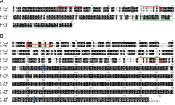
contain (34). YsrR and DygR belong to the LuxR class and contain a helix-turn-helix DBD. These proteins are 81% identical, but the amino acid differences are primarily clustered into two regions (Fig. 1A). In the receiver domain, YsrR and DygR contain two short regions that appear to be insertions compared to other pro-teins of the same class (23). One of these insertion regions harbors one of the clusters of differences, and the second cluster is located just at the beginning of the DBD. The putative phosphorylation site (D75) and other residues important for the phosphorylation and signal transduction (E13, D14, T115, and K137) are con-served, as are other key structural residues (P95, G95, and A116) (35). The conservation of these important residues suggests that the receiver domain of DygR is functional.
Sensor kinase alignment.The alignment of YsrS and DygS shows a strikingly high identity between the two proteins (Fig. 1B). Within the cytoplasmic portion of these proteins, they are 97.8% identical, differing only in five amino acids just C terminal of the second transmembrane domain, three residues in the mid-section, and the very C terminus. The DNA sequences encoding these cytoplasmic domains are 98.3% identical; there are only five synonymous changes, and these are located adjacent to the regions where amino acids are different. In contrast, the periplasmic re-gions, including the two transmembrane domains, are only 68.2% identical. While there are⬃20-residue stretches of identity, the
differences are evenly distributed within this domain. Based on examination of the DygS amino acid sequence, it can be inter-preted that DygS has similar phosphorelay capabilities but re-sponds to a signal different from the signal that YsrS rere-sponds to (NaCl).
Genomic context. The ⬃30-kb ysa locus contains a long operon with genes encoding the secretion apparatus, translocon proteins, and one effector protein. Adjacent to this operon, lo-cated on the opposite strand, are a few open reading frames en-coding the recently identified YsaP pilotin protein (36) and other proteins that are likely part of the secretion apparatus. TheysrRST genes are just downstream of this region, and it was their proxim-ity to theysagenes that led us to investigate their role inysagene expression (22). ThedygRSgenes are approximately 10 kb down-stream ofysrRST. The region duplicated seems only to include a few base pairs upstream of the YsrR/DygR start codon through a few base pairs upstream of the stop codon of YsrS/DygS. The pro-moter region forysrRSTshares very little with the region upstream ofdygR, and there is no gene encoding a histidine phosphotrans-ferase (HPt)-containing protein downstream ofdygS. Between the ysrRSTgenes and thedygRSgenes are genes encoding a type II secretion system (T2SS) that is important for virulence (37) and one of the three T2SS-secreted proteins (38). It is somewhat curi-ous that there is a sizable distance between theysranddyggenes, as
TABLE 2Primers used in this work
Name Sequencea(5=
¡3=)
MWO-019 CATATCGTCGACCTGATCCGCTGGGGCATTGATGC
MWO-020 CTGTGGATCCCGTTTCTATCATAATTCCGAC
MWO-021 AGACGGATCCGCCTCAGGAAAATAACAACAAG
MWO-022 CAGTAGCGGCCGCCTGTTCAGAATCGGAAAAACC
MWO-056 GGGGAATCTAGAGGGTTATTGCATGTGGTGGCG
MWO-057 GGCAAGTAGATCTGCGCAAGGCGACTGAACGG
MWO-102 GCGTCGACGCGCTTTCAAAAAACTGGGG
MWO-103 CGGAATTCTTATTTTCCTGAGGCGAAATAGTC
MWO-104 GCGTCGACCCCACCACTCCTCAATGCCAC
MWO-105 CGGGATCCGGTTTTATAAAGAAATTTCATGGGC
MWO-106 AGACGGATCCCGCGCTTTCAAAAAACTGGGG
MWO-107 CAGTAGCGGCCGCCGACGGCAGCTTTTGCTCGC
MWO-108 CATATCGTCGACGCTGATTTACAGCGCCTTTTC
MWO-109 CTGTGGATCCTTTATAAAGAAATTTCATGGGCATAATGG
MWO-110 CATATCTAAGCTTGTGCCTTCAAAAAACTGGGG
MWO-111 CCATTTTTAGCTGCCGGTTACGATAGAAAACGAATAACAGCAAGC
MWO-112 GCTTGCTGTTATTCGTTTTCTATCGTAACCGGCAGCTAAAAATGG
MWO-113 CATATCCTGCAGTTATTTTCCTGAGGCGAAATAGTC
KW223 AAGGATCCGATGACACAAACGAAAACGCTCAATATAG
KW224 CGGGTACCTTATAGAGAAATTTCATGAGCATATTTAAAG
KW227 CCGCTCGAGGATGACACAAACGAAAACGCTCAATATAG
KW228 CCCCCAAGCTTCCTAGAGAAATTTCATGAGCATATTT
KW251 AACTGCAGGGTATCGCTACCGGCGATTAAAAATGG
KW252 GGGGTACCTCAGTCATGTTCTTTTTCCTTAG
KW253 AACTGCAGGGTATCGTAACCGGCAGCTAAAAATGGC
KW259 GGGGTACCTTATTTTCCTGAGGCGAAATAGTC
KW254 CCGCTCGAGCATGACTGATGCCACCTTCAGCGCAC
KW255 GCGTCGACGCGCTGTTATCTAGCAAGGCATAAAATTGC
KW263 AACTGCAGGGATGACTGATGCCACCTTCAGCGCAC
KW264 CGGGATCCTTAGCTGTTATCTAGCAAGGCATAAAATTGC
KW256 AACTGCAGGGATGATAGAAACGAAAATGTTAAATATAGCC
KW257 CCGCTCGAGCATGATAGAAACGAAAATGTTAAATATAGCC
KW265 CCCAAGCTTGCTAAAGAAATTTCATGGGCATAATGG
duplication events typically generate tandem genes (39). One pos-sible explanation is that, following the duplication event, this seg-ment of DNA encoding the T2SS inserted itself between the tan-dem repeats.
Given the pronounced differences in DNA sequence immedi-ately upstream of thedygRstart codon, we investigated if there was a functional promoter by constructing a chromosomallacZ re-porter in the region upstream ofdygRin our wild-typeY. entero-coliticastrain. When grown at 26°C in L-broth and LB-290,-gal activity was measured at 755⫾103 and 1,841⫾117 Miller units (MU; averages⫾standard deviations), respectively. We know this is an indication of transcriptional activity, as the parent strain lacking any reporter produces less than 50 MU (data not shown); this low-level activity is likely derived from the intact nativelac operon. It is curious to note the⬃2-fold increase in the presence of 290 mM NaCl. We also tested thedygR-lacZreporter strain following growth under several other conditions, such as LB with high sucrose, iron depletion, acidic and basic pHs, and growth at 37°C, and we found that it was always expressed and with little variation in levels (data not shown). If this DNA region upstream ofdygRwas part of the T2SS-containing insertion event men-tioned above, it is perhaps somewhat serendipitous that it in-cluded a functional promoter.
DygS can compensate for loss of YsrS.In order to determine if DygS plays a role in regulating theysaT3SS genes, we examined expression of the Ysr-dependentysaEpromoter, a key promoter that drives transcription of a long operon that contains many of theysaapparatus genes (13,22). An in-frame deletion ofdygSwas constructed, and-gal activity was measured from a chromo-somally encodedysaE-lacZ fusion following growth under the
known Ysa-inducing condition, LB with 290 mM NaCl (LB-290). We found that deleting thedygS gene had no impact on ysaE expression (Fig. 2A). Given that the putative promoter region is active under these growth conditions, this suggests that DygS plays no role in regulatingysaEtranscription when cultured in LB-290. A preliminary screen of easily tested growth conditions, including pH, 1% sucrose, magnesium, and iron levels (supplemented and depleted), failed to reveal any role for DygS inysaE-lacZ expres-sion (data not shown). However, because it was still possible that there were conditions under which DygS could regulateysaE ex-pression, we constructed a plasmid constitutively expressingdygS (pDygS) to assess if overproduction of DygS could modulate ysaE-lacZexpression. pDygS was transformed into the wild-type and
⌬ysrS,⌬dygS, and⌬ysrS⌬dygSmutant strains carrying the ysaE-lacZreporter, and the resulting strains were assayed for-gal ac-tivity following growth in LB-290. As controls, the vector pWKS130 and the ysrScomplementing clone, pYsrS, were in-cluded. When overproduced in trans, strains carrying pDygS yielded between 1,300 and 1,950 MU, comparable to the activity in the wild-type strain carrying the vector pWKS130 (Fig. 2A). Thus, pDygS restored expression in the⌬ysrSand⌬ysrS⌬dygSmutant strains, indicating that DygS has the capacity to compensate for loss of YsrS.
Because the cytoplasmic domains of YsrS and DygS are so highly conserved, we hypothesized that the cytoplasmic domain of DygS, which is nearly identical to YsrS, could participate in the Ysr phosphorelay. To test this, we constructed a plasmid with a chi-meric gene encoding the YsrS sensing (periplasmic) domain with the DygS cytoplasmic domains, called pChimera. As predicted, this plasmid complemented theysrSgene deletion, yielding 2,208
A.
B.
and 3,513 MU in the⌬ysrSand⌬ysrS⌬dygSstrains, respectively (Fig. 2A). These expression levels were higher than those obtained when pDygS was transformed into these same strains but similar to those obtained with pYsrS; this may be due to the presence of the salt-responsive sensor domain on pChimera and pYsrS.
The results with pDygS and pChimera suggest that DygS is
capable of transferring the phosphoryl group to YsrT, which then transfers to YsrR, resulting inysaEtranscription activation. To verify that these phenotypes are from DygS substituting for YsrS and not some other indirect effect, we transformed pDygS into the
⌬ysrRand⌬ysrTstrains and tested forysaE-lacZexpression (Fig. 2B). The presence of pDygS in these strains could not restoreysaE
0 1000 2000 3000 4000 5000
pWKS130
Miller Units
wild type
ysrS
dygS
ysrS dygS
pYsrSWT pDygS pYsrSpChimera D714A
A.
LB-290
0 500 1000 1500
wild type
ysrR
ysrT
pWKS130 pDygS
B.
Miller Units
LB-290
0 500 1000 1500 2000 2500
0 50 100 150 1000 1200 1400
C.
Miller Units
pMWO-034 pMWO-034 piYsrSD714A piYsrSD714A
LB-290 L-broth
wild type
ysrS
dygS
ysrS dygS
FIG 2DygS compensates for loss of YsrS. (A)-Galactosidase assay results for wild-type,⌬ysrS,⌬dygS, and⌬ysrS⌬dygSstrains carrying aysaE-lacZfusion. Each strain was transformed with a plasmid expressingysrS(pYsrS),dygS(pDygS), theysrS-dygSchimera (pChimera), orysrSD714A(pYsrSD714A) or with the vector (pWKS130). (B) pDygS was transformed into the⌬ysrRand⌬ysrTstrains to test the roles of YsrR and YsrT in the phosphorelay initiating with DygS. (C)
expression, further supporting our hypothesis that the observed complementation of the⌬ysrSmutant by pDygS and pChimera is due to DygS participating in the Ysr phosphorelay that leads to activation ofysagene expression.
Further evidence of a putative role for DygS in regulation of ysaEexpression came as we pursued a peculiar result from our genetic analysis of the Ysr phosphorelay. In a previous study, we made alanine substitutions in each of the aspartate and histidine residues predicted to be phosphorylated in YsrS, YsrT, and YsrR (23). All of the mutants behaved as expected, in thatysaE expres-sion was not activated, with one striking exception. Mutation of the conserved aspartate residue in the receiver domain of YsrS to alanine (D714A) actually resulted in constitutiveysaE-lacZ ex-pression, and this was dependent on the presence of wild-type YsrT and YsrR (23). Wild-type and⌬ysrSstrains transformed with a plasmid expressing the mutantysrSgene (pYsrSD714A) yielded
equally high levels of-gal activity after growth in L-broth and LB-290 (23). After obtaining the results above that suggested that DygS had the capacity to participate in the Ysr phosphorelay, we transformed pYsrSD714Ainto the⌬dygSand⌬ysrS⌬dygSstrains.
In the⌬dygSstrain with pYsrSD714A,ysaE-lacZ expression was
exceptionally high, as observed in the⌬ysrSstrain, yielding 3,905 and 4,659 MU, respectively (Fig. 2A). However, in the ⌬ysrS
⌬dygSstrain with pYsrSD714A, theysaE-lacZexpression level was
41 MU, approximately the same as the level with pWKS130 (49 MU). This is consistent with the activity measured from strains with noysaEexpression and is what would normally be expected from the D714A mutation. To ascertain if the unusual phenotypes with pYsrSD714Awere a consequence of constitutive expression of
the mutant gene, we subcloned the insert into a vector with an inducible promoter, creating piYsrSD714A. We then examined
ysaE-lacZlevels when strains carrying this plasmid were cultured with a very low concentration of inducer (5 ng/l of ATc). In this situation, we still saw that YsrSD714Awas capable of
complement-ing theysrSdeletion, yielding 1,450 MU (roughly equivalent to the wild-type strain), and was still constitutive, in that it resulted in high levels ofysaEexpression (1,075 MU) in the⌬ysrSstrain in the absence of NaCl (Fig. 2C). Thus, it appears that, in a strain with a chromosomal copy of eitherysrSordygS, YsrSD714Amay form a
dimer with YsrS or DygS, and the phosphorelay travels from H320 of YsrSD714Ato D714 (YsrS) or D712 (DygS). In the absence of a
chromosomal copy ofysrSordygS, there is no aspartate residue in the sensor receiver domain and, thus, no way to complete the phosphorelay. Why this creates a constitutively active phosphore-lay in⌬ysrSis still a mystery. The high levels of expression under noninducing conditions are only evident whendygSis present and ysrSis absent, and it is possible that the backward movement of the phosphoryl group, a carefully controlled step in phosphorelay sys-tems, is disrupted. Taken together, these data indicate that YsrS and DygS are similar enough to play nearly identical roles and suggest that, under certain conditions, DygS has the capacity to activateysaEexpression and, thus, production of the Ysa T3SS.
DygR does not compensate for loss of YsrR.Having deter-mined that DygS can participate in the pathway leading toysaE activation, we set out to examine if DygR could also activate ysaE-lacZexpression. We performed a similar set of experiments using adygRin-frame deletion strain (⌬dygR) anddygR-overexpressing plasmid (pDygR). As was the case with⌬dygS, we found that de-leting thedygRgene had no impact onysaEexpression (770 MU with pWKS130), suggesting that DygR plays no role in regulating
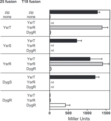
ysaEtranscription in LB-290 (Fig. 3). However, the results from overexpressing DygR differed from DygS. In this case, we found that transformation with pDygR had no impact onysaE expres-sion in any strain background examined, yielding about 1,300 MU in strains withysrRand⬍50 MU in strains lackingysrR. Thus, activation ofysaE-lacZby DygS most likely occurs via a mecha-nism other than the canonical sensor-response regulator pathway. DygS, but not DygR, interacts with YsrT.The data presented inFigure 2Bshow that YsrT and YsrR are necessary for pDygS to enableysaEactivation. Presumably, for DygS to facilitate activa-tion ofysaE, it must transfer the phosphoryl group to an HPt domain. There does not appear to be a gene encoding an HPt-containing protein in thedyglocus, and our data implicate YsrT in this role. Thus, we turned to the Ladant bacterial 2-hybrid system (B2H) (25) to examine protein-protein interactions that phos-phoryl group transfer would necessitate. In this system, two vec-tors, each encoding a domain of theBordetella pertussisadenylate cyclase (AC), are used to make AC fusions to proteins of interest and are cotransformed into E. coliBTH101 cells. If the fusion proteins interact, it brings the two AC domains together, allowing synthesis of cAMP. Then, cAMP binds to CRP, leading to the activation of maltose and lactose operons. BTH101 has acya mu-tation that produces only very small amounts of cAMP, thus mak-ing the increased cAMP due to interaction of the two fusion pro-teins readily visible through enzymatic analysis of the maltose or lactose fermentation and-gal activity. While we had genetic ev-idence of phosphoryl transfer from YsrS to YsrT and then to YsrR (23), we had yet to examine if these proteins interacted. Because YsrS and DygS are most likely anchored in the inner membrane, we cloned only sequences encoding their cytoplasmic domains into pT25, assuming that the N-terminal AC fusion would not interfere with the interaction regions. The genesysrR,ysrT, and dygRwere cloned into both pT18 and pT25 vectors for analysis of homodimerization as well as interactions with YsrS and DygS.E. colistrain BHT101 was cotransformed with one pT18-based plas-mid and one pT25-based plasplas-mid, plated on MacConkey agar plates containing 5% maltose (MacMal), and incubated at 30°C for a qualitative assessment of interactions (positive interactions yielded red-colored colonies). Quantitative assessment via-gal assays was performed after 3 days on MacMal plates. Our positive-control strain contained pT18-zip and pT25-zip, each containing
0 500 1000 1500 2000 2500
FIG 3-Galactosidase assay results for wild-type,⌬ysrR,⌬dygR, and⌬ysrR
a leucine zipper domain (25); the negative-control strain con-tained the vectors only. This assay revealed that both YsrS and DygS interact with YsrT, as strains carrying these pairs of plasmids produced 722 and 1,210 MU, respectively (Fig. 4). The-gal ac-tivity from the YsrT-DygS strain was slightly higher than from the YsrS strain; this can be interpreted to mean that the YsrT-DygS interaction was strong enough to facilitate phosphoryl transfer. Many response regulators form homodimers in their ac-tive state (reviewed in reference40). As predicted, BHT101 trans-formed with each plasmid carryingysrRyielded 1,408 MU, dem-onstrating that YsrR forms homodimers.
We included DygR in the B2H analysis to address (i) whether it could interact with YsrT, which could mean that it can participate in a phosphorelay with DygS-YsrT and regulate other genes, and (ii) whether the level of similarity with YsrR was sufficient for heterodimer formation.-gal assays following cotransformation with DygR constructs indicate that DygR does not interact with YsrT or with YsrR; the measured activity was just slightly above the negative control at 11 and 16 MU, respectively (Fig. 4). How-ever, the-gal activity from the DygR-DygR strain (420 MU) indicates that it does homodimerize. This last result also indicates that these DygR constructs are functional, strengthening the in-terpretation that it does not interact with YsrT or YsrR.
DISCUSSION
DygS and DygR are putative phosphorelay proteins that share a strikingly high similarity to YsrS and YsrR. The Ysr phosphorelay
system, comprised of the sensor kinase YsrS, the histidine phos-photransferase (HPt) YsrT, and the response regulator YsrR, is required for activation of theysaEpromoter that drives transcrip-tion of the chromosomally encoded Ysa type III secretranscrip-tion system (T3SS) (13,22). ThedygSanddygRgenes (YE3579 and YE3578, respectively [12]) are located about 10 kb downstream of theysa/ ysrlocus and most likely were acquired by a gene duplication event. Because of the similarity to YsrS and YsrR, we investigated whether DygS and DygR could also activateysaEexpression. We considered several hypotheses. First, the differences within DygR and the periplasmic region of DygS could have rendered them nonfunctional and the Dyg phosphorelay system a dying remnant of duplication. However, the nearly 100% conservation of the cy-toplasmic region of DygS suggested that it likely was at least a partially functional histidine kinase. With the assumption that DygR and DygS both retain function, a second hypothesis we con-sidered is that DygS autophosphorylates in response to a signal (other than NaCl) and transfers the phosphoryl group first to YsrT and then to YsrR, which could then activateysaEexpression. A third possibility is that the DygS-YsrT-DygR phosphorelay com-prises a completely separate regulon that may or may not include ysaE. Our data suggest that what occurs is a combination of the first and second hypotheses: that DygS can initiate a phosphorelay with YsrT and YsrR to activateysaEexpression, but that DygR may have acquired enough deleterious mutations to lose function.
The data we obtained for DygR showed that it does not interact with YsrT and cannot promoteysaE transcription, even when overexpressed. We conclude that the differences between YsrR and DygR are mutations that have altered the function of DygR such that it can no longer become phosphorylated by YsrT, can no longer bind theysaEpromoter, or both. One cluster of differences lies near the DNA-binding domain. These differences could lead to changes in structure or binding specificity, such that it cannot bind theysaEpromoter. The second cluster of differences is in one of the two insertion sequences; these residues could be in the re-gion that facilitates phosphotransfer from YsrT. The lack of inter-action between DygR and YsrT suggests that, if it does participate in a phosphorelay with DygS, it does so with another HPt protein. Given thatY. enterocoliticacontains only six HPt-containing pro-teins and that they are all part of characterized phosphorelay sys-tems, we suspect that the amino acid changes acquired by DygR have caused a loss of function.
Through the use of gene deletions andtranscomplementation with a variety of plasmids expressing different forms ofysrSand dygS, we provide several lines of evidence indicating that DygS is capable of facilitating transcription of theysaEpromoter by par-ticipating in a phosphorelay with YsrT and YsrR. First, while loss of thedygSgene had no impact onysaEexpression, introducing a plasmid expressing either the wild-typedygSgene or theysrS-dygS chimeric gene could compensate for loss of theysrSgene. Second, the bacterial two-hybrid experiment demonstrated an interaction between DygS and YsrT with strength similar to that of the YsrS-YsrT interaction. Third, activation by DygS requires theysrTand ysrRgenes. Finally, the constitutive-on phenotype observed when complementing the⌬ysrSstrain with pYsrSD714Arequires thedygS
(orysrS) gene, providing strong genetic evidence that YsrS and DygS can heterodimerize. This is not surprising, given the 100% identity conservation of the HisKA domain (residues 313 to 366), which is known to promote dimerization (41).
If DygS does indeed activateysagene expression in response to
0 500 1000 1500
nd
nd
nd
nd nd
T25 fusion T18 fusion
YsrT
YsrS
YsrR
DygS
DygR
none none zip zip
YsrT YsrR DygR YsrT YsrR DygR YsrT YsrR DygR
YsrT YsrR DygR
YsrT YsrR DygR
Miller Units
a signal other than NaCl, it indicates that theysasystem has two sensors that can independently lead to expression (Fig. 5). This is not unlike theVibrioquorum sensing phosphorelay systems re-sponding to autoinducers. In this case, there are two sensors (LuxN and LuxQ), each responding to a different autoinducer, each relaying the phosphoryl group to the HPt-containing LuxU protein, which leads to phosphorylation of LuxO that regulates the transcription of downstreamluxgenes (reviewed in reference 42). InBacillus subtilis, five separate sensor kinases (KinA through KinE) all lead to the phosphorylation of the response regulator Spo0A. These kinases respond to different signals and result in different cellular fates: sporulation, matrix production, and bio-film production (reviewed in reference43). Thus, it is plausible that, under conditions yet to be identified, DygS initiates a phos-phorelay signal that leads to activation ofysagene expression and possibly other genes as well.
The data provided herein suggest that DygS has undergone evolution by the innovation-amplification-divergence model (44). In this model, there is an assumption that a gene that is duplicated has a side function (innovation). In this situation, it could be a trigger other than NaCl that initiates the Ysr phospho-relay. This side function is advantageous enough that the gene is duplicated to generate a larger dose of this function (amplifica-tion). In the duplicated gene, this side function is selected for through the acquisition of mutations that enhance this function (divergence). Evidence exists that other salts can activateysaE ex-pression (13) (K. A. Walker and V. L. Miller, unpublished data). While it has not been determined if these other salts initiate a phosphorelay through YsrS or DygS, it is tempting to speculate that one side function of YsrS was response to a secondary signal that became the primary signal for DygS. Because we have yet to find an environmental signal that triggers DygS-dependent
acti-vation ofysaEexpression, we cannot rule out that the sensing periplasmic region acquired deleterious mutations that render it nonfunctional. However, the preservation of the cytoplasmic re-gion at not just the amino acid sequence level but also the DNA sequence level suggests that DygS provides a vital function during some phase of theY. enterocoliticalife cycle and further supports the notion that the Ysa T3SS is a critical component for the sur-vival of this pathogen.
ACKNOWLEDGMENTS
We thank Ruth E. Silversmith and Bob Bourret for stimulating and in-sightful discussions and Rita Tamayo for critical review of the manuscript. We appreciate the efforts of Jordan Walters and Stephan Dickgiesser in generating several plasmids used in this work.
This work was supported by grant A14-1440 from the HHMI-BFW Medical Research Fellows Program (to L.A.G.).
FUNDING INFORMATION
This work, including the efforts of Lauren A. Griggs, was funded by HHMI-BFW Medical Research Fellows Program (A14-1440).
REFERENCES
1.Bottone EJ.1997.Yersinia enterocolitica: the charisma continues. Clin Microbiol Rev10:257–276.
2.Cover TL, Aber RC.1989.Yersinia enterocolitica. N Engl J Med321:16 – 24.http://dx.doi.org/10.1056/NEJM198907063210104.
3.Porter CK, Choi D, Cash B, Pimentel M, Murray J, May L, Riddle MS.
2013. Pathogen-specific risk of chronic gastrointestinal disorders follow-ing bacterial causes of foodborne illness. BMC Gastroenterol13:46.http: //dx.doi.org/10.1186/1471-230X-13-46.
4.Verdu EF, Riddle MS.2012. Chronic gastrointestinal consequences of acute infectious diarrhea: evolving concepts in epidemiology and patho-genesis. Am J Gastroenterol107:981–989.http://dx.doi.org/10.1038/ajg .2012.65.
5.Scallan E, Hoekstra RM, Angulo FJ, Tauxe RV, Widdowson M-A, Roy SL, Jones JL, Griffin PM.2011. Foodborne illness acquired in the United States: major pathogens. Emerging Infect Dis17:7–15.http://dx.doi.org /10.3201/eid1701.P11101.
6.Lamps LW, Havens JM, Gilbrech LJ, Dube PH, Scott MA. 2006. Molecular biogrouping of pathogenicYersinia enterocolitica: development of a diagnostic PCR assay with histologic correlation. Am J Clin Pathol
125:658 – 664.http://dx.doi.org/10.1309/A8JJPGGGWXYLF48A. 7.McNally A, Cheasty T, Fearnley C, Dalziel RW, Paiba GA, Manning G,
Newell DG.2004. Comparison of the biotypes ofYersinia enterocolitica
isolated from pigs, cattle, and sheep at slaughter and from humans with yersiniosis in Great Britain during 1999-2000. Lett Appl Microbiol39:
103–108.http://dx.doi.org/10.1111/j.1472-765X.2004.01548.x. 8.Rosner BM, Stark K, Werber D.2010. Epidemiology of reportedYersinia
enterocoliticainfections in Germany, 2001-2008. BMC Public Health10:
337.http://dx.doi.org/10.1186/1471-2458-10-337.
9.Sihvonen LM, Haukka K, Kuusi M, Virtanen MJ, Siitonen A, YE study group.2009.Yersinia enterocoliticaandY. enterocolitica-like species in clinical stool specimens of humans: identification and prevalence of bio/ serotypes in Finland. Eur J Clin Microbiol Infect Dis28:757–765.http://dx .doi.org/10.1007/s10096-008-0696-y.
10. Huovinen E, Sihvonen LM, Virtanen MJ, Haukka K, Siitonen A, Kuusi M.2010. Symptoms and sources ofYersinia enterocolitica-infection: a case-control study. BMC Infect Dis 10:122. http://dx.doi.org/10.1186 /1471-2334-10-122.
11. Consumer Reports.13 January 2013. What’s in that pork? p 44 – 46. Consumer Reports, Yonkers, NY.
12. Thomson NR, Howard S, Wren BW, Holden MTG, Crossman L, Challis GL, Churcher C, Mungall K, Brooks K, Chillingworth T, Feltwell T, Abdellah Z, Hauser H, Jagels K, Maddison M, Moule S, Sanders M, Whitehead S, Quail MA, Dougan G, Parkhill J, Prentice MB.2006. The complete genome sequence and comparative genome analysis of the high pathogenicityYersinia enterocoliticastrain 8081. PLoS Genet2:e206.http://dx.doi.org/10.1371/journal.pgen.0020206. 13. Venecia K, Young GM.2005. Environmental regulation and virulence YsrR~P
YsrT
H2
P
D2
P
D2
YsrR
YsrS DygS P
P
D1 H1
D1 H1
ADP ATP
NaCl
P
P
D1 H1
D1 H1
ADP ATP ???
ysa operon other genes?
FIG 5Model of potential phosphorelay events leading to the activation ofysa
attributes of the Ysa type III secretion system ofYersinia enterocolitica
biovar 1B. Infect Immun73:5961–5977.http://dx.doi.org/10.1128/IAI.73 .9.5961-5977.2005.
14. Walker KA, Maltez VI, Hall JD, Vitko NP, Miller VL.2013. A phenotype at last: essential role for theYersinia enterocoliticaYsa type III secretion system in aDrosophila melanogasterS2 cell model. Infect Immun81:
2478 –2487.http://dx.doi.org/10.1128/IAI.01454-12.
15. Haller JC, Carlson S, Pederson KJ, Pierson DE.2000. A chromosomally encoded type III secretion pathway inYersinia enterocoliticais important in virulence. Mol Microbiol36:1436 –1446.
16. Foultier B, Troisfontaines P, Müller S, Opperdoes FR, Cornelis GR.
2002. Characterization of theysapathogenicity locus in the chromosome
ofYersinia enterocoliticaand phylogeny analysis of type III secretion
sys-tems. J Mol Evol55:37–51.http://dx.doi.org/10.1007/s00239-001-0089-7. 17. Matsumoto H, Young GM.2006. Proteomic and functional analysis of the suite of Ysp proteins exported by the Ysa type III secretion system of
Yersinia enterocoliticabiovar 1B. Mol Microbiol59:689 –706.http://dx.doi
.org/10.1111/j.1365-2958.2005.04973.x.
18. Bent ZW, Branda SS, Young GM.2013. TheYersinia enterocoliticaYsa type III secretion system is expressed during infections bothin vitroandin vivo. Microbiologyopen2:962–975.http://dx.doi.org/10.1002/mbo3.136. 19. Bent ZW, Poorey K, Brazel DM, LaBauve AE, Sinha A, Curtis DJ, House SE, Tew KE, Hamblin RY, Williams KP, Branda SS, Young GM, Meagher RJ.2015. Transcriptomic analysis ofYersinia enterocolitica bio-var 1B infecting murine macrophages reveals new mechanisms of extra-cellular and intraextra-cellular survival. Infect Immun83:2672–2685.http://dx .doi.org/10.1128/IAI.02922-14.
20. Walker KA, Miller VL.2009. Synchronous gene expression of theYersinia
enterocoliticaYsa type III secretion system and its effectors. J Bacteriol
191:1816 –1826.http://dx.doi.org/10.1128/JB.01402-08.
21. Petersen S, Young GM.2002. Essential role for cyclic AMP and its recep-tor protein inYersinia enterocoliticavirulence. Infect Immun70:3665– 3672.http://dx.doi.org/10.1128/IAI.70.7.3665-3672.2002.
22. Walker KA, Miller VL.2004. Regulation of the Ysa type III secretion system ofYersinia enterocoliticaby YsaE/SycB and YsrS/YsrR. J Bacteriol
186:4056 – 4066.http://dx.doi.org/10.1128/JB.186.13.4056-4066.2004. 23. Walker KA, Obrist MW, Mildiner-Earley S, Miller VL.2010.
Identifi-cation of YsrT and evidence that YsrRST constitute a unique phosphorelay system inYersinia enterocolitica. J Bacteriol192:5887–5897.http://dx.doi .org/10.1128/JB.00745-10.
24. Miller VL, Mekalanos JJ.1988. A novel suicide vector and its use in construction of insertion mutations: osmoregulation of outer membrane proteins and virulence determinants inVibrio choleraerequirestoxR. J Bacteriol170:2575–2583.
25. Karimova G, Pidoux J, Ullmann A, Ladant D.1998. A bacterial two-hybrid system based on a reconstituted signal transduction pathway. Proc Natl Acad Sci U S A95:5752–5756.http://dx.doi.org/10.1073/pnas.95.10 .5752.
26. Kinder SA, Badger JL, Bryant GO, Pepe JC, Miller VL.1993. Cloning of the YenI restriction endonuclease and methyltransferase fromYersinia
enteroco-liticaserotype O8 and construction of a transformable r⫺m⫹mutant. Gene
136:271–275.http://dx.doi.org/10.1016/0378-1119(93)90478-L.
27. Merriam JJ, Mathur R, Maxfield-Boumil R, Isberg RR.1997. Analysis of
theLegionella pneumophila fliIgene: intracellular growth of a defined
mu-tant defective for flagellum biosynthesis. Infect Immun65:2497–2501. 28. Wang RF, Kushner SR.1991. Construction of versatile low-copy-number
vectors for cloning, sequencing and gene expression inEscherichia coli. Gene
100:195–199.http://dx.doi.org/10.1016/0378-1119(91)90366-J.
29. Obrist MW, Miller VL.2012. Low copy expression vectors for use in
Yersiniasp. and related organisms. Plasmid68:33– 42.http://dx.doi.org
/10.1016/j.plasmid.2012.02.003.
30. Ellison DW, Young B, Nelson K, Miller VL.2003. YmoA negatively regulates expression of invasin fromYersinia enterocolitica. J Bacteriol185:
7153–7159.http://dx.doi.org/10.1128/JB.185.24.7153-7159.2003. 31. Kearse M, Moir R, Wilson A, Stones-Havas S, Cheung M, Sturrock S,
Buxton S, Cooper A, Markowitz S, Duran C, Thierer T, Ashton B, Meintjes P, Drummond A. 2012. Geneious Basic: an integrated and extendable desktop software platform for the organization and analysis of sequence data. Bioinformatics28:1647–1649.http://dx.doi.org/10.1093 /bioinformatics/bts199.
32. Miller JH.1992. A short course in bacterial genetics. Cold Spring Harbor Laboratory Press, Cold Spring Harbor, NY.
33. Karimova G, Ullmann A, Ladant D.2000. A bacterial two-hybrid system that exploits a cAMP signaling cascade inEscherichia coli. Methods Enzy-mol328:59 –73.http://dx.doi.org/10.1016/S0076-6879(00)28390-0. 34. Galperin MY.2010. Diversity of structure and function of response
reg-ulator output domains. Curr Opin Microbiol13:150 –159.http://dx.doi .org/10.1016/j.mib.2010.01.005.
35. Bourret RB.2010. Receiver domain structure and function in response regulator proteins. Curr Opin Microbiol13:142–149.http://dx.doi.org/10 .1016/j.mib.2010.01.015.
36. Rau R, Darwin AJ.2015. Identification of YsaP, the pilotin of theYersinia
enterocoliticaYsa type III secretion system. J Bacteriol197:2770 –2779.
http://dx.doi.org/10.1128/JB.00238-15.
37. Iwobi A, Heesemann J, Garcia E, Igwe E, Noelting C, Rakin A.2003. Novel virulence-associated type II secretion system unique to high-pathogenicityYersinia enterocolitica. Infect Immun71:1872–1879.http: //dx.doi.org/10.1128/IAI.71.4.1872-1879.2003.
38. Shutinoski B, Schmidt MA, Heusipp G.2010. Transcriptional regulation of the Yts1 type II secretion system ofYersinia enterocoliticaand identifi-cation of secretion substrates. Mol Microbiol75:676 – 691.
39. Andersson DI, Jerlström-Hultqvist J, Näsvall J.2015. Evolution of new functions de novo and from preexisting genes. Cold Spring Harb Perspect Biol7:a017996.http://dx.doi.org/10.1101/cshperspect.a017996. 40. Gao R, Stock AM. 2010. Molecular strategies for
phosphorylation-mediated regulation of response regulator activity. Curr Opin Microbiol
13:160 –167.http://dx.doi.org/10.1016/j.mib.2009.12.009.
41. Dutta R, Qin L, Inouye M.1999. Histidine kinases: diversity of domain organization. Mol Microbiol 34:633– 640. http://dx.doi.org/10.1046/j .1365-2958.1999.01646.x.
42. Bassler BL.1999. How bacteria talk to each other: regulation of gene expression by quorum sensing. Curr Opin Microbiol2:582–587.http://dx .doi.org/10.1016/S1369-5274(99)00025-9.
43. López D, Kolter R.2010. Extracellular signals that define distinct and coexisting cell fates inBacillus subtilis. FEMS Microbiol Rev34:134 –149. http://dx.doi.org/10.1111/j.1574-6976.2009.00199.x.
44. Bergthorsson U, Andersson DI, Roth JR.2007. Ohno’s dilemma: evo-lution of new genes under continuous selection. Proc Natl Acad Sci U S A
104:17004 –17009.http://dx.doi.org/10.1073/pnas.0707158104. 45.Ulrich LE, Zhulin IB. 2010. The MiST2 database: a comprehensive
genomics resource on microbial signal transduction. Nucleic Acids Res