THE INFLUENCE OF CAMERA PERSPECTIVE ON DIAGNOSTIC ACCURACY USING ORTHODONTIC RECORDS
Christopher J. Kirk
A thesis submitted to the faculty at the University of North Carolina at Chapel Hill in partial fulfillment of the requirements for the degree of Master of Science in the School of Dentistry
(Orthodontics).
Chapel Hill 2016
ABSTRACT
Christopher J. Kirk: The influence of camera perspective on diagnostic accuracy using photographic records
(Under the direction of Tate Jackson)
Introduction: Dr. Edward Angle, the “Father of Orthodontics” was also the first orthodontist to incorporate photography into the practice of orthodontics1. With the advent of digital cameras, clinical photography is easier, faster and more cost effective than ever before. Despite technological advances, difficulties with patient-camera positioning and the resultant parallax effect still persist. This cross-sectional survey evaluated the influence of camera perspective changes on the ability of orthodontists to make diagnostic judgments using photographic records.
Methods: Practicing orthodontists (n=205) assessed, via an electronic survey tool, 12 patient casts photographed at three angulations in the axial plane of one of two views: buccal view and frontal view. Participants judged midlines or canine and molar classification based on the images provided at different camera angulations. The relationships between correct responses and demographic information, degree of angulation, and clinical photography practice were assessed using Chi-square for bivariate analyses and conditional logistic regression for multivariate analyses.
midline deviation as camera perspective in the axial plane changes from ideal to non-ideal angulations.
ACKNOWLEDGEMENTS
Thank you to my committee members, Dr. Phillips, Dr. Jackson and Dr. Koroluk, for your expertise, guidance, and advice throughout my project. Thank you to the Southern Association of Orthodontists and the Dental Foundation of North Carolina, Inc. for the
TABLE OF CONTENTS
LIST OF TABLES ... viii
LIST OF FIGURES ... ix
LIST OF ABBREVIATIONS ...x
LIST OF SYMBOLS ... xi
REVIEW OF THE LITERATURE ...1
Historical Perspective ...1
Records and Orthodontics ...2
Photographic Armamentarium ...5
Photographic Technique ...5
Medicolegal Requirements ...8
Communication ...10
Conclusion ...11
References ...12
THE INFLUENCE OF CAMERA PERSPECTIVE ON DIAGNOSTIC ACCURACY USING PHOTOGRAPHIC RECORDS ...14
Introduction ...14
Participants ...17
Stimuli Construction ...18
Tasks ...20
Survey Construction ...21
Analysis...22
Results ...22
Descriptive Statistics ...22
Reliability ...23
Univariate Analysis ...23
Bivariate and Covariate Analysis ...25
Discussion ...26
Conclusions ...33
Tables ...34
Figures...39
LIST OF TABLES
Table 1 – Distribution of Participants by Region and Gender ...34
Table 2 – Participant Gender by Age Group ...34
Table 3 – Participant Demographics by Region ...34
Table 4 – Buccal Question Block Intra-observer Reliability ...35
Table 5 – Frontal Question Block Intra-observer Reliability ...35
Table 6 – Distribution of Photographic Armamentarium ...36
Table 7 – Distribution of Photographic Protocols ...36
Table 8 – Accuracy of Angle Classification by Degree of Deviation and Demographics ...37
Table 9 – Odds Ratio of Buccal Questions for Deviation ...37
Table 10 – Accuracy of Frontal Responses by Degree of Deviation and Demographics ...38
LIST OF FIGURES
LIST OF ABBREVIATIONS AAO American Association of Orthodontists
ABO American Board of Orthodontics ALARA As Low as Reasonably Achievable BOR Buccal Object Rule
CBCT Cone Beam Computed Tomography
CO Centric Occlusion
CT Computed Tomography
LIST OF SYMBOLS
A REVIEW OF THE LITERATURE
Historical Perspective:
Although photographic technology has changed significantly in the past 175 years, photography and dentistry have been irreversibly linked since the inception of the camera. An early influence was Dr. Alexander Wolcott, a dentist from New York City who patented a camera and was responsible for opening the first commercial photographic studio1. As cameras became more accessible, American dentists recognized their value and were early adopters of the technology. These pioneers began recording their treatment outcomes using photography.
Although the American Journal of Dental Science began publication in 1839 as the first dental journal, it wasn’t until 1850 when photography was first used to show pre- and post-surgical outcomes1. Drs. R. Thompson and W.E. Ide of Columbus, Ohio surgically removed a maxillary tumor and fitted a prosthesis of gutta-percha, photographing the procedure and later submitting for publication in the American Journal of Dental Science1.
As photography became integral to orthodontic treatment planning, standardization was required to achieve consistent results. Since changes and improvements in equipment have been continuous, many articles have been written outlining correct technique and protocol. In 1979, Dr. Goodlin created a standardized guide for various views, magnification ratios and procedures in an attempt to improve outcomes3. The current ‘Gold Standard’ for intraoral photographic records is outlined by the American Board of Orthodontics (ABO) for the purpose of board certification and is generally accepted as the standard for orthodontics in general. It includes right and left buccal photographs, occlusal, and frontal views with the teeth in maximum intercuspation (MI)1,4,5.
Records and Orthodontics
With rapidly advancing innovations in dentistry and orthodontics, are photographs still relevant? The first method of recording patient encounters were written chart notes and
sketches1. Today they are more commonly typed and stored electronically, but the same limitations apply. The clinician may find it difficult to accurately describe a clinical finding or may not record a seemingly insignificant finding at all. It would not be possible for one to record all the minutia present in photograph with clinical notes2. Therefore, photographs serve as an efficient and non-invasive method of recording the current intraoral environment and to supplement clinical notes.
capture soft as well. A lateral cephalogram shows soft tissue profile and a CBCT records a three-dimensional volume of soft tissue.
Although this data is clinically useful and provides some of the same information as photographs, radiographic imaging falls short in several areas. A recent systematic review by Olivier et al determined that for most straightforward cases, a CBCT was not justified and a diagnosis could be made using traditional records7. Exposing the patient to additional radiation comes with risks and the ALARA principle should be kept in mind7. Additional radiographic images also come at a greater financial cost than photographs8. Lastly, due to the nature of the images, there is much clinical information missing in radiographs that is better recorded with photography such as soft tissue and facial esthetics, presence of white spot lesions, periodontal condition, and oral hygiene.
Intraoral scanners have been developed as a method of eliminating the need for plaster casts and alginate impressions9. Although developing at a rapid pace, these scanners remain somewhat cumbersome, time-consuming and expensive. Additionally, the resultant scan only records surface forms and textures and is unable to accurately record the color and condition of the dentition and soft tissues. Other possible methods of recording data include
three-dimensional photographs and video. Although these methods are accurate and realistic, they fall short in terms of user-friendliness, ease of use and cost10.
formulate a treatment plan11. Although laws may vary state-to-state, the general current standard of care for an initial pre- and post-treatment patient record includes a lateral cephalometric and panoramic radiograph, dental casts and clinical photographs; both intraoral and extraoral8,12. As mentioned above, the ABO has set photographic requirements in order to submit for board certification. The ABO standard is accepted as the general guideline for intra-oral
photography4,5,12. Depending on the patient’s age, malocclusion and oral health, additional radiographs and photographs may be indicated. The same records should be repeated at the conclusion of treatment as well8.
Another important factor is the frequency of record taking. Although not a requirement, some practitioners elect to take additional photographs at time points during treatment.
Additional images may include only the standard five intra-oral photographs or may involve additional views depending on the clinical situation. Images taken mid-treatment are useful for many reasons, especially for creating a permanent record where compliance and lack of oral-hygiene are problematic. They are also useful to record unfavorable growth or treatment side-effects allowing the practitioner to further examine the patient’s treatment progress without having them be present in the dental chair12.
According to State Dental Boards, prior to beginning and at the conclusion of treatment, sufficient records must be taken to diagnose, formulate and execute a treatment plan11,12.
Photographic Armamentarium:
Proper equipment is critical to produce images of diagnostic quality1,13. Moving from film to digital has allowed photographs to be developed instantly and at no cost after the initial investment of the camera equipment. Instant feedback from the digital camera allows for retakes and on-the-fly adjustment of camera settings and flash13.
Inexpensive compact digital cameras and mobile phone cameras lack the quality and adjustability required to take proper clinical records2. The typical modern setup today consists of a DSLR camera, macro lens and ring-flash for adequate exposure1,13. A macro lens is preferred for taking clear, undistorted images. The ring-flash provides even lighting and eliminates
unwanted shadows2. Camera and flash settings vary by manufacturer and specifications, but once set up correctly, consistent results can be obtained2.
Additionally, cheek retractors and mirrors are required in order to take images of diagnostic quality1,13. There are a wide variety of retractors available for buccal, frontal and occlusal photographs. Occlusal photographs are typically taken using a mirror to capture an image perpendicular to the occlusal plane. Some practitioners prefer to use a mirror to capture the buccal shots allowing them to include the second molars. Warming the mirror is a simple method of reducing distracting mirror fog in photographs.
Photographic Technique
Although various elements are required for a complete clinical record, basic intraoral and extraoral clinical photography is considered one of the pillars. The accepted standard for
and frontal photographs, although additional images may be taken depending on the provider’s preference4,8. Having established the importance of clinical photography, it is important to consider what aspects of a patient’s occlusion are critical to have captured in each image. Proper patient positioning and camera angulation will allow for consistent image quality.
When considering the ideal occlusion, there are many factors that are relevant. One of the most important aspects of a ‘normal’ occlusion is the molar relationship14 and as an extension of such, the canine relationship. Molar and canine relationship are always relevant when
determining an initial diagnosis, are monitored throughout treatment and improvement of molar and canine relationship is often a goal of orthodontic treatment. Its importance is highlighted by its inclusion in both the Discrepancy Index (DI) and final ABO Cast/Radiograph Evaluation4.
Since a Class I occlusion is often a treatment objective, any deviation from Angle molar and canine Class I would be considered a less than ideal outcome. Photographically, the most accurate way to record this relationship is a photograph perpendicular to the tangent of the buccal occlusion from canine posteriorly to the second molar8. Mckeown et al notes in their 2005 paper that photographs of inadequate quality can misrepresent the malocclusion13. An example of this is that as the angle between the long-axis of the camera lens and the tangent to the buccal segment decreases, the molar and canine relationship appears skewed, resulting in a ‘parallax effect’ and an inaccurate representation of the molar and canine relationship13. Although this is widely known, no data exists to determine at which point the parallax effect becomes clinically significant.
correct camera angulation superiorly and inferiorly. The anteroposterior angulation of the camera is based on the requirement of being able to see the entire first molar and ipsilateral central incisor while remaining perpendicular to a tangent line to buccal segment4,13.
Another important consideration is the coincidence of the midlines, both the maxillary midline to facial midsagittal plane (MSP) and maxillary midline relative to the mandibular midline. According to the ABO, correct camera orientation is based on the occlusal plane bisecting the photograph, the teeth in centric occlusion and an equal display of posterior dentition, controlling for rotation about the sagittal plane with the midline centered in the frame13. Much research has been done examining the effect of maxillary midline deviation from the MSP and perceived esthetics. Orthodontists consider smaller deviations of the maxillary midline less esthetic than both dentists and lay-people15.
Examining the deviation of the maxillary midline to the mandibular midline and its relationship to the parallax effect has not been previously studied. Although a patient’s teeth are rarely in maximum intercuspation, coincident dental midlines are often a treatment objective. Dental midlines are also more easily measured than the maxillary midline to MSP. Despite this, due to overjet and camera positioning, frontal view photographs which should accurately depict the dental midline relationship are still subject to the parallax effect.
Another source of error is linked to the skill level of the photographer. Although some orthodontists take their own records, many times it is delegated to support staff, clinical staff or a professional photography studio. The result may be inconsistent photographic quality. A
dark buccal corridors, insufficient tongue retraction and saliva bubbles16. Despite these findings, with adequate training, every member of the dental office should be competent at clinical
photography2,13. Proximity of the camera to the subject and magnification are not crucial as photographs can be cropped later with photo editing software or orthodontic imaging software such as Dolphin13.
Photographic technique is extremely important in order to maintain complete and accurate patient records. Deviation from the ideal camera position results in the parallax effect and the misrepresentation of molar/canine classification and dental midlines. Having an accurate record of the patient’s occlusion is extremely important for many reasons including but not limited to medicolegal, diagnostic treatment planning, communication with professionals, monitoring treatment progress, stability and growth.
Medicolegal Requirements
A recent study found that poor record keeping and lack of consent were the two most common factors resulting in litigious action against a dental practitioner for crown and bridge treatment2. The use of written notes is time consuming and open to interpretation when recording existing pathology and disease progression2. Another example of patient claims involves
orthodontic patients with a history of periodontal bone loss. Wander stated that there has been an increase in the number of claims regarding ‘black triangles’ at the end of orthodontic treatment as a result of loss of periodontal support. In addition to patient education regarding current periodontal condition and likelihood of black triangles, photographic records are important to record the pre-treatment and post-treatment condition2.
Final record photographs are required at debond to record the final occlusion. They are especially valuable in cases where retainer compliance has been inadequate and relapse occurs. Having a final record of the occlusion at the time of retainer delivery helps to prove that poor compliance is the likely culprit.
Another potential use of photographic records is forensic dentistry. In tandem with radiographic records, intraoral and extraoral photographs may be used in the identification of disaster victims or other causes of death2,17.
There are a multitude of reasons to take records before, during and at the end of orthodontic treatment. Not only is it a medicolegal requirement, it also provides a safety-net protecting the orthodontist from legal action.
The ability to show a patient how their treatment has progressed can be invaluable. Many patients simply forget the severity of their initial malocclusion2. Wander points out that
photographs are an invaluable tool in providing a visual aid for patient education regarding current oral health status and malocclusion2. With proper consent, before and after photographs may be shown on websites and advertisements to demonstrate to patients the possible treatment outcomes and services provided2. Digital photography has allowed the practitioner to show photos to the patient immediately3. The initial photographic record and mid-treatment photos may prove invaluable to support the justification of a treatment plan should the patient transfer to a different provider2.
The advent of teledentistry underscores the importance of good clinical photography as initial assessments and diagnoses may be made in remote locations via the internet18.
Teledentistry may help to reduce costs and facilitate treatment in areas of need. Morosini et al published a paper recently examining the validity of screening for dental caries of inmates via teledentistry18. The same techniques have been used for orthodontic screening in remote areas in the UK19.
Patient records are of great importance for communication between dental and medical professionals17. They may be used for referrals, research, journal submissions and study clubs and presentations5,17. They are also used for submission to dental insurance companies for approval of various procedures or treatment17.
REFERENCES
1. Galante DL. History and current use of clinical photography in orthodontics. J Calif Dent Assoc. 2009;37(3):173-174.
2. Wander P. Dental photography in record keeping and litigation. Br Dent J. 2014;216(4):207-208. doi: 10.1038/sj.bdj.2014.141 [doi].
3. Goodlin R. Photographic-assisted diagnosis and treatment planning. Dent Clin North Am. 2011;55(2):211-27, vii. doi: 10.1016/j.cden.2011.02.001 [doi].
4. American board of orthodontics: Ideal photographs and radiographs.
https://www.americanboardortho.com/media/1206/example-photos-radiographs.pdf. Updated 2016. Accessed 01/18, 2015.
5. Sandler J, Murray A. Recent developments in clinical photography. Br J Orthod. 1999;26(4):269-272.
6. van Vlijmen OJ, Kuijpers MA, Berge SJ, et al. Evidence supporting the use of cone-beam computed tomography in orthodontics. J Am Dent Assoc. 2012;143(3):241-252. doi: 143/3/241 [pii].
7. Carter JB, Stone JD, Clark RS, Mercer JE. Applications of cone-beam computed tomography in oral and maxillofacial surgery: An overview of published indications and clinical usage in united states academic centers and oral and maxillofacial surgery practices. J Oral Maxillofac Surg. 2016;74(4):668-679. doi: 10.1016/j.joms.2015.10.018 [doi].
8. Sandler J, Murray A. Clinical photographs--the gold standard. J Orthod. 2002;29(2):158-161. doi: 10.1093/ortho/29.2.158 [doi].
9. Naidu D, Freer TJ. Validity, reliability, and reproducibility of the iOC intraoral scanner: A comparison of tooth widths and bolton ratios. Am J Orthod Dentofacial Orthop.
2013;144(2):304-310. doi: 10.1016/j.ajodo.2013.04.011 [doi].
10. Plooij JM, Maal TJ, Haers P, Borstlap WA, Kuijpers-Jagtman AM, Berge SJ. Digital three-dimensional image fusion processes for planning and evaluating orthodontics and orthognathic surgery. A systematic review. Int J Oral Maxillofac Surg. 2011;40(4):341-352. doi:
10.1016/j.ijom.2010.10.013 [doi].
11. Clinical practice guidelines for orthodontics and dentofacial orthopedics.
https://www.aaoinfo.org/system/files/media/documents/2014%20Cllinical%20Practice%20Guid elines.pdf. Updated 2016. Accessed 01/18, 2015.
13. McKeown HF, Murray AM, Sandler PJ. How to avoid common errors in clinical photography. J Orthod. 2005;32(1):43-54. doi: 32/1/43 [pii].
14. Andrews LF. The six keys to normal occlusion. Am J Orthod. 1972;62(3):296-309.
15. Kokich VO,Jr, Kiyak HA, Shapiro PA. Comparing the perception of dentists and lay people to altered dental esthetics. J Esthet Dent. 1999;11(6):311-324.
16. Sandler J, Dwyer J, Kokich V, et al. Quality of clinical photographs taken by orthodontists, professional photographers, and orthodontic auxiliaries. Am J Orthod Dentofacial Orthop. 2009;135(5):657-662. doi: 10.1016/j.ajodo.2007.04.038 [doi].
17. Brown L. Inadequate record keeping by dental practitioners. Aust Dent J. 2014. doi: 10.1111/adj.12258 [doi].
18. Morosini Ide A, de Oliveira DC, Ferreira Fde M, Fraiz FC, Torres-Pereira CC. Performance of distant diagnosis of dental caries by teledentistry in juvenile offenders. Telemed J E Health. 2014;20(6):584-589. doi: 10.1089/tmj.2013.0202 [doi].
THE INFLUENCE OF CAMERA PERSPECTIVE ON DIAGNOSTIC ACCURACY USING PHOTOGRAPHIC RECORDS
Introduction
Background
Although photographic technology has changed significantly in the past 175 years, photography and dentistry have been irreversibly linked since the inception of the camera. An early influence was Dr. Alexander Wolcott, a dentist from New York City, who patented a camera and was responsible for opening the first commercial photographic studio1. As cameras became more accessible, American dentists recognized their value and were early adopters of the technology to record treatment outcomes1. Although the first dental journal, the American
Journal of Dental Science, was published in 1839, it wasn’t until 1850 when photography was first used to show pre- and post-surgical outcomes1. Drs. R. Thompson and W.E. Ide of Columbus, Ohio surgically removed a maxillary tumor and fitted a prosthesis of gutta-percha, photographing the procedure and later submitting for publication in the American Journal of Dental Science1.
Since Angle’s day, the world of photography advanced from film to the first digital camera produced in 1975 by Eastman Kodak2. As photography became increasingly
commonplace in orthodontic treatment planning, many practitioners attempted to standardize procedures to achieve consistent results. Since changes and improvements in equipment have been continuous, many articles have been written outlining correct technique and protocol. In 1979, Dr. Goodlin created a standardized guide for various views, magnification ratios and procedure in an attempt to improve outcomes3.
The AAO does not currently have a national standard of care for orthodontic records and has left it to the State Dental Boards to regulate the ‘Gold Standard’4. Although required
orthodontic records are not outlined, the AAO states that the records must be sufficient to
identify problems, accurately diagnose and formulate a treatment plan4. Although laws may vary state-to-state, the general current standard of care for an initial pre- and post-treatment patient record includes a lateral cephalometric and panoramic radiograph, dental casts, and both intraoral and extraoral clinical photographs 5,6. The current accepted standard for intraoral photographic records include right and left buccal photographs, occlusal, and frontal views with the teeth in MI6-8. In North America, the current accepted standard for extraoral photographic records include right lateral, frontal, frontal animated smile7. In Europe, the standard extraoral lateral head photograph is left-facing6.
Significance
Very little has been written on how the accuracy of intraoral photographs affects
requirement for records was still undefined, despite advances in technology9. A study by Mandall in the U.K. aimed to determine if photographic records were reliable for orthodontic screening10. He concluded that using intraoral photographs was comparable to other methods of orthodontic screening, however no information was provided on photographic protocol10. Despite the lack of a formal medicolegal protocol to validate the use of intraoral photographs for orthodontic
diagnosis, the contemporary accepted standard in the United States is best defined by the ABO. The ABO outlines which photographs are required to meet standards for board certification and provides basic guidelines for photo composition and quality.
Despite guidelines from the ABO and other resources on how best to record intraoral photographic images, no information exists regarding the potential impact that photographic quality may have on orthodontic diagnosis and treatment planning. Variations such as camera angulation in the axial, coronal, and sagittal planes could potentially influence one’s perception of the malocclusion. The parallax effect is an example. Ideal camera angulation is perpendicular to a tangent on the buccal segment. As camera angulation changes in the axial plane, moving away from perpendicular to the buccal occlusion, perception of the molar and canine
classification changes11.
A study designed to analyze common photography errors, such as camera angulation, and their effect on diagnosis would be useful to help orthodontists better understand how
photographic record quality might have an effect on the quality of patient care.
Specific Aims
To better understand the relationship between common intraoral photographic errors and the clinician’s ability to correctly diagnose malocclusion, a cross-sectional survey was
conducted. We examined two key aspects of malocclusion that are affected by the parallax effect: midline discrepancy and Angle classification of buccal occlusion.
The specific aims were:
a. Assess the influence of camera angulation in the axial plane on the orthodontist’s ability to correctly determine molar classification from the buccal views.
b. Assess the influence of camera angulation in the axial plane on the orthodontist’s ability to correctly determine canine classification from the buccal views.
c. Assess the influence of camera angulation in the axial plane on the orthodontist’s ability to correctly determine the dental midline discrepancy from the frontal view.
Materials and Methods
Participants
members with a link to the survey. Two weeks after the initial email, a follow-up email was sent. Orthodontic residents and retired practitioners were excluded from the survey.
Stimuli construction
The initial diagnostic casts of consecutive patients who were undergoing orthodontic treatment at the University of North Carolina at Chapel Hill Graduate Orthodontic Clinic were examined and 12 patient casts were selected that met the following inclusion criteria: adult dentition; buccal segments fully erupted (excluding 2nd and 3rd molars); Class I or Class II molar and canine occlusion; end-to-end or greater overbite and reasonably compatible arches. Casts were excluded if the subject had missing teeth other than 2nd or 3rd molars, erupted
supernumerary teeth, posterior crossbite, or Class III occlusion. The consecutive casts were from between August to December 2013.
The maxillary and mandibular dental casts of the sample cases were articulated using a wax bite and marked with a scoring tool to facilitate exact rearticulation throughout the study. The articulated casts were directly assessed by three independent 3rd year orthodontic residents to determine the midline discrepancy, molar and canine classification to be used as “Gold”
standard. The assessment was repeated one week later and any discrepancies among examiners were settled by discussion.
In order to standardize the photographs at the specified angles, an apparatus was
constructed to position the camera and models. A rotating plinth was constructed as the platform on which the dental casts were photographed. Specific angulations as well as the center of rotation were marked on the surface of the plinth which allowed for precise rotation in the axial plane. The camera (Canon T3i DSLR, Canon MR-14EX ring flash and Canon EF 100mm f/2.8 IS USM) was mounted on a tripod with the center of the lens (point of focus) leveled with the vertical height of the occlusal plane. The distance from the lens to the center of rotation was standardized and measured at 310mm for all photographs. The photos were exposed at ISO 200, 100mm, 0 ev, f/32, 1/200 and saved as a JPEG.
For the buccal view photographs, the casts were positioned on the plinth such that the center of rotation of the plinth coincided with the midpoint on a tangent from the cusp tip of the mandibular canine to distobuccal cusp of the mandibular first molar. The buccal view
photographs were repeated in three axial inclinations relative to a tangent of right-side buccal segment: zero deviation (perpendicular to the buccal segment, the Gold Standard), 15 degrees of deviation (75 degrees anterior to the buccal segment tangent), 30 degrees of deviation (60 degrees anterior to the buccal segment tangent).
zero degrees to midsagittal (Gold Standard), 4 degrees left of midsagittal, and 8 degrees left of midsagittal. One calibration photograph was exposed that included a ruler in the frame.
The raw image files were imported into the graphic design software Affinity Designer (Serif Europe Limited). An asterisk was added to each buccal photo highlighting the canines and first molars. This was done to draw attention to the teeth that would be referred to in the question below the image.
The frontal view photos were also modified. Using the calibration photograph which included a ruler as a guide, a digital 5 millimeter ruler with 1 millimeter increments was created using the graphic design software. The ruler was incorporated into each frontal view photo over the left lateral and central incisor, close to the midline. The modified images were imported into Dolphin and cropped maintaining their original aspect ratio and relative size. Following
modification and cropping, the photos were exported from Dolphin and utilized in the questionnaire.
Tasks
Participants were randomly assigned to one of two independent blocks of questions: the
For the frontal view question block, participants were shown an image of an articulated maxillary and mandibular dental cast. The photos were exposed at one of three angulations: coincident with the mid-sagittal plane, 4 degrees or 8 degrees left of center. The participant rated both the degree of dental midline discrepancy and the direction of the discrepancy by filling out radio buttons on the questionnaire. The degree of midline discrepancy was rated from 0 to 5mm in 0.5mm increments. The direction of mandibular midline deviation was rated as Right, Left, or Coincident. The direction of midline deviation was recorded but not reported in the study. It was included to enhance clarity and give the participant a frame of reference for determining the midline discrepancy. The series of questions were randomized using a random number generator with the condition that identical casts were not shown consecutively.
Survey Construction
The electronic survey questionnaire was constructed using Qualtrics research software
(Qualtrics Research Suite, Provo, UT). The software enabled the creation of an anonymous
survey that was distributed to the participants by the AAO through the Partners in Research
Program. A reminder email was sent two weeks after the initial email. Data were downloaded
after closure of the survey into an excel spreadsheet.
For all participants, demographic data included age, sex, and ethnicity were recorded.
Participants were also asked to record their practice location, number of years of practice and
practice hours per week.
Both the buccal view and frontal view question blocks consisted of forty images;
thirty-six unique and four replicates (10%) to establish intra-rater reliability. The Qualtrics research
asked participants to assess either the degree of midline deviation or the Angle classification of
both the canine and molar relationship.
The survey concluded with a series of questions regarding photographic protocols and
armamentarium used in the participant’s practice.
Analysis
Kappa and extended McNemar analysis were used to determine the intra-observer
reliability. Bivariate analysis as well as conditional logistic regression were used both to compare accuracy and to assess the influence of covariates on visual judgments across groups. Level of significance was set at 0.05. Statistical analysis was performed using SAS v 9.3 (Cary, NC).
Results
Descriptive statistics
The survey was distributed to 2300 members of the AAO. A total of 206 participants consented to participate and completed the survey, resulting in an 8.96% response rate. 94.3% of participants were from the United States while the remainder were AAO members from other areas of the world. The United States was further sub-divided into regions: Northeast, Midwest, South and West (See Table 1). Within the United States, the majority of subjects hailed from the South while the least were from the Northeast at 40.6% and 10.9% respectively. The majority of participants were male (87.2%). Subject ethnicity was also recorded with the majority (82.0%) identifying themselves as Caucasian.
Looking at the distribution of age group and gender by region (See Table 3), there was a statistically significant difference between regions (P=0.05).
The participants who completed the survey had a median age of 54, with an interquartile range of 39-66. The median number of hours a subject worked each week was 32 hours (IQR: 28-36), while the median number of years of practice experience was 25 (IQR: 7-35). Due to the survey questions of age, hours worked per week and years of experience not being a forced response, we found that the age was the most commonly reported value. As such, participant age was used as an approximation for years of experience.
Reliability
The buccal and frontal question blocks each had 36 unique questions with four questions repeated to assess participant reliability (10%). For the buccal questions, all
intra-participant reliabilities were considered at least moderate strength of agreement (K>0.40) except the third canine question which was considered ‘fair (K>0.20)’ (See Table 4)14. The third and last molar questions had statistically significant discordance (P=0.02 and P=0.05 respectively)
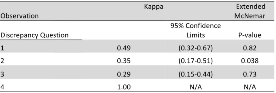
indicating a systematic difference between the 1st and 2nd replicate responses. For the midline questions, all intra-participant reliabilities were considered at least moderate strength of
agreement except the second and third questions which were considered ‘fair’ (See Table 5). The second midline question had statistically significant discordance (P=0.02).
Univariate Analysis
The majority of participants (67.8%) reported using a Digital Single Lens Reflex (DSLR) camera for photographic records. Point and shoot cameras were also used, while 1.1% of
most commonly used by participants at 44.4% and 30.3% respectively. Interestingly, 31.3% of participants used mirrors for their buccal photographs while the remainder did not.
Although orthodontic practices have different protocols for record-taking, most commonly, a dental assistant was exclusively responsible for exposing photographs (38.0%) while 2% of participants indicated that it may either be the orthodontist, dental assistant or treatment coordinator taking photos (See Table 7).
The frequency of record taking and the number of records taken also varied between participants (See Table 7). Over 77% of participants indicated that they took initial and final records while only 1.95% took photographs at every appointment. Similar numbers of
participants indicated that they exposed the standard frontal/buccal/occlusal views at initial and final records (64.6% and 65.1% respectively), while much fewer indicated taking photographs during active treatment (29.6%).
When asked which types of clinical records participants used for treatment planning, participants reported that clinical notes, photographs, followed by dental casts were used (See Figure 1). When the three types of records were ranked (See Figure 2), participants placed the greatest importance on clinical notes, followed by photographs and ranked dental casts the lowest. Interestingly, in the ‘most important’ category, dental casts were ranked higher than
photographs. Despite being ranked second most-important, when asked specifically to rank the importance of photographs for treatment planning on a visual analog scale, 47.32% indicated that photographs were 100% important and 26.7% reported that they were 75% important for
treatment planning (See Figure 3).
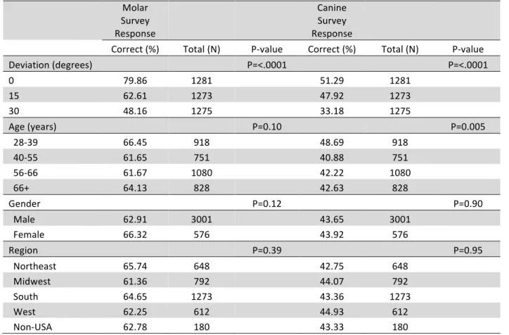
Using bivariate analysis, we found that neither gender nor region significantly influenced the likelihood of a correct response, while deviation angle resulted in a statistically significant difference (P=<.0001) for all buccal questions (See Table 8). This finding was confirmed using a conditional logistical regression model assessing the influence of deviation while controlling for the remaining factors, age, sex and region (See Table 9). The odds ratios show that as the camera angle deviated away from ideal, the likelihood of a correct response decreased. For the molar questions, participants were 3.21 and 7.18 times more likely to have a correct response at ideal angulation versus 15 degrees and 30 degrees respectively. For the canine questions, participants were 1.23 and 3.07 times more likely to have a correct response at ideal angulation versus 15 degrees and 30 degrees respectively. For the frontal questions, participants were 1.60 and 2.20 times more likely to have a correct response at ideal angulation versus 4 degrees and 8 degrees respectively
Although age was statistically significant for correct identification of canine classification, with younger orthodontists scoring better than the other age categories, a
conditional logistical regression indicated that age was not a significant factor when angulation was controlled for.
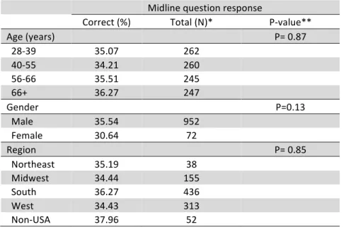
The frontal question responses were similar to the buccal questions. Using bivariate analysis, neither age, sex nor region significantly influenced the likelihood of a correct response, while deviation angle resulted in a statistically significant difference (P=<.0001) for the midline questions (See Table 10). Camera angle deviation was found to be the only statistically
Discussion
Demographics
Survey distribution through the AAO Partners in Research program (n=2300) resulted in a response rate of 8.96% with 206 respondents. The response rate is similar to other studies that used the Partners in Research program15-17. The subjects were divided to four regions of the United States plus non-US subjects for a total of five regions (See Table 1). The majority of respondents were from the South, while the minority were from outside of the US. This may be explained by motivation bias due to the increased likelihood that Southern orthodontists may have gone to the University of North Carolina and felt a connection with the program.
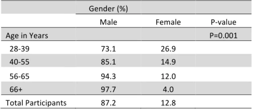
Interestingly, a trend of increasing numbers of female orthodontists was noted as age decreases (See Table 2). It has been well documented that orthodontic programs in North America are enrolling an increasing number of female residents18.
Buccal questions
As mentioned previously, two blocks of questions were equally, but randomly distributed amongst the participants: buccal and frontal question blocks.
For the buccal questions, all intra-participant reliabilities were considered to be of at least moderate strength of agreement except the third canine question which was considered ‘fair’ (See Table 4). This was likely due to the Angle classification of the dental cast appearing to be very close to the midpoint between two measurements, for example between class I and ¼ cusp class II. This borderline judgment could have made the determination of the correct
The third and last molar questions had statistically significant discordance, meaning that there was a systematic shift in their responses to the replicate questions. This may have meant that the question was unclear or that again, it was simply a challenging question, and it was difficult to make a judgment.
Based on our results, we found that as camera angulation increased from 0 to 15 degrees and again from 15 to 30 degrees anterior to ideal, there were statistically significant differences in the respondent's ability to correctly identify molar or canine Angle classification (See Figure 4). In addition to being statistically significant, this finding is also clinically significant. With as little as 15 degrees of camera deviation, the extent of the malocclusion may be incorrectly categorized in such a way as to influence the treatment modality, biomechanics, or extraction pattern chosen for a particular case.
The explanation for this observation is known as the ‘parallax effect’, and in some ways it is very similar to the ‘buccal object rule’ (BOR)19. The BOR is used when interpreting
radiographs to determine whether an object is buccal or lingual to a point of reference. In this case, when explaining the parallax effect using the BOR, the mandibular dentition was
considered lingual to the maxillary dentition. When assessing the molar and canine classification, the mandibular reference points were lingual to the maxillary reference points. When the camera lens, instead of the radiographic tube, was positioned at 15 or 30 degrees anterior to ideal, it had the visual effect of making the mandibular dentition appear more anterior relative to the
Referring to the odds ratio for the buccal questions, it was interesting to note that participants found it much more difficult to correctly determine molar versus canine
classification as camera angulation increased (See Table 9). This may be explained by variations in molar anatomy and lack of defined reference points as compared to canine cusp tips or an increased overjet in the molar region.
As we expected, there was no relation between the proportion of correct responses and different subgroups including age, gender and region that were not overwhelmingly explained by camera angulation.
Frontal questions
Our results showed that the frontal questions were also acceptably reliable. For the frontal questions, all intra-participant reliabilities were considered of at least moderate strength of
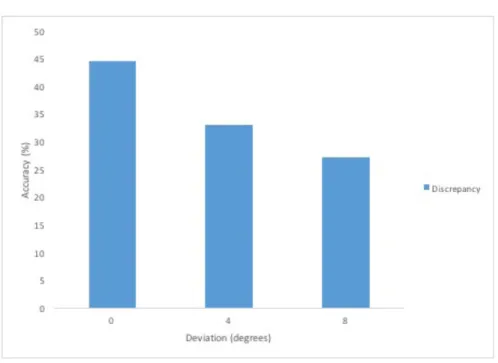
Based on our results, we found that as camera angulation increased from 0 to 4 degrees and again from 4 to 8 degrees away from ideal, there were statistically significant differences in the respondent’s ability to correctly identify the amount of midline discrepancy (See Figure 5). The odds ratio for the midline questions also confirmed this trend (See Table 11). In addition to being statistically significant, it is also clinically significant in that misinterpreting the degree midline discrepancy may influence the treatment modality, biomechanics or extraction pattern chosen for a particular case. Like the buccal questions, this may also be explained by the parallax effect
As we expected, there was no relation between the proportion of correct responses and different subgroups including age, gender and region that were not overwhelmingly explained by camera angulation.
When comparing the buccal versus the frontal questions, it was apparent that the
participants answered the buccal questions to a much higher level of accuracy. When the camera angulation was ideal for the buccal questions, the participants were 79.9% and 51.3% correct for the molar and canine questions respectively. At ideal angulation, participants were 44.3% correct for midline discrepancy. This demonstrates that even at ideal camera angulation, the overall accuracy is not particularly high. This finding indicates that photographs alone may be
insufficient for correct diagnosis and should be supplemented with clinical notes or dental casts.
One area where poor intraoral photography may have clinical implications is with new start-up orthodontic clear aligner companies that offer a do-it-yourself service. These companies, such as Smile Care Club®20 offer individuals a kit and instructions on how to take their own dental impressions. Before being sent the kit, the individual is instructed to send photographs taken with a cell phone or digital camera to the company to be assessed for treatment
complexity. It would be interesting to analyze the quality of these intraoral images taken by non-professionals.
Trends in intraoral photography
In the past several decades, there have also been changes in photographic
two most popular amongst respondents being Canon (44.4%) and Nikon (30.3%). Both of these manufacturers have affordable, high-quality DSLR cameras that are user-friendly.
Only 31.1% of participants used buccal mirrors for their photographs. This may be tied to our previous finding that molar classification is more difficult to determine than canine and may be an indication that orthodontists should be using buccal mirrors more frequently. Using a buccal mirror may help ensure that the photograph is captured at ideal angulation and the entire first molar is captured. Buccal mirror use may be less common due to the need for additional armamentarium, additional staff required and resultant expenses.
We also analyzed the frequency at which participants were taking photographs during treatment. The majority took initial and final records which are important for medicolegal reasons as well as diagnosis and treatment planning, however it was interesting to note how many people took additional series of photos during treatment.
Taking additional photos mid-treatment may be due to a case being particularly difficult or a major change in the treatment plan such as going from non-extraction to extraction. It may also indicate that a participant more highly values having a more complete photographic record to monitor case progression. It is interesting to note that 2% of participants take photos at every visit, similar to some residency programs. These respondents likely place a very high value on photos and would want to ensure that they are done to a high standard.
office were responsible. This may show that in the first group, the respondent was more
concerned with the quality of the records and has specifically trained one person to take photos.
In the cases where the orthodontist solely took the photos, this likely shows that they place a high value on the accuracy of their clinical photographs and do not want to delegate the task to any other staff members. The respondent may have also been a newer graduate or a part of a smaller start-up practice where there is less staff delegation. Additionally, having exposed the photos themselves, the orthodontist may have a better sense of how good an image is versus if they looked at an image someone else took; they are most aware how far they are able to retract and how they position the camera relative to the patient.
We also examined how the participants used their photographic records relative to other common records such as clinical notes and dental casts. When asked which type of record they primarily used for treatment planning, over 80% reported using mostly clinical notes and photographs while only about 55% use dental casts (See Figure 1). The decreased use of dental casts may be due to the redundancy of already having the information from clinical and
photographic records. It may also be explained by practices are doing more one-step starts and not having the time or need to pour a model.
When the three types of records were ranked from most to least important, we found that clinical notes were most important, photographs were second most and dental casts were least important (See Figure 2). Although clinical notes were ranked most important, photographs were still a key supplement for diagnosis.
finding may be linked to practitioners only taking casts on select patients. Patients that have impressions taken may be more likely to be complicated cases so the diagnostic information yield from dental casts is more crucial on those select patients. These types of more difficult cases may be surgical patients, borderline extraction cases, adults or cases with TMD or missing multiple posterior teeth.
Conclusions
1. Molar and canine classification is most accurately assessed at ideal camera angulation; as angulation becomes less ideal, accuracy decreases significantly.
• This can negatively impact a clinician’s ability to correctly diagnose at as little as
15 degrees anterior to ideal.
2. Amount of midline discrepancy is most accurately assessed at ideal camera angulation; as camera angulation becomes less ideal, accuracy decreases significantly.
• This can negatively impact a clinician’s ability to correctly diagnose at as little as
Table 1. Distribution of Participants by Region and Gender
Region N (%)
Northeast 21 10.9
Midwest 37 19.3
South 78 40.6
West 45 23.4
Non-USA 11 5.7
Gender
Male 172 87.3
Female 25 12.7
Table 1 displays the regional and gender distribution of the participants.
Table 2. Participant Gender by Age Group
Gender (%)
Male Female P-value
Age in Years P=0.001
28-39 73.1 26.9
40-55 85.1 14.9
56-65 94.3 12.0
66+ 97.7 4.0
Total Participants 87.2 12.8
Table 3. Participant Demographics by Region
Region N
(%) P-value
Characteristic Northeast Midwest South West Non-USA Total Number of
participants 21 (10.9) 37 (19.3) 78 (40.6) 45 (23.4) 11 (5.7) (100.0) 192
Age in Years P=0.05
28-39 1 (0.5) 9 (4.7) 21 (11.0) 13 (6.8) 6 (3.1) 50 (26.2) 40-55 4 (2.1) 4 (2.1) 20 (10.5) 14 (7.3) 2 (1.1) 44 (23.0) 56-65 8 (4.2) 16 (8.3) 19 (10.0) 8 (4.2) 2 (1.1) 53 (27.8) 66+ 8 (4.2) 8 (4.2) 17 (8.9) 10 (5.2) 1 (0.5) 44 (23.0)
Gender P=0.45
Table 4. Buccal Question Block Intra-observer Reliability
Observation Kappa Extended McNemar
Molar Question 95% Confidence Limits P-value
1 0.48 (0.23-0.73) 0.61
2 0.57 (0.43-0.71) 0.44
3 0.56 (0.40-0.73) 0.02
4 0.49 (0.34-0.64) 0.05
Canine Question
1 0.97 N/A 0.75
2 0.57 (0.43-0.71) 1.00
3 0.24 (-0.16-0.64) 0.57
4 0.56 (0.38-0.74) 0.12
Table 4 displays concordance with a weighted Kappa and simple Kappa for Molar 3. Kappa could not be calculated given the near-perfect agreement. Percent raw agreement is shown instead.
Table 5. Frontal Question Block Intraobserver Reliability Observation
Kappa Extended
McNemar
Discrepancy Question 95% Confidence Limits P-value
1 0.49 (0.32-0.67) 0.82
2 0.35 (0.17-0.51) 0.038
3 0.29 (0.15-0.44) 0.73
Table 6. Distribution of Photographic Armamentarium
Camera Type (%)
DSLR 67.8
Point and Shoot 31.1
Film 1.1
Brand
Canon 44.4
Nikon 30.3
Other 12.3
Pentax 9.0
Sony 2.8
Panasonic 1.1
Buccal Mirror
No 68.9
Yes 31.1
Table 7. Distribution of Photographic Protocols
Frequency of Photographic Records (%)
Initial and Final 77.07
One Series Mid-treatment 13.17 2-5 Series Mid-treatment 17.07
Every Appointment 1.95
Photographer (%)
Dental Assistant 38.30
Other 21.80
Only Orthodontist 14.60
DA or TC* 8.70
Orthodontist/DA/TC 2.00
Table 8. Accuracy of Angle Classification by Degree of Deviation and Demographics Molar Survey Response Canine Survey Response
Correct (%) Total (N) P-value Correct (%) Total (N) P-value Deviation (degrees) P=<.0001 P=<.0001
0 79.86 1281 51.29 1281
15 62.61 1273 47.92 1273
30 48.16 1275 33.18 1275
Age (years) P=0.10 P=0.005
28-39 66.45 918 48.69 918
40-55 61.65 751 40.88 751
56-66 61.67 1080 42.22 1080
66+ 64.13 828 42.63 828
Gender P=0.12 P=0.90
Male 62.91 3001 43.65 3001
Female 66.32 576 43.92 576
Region P=0.39 P=0.95
Northeast 65.74 648 42.75 648
Midwest 61.36 792 44.07 792
South 64.65 1273 43.36 1273
West 62.25 612 44.93 612
Non-USA 62.78 180 43.33 180
Table 9. Odds Ratio of Buccal Questions for Deviation Molar question
responses Canine question responses
Variable Odds ratio 95% confidence interval Odds ratio 95% confidence interval
Deviation (degrees)
0 vs 15 3.21 (2.59-3.98) 1.23 (1.01-1.51)
0 vs 30 7.18 (5.58-8.97) 3.07 (2.50-3.78)
38
Table 10. Accuracy of Frontal Responses by Degree of Deviation and Demographics
Midline question response
Correct (%) Total (N)* P-value**
Age (years) P= 0.87
28-39 35.07 262
40-55 34.21 260
56-66 35.51 245
66+ 36.27 247
Gender P=0.13
Male 35.54 952
Female 30.64 72
Region P= 0.85
Northeast 35.19 38
Midwest 34.44 155
South 36.27 436
West 34.43 313
Non-USA 37.96 52
Table 11. Odds Ratio of Frontal Questions for Deviation Midline question response
Variable Odds ratio 95% confidence interval Deviation (degrees)
Figure 1. Records Primarily Used for Orthodontic Treatment Planning
Figure 3. Treatment Time (in months) and Number of Treatment Appointments
REFERENCES
1. Galante DL. History and current use of clinical photography in orthodontics. J Calif Dent Assoc. 2009;37(3):173-174.
2. Wander P. Dental photography in record keeping and litigation. Br Dent J. 2014;216(4):207-208. doi: 10.1038/sj.bdj.2014.141 [doi].
3. Goodlin R. Photographic-assisted diagnosis and treatment planning. Dent Clin North Am. 2011;55(2):211-27, vii. doi: 10.1016/j.cden.2011.02.001 [doi].
4. Clinical practice guidelines for orthodontics and dentofacial orthopedics.
https://www.aaoinfo.org/system/files/media/documents/2014%20Cllinical%20Practice%20Guid elines.pdf. Updated 2016. Accessed 01/18, 2015.
5. Sandler J, Murray A. Clinical photographs--the gold standard. J Orthod. 2002;29(2):158-161. doi: 10.1093/ortho/29.2.158 [doi].
6. Proffit w, Fields h, Sarver d. Contemporary orthodontics. 5e ed. Elsevier; 2012. 7. American board of orthodontics: Ideal photographs and radiographs.
https://www.americanboardortho.com/media/1206/example-photos-radiographs.pdf. Updated 2016. Accessed 01/18, 2015.
8. Sandler J, Murray A. Recent developments in clinical photography. Br J Orthod. 1999;26(4):269-272.
9. Rischen RJ, Breuning KH, Bronkhorst EM, Kuijpers-Jagtman AM. Records needed for orthodontic diagnosis and treatment planning: A systematic review. PLoS One.
2013;8(11):e74186. doi: 10.1371/journal.pone.0074186 [doi].
10. Mandall NA. Are photographic records reliable for orthodontic screening? J Orthod. 2002;29(2):125-127. doi: 10.1093/ortho/29.2.125 [doi].
11. McKeown HF, Murray AM, Sandler PJ. How to avoid common errors in clinical photography. J Orthod. 2005;32(1):43-54. doi: 32/1/43 [pii].
12. Sandler J, Dwyer J, Kokich V, et al. Quality of clinical photographs taken by orthodontists, professional photographers, and orthodontic auxiliaries. Am J Orthod Dentofacial Orthop. 2009;135(5):657-662. doi: 10.1016/j.ajodo.2007.04.038 [doi].
14. Hallgren KA. Computing inter-rater reliability for observational data: An overview and tutorial. Tutor Quant Methods Psychol. 2012;8(1):23-34.
15. Sturgill J, Park JH. Changes in orthodontists' retirement planning and practice operations due to the recent recession. J Clin Orthod. 2015;49(4):240-248.
16. Neill CC, Migliorati C, Trojan T, et al. Experience and expertise regarding orthodontic management of childhood and adolescent cancer survivors. Am J Orthod Dentofacial Orthop. 2015;148(5):765-770. doi: 10.1016/j.ajodo.2015.05.027 [doi].
17. conaway D, Kim K, Ueno H. The attitudes, awareness and perceptions of orthodontists with regards to orthodontic literature on interceptive treatment of class ii malocclusions in the mixed dentition. [MSc]. St. Louis University; 2015.
18. Blasius JJ, Pae EK. Work-pattern differences between male and female orthodontists. Am J Orthod Dentofacial Orthop. 2005;128(3):283-90; discussion 290-1. doi: S0889-5406(05)00671-2 [pii].
19. Gutmann JL, Endo C. Clark's rule vis a vis the buccal object rule: Its evolution & application in endodontics. J Hist Dent. 2011;59(1):12-15.