CELLULAR MECHANISMS OF IMMUNE AND HEMATOPOIETIC DYSFUNCTION FOLLOWING RADIATION AND BURN INJURIES
Brandon Michael Lee Linz
A dissertation submitted to the faculty at The University of North Carolina at Chapel Hill in partial fulfillment of the requirements for the degree of Doctor in Philosophy in the Department
of Microbiology and Immunology.
Chapel Hill 2016
ABSTRACT
Brandon Michael Lee Linz: Cellular Mechanisms of Immune and Hematopoietic Dysfunction Following Radiation and Burn Injuries
(Under the direction of Bruce A. Cairns and Robert Maile)
The immune system has evolved to protect the body against damage from infection, disease, or injury. Severe injuries, such as large burns or radiation exposures, induce profound immune dysfunctions at the cellular and humoral levels that heighten the body’s susceptibility to infections. Despite progress made toward reducing the consequences of burn shock, translocation of intestinal bacteria and wound and pulmonary infections leading to sepsis are still major causes of mortality following a traumatic injury. Following a severe burn or radiation injury, the body must respond rapidly to activate or produce new immune cells to challenge the insult and to restore homeostasis also while preventing any bacteria from establishing an infection. Therefore, to improve patient outcomes, it is important to understand not only the immune but also the hematopoietic responses to injury and infection.
NLRP12 is a member of the NLR family of proteins that are responsible for coordinating inflammatory responses upon recognition of invading pathogens and damage signals. Mutations in human NLRP12 have been linked to atopic dermatitis and hereditary periodic fevers with skin, however the mechanisms by which NLRP12 affects these conditions remain to be fully
animals induced the stem cells responsible for the bulk of myeloid cell production to undergo apoptosis. This defect in repopulation of the peripheral immune system lead all Nlrp12 knockout animals to quickly succumb to an infectious challenge, thus highlighting the importance of Nlrp12 in responding to infection or injury.
Following a radiation-thermal combined injury, wild type myeloid progenitor cells underwent apoptosis at a low level. Administration of the glycoprotein granulocyte-monocyte colony stimulating factor was evaluated as a therapeutic to stimulate stem cell maturation and production of myeloid cells following injury. Treatment resulted in increased myeloid cell production: including increases in platelets, red blood cells, immature monocytes, dendritic cells, neutrophils, and macrophages. Notably, platelets and monocytes displayed increased function, in turn decreasing mortality and response to an infectious challenge. The innate immune response was then assessed early after only burn injury. Burn mice were susceptible to an early wound infection with Pseudomonas aeruginosa as shown with increased mortality and systemic bacterial colonization. The defective bacterial clearance was associated with a neutrophil anti-inflammatory polarization phenotype (N2; IL-10+ IL-12-). This work expands on our
ACKNOWLEDGEMENTS
A PhD is a labor of love and like anything it’s better when you do not have to go at it alone. I would like to generously acknowledge all members of the Burn Basic Science Research Lab. Bruce Cairns and Rob Maile have been great mentors, allowing access to wealths of
knowledge and guidance when the science gets you down. My fellow graduate students in crime – CJ Neely, Laurel Kartchner, and Julia Dunn– life could not have been possible without the
daily moral support from you. Rebecca Drapp, Sha’Leema Miller, Steven Mouro, and Lindsey Glenn thank you for all your invaluable technical support. April Mendoza, Amal Khoury, and CJ – I would like to thank you for helping me get this project off the ground and establishing this
beast of a model. The collaboration with the laboratory of Jenny Ting: Jenny, Aga Truax and June Brickey, your steady and wonderful supply of knock-out mice was extraordinary and bountiful and just as helpful as your advice. I would like to thank my committee for their sage advice and thorough scientific knowledge as well as our fruitful discussions. The
Microbiology/Immunology administrative staff, without whom everything would fall apart. Next I would like to thank my family: my parents, Rhonda and Stewart Linz; and my siblings,
TABLE OF CONTENTS
LIST OF FIGURES xiii
LIST OF ABBREVIATIONS xv
CHAPTER 1: INTRODUCTION 1
1.1Burn Injury 1
1.2Grading of burn wounds 1
1.3Acute radiation exposure 2
1.4Radiation-thermal combined injury 3
1.5Systemic response to burn injury 3
1.6Nucleotide-binding oligomerization domain family of receptors 4
1.7NLRP12 6
1.8NFkB signaling 6
1.9Tumor necrosis factor 7
1.10 Hematopoieis following injury 8
1.11 Pulmonary Pseudomonas aeruginosa infections 9
1.12 Immune response to infection 10
1.13 Granulocyte-monocyte colony stimulating factor 10
1.14 Platelets 11
1.15 Neutrophils 11
1.16 Macrophages 12
1.18 Toll-like receptors 13
1.19 Remaining questions 14
CHAPTER 2: INNATE IMMUNE CELL RECOVERY IS POSITIVELY
REGULATED BY NLRP12 DURING EMERGENCY HEMATOPOESIS 15
2.1 Summary 15
2.2 Introduction 15
2.3 Methods and Materials 18
Mice and Combined Irradiation and Burn Injury procedure 18
Quantitative RT-PCR 18
Histology 19
Pseudomonas aeruginosa infection 19
Serum Collection and Cytokine ELISA 19
Flow Cytometry 19
Intracellular Staining and Phospho-flow 20
TNF-Depletion 20
Statistical Analysis 20
2.4 Results 20
NLRP12 limits morbidity and mortality following RCI 20 Splenic and pulmonary immune repopulation is impaired following
RCI in Nlrp12-/- mice 21
Nlrp12-/- mice show decreased bone marrow and peripheral
cell numbers following RCI 22
Defects in myelopoiesis following RCI are not observed in
inflammasome-deficient animals 23
Nlrp12-/-mice display increased serum TNF, IL-6 and IL-12
IκBa activity is increased in CD34+ cells Nlrp12-/- animals after RCI 24
RCI of Nlrp12-/- animals leads to increased granulocyte/monocyte
progenitor apoptosis 25
Anti-TNF antibody administration prevents NLRP12-associated
GMP apoptosis after combined injury 26
Nlrp12-/- mice lack control of pulmonary infection following
radiation-thermal combined injury 27
2.5 Conclusions 28
CHAPTER 3: DELETION OF NLRP12 AN IMPARES INNATE IMMUNE REPONSES DURING RADIATION-THERMAL
COMBINED INJURY AND SHOCK 41
3.1 Summary 41
3.2 Introduction 41
3.3 Methods and Materials 43
Mice and Combined Irradiation and Burn Injury procedure 43
Murine Chimeras 43
Pseudomonas aeruginosa infection 44
Serum Collection and Cytokine ELISA 44
Flow Cytometry 44
Intracellular Staining 45
TNF-Induced Shock 45
LPS- and PolyIC-induced shock 45
Statistical Analysis 45
3.4 Results 46
Adoptive transfer of chimera bone marrow (Wt:Nlrp12-/-) reveals
NLRP12 deficiency leads to higher TNFR1 but not TNFR2 on
GMPs after RCI 47
RCI-NLRP12 Have Greater M1 Balance and More Polarization following Infection AND NLRP12 Controls TLR Expression
following RCI on iMos 47
Wild type animals are less susceptible to TNF-shock compared
to Nlrp12-/- animals 48
LPS, but not PolyIC, induces granulocyte monocyte
progenitor expansion 49
3.5 Conclusions 49
CHAPTER 4: GM-CSF TREATMENT FOLLOWING RADIATION- THERMAL COMBINED INJURY IMPROVES PLATELETS AND
MONOCYTE RECOVERY AND FUNCTION 56
4.1 Summary 56
4.2 Introduction 57
4.3 Methods and Materials 58
Mice and combined irradiation and burn injury procedure 58
GM-CSF administration 59
Pseudomonas aeruginosa infection 59
Serum collection and cytokine quantification 59
Flow cytometry 59
Statistical analysis 60
4.4 Results 60
GM-CSF increases survival and weight following RCI 60 GM-CSF increases granulocyte-monocyte progenitor numbers
following RCI 61
Progenitor increase is attributed to decreased apoptosis of
Peripheral innate immune cells and platelets expand following
GM-CSF treatment after RCI 62
Platelets display normalized phenotype following GM-CSF
treatment and RCI 62
GM-CSF treated animals show a reduction in serum IL-6 and
an increase in IL-10 64
GM-CSF treatment improves systemic infection responses to
Pseudomonas aeruginosa 64
4.5 Discussion 65
CHAPTER 5: FLAGELLIN TREATMENT PREVENTS INCREASED SUSCEPTIBILITY TO SYSTEMIC BACTERIAL INFECTION AFTER
INJURY BY INHIBITING IL-10+ IL-12- NEUTROPHIL POLARIZATION 74
5.1 Summary 74
5.2 Introduction 75
5.3 Methods and Materials 78
Animals 78
Mouse Burn Injury 78
Bacterial strains and preparation 79
Animal infections 79
Determination of bacterial infection 79
CD11b+ cell enrichment 80
In vitro stimulation 80
Flow cytometric analysis 80
Serum cytokine analysis 81
Statistical analysis 81
Burn mice, but not sham mice, developed a systemic infection
following wound inoculation with wildtype P. aeruginosa 81 Innate cell populations had altered TLR expression with the
combination of burn injury and infection 82
Infection following burn injury resulted in a systemic increase
in IL-10 83
Infected burn mice had an increased polarization of neutrophils,
but not macrophages, into an IL-10+ IL-12− phenotype 83 Increased resistance of burn mice to systemic infection with an
attenuated strain (ΔCyaB) P. aeruginosa correlated with reduced
N2 polarization of neutrophils 85
Treatment of mice with flagellin after burn injury enhanced
clearance of wildtype P. aeruginosa 86
5.5 Discussion 87
CHAPTER 6: CONCLUSIONS AND FUTURE DIRECTIONS 98
6.1 A clinical need 98
6.2 Cellular mechanisms of immune dysfunction following
radiation-thermal combined injury in the absence of NLRP12 99 6.3 Future directions: Characterization of NLRP12 impact on
canonical NFκB induction of apoptosis 101
6.4 Rescue of platelet and myeloid populations after
radiation-thermal combined injury 102
6.5 Future directions: Investigate contribution of platelets to
radiation-thermal combined injury 103
6.6 Cellular mechanisms of increased susceptibility to early
wound infection after thermal injury 103
6.7 Future directions: characterization of neutrophil response
early after burn injury 106
6.8 Closing remarks 107
LIST OF FIGURES
Figure 2.1 NLRP12 expression is increased after combined injury, and acts
to limits mortality and weight loss 30
Figure 2.2 NLRP12 regulates peripheral immune repopulation after
combined injury 31
Figure 2.3 NLRP12 regulates pulmonary immune repopulation and bone
marrow cell numbers after combined injury 32
Figure 2.4 NLRP12 deficiency does not result in macrophage, B or T cell
changes after injury 33
Figure 2.5 Defects in myelopoiesis following RCI are not observed in
inflammasome-deficient animals 34
Figure 2.6 Nlrp12-/- animals have increased serum cytokine and bone marrow
receptor expression following combined injury 35
Figure 2.7 NLRP12 regulates serum cytokines 36
Figure 2.8 Nlrp12-/- animals have increased granulocyte/monocyte progenitor
apoptosis after combined injury 37
Figure 2.9 NLRP12 deficiency results in no changes in lymphoid progenitors 38
Figure 2.10 Anti-TNF antibody administration prevents NLRP12-associated
GMP apoptosis after combined injury 39
Figure 2.11 Nlrp12-/- mice lack control of pulmonary infection following
combined injury 40
Figure 3.1 NLRP12 acts intrinsically to granulocyte monocyte progenitor
apoptosis signaling in bone marrow chimeras 51
Figure 3.2 TNFR2 expression, but not TNFR1, is increased on GMPs after injury 52
Figure 3.3 Nlrp12-/- show greater monocyte inflammation after injury 53
Figure 3.4 A model of TNF-shock replicates granulocyte monocyte progenitor
Figure 3.5 LPS treatment, but not PolyIC, reduces granulocyte monocyte
progenitor numbers 55
Figure 4.1 GM-CSF treatment increases temperature, survival and weight
following RCI 67
Figure 4.2 GM-CSF administration increases progenitor cell numbers
following RCI 68
Figure 4.3 GM-CSF reduces progenitor apoptosis following RCI 69
Figure 4.4 GM-CSF increases circulating platelet, neutrophil, and monocyte
numbers after RCI 70
Figure 4.5 GM-CSF increases platelet function after RCI 71
Figure 4.6 GM-CSF treatment reduces IL-6 and increases IL-10 after RCI 72
Figure 4.7 GM-CSF mice show greater bacterial clearance than vehicle treated 73
Figure 5.1 Burn mice, but not sham mice, exhibit dose-dependent mortality and develop a systemic infection following a P. aeruginosa
wound inoculation 92
Figure 5.2 TLR expression is decreased on splenic neutrophils and Ly6G+ CD11b+ myeloid cells, but not macrophages, after burn injury
with infection 93
Figure 5.3 Burn mice, but not sham mice, mount a robust serum IL-10
response after P. aeruginosa wound inoculation 94 Figure 5.4 Infected burn mice have a higher percentage of IL-10+ neutrophils and
a lower percentage of IL-12+ neutrophils, dendritic cells, and
macrophages than infected sham mice 95
Figure 5.5 Reduced bacterial load at distal organs following wound inoculation with an attenuated P. aeruginosa strain (ΔCyaB) is associated with an increased serum IL-12 and pro-inflammatory neutrophil
(N1; IL-10−IL-12+) response in burn mice 96
Figure 5.6 Administration of flagellin at burn resuscitation and prior to wound infection with wildtype P. aeruginosa (PAK) reduces bacterial load in the periphery and increases the percentage of IL-12
LIST OF ABBREVIATIONS
ASC: apoptosis-associated speck-like protein containing a CARD domain
ATP: adenosine triphosphate
C-terminal: carboxy-terminal
cAMP: cyclic adenosine 3’,5’-monophosphate
CAPS: Cryopyrin-Associated Periodic Syndromes
CARD: caspase activation and recruitment domain
CATERPILLER: caspase activation and recruitment domains [CARD], transcription enhancer, R [purine]-binding, lots of leucine repeats
CDX: e.g. cluster of differentiation 3, cluster of differentiation 40
CD40L: CD40 ligand
CLP: common lymphoid progenitor
DAMP: damage associated molecular pattern
DC: Dendritic cell
FCAS: familial cold auto-inflammatory syndrome
GCSF: granulocyte colony stimulating factor
GM-CSF: granulocyte monocyte colony stimulating factor
Hsp70: heat shock protein 70
Hsp90: heat shock 90
IFNα/β/γ: interferon alpha/beta/gamma
IL-X: e.g. interleukin 1-beta, interleukin-2, interleukin-18
IL1ra: interleukin 1 receptor-a
IκB: inhibitor of NFκB
IKK: inhibitor of NFκB kinase
IRAK1: interleukin-1 receptor-associated kinase
LPS: lipopolysaccharide
LRR: leucine-rich repeats
MAPK: mitogen-activated protein kinase
MEP: megakaryocyte erythrocyte progenitor
N-terminal: amino-terminal
NBD: nucleotide bind domain
NFκB: nuclear factor kappa B
NIK: NFκB inducing kinase
NK: Natural killer
NLRC4: nucleotide binding domain leucine-rich repeat CARD protein 4
NLRP12: nucleotide binding domain leucine-rich repeat containing a pyrin domain 12
PRR: pattern recognition receptor
TAK1: transforming growth factor beta-activated kinase 1
TCR: T cell receptor
TGFβ: tumor growth factor-beta
TLRX: e.g. Toll-like receptor-2, Toll-like receptor-4
TNF: tumor necrosis factor
Wt: Wild type
CHAPTER 1: INTRODUCTION 1.1Burn injury
Burn injuries occur when a hot liquid (scald), solid (contact burn), or flame are exposed to a tissue and cause subtotal or total destruction of the cells present in the skin or below. Burns can also be caused by exposure to radiation sources, electricity, and caustic chemicals
(Gabrielsen, 2003). In 2007, there were 11 million cases of burns that required medical
treatment, making it the fourth most common injury world-wide ("World Health Organization. The Global Burden of Disease: 2004 Update," 2008). In the US alone, there were 450,000 emergency room visits and 3,500 deaths from burn injuries in 2010 (Miller et al., 2008). The rapid onset of burn shock following injury necessitates immediate, specialized care to reduce morbidity and mortality. Aggressive fluid resuscitation, regulation of body temperature, analgesics, wound debridement, wound excision and closure, and preventative infection are measures in which certified American Burn Associated burn centers specialize (Association, 2015). The cost of treatment at one of these centers can be greater than $200,000 (Association, 2015). However, this does not include expenses from rehabilitation, occupational and physical therapies, and any chronic complications.
1.2Grading of burn wounds
blisters that will often heal and scar over within 5 weeks (Roth & Hughes, 2015). A third-degree or full thickness burn extends through all layers of the skin and into the subcutaneous tissue. Since this depth includes the basement membrane and progenitor cells responsible for dermal maintenance and cell production, treatment of these wounds requires excision of the damaged tissue and a skin-graft to close the now open wound. Lastly, a fourth-degree burn involves other organ tissue below the skin and soft tissue, such as muscle, connective tissue, and bone. Burn injuries are also measured by size or area of injury (Gabrielsen, 2003). The percent total body surface area (TBSA) of an adult patient is typically assessed by the rule of nines where most areas of the body (i.e. an arm, back and front of the legs, half of the chest or back) can be
assumed to be approximately 9% of the total body surface area of the adult. The overall burn size and depth help to guide fluid resuscitation and wound management within the burn unit (Roth & Hughes, 2015).
1.3Acute radiation exposure
Exposure to high amounts of ionizing radiation can lead to numerous cellular and
systemic damages. Ionization of DNA from radiation exposure leads to decreased ability for cells to divide due to ionization-induced mutation of key factors involved in cell division (Coleman, Stone, Moulder, & Pellmar, 2004). This elimination of rapidly dividing cells – namely
gastrointestinal, and hematopoietic distress (Chua et al., 2012; Gaugler, 2014). The apoptosis of the majority of hematopoietic progenitor cells leads to severe leukopenia and subsequent
increased susceptibility to infections (Chua et al., 2012; Heslet, Bay, & Nepper-Christensen, 2012; Heylmann, Rödel, Kindler, & Kaina, 2014).
1.4Radiation-thermal combined injury
Greater than 30%, and predicted to potentially be as high as 65-70%, of accidental or incidental exposures to high doses of radiation are coupled with a secondary burn injury either from the heat from the primary radiation exposure source or from a secondary fire created by an industrial accident or incidental exposures (Fushiki, 2013; Hasegawa et al., 2015; Shaw, 2014). Patients who receive a radiation-thermal combined injury (RCI) undergo physiologic changes and display cytokine profiles consistent with burn shock, but uniquely show immune and hematopoietic cell destruction consistent with total body radiation injury ablation of rapidly dividing cells (Cherry, Williams, O'Banion, & Olschowka, 2013; Chua et al., 2012; Coleman et al., 2004; Hasegawa et al., 2015). This combination of unique injuries leads to unique challenges for medical interventions that have been utilized for the individual injuries which may be
counter-indicated in RCI in the absence of research into RCI-relevant treatments (Basile et al., 2012; Browne, 2013). Despite no to low numbers of cases each year, the heightened security concerns of today coupled with the lack of clinically proven treatments necessitate inquiry. 1.5Systemic response to burn injury
magnitude of immune impairment is proportional to the size of burn (Baker et al., 1979). The massive release of cellular debris and damage associated molecular patterns (DAMPs) trigger a systemic immune response (Finnerty, Przkora, Herndon, & Jeschke, 2009). This massive release of inflammatory mediators from the wound and other tissues is believed to impact and/or trigger multi-organ dysfunction (Santaniello et al., 2004). Following the initial systemic inflammatory response, there is also long-term immune suppression demonstrated by prolonged allograft skin survival on burn wounds (Lagus, Sarlomo-Rikala, Böhling, & Vuola, 2013; Mowlavi, Andrews, Milner, Herndon, & Heggers, 2000). Since burn injury impairs all parts of the immune system – hematopoietic, innate, and adaptive systems – patients are extremely susceptible to infection (Church, Elsayed, Reid, Winston, & Lindsay, 2006; Manson, Pernot, Fidler, Sauer, & Klasen, 1992).
1.6Nucleotide-binding oligomerization domain family of receptors
Pattern recognition receptors (PRR) play a key role in regulating acute and chronic innate immune responses to tissue damage or infection (Broz & Monack, 2013). In addition to toll-like receptors (TLRs), the more recently discovered family of intracellular receptors, the nucleotide-binding oligomerization domain-like receptors (NLRs), have been found to not only cooperate with TLRs, but also both positively and negatively regulate inflammatory responses, initiate enzymatic cleavage of cytokines, as well as regulate apoptotic responses (Duran, Alvarez-Mon, & Valero, 2014). The majority of study has focused on a key members’ function as
adaptor protein apoptosis-associated speck-like protein containing a c-terminal caspase
recruitment domain (ASC) (Franklin et al., 2014; Hara et al., 2013). Following binding to ASC, the inchoate inflammasome will recruit either caspase-1 or caspase-11 (Aachoui et al., 2013; Guey, Bodnar, Manié, Tardivel, & Petrilli, 2014; Hagar, Powell, Aachoui, Ernst, & Miao, 2013; Kuida et al., 1995). Then, utilizing either its own caspase activation and recruitment domain (CARD) or the CARD adaptor on ASC, will oligomerize into a heptamer. This now active inflammasome will then begin to catalytically cleave pro-interleukin (IL)-1β (IL-1β) and pro-IL-18 into their active forms (Aachoui et al., 2013; Groß et al., 2012; Guey et al., 2014; Hagar et al., 2013; Kuida et al., 1995; Pilla et al., 2014). Active IL-1β is a pleiotropic cytokine that initiates cellular proliferation, cytokine production and secretion, induction of proptosis, and an anti-viral state(Ali, Karin, & Nizet, 2015); whereas IL-18 serves to increase interferon-γ (IFNγ) production by natural killer cells (NK cells) and T cells (Takeda et al., 1998; Wong, Muthuswamy, &
Kalinski, 2012). In addition to the inflammasome subfamily, other NLRs have been primarily found to be involved in regulation of innate immune responses, most significantly through regulation of the NFκB signaling cascade proteins: NOD1 and NOD2 have been found to
activate the kinase RIP2 that in turn activates IκB kinase, which in turn leads to NFκB activation (Caruso, Warner, Inohara, & Núñez, 2014; Lich & Ting, 2007); NLRX1 amplifies NFκB
signaling and JNK signaling by increasing production of radical oxygen species (Irving C Allen et al., 2011; Tattoli et al., 2008); NLRP12 has been shown to interact with heat shock protein 90 (Hsp90) and in turn with NFκB inhibitory kinase (NIK), this complex then in turn suppresses NFκB signaling (Arthur, Lich, Aziz, Kotb, & Ting, 2007; Krauss et al., 2015; Vitale et al., 2013;
1.7NLRP12
Mutations in Nlrp12, formerly known as Monarch-1 or Pypaf-7, have been shown to be associated with familial cold auto-inflammatory syndrome (FCAS), an extremely rare autosomal dominant disease that results in recurrent fever and skin urticarial due to cold conditions (Lich & Ting, 2007; Liu et al., 2013; Vitale et al., 2013; Xia et al., 2016). NLRP12 is expressed in innate immune cells (specifically monocytes and macrophages), intestinal cells, bone and bone marrow cells, as well as liver cells (Lech, Avila-Ferrufino, Skuginna, Susanti, & Anders, 2010). This is achieved by inducing proteasome-mediated degradation of NF-κB inducing kinase (NIK) in response to pathogens and activation through pro-inflammatory receptors (Arthur et al., 2007; Lord et al., 2009; Zaki et al., 2011). NLRP12 is stabilized by interaction with Hsp90, thus allowing for suppression of NFκB inhibitory kinase (NIK) (Arthur et al., 2007; Ataide et al., 2014; Vladimer et al., 2012). Because NLRP12 functions to dampen these signals, it is clear that NLRP12 must be controlled in order to mount an adequate cellular response to such insults. However, NLRP12 has also been found to act as an inflammasome by oligomerizing with ASC and Caspase-1 during Yersinia pestis and malaria infections and mediate cleavage of IL-18 into its active form (Ataide et al., 2014; Vladimer et al., 2012). Additionally, molecular analysis reveals that in the absence of NLRP12, dendritic cells display an inappropriate activation of NIK, resulting in high levels of NIK dependent gene expression (Arthur et al., 2010; Krauss et al., 2015). Taken together, NLRP12 function within the immune system has yet to be fully explained.
1.8NFkB signaling
caused by reactive oxygen species (ROS), lipopolysaccharides (LPS) from Graham – bacteria, interleukin-1β (IL-1β), ionizing radiation, and tumor necrosis factor (TNF) (Van Antwerp, Martin, Kafri, Green, & Verma, 1996; Ward-Kavanagh, Lin, Šedý, & Ware). Canonically, NFkB signaling cannot be achieved without activation by IkB kinase (IKK) composed of the two subunits IKKα and IKKβ in addition to the master regulatory IKKγ. IKK phosphorylates IkB, altering its quaternary structure and allowing ubiquitination and subsequent destruction through the proteasome (S.-C. Sun, 2011). Non-canonical signaling begins when the lympotoxin β-receptor (LTβR), BAFF, or RANK activates NFkB inducing NIK (NIK) allowing IKKA to cleave p100 into the mature p52 subunit (S.-C. Sun, 2011).
1.9Tumor necrosis factor
Following tissue damage, infection, or trauma, initial responses are initiated by the cytokine tumor necrosis factor (TNF, also known as TNFα) (Beg & Baltimore, 1996; Peschon et al., 1998). TNF can be secreted by macrophages, monocytes, CD4+ T cells, neutrophils, mast cells, and eosinophils (Schindler et al., 1990). Primarily, TNF acts as a pyrogen, mediates acute liver responses to damage, acts as a chemoattractant for neutrophils, stimulates increased
macrophages phagocytosis, and pro-inflammatory cytokine production and expression (Kapas et al., 1992). High concentrations of TNF induce shock-like symptoms, with prolonged exposure resulting in cachexia, as typified by latent tuberculosis infections (Croft, Benedict, & Ware, 2013; Di Paolo et al., 2016; Hayden & Ghosh, 2014). Moreover, high serum or tissue
2014). Following prolonged, high concentrations of serum TNF, leukocytes will downregulate their expression of TNFR2 and maintain or increase expression of TNFR1 (Wicovsky et al., 2009). Prolonged signaling through TNFR1 on myeloid and myeloid progenitor cells will causes the cell to no longer signal through TNF receptor associated factor 2 (TRAF2) and (nuclear factor k B) NFκB signaling cascade, but rather to recruit TNFRSF1A-Associated via Death Domain (TRADD) and fas-associated protein with Death Domain (FADD) which in turn recruits the cysteine protease procaspase 8 (Micheau & Tschopp, 2003; Van Antwerp et al., 1996). This recruitment will activate proteolytically cleave caspase 3 into its active form. From which, the now active caspase 3 and 9 will lead to Bid-associated apoptosis (Cai et al., 2014; Croft et al., 2013; Di Paolo et al., 2016; Hayden & Ghosh, 2014; Micheau & Tschopp, 2003; Wei et al., 2014; Zhao et al., 2012).
1.10 Hematopoiesis following injury
blood, spleen, lungs, and liver – is a cell type that by its cell surface protein expression (Ly6G+Ly6C+CD11b+) resembles myeloid derived suppressor cells (MSDCs) (Carter et al., 2013; Mendoza et al., 2012; Palmer et al., 2013). However, upon further study, the cells promote, albeit weakly, T cell proliferation and IFNγ production (Mendoza et al., 2012). We have identified these cells as inflammatory monocytes and have shown that they are vital to responding to an infectious challenge after injury (Mendoza et al., 2012).
1.11 Pulmonary Pseudomonas aeruginosa infections
A major cause of mortality for patients who survive the initial shock of a burn injury are pulmonary bacterial infections. Prolonged ICU stays and ventilation increase a patient’s risk of developing a pulmonary infection, often termed ventilator-associated pneumonia (VAP). VAP typically occurs when a patient is mechanically ventilated for more than 48 hours (Chastre & Fagon, 2007; Fabian, 2000; Hollaar et al., 2016; Shorr, Sherner, Jackson, & Kollef, 2005). The Gram negative saprophyte, Pseudomonas aeruginosa, is one of the most common infectious agents in United States’ burn centers due to its ability to survive on many hospital surfaces (Lyczak, Cannon, & Pier, 2000). While the innate immune system of a healthy adult is able to easily clear P. aeruginosa infections, immunocompromised hosts are more susceptible to infection and mortality (Chitkara & Feierabend, 1980; Lavoie, Wangdi, & Kazmierczak, 2011; Lyczak et al., 2000). During infection, P. aeruginosa will downregulate synthesis of the motility protein flagellin, the major component of bacterial flagellum. This allows the bacterium to avoid detection by host Toll-like receptor 5 (TLR5), thus limiting humoral and cellular immune
1.12 Immune response to infection
The immune system is the body’s defense against infection and disease. It detects a wide variety of antigens derived from invading pathogens and distinguishes them from the host’s own tissue. The immune response can be divided into innate and adaptive immunity. The innate immune system is non-specific, meaning it recognizes and responds to pathogens in a generic way. More specifically, it depends upon germline-encoded receptors (e.g. TLRs) to recognize features that are common to many microbes (Duran et al., 2014). Most pathogens are detected and destroyed within minutes to hours of invasion by innate immune cells, which includes the neutrophils and macrophages. However, if a pathogen persists, the adaptive immune response ensues (Angus & Van der Poll, 2013; Baker et al., 1979). The adaptive immune system is
specific and consists of T and B lymphocytes. It targets a precise pathogen by utilizing pathogen-specific receptors, such as T cell receptors (TCR), which are acquired during the lifetime of the host (Ohkura et al., 2012). Induction of an adaptive immune response leads to immunological memory, which ensures a more rapid and effective response on subsequent encounters with the same pathogen (McHeyzer-Williams, Okitsu, Wang, & McHeyzer-Williams, 2012; Mueller, Gebhardt, Carbone, & Heath, 2013).
1.13 Granulocyte-monocyte colony stimulating factor
to stimulating stem cell function, GM-CSF inhibits neutrophil migration, increases reactive oxygen species production, and acts as an embryokine. Administration of GM-CSF to mice and humans results in the production of leukocytes following chemotherapy to prevent neutropenia (Dragon, Saffar, Shan, & Gounni, 2008; Dugan et al., 2002; Gardner et al., 2014; J. G. Noel et al., 2005; J. G. Noel et al., 2002; Reeves, 2014). Additionally, GM-CSF has been used clinically under the name Sargramostin as a therapeutic for inflammatory bowel disease, leukemia, and acute lung injury (Campo et al., 2012; J. B. Cohen et al., 2015; Danese, 2012).
1.14 Platelets
Small fragments of megakaryocyte cytoplasm, platelets, or thrombocytes, play important roles in primary and secondary hemostasis as crucial steps of the coagulation cascade occur on their cell surfaces (Hess et al., 2014; Vieira-de-Abreu, Campbell, Weyrich, & Zimmerman, 2012). Despite their primary function as hemostatic regulators, platelets can also act as inflammatory cells through the release of cytokines, chemokines, expression of
pro-inflammatory surface markers, interactions with leukocytes and endothelial cells, and release of inflammatory mediators through degranulation (Vieira-de-Abreu et al., 2012).
1.15 Neutrophils
phagocytosis, activated and recruited neutrophils will generate a variety of toxic byproducts to help destroy invading pathogens through an oxidative burst. Degranulation of secretory vesicles called granules releases cytotoxic molecules to aid in bacterial killing (Borregaard, 1997). One study showed that neutrophils have decreased Fc receptor mediated phagocytosis, as well as a 50% reduction in intracellular killing, after burn injury (Adediran et al., 2010; Bjerknes,
Vindenes, & Laerum, 1989). This group also showed that the ability of circulating neutrophils to undergo oxidative burst gradually declines during the first two weeks after burn injury. Another study reported that there is an increased number of neutrophils in the peritoneal cavity and an increase in neutrophil oxidative burst at one day after burn (Bjerknes et al., 1989; L. W. Chen, Huang, Lee, Hsu, & Lu, 2006). Neutrophils have also been reported to be immunosuppressive after burn injury as demonstrated by their secretion of IL-10, a potent anti-inflammatory
cytokine, upon TLR2 stimulation (S. W. Jones et al., 2013; G. Noel et al., 2010; Greg Noel et al., 2011).
1.16 Macrophages
Macrophages are capable of phagocytosis, cytokine production, oxidative bursts, and antigen presentation (Fujiwara & Kobayashi, 2005). With their ability to rapidly respond to infection as circulating monocytes recruited to tissues, or as resident macrophages, these cells play central roles in the initiation and resolution of injury and infection. Antigen presentation consists of the macrophages internally digesting pathogens or antigens and processing these proteins for presentation as fragments on MHC-complexes, to in turn. Upon activation,
macrophages can produce significant amounts of IL-1β, IL-6, and transforming growth factor-β (TFGβ) (Fujiwara & Kobayashi, 2005; J. G. Noel et al., 2005). Macrophage activation represents
interferon-gamma (IFN-γ) compared to anti-inflammatory macrophages (M2) that produce IL-10. This shift toward an M2 phenotype is achieved by exposure to IL-10, glucocorticoids, or TLR ligation (N. Wang, Liang, & Zen, 2015). Macrophage hyperactivity and shifts in
macrophage polarization toward M2 phenotypes have been suggested as a cause of increased bacterial susceptibility following injury (Greg Noel et al., 2011; J. G. Noel et al., 2005). 1.17 Dendritic cells
Similar to macrophages, dendritic cells (DCs) are capable of both phagocytosis and antigen presentation. The primary function of DCs are to bridge innate immune cell activation and responses to adaptive and humoral efforts (Auffray, Sieweke, & Geissmann, 2009; Savina & Amigorena, 2007). DCs achieve this by presenting foreign antigens to stimulate T cell activation directly as well as through secretion of a variety of cytokines, namely IL-12 (Auffray et al., 2009). A significant reduction in the total number of DCs have been reported after burn injury, specifically myeloid DCs (mDCs) and plasmacytoid DCs (pDCs); this reduction is directly correlated with burn size and depth.
1.18 Toll-like receptors
TLRs are PRRs that respond to pathogen associated molecular patterns (PAMPs) and endogenous stress signals termed danger associated molecular patterns (DAMPs) (Matzinger, 2012; O'Neill, Golenbock, & Bowie, 2013). TLRs, expressed by both immune and non-immune cells, are activated by binding to their specific ligand: TLR2 binds membrane bound
an inflammatory response (Buechler, Teal, Elkon, & Hamerman, 2013; L. W. Chen et al., 2006; Duran et al., 2014; Lee, Avalos, & Ploegh, 2012; Moore et al., 2007; O'Neill et al., 2013; Paterson et al., 2003). Both in vitro and in vivo studies have shown that TLR2 and TLR4 responses are heightened between 1-7 days after burn injury (B. A. Cairns, C. M. Barnes, S. Mlot, A. A. Meyer, & R. Maile, 2008). Upon TLR stimulation, macrophages, dendritic cells, and γδ T cells from burn mice have increased cytokine production compared to non-burn controls
(Neely et al., 2011). The precise mechanism responsible for TLR hyper-responsiveness after burn injury is unknown. However, there is evidence to suggest that increased cell surface expression of TLR proteins and increased phosphorylation of p38 MAP kinase, a component of the TLR signaling cascade, both contribute (B. A. Cairns et al., 2008; Hagar et al., 2013; Moresco, LaVine, & Beutler, 2011).
1.19 Remaining questions
CHAPTER 2: INNATE IMMUNE CELL RECOVERY IS POSITIVELY REGULATED BY NLRP12 DURING EMERGENCY HEMATOPOESIS1
2.1 Summary
With enhanced concerns of terrorist attacks, dual exposure to radiation and thermal combined injury (RCI) has become a real threat with devastating immunosuppression. NLRP12, a member of the NOD-like receptor family, is expressed in myeloid and bone marrow cells and has been implicated as a checkpoint regulator of inflammatory cytokines as well as an
inflammasome activator. We show that NLRP12 has a profound impact on hematopoietic recovery during RCI by serving as a checkpoint of TNF signaling and preventing hematopoietic apoptosis. Using a mouse model of RCI, increased NLRP12 expression was detected in target tissues. Nlrp12-/- mice exhibited significantly greater mortality, inability to fight bacterial infection, heightened levels of pro-inflammatory cytokines, overt granulocyte/monocyte progenitor cell apoptosis and failure to reconstitute peripheral myeloid populations. Anti-TNF antibody administration improved peripheral immune recovery. These data suggest that NLRP12 is essential for survival after RCI by regulating myelopoiesis and immune reconstitution.
2.2 Introduction
The hematopoietic system is capable of rapidly increasing myeloid cell production in response to tissue damage and is critical for wound healing and infection clearance (Baldridge, King, & Goodell, 2011; Dugan et al., 2002; Gardner et al., 2014; Manz & Boettcher, 2014; J. G. Noel et al., 2005; J. G. Noel et al., 2002; Santangelo et al., 2000; Serafini et al., 2007;
Kinsky, Lin, Herndon, & Sherwood, 2003; H. Zhang et al., 2010). While the factors that initiate emergency myelopoiesis are not fully elucidated, it is generally accepted that emergency
myelopoiesis is tightly coupled with cytokine and growth factor production, namely TNF and IL-6, and is mediated by NF-κB and other immune regulatory transcription factors.
Rare mutations in Nlrp12, a nucleotide-binding leucine rich repeat and pyrin domain-containing receptor (NLR, also known as NOD-like receptor), have been associated with periodic fevers in humans although the association needs to be further studied. Nonsense and splice mutations within human-Nlrp12 have been shown to diminish suppression of NF-κB signaling (Jeru et al., 2008), however some variants do not exhibit such activity but are associated with modestly enhanced or more rapid inflammasome activation (Borghini et al., 2011). The different functions observed with NLRP12 may be consistent with NLRP12
exhibiting an inflamamsome function in certain infections (Ataide et al., 2014; Vladimer et al., 2012) but not other infections or inflammatory conditions (Zaki, Man, Vogel, Lamkanfi, & Kanneganti, 2014). While the pyrin-domain containing members of the NLR family have largely been studied in the context of the inflammasome (Wen, Miao, & Ting, 2013), there is growing evidence that a few play an important role in regulating inflammatory signaling. Some NLR proteins have been shown to be positive regulators of NF-κB, while NLRP12 has been
The cytokines that regulate hematopoietic stem cell (HSC) function, such as IFNα/β, IFNγ, IL-12, and TNF, are tightly controlled elements of cell expansion. Type I IFNs and TNF,
induced by TLR signaling, can act upon myeloid progenitors to promote the expansion of granulocyte/monocyte progenitors (GMP), leading to systemic myeloid expansion (Buechler et al., 2013). Alternatively, excessive TNF signaling reduces myelopoiesis by inducing caspase-3/caspase-8-dependent progenitor cell apoptosis (Wei et al., 2014). Excessive TNF, TLR signaling, and deficiencies in negative regulation of NF-κB lead to apoptosis of HSCs and defects in myeloid progenitor function (Buechler et al., 2013; Stein & Baldwin, 2013).
We and others have shown that burn and radiation injuries lead to increased susceptibility to infection within survivors (Dugan et al., 2002; Mendoza et al., 2012; J. G. Noel et al., 2005). This is a pressing clinical problem in the face of nuclear accidents and possible incorporation of nuclear materials within explosives. This susceptibility has been attributed to a loss of
inflammatory regulation, incomplete immune restoration and a systemic anti-inflammatory response following sepsis and shock (S. W. Jones et al., 2013; Neely et al., 2014; Neely et al., 2011). Following a radiation-thermal combined injury (RCI), an immature monocyte population (iMo) rapidly expands and predominates the periphery (Mendoza et al., 2012). Using this model, we observed that TNF is significantly increased in RCI compared to burn, radiation, and sham alone.
2.3 Methods and Materials
Mice and Combined Irradiation and Burn Injury procedure
The Nlrp12-/-,Caspase1/11-/-, Asc-/- and IL-1Ra-/-mouse strains have been described (Arthur et al., 2007; Hirsch, Irikura, Paul, & Hirsh, 1996; Kuida et al., 1995; Takeda et al., 1998). All experiments were conducted with female mice housed under SPF conditions that were age-matched and backcrossed for at least nine generations onto the C57BL/6 background. All studies were conducted in accordance with the IACUC guidelines of the University of North Carolina at Chapel Hill and NIH Guidelines for the Care and Use of Laboratory Animals. Our model of RCI has been previously described; briefly, mice received a subcutaneous injection of morphine (3mg/kg body weight) for pain control immediately before burn injury. A
full-thickness contact burn of 20% total body surface area (TBSA) was produced and within 1 hour, mice received a 5Gy (dose rate of 0.98 Gy/min) whole-body dose of ionizing radiation and were maintained on oral morphine for the duration of the experiment. Sham controls with 0% TBSA underwent all described interventions except for the burn and γ-irradiation exposure.
Quantitative RT-PCR
RNA was extracted from organ homogenates, suspended in TRIzol and isolated according to the manufacturer’s protocol (Life Technologies, Carlsbad, CA). qPCR was
Histology
Mouse femurs were extracted and muscle and connect tissue were removed and initially preserved in 10% formalin. Femurs were then decalcified with Immunocal, water washed, and paraffin infused. Following sectioning and processing, sections were then stained by
hematoxylin and eosin. Samples were processed using ImageJ to determine area of cell loss within each femur.
Pseudomonas aeruginosa infection
A wildtype strain (PAK) of P. aeruginosa was obtained from M. Wolfgang (University of North Carolina, Chapel Hill, NC). 106 bacteria were then aerosolized intratracheally as described previously.
Serum Collection and Cytokine ELISA
Animals underwent a submandibular bleed and systemic cytokines were measured by single-plex ELISA (eBioscience, CA, USA or Biolegend, CA, USA) according to the
manufacturer’s instructions or by Cytokine Mouse 20-Plex Panel (Life Technologies, Carlsbad,
CA) on Luminex Bead Array technology. Flow Cytometry
cKit-BUV395, FcγR-BV605, CD34-Alexa647, and Annexin V-Pacific Blue. In each case, a million cells per organ were used for flow cytometric analysis.
Intracellular Staining and Phospho-flow
Intracellular staining was performed using a BD Bioscience Cytofix/Cytoperm kit. Antibodies used were TNF-PE (BD Biosciences), phosphor-p65 S528 (BD Biosciences), IκBa S32/536-eFlour 660 (eBiosciences), p38 ST180/Y182-PE and phospho-IKKα/β S176/180-PE (Cell Signaling Technologies). In each case, a million cells per organ were
used for flow cytometric analysis. TNF-Depletion
Immediately following combined irradiation and burn injury, mice were given 25mg/kg of rat IgG1, kappa anti-mouse TNF, clone MP6-XT3 or ratIgG1 isotype control (eBioscience, CA, USA) intraperitoneally dissolved in PBS (Sigma, CA, USA).
Statistical Analysis
Analysis was carried out with Prism 7.0 for Windows. All data are presented as the mean +/- standard error of the mean (SEM). Complex data sets were analyzed by analysis of variance (ANOVA) with a Tukey-Kramer post-test HSD for multiple comparisons. Single data points were assessed by the Student’s two-tailed t test. The product limit method of Kaplan-Meier was utilized for generating the survival curves, which were compared using the log rank test. A p value less than 0.05 was considered statistically significant for all data sets.
2.4 Results
NLRP12 limits morbidity and mortality following RCI
2010; Moore et al., 2007; Vladimer et al., 2012; Zaki et al., 2014; Zaki et al., 2011). We therefore investigated whether NLRP12 was acting to limit excessive inflammatory signaling and consequently promote peripheral immune reconstitution in our model of emergency myelopoiesis.
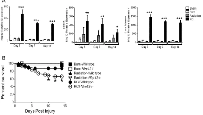
Wild type and Nlrp12-/- mice received a 20% TBSA burn and were irradiated with 5-Gy of γ-irradiation within an hour of burn injury. In wild type mice, we observed elevated NLRP12 expression in spleen, bone marrow and lung tissues early (3, 7, and 14 days post-injury) after RCI (Figure 2.1A) compared to burn or radiation alone and sham controls. Mortality among NLRP12-deficient mice was significantly elevated following RCI, but not following burn or radiation alone (Figure 2.1B). While RCI-wild type animals lost weight initially, they were able to return to a baseline weight by seven days after injury and exceed their baseline weight by 14 days post injury; RCI-Nrlp12-/- animals lost more weight and did not fully recover weight in comparison to wild type animals (Figure 2.1C). These data suggest that NLRP12 protected against morbidity after RCI.
Splenic and pulmonary immune repopulation is impaired following RCI in Nlrp12-/- mice
During events that induce enhanced myelopoiesis and inflammation, specifically RCI, we have shown that immature monocytes with high granularity comprise the majority of the
the repopulation of lung immune cells—common sites of opportunistic infection in burn
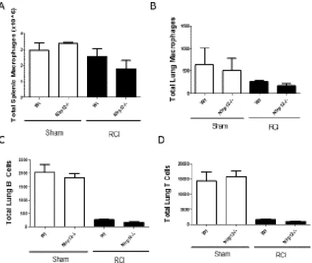
patients—after RCI. Nlrp12-/- mice displayed a reduced ability to repopulate the lung after RCI. This inability was characterized by a decrease in total CD45+ leukocytes and by the absence of the immature monocyte accumulation normally observed following RCI at two weeks post-injury (Figure 2.3A-B). There were no differences in macrophage (CD11b+ Ly6C+Ly6GloF4/80hi) accumulation in Nlrp12-/- mice when compared to wild type (Figure 2.4A). The total number of pulmonary macrophages, B and T cells were similar in Nlrp12-/-and wild type mice (Figure 2.4B-D). These data implicate a role for NLRP12 in regulating emergency hematopoiesis following RCI.
Nlrp12-/- mice show decreased bone marrow and peripheral cell numbers following RCI NLRP12 has been shown to be expressed constitutively in bone marrow cells (Arthur et al., 2010; Savic et al., 2012; Vitale et al., 2013; Zaki et al., 2014). We hypothesized that reduced immune repopulation in the periphery of injured Nlrp12-/- mice was due to reduced cell
generation and output by the bone marrow. To test this, we investigated the impact of NLRP12 deficiency on bone marrow populations after RCI. Nlrp12-/- mice had reduced total bone marrow cells compared to wild type mice after RCI. We also observed a decrease in total iMO and neutrophils (Figure 2.3C) within the bone marrow of Nlrp12-/- mice as early as seven days post
injury compared to wild type mice. Additionally, we observe decreases in the total numbers of monocytes and neutrophils in the peripheral blood (Figure 2.3D). These data suggest that
Defects in myelopoiesis following RCI are not observed in inflammasome-deficient animals
NLRP12 is also found to form an inflammasome complex or regulate caspase-1 activity (I. C. Allen et al., 2012; Vladimer et al., 2012; Ye et al., 2008) and regulates IL-1β processing by complexing with ASC during infection with Yersinia or malaria (Vladimer et al., 2012). To examine whether NLRP12 is important for inflammasome activation following RCI, we assessed IL-1β levels after RCI in wild type or Nlrp12-/- mice. There were no detectable levels of IL-1β at any time point measured (Figure 2.5A) nor was than any differences in serum IL-18 (data not shown). Due to inability to capture IL-1β levels in serum because of its high turnover, we examined the role of the inflammasome in RCI. We applied the RCI model to various mice strains lacking key components of genes encoding proteins that encode common shared
components of the inflammasome. These include Caspase1/11-/- which lacks both canonical and noncanonical inflammasome caspases, Asc-/- which lacks the common adaptor shared by
multiple inflammasome NLRs and AIM2, or Il1r-/- which lacks the IL-1 receptor protein.
Following RCI, Caspase1/11-/-, Asc-/- and Il1r-/- mice had a similar immune repopulation in the lung and spleen as wild type mice (Figure 2.5B-C). We also saw no significant differences in bone marrow populations in Caspase-1/11-/-, Asc-/- or Il1r-/- mice following RCI (Figure 2.5D). Additionally, injured Caspase-1/11-/-, Asc-/- or Il1r-/- animals did not show an increase in
mortality compared to wild type (Figure 2.5E). Together, these results suggest that NLRP12 controls myelopoiesis in an inflammasome-independent pathway.
Nlrp12-/-mice display increased serum TNF, IL-6 and IL-12 cytokine and bone marrow TNF-
receptor expression
NLRP12-dependent production of selected cytokines and their receptors after RCI. In wild type animals, serum TNF expression increased early following injury and declined over time (Mendoza et al., 2012). In sham-treated Nlrp12-/-animals, the TNF level was similar to sham wildtype controls. However, during RCI in Nlrp12-/-animals, the TNF increased initially and was maintained over time – a significant elevation when compared to wild type mice (Figure 2.6A). In addition, Nlrp12-/- bone marrow cells displayed increased TNFR expression (Figure 2.6B) as well as CD40 and RANK (Figure 2.6C). Using intracellular straining, we observed that monocyte production of TNF after RCI is increased when compared to burn and radiation controls; however, in the absence of NLRP12, monocyte production of TNF is significantly elevated compared to wild type controls (Figure 2.6D). As well as TNF, other cytokines such as IL-6, IL-12, IFNα, and IFNγ were increased in Nlrp12-/- mice compared to wild type mice but less so than TNF (Figure
2.6A). This elevation is potentially derived from the initial shock and the selective apoptotic environment induced by the absence of NLRP12 and necessary myelopoiesis. Heightened levels of IL-6 following trauma have been shown to be the major predictor of poor outcome (bacterial infection) following a traumatic injury (Gebhard, Pfetsch, Steinbach, & Strecker, 2000). Other cytokines and growth factors (IL-4, IL-10, and GM-CSF) were measured but showed no significant differences between wild type and Nlrp12-/- animals (Figure 2.6B).
IκBa activity is increased in CD34+ cells Nlrp12-/- animals after RCI
Both the canonical and non-canonical pathways of NFκB have been shown to be
Increased phosphorylation is indicative of increased canonical NFκB signaling in the absence of NLRP12. However, pIKKα/β and the downstream p38/MAPK showed no changes in activity when comparing wild type to Nlrp12-/- injured animals. Taken with the increased TNFR expression on marrow cells, these results suggest that NLRP12 is negatively regulating the canonical NFκB signaling cascade.
RCI of Nlrp12-/- animals leads to increased granulocyte/monocyte progenitor apoptosis
We observed that NLRP12 regulates reconstitution of granulocytic and monocytic bone marrow and peripheral cells in Nlrp12-/- mice following RCI. We therefore hypothesized that NLRP12 regulates bone marrow granulocyte/monocyte progenitors (GMP), the source of granulocytes and immature monocytes. To test this, we utilized flow cytometric analysis to evaluate the number of GMP (Lin- IL7R- Sca1- cKit+ FcγRhi CD34+) in Nlrp12-/- mice following RCI.
We detected a similar number of bone marrow GMP in Nlrp12-/- and wild type mice at 3 days after injury. However, at 7 and 14 days after injury, wild type GMP expanded and increased in numbers while Nlrp12-/-GMP expansion was attenuated (Figure 2.8A). There were no
measured differences in lymphoid lineage progenitors (Figure 2.9A). We tested the hypothesis that the significant decrease in GMP in Nlrp12-/- mice is due to increased apoptosis. To
Increased apoptosis and decreased bone marrow cellularity was confirmed by histological staining. H&E femur sections were obtained at 14 days post injury. There were no histological changes from wild type to Nlrp12-/- mice in sham, burn, or radiation alone animals. However, RCI-Nlrp12-/- mice displayed medial patches of cell loss within the femurs, which was not present in RCI-wild type femurs (Figure 2.8D). Collectively, our findings imply that NLRP12 prevents progenitor cell apoptosis, thus allowing myelopoiesis and peripheral immune cell reconstitution to occur in wild type animals.
Leukopenia can have complex etiologies in both inflammatory and non-inflammatory conditions, many of which involve alterations in HSC steady-state hematopoiesis (T. D. Jones, Morris, Young, & Kehlet, 1993; Serafini et al., 2007; Toliver-Kinsky et al., 2003). Our data show that NLRP12 limits TNF following RCI, resulting in expansion of myeloid precursors and monocyte populations throughout the periphery. Previous studies showed increased
hematopoiesis following total body irradiation; however, our results are novel because we have shown that NLRP12 promotes hematopoiesis of specific lineages during RCI (Baldridge et al., 2011; Mendoza et al., 2012).
Anti-TNF antibody administration prevents NLRP12-associated GMP apoptosis after combined injury
After observing significantly elevated levels of TNF and reduced myelopoiesis in injured Nlrp12-/- mice, we hypothesized that increased levels of TNF were leading to pathology through TNF-mediated apoptosis of immune progenitor cells as seen in other models of excessive
TNF(Micheau & Tschopp, 2003). Specifically, we hypothesized that GMP were undergoing TNF-mediated apoptosis with reduced peripheral neutrophil and inflammatory monocyte
We observed significantly fewer GMP in the Nlrp12-/- mice given the isotype control compared to wild type mice. However, Nlrp12-/- mice given the anti-TNF antibody had similar numbers of GMP compared to isotype and anti-TNF treated wild type mice (Figure 2.9A). Additionally, the proportion of GMP actively undergoing apoptosis was higher in the Nlrp12-/- isotype treated animals compared to Nlrp12-/- mice treated with anti-TNF (Figure 2.9B). This is correlated with a decrease in the total CD45+ pulmonary cells as well as pulmonary iMO (Figure 2.9C-D). These data indicate that in the absence of NLRP12, TNF mediates the enhanced bone marrow death during RCI and resultant incomplete restoration of the peripheral immune system. Nlrp12-/- mice lack control of pulmonary infection following radiation-thermal combined injury
In a clinical setting, patients that are able to survive initial shock from a burn or radiation-thermal-combined injury will often succumb to a pulmonary infection associated with the
prolonged hospital stay (Moore et al., 2007). We sought to evaluate the role NLRP12 deficiency plays in a clinically relevant model of a lung infection following injury. Wild type and Nlrp12-/- animals were subjected to either sham, burn, or radiation only, and RCI. Mice were then
sustained for two weeks in individual housing wherein they were infected intratracheally with 1x10⁶ CFU of Pseudomonas aeruginosa (PAK).
We next sought to determine the immune response to infection following RCI. Nlrp12-/- mice showed a decrease in innate, pulmonary immune cell populations following RCI and infection (Figure 2.10C), leading us to conclude that NLRP12 results in increased hematopoietic recovery which is likely crucial to the effective control of infection after traumatic injury. 2.5 Conclusions
Our study demonstrates that NLRP12 suppresses TNF signaling in vivo during
inflammation-induced emergency myelopoiesis. Most importantly, our research indicates a role for NLRP12 in hematopoietic progenitor cells by limiting TNF-induced apoptosis of these cells. TNF inflammation initiated by RCI without NLRP12 leads to the apoptosis of progenitor cells and a defective peripheral immune reconstitution, associated with increased mortality and inability to control an infectious challenge.
part, by suppression of the non-canonical NFκB pathway and cross talk with the canonical pathway (I. C. Allen et al., 2012; Arthur et al., 2007; Lich & Ting, 2007; Lich et al., 2007). NLRP12-mediated NFκB suppression likely limits TNF and cellular death during inflammation and hematopoiesis. Our data in NLRP12-deficient mice shows compromised hematopoiesis due to enhanced TNF production, leading to flagrant HSC/GMP apoptosis. This lack of HSC
Figure 2.1. NLRP12 expression is increased after combined injury, and acts to limits mortality and
weight loss. Wildtype C57BL/6 mice were subjected to sham, 5Gy of γ-irradiation, a 20% total body
surface area burn or a combined injury (RCI). (A) mRNA was isolated from spleen, bone marrow, and
whole lung at 3, 7, and 14 days post injury. Relative Nlrp12 - expression was determined by qRT-PCR.
(n=6/timepoint). Wildtype C57BL/6 or Nlrp12-/- mice were subjected to sham, 5Gy of γ-irradiation, a
Figure 2.2. NLRP12 regulates peripheral immune repopulation after combined injury. Wildtype
C57BL/6 or Nlrp12-/- mice were subjected to sham or combined radiation and burn injury (RCI). Spleens
were harvested 3, 7 and 14 days post injury and the total number of (A) splenocytes, neutrophils (CD11b+
Ly6Cint Ly6G+ F4/80-) and immature monocytes (iMOs; CD11b+ Ly6C+ Ly6GhiF4/80hi) were quantified
Figure 2.3. NLRP12 regulates pulmonary immune repopulation and bone marrow cell numbers
after combined injury. Wildtype C57BL/6 or Nlrp12-/- mice were subjected to sham or combined
radiation and burn injury (RCI). Lungs were harvested 14 days post injury and the total number of (A)
CD45+ cells, (B) neutrophils (CD11b+ Ly6Cint Ly6G+ F4/80-) and immature monocytes (iMOs; CD11b+
Ly6C+ Ly6GhiF4/80hi) were quantified by flow cytometry analysis. Wildtype C57BL/6 or Nlrp12-/- mice
were subjected to sham or combined radiation and burn injury (RCI). Bone marrow from femurs and tibias and blood from a cheek bleed were harvested 3, 7 and 14 days post injury and the total number of
(C) bone marrow cells, immature monocytes (iMOs; CD11b+ Ly6C+ Ly6GhiF4/80hi), neutrophils
(CD11b+ Ly6Cint Ly6G+ F4/80-) from the bone marrow and platelets (CD62P+TER119-), monocyte and
Figure 2.4. NLRP12 deficiency does not result in macrophage, B or T cell changes after injury. Wildtype C57BL/6 or Nlrp12-/- mice were subjected to sham, burn, radiation, or
combined radiation and burn injury (RCI). Spleens were harvested at 14 days post injury and the total number of (A) splenic and (B) pulmonary macrophages (CD11b+ Ly6C+ Ly6G-F4/80hi), and (C) pulmonary B and (D) T cells were quantified by flow cytometry analysis. Data
Figure 2.5. Defects in myelopoiesis following RCI are not observed in inflammasome-deficient animals. Caspase1/11-/-, adaptor Asc-/-, or receptor Il1r-/- mouse strains were
Figure 2.6. Nlrp12-/- animals have increased serum cytokine and bone marrow receptor expression
following combined injury. Wildtype C57BL/6 or Nlrp12-/- mice were subjected to sham or combined
radiation and burn injury (RCI). The concentration of (A) TNF was quantified using ELISA in serum 3, 7, and 14 days post injury. We also analyzed mean fluorescent intensity of (B) TNFR, CD40, and RANK on bone marrow cells harvested at 14 days post injury using flow cytometry. (D) The percentage of TNF producing iMos was determined using intracellular staining and flow cytometry. (E) The level of phospho-IκBα, phospho-IKKα/β, phosphor-p65, and phospho-p38 was quantified using intracellular
staining and flow cytometry. Data represented as mean ± SEM, with statistical significance defined as**
Figure 2.8 Nlrp12-/- animals have increased granulocyte/monocyte progenitor apoptosis after
combined injury. Wildtype C57BL/6 or Nlrp12-/- mice were subjected to sham or combined radiation
and burn injury (RCI). Bone marrow was collected from wild type and Nlrp12-/- mice at 3, 7, and 14 days
post RCI or sham treatment (n= 6/group). Using flow cytometric analysis, (A) the total number of bone
marrow Granulocyte/Monocyte Progenitors (GMP, Lin- IL7R- Sca1- ckit+ FcγRhi CD34+) and (B) the
percentage of GMP cells undergoing apoptosis was determined by positive Annexin V staining in the
absence of 7-AAD- staining cells; representative flow staining from Nlrp12-/- mice is shown is shown in
(C). Data represented as mean ± SEM, with statistical significance defined *, p<0.05, **, p<0.005 and ***, p<0.001 by Student’s t test with n=5 mice per group. In separate experiments, wildtype C57BL/6 or
Figure 2.10. Anti-TNF antibody administration prevents NLRP12-associated GMP apoptosis after
combined injury. Wild type and Nlrp12-/- C57/BL6 mice received a single administration of anti-TNF
or isotype control antibody immediately following combined radiation and burn injury (RCI). We
harvested bone marrow and lung from these mice 14 days after injury. We quantified (A) the total number
of bone marrow Granulocyte/Monocyte Progenitors (GMP, Lin- IL7R- Sca1- ckit+ FcγRhi CD34+) and
(B) the percentage of GMP cells undergoing apoptosis by 7-AAD- Annexin V+ staining by flow cytometry. We measured (C) the total number of pulmonary CD45+ cells and immature monocytes
(iMOs; CD11b+ Ly6C+ Ly6GhiF4/80hi) by flow cytometry analysis. Data represented as mean ± SEM,
Figure 2.11 Nlrp12-/- mice lack control of pulmonary infection following combined injury. Wildtype
C57BL/6 or Nlrp12-/- mice were subjected to sham or combined radiation and burn injury (RCI). Mice
were inoculated 14 days post-injury intratracheally with 1x10⁶ CFU of Pseudomonas aeruginosa (PAK).
We quantified (A) survival, (B) bacterial load within lungs and liver by culture, and (C) number of
splenic CD45+ cells, neutrophils (CD11b+ Ly6Cint Ly6G+ F4/80-) and immature monocytes (iMOs;
CD11b+ Ly6C+ Ly6GhiF4/80hi) harvested two days after inoculation. Data represented as mean ± SEM,
CHAPTER 3: DELETION OF NLRP12 IMPARES INNATE IMMUNE REPONSES DURING RADIATION-THERMAL COMBINED INJURY AND SHOCK
3.1 Summary
During a traumatic event that induces emergency myelopoiesis, innate immune signaling helps to control how inflammatory cells respond and what new immune cells the hematopoietic system will produce. The severe immune impairment after a radiation thermal combined injury is exacerbated in the absence of NLRP12 as NLRP12 has been shown to play a role in limiting TNF-induced apoptosis of progenitors. Using a bone marrow chimera, a mixture of wild type and Nlrp12-/- cells, we show that NLRP12 acts intrinsically within the bone marrow progenitor population to directly limit induction of TNF and to limit TNF- and inflammation-induced apoptosis of progenitor cells after a radiation-thermal combined injury. Additionally, that excessive TNF signaling leads to further defects in innate immune signaling, by altering TLR signaling. Furthermore, TNF administration without further injury is sufficient to induce
progenitor apoptosis. Taken together, these data show the NLRP12 acts as an intrinsic regulator of innate inflammation with progenitors during emergency hematopoiesis.
3.2 Introduction
system after RCI – as shown in Chapter 1. This increased TNF and reduction of peripheral innate immune cells leads to an increased susceptibility to infection. Several open questions remain regarding NLRP12: Is NLRP12 acting within the hematopoietic compartment to limit
inflammation, or acting with peripheral immune cells to reduce TNF production? Is NLRP12 impacting other inflammatory pathways? Can the increased apoptosis of progenitors be seen in other models of shock?
Use of bone marrow chimeras has been an integral part of studying basic immunology using mouse models(Kuida et al., 1995; Onoe, Fernandes, & Good, 1980). This allows scientist to differentiate between affects non-hematologic cells play and those from the bone marrow compartment. More importantly, chimers allow to determine the affect an immune environment plays on two different cells (i.e. Wt and Nlrp12-/-progenitors) after injury.
TNFR1 ligation of TNF signals through p65/NFκB and induces production of pro-inflammatory cytokines like IL-6, pro-IL1β, chemoattractants, and further promotes cell
survival(Rothe, Pan, Henzel, Ayres, & Goeddel, 1995; Wicovsky et al., 2009). Contrastingly, the TNFR2 variant signals through the Fas-associated protein with a death domain (FADD) adaptor protein to engage Caspases 3 and 8 and causes the cell to undergo apoptosis(Rothe et al., 1995; F. Wang et al., 2006).
Given that NLRP12 suppresses apoptosis of GMPs during emergency hematopoiesis, we sought to determine NLRP12 impact on innate immune signaling and reconstitution using RCI and other models of shock. We demonstrate that NLRP12 works intrinsically within the bone marrow compartment and works to limit pro-inflammatory responses during an infection and that a model of TNF-shock can replicate the apoptosis phenotype seen in Nlrp12-/- animals after RCI.
3.3 Methods and Materials
Mice and Combined Irradiation and Burn Injury procedure
The Wt and Nlrp12-/- C57/B6 mouse strains have been described (Arthur et al., 2010; Honda et al., 2005; Kaisho & Akira, 2001). All experiments were conducted with female mice housed under SPF conditions that were age-matched and backcrossed for at least nine
generations onto the C57BL/6 background. All studies were conducted in accordance with the IACUC guidelines of the University of North Carolina at Chapel Hill and NIH Guidelines for the Care and Use of Laboratory Animals. Our model of RCI has been previously described; briefly, mice received a subcutaneous injection of morphine (3mg/kg body weight) for pain control immediately before burn injury. A full-thickness contact burn of 20% total body surface area (TBSA) was produced and within 1 hour, mice received a 5Gy (dose rate of 0.98 Gy/min) whole-body dose of ionizing radiation and were maintained on oral morphine for the duration of the experiment. Sham controls with 0% TBSA underwent all described interventions except for the burn and γ-irradiation exposure.
Murine Chimeras