Introduction
Elevated lead exposure in early life is associated with growth retardation, neuro-toxicity, impaired cognitive development in infancy, and deficits in attention and execu-tive function (Bellinger et al. 1986, 1987). Accumulating evidence also indicates that the effect of lead exposure in early childhood can change neurochemistry (Binns et al. 2007), cause neurobehavioral and cognitive deficits in later life (Finkelstein et al. 1998; Sanders et al. 2009; Winneke et al. 1983; Zahran et al. 2009), decrease brain volume, and increase the rate of criminal arrest in adult-hood (Cecil et al. 2008; Wright et al. 2008). Although the current actionable concentra-tion for lead is 5 μg/dL in the United States,
the threshold for its toxicity is unknown. Furthermore, the mechanisms by which lead exposure affects diverse neuropathological outcomes is not clearly defined, although epigenetic mechanisms have been proposed (Wright et al. 2010).
Environmental exposures to both physical and chemical agents, especially during early development, can induce alterations in DNA methylation that alter disease suscep-tibility in adulthood (Bernal et al. 2013; Dolinoy et al. 2006, 2007; Waterland and Jirtle 2003). Animal studies likewise suggest that epigenetic modifications may link lead exposure to neurotoxicity and attention deficit disorders (Faulk et al. 2013; Luo et al. 2014), but stable epigenetic targets
responsive to early lead exposure in humans remain uncharacterized.
Because of the relative ease of measuring genomic DNA cytosine methylation at CpG dinucleotides, this end point is the most commonly investigated epigenetic modifi-cation in epidemiologic studies. In vitro and in vivo studies demonstrate that DNA methyla tion is altered by exposure to toxic metals, including arsenic, cadmium, and lead (Bolin et al. 2006; Reichard et al. 2007; Takiguchi et al. 2003). The only epigenetic studies conducted in humans thus far have evaluated DNA methylation at Alu and LINE repeat elements. They showed associations between maternal patella lead levels and global DNA hypomethylation in newborns (Pilsner et al. 2009) and adult males (Wright et al. 2010). Although the biological signifi-cance of reduced DNA methylation at repetitive elements in lead-exposed humans is unknown, this epigenetic change also occurs frequently in cancer, where it is believed to result in chromosomal instability and genomic mutations (Wilson et al. 2007).
*These authors contributed equally to this work. Address correspondence to C. Hoyo, Department of Biological Sciences, Center for Human Health and the Environment (CHHE), Program of Epidemiology and Environmental Epigenomics, North Carolina State University (NCSU), Raleigh, NC 27695-7633 USA. Telephone: (919) 515-0540. E-mail: [email protected], or R.L. Jirtle, Department of Biological Sciences, CHHE, NCSU, Raleigh, NC 27695-7633 USA. Telephone: (919) 399-3342. E-mail: [email protected]
Supplemental Material is available online (http:// dx.doi.org/10.1289/ehp.1408577).
We gratefully acknowledge financial sup-port from the National Institutes of Health (grants R01ES016772, R01DK085173, R01ES015559, and 8 UL1 TR000077) and the Department of Energy (grants DE-FG02-10ER64931 and R21ES020048) and gifts from the Triangle Community Foundation, Howard and Julia Clark, and Fred and Alice Stanback.
The authors declare they have no actual or potential competing financial interests.
Received: 18 April 2014; Accepted: 24 June 2015; Advance Publication: 26 June 2015; Final Publication: 1 May 2016.
Lead Exposure during Early Human Development and DNA Methylation
of Imprinted Gene Regulatory Elements in Adulthood
Yue Li,1,2* Changchun Xie,3* Susan K. Murphy,1,2 David Skaar,4 Monica Nye,5 Adriana C. Vidal,1,2 Kim M. Cecil,6,7,8,9,10 Kim N. Dietrich,11 Alvaro Puga,9,10 Randy L. Jirtle,4,12,13 and Cathrine Hoyo1,2,4
1Department of Community and Family Medicine, and 2Department of Obstetrics and Gynecology, Duke University Medical Center, Durham, North Carolina, USA; 3Division of Epidemiology and Biostatistics, Department of Environmental Health, Center for Clinical and Translational Science and Training, University of Cincinnati (UC), Cincinnati, Ohio, USA; 4Department of Biological Sciences, Center for Human Health and the Environment, North Carolina State University (NCSU), Raleigh, North Carolina, USA; 5Department of Epidemiology, University of North Carolina at Chapel Hill, Chapel Hill, North Carolina, USA; 6Cincinnati Children’s Environmental Health Center, Cincinnati Children’s Hospital Medical Center, UC College of Medicine, Cincinnati, Ohio, USA; 7Department of Radiology, 8Department of Pediatrics, 9Department of Environmental Health, 10Center for Environmental Genetics, and 11Division of Epidemiology and Biostatistics, UC College of Medicine, Cincinnati, Ohio, USA; 12Department of Oncology, McArdle Laboratory for Cancer Research, University of Wisconsin-Madison, Madison, Wisconsin, USA; 13Department of Sport and Exercise Sciences, Institute of Sport and Physical Activity Research, University of Bedfordshire, Bedford, Bedfordshire, United Kingdom
Background: Lead exposure during early development causes neurodevelopmental disorders by
unknown mechanisms. Epidemiologic studies have focused recently on determining associations between lead exposure and global DNA methylation; however, such approaches preclude the identification of loci that may alter human disease risk.
oBjectives: The objective of this study was to determine whether maternal, postnatal, and early
childhood lead exposure can alter the differentially methylated regions (DMRs) that control the monoallelic expression of imprinted genes involved in metabolism, growth, and development.
Methods: Questionnaire data and serial blood lead levels were obtained from 105 participants (64 females, 41 males) of the Cincinnati Lead Study from birth to 78 months. When participants were adults, we used Sequenom EpiTYPER assays to test peripheral blood DNA to quantify CpG methylation in peripheral blood leukocytes at DMRs of 22 human imprinted genes. Statistical analyses were conducted using linear regression.
results: Mean blood lead concentration from birth to 78 months was associated with a
signifi-cant decrease in PEG3 DMR methylation (β = –0.0014; 95% CI: –0.0023, –0.0005, p = 0.002), stronger in males (β = –0.0024; 95% CI: –0.0038, –0.0009, p = 0.003) than in females (β = –0.0009; 95% CI: –0.0020, 0.0003, p = 0.1). Elevated mean childhood blood lead concentration was also asso-ciated with a significant decrease in IGF2/H19 (β = –0.0013; 95% CI: –0.0023, –0.0003, p = 0.01) DMR methylation, but primarily in females, (β = –0.0017; 95% CI: –0.0029, –0.0006, p = 0.005) rather than in males, (β = –0.0004; 95% CI: –0.0023, 0.0015, p = 0.7). Elevated blood lead concen-tration during the neonatal period was associated with higher PLAGL1/HYMAI DMR methylation regardless of sex (β = 0.0075; 95% CI: 0.0018, 0.0132, p = 0.01). The magnitude of associations between cumulative lead exposure and CpG methylation remained unaltered from 30 to 78 months.
conclusions: Our findings provide evidence that early childhood lead exposure results in
sex-dependent and gene-specific DNA methylation differences in the DMRs of PEG3, IGF2/H19, and
PLAGL1/HYMAI in adulthood.
Imprinted genes are characterized by parent-of-origin–dependent monoallelic expression, with the functionally haploid state controlled by differentially methyl-ated regions (DMRs). The inherited imprint methylation marks at these DMRs are estab-lished during gametogenesis (i.e., gametic imprints) or early in embryogenesis (i.e., somatic imprints) (Barlow 2011; Reik and Walter 2001). Epigenetic dysregulation of imprinted genes is associated with diseases, including cancer, diabetes, obesity, and devel-opmental and neurological disorders (Ishida and Moore 2013; Murphy and Jirtle 2003). DNA methylation marks at imprinted DMRs are generally maintained in tissues from the three germ layers (Murphy et al. 2012b; Waterland et al. 2010; Woodfine et al. 2011). Furthermore, with imprinted genes often occurring in clusters (Edwards et al. 2007) and with the potential for network regulation (Varrault et al. 2006), the methylation status of a single DMR could affect the expression of multiple genes.
The availability of childhood blood lead concentration data and adult peripheral blood DNA from the Cincinnati Lead Study cohort (Cecil et al. 2008; Dietrich et al. 1987, 1993, 2001) provided the impetus for the first determination of prenatal and postnatal lead exposure associations with DNA methylation in adulthood for the DMRs of 22 imprinted genes, as recently described (Skaar et al. 2012).
Materials and Methods
Study population. Participants comprised 41 men and 64 women born between 1979 and 1984 who were enrolled in the Cincinnati Lead Study and were successfully recontacted in 2008–2010. Accrual and lead measure-ment methods have been described in detail (Cecil et al. 2008; Dietrich et al. 1987). Briefly, pregnant women living in neigh-borhoods with high prevalence of pediatric lead poisoning were eligible. Women with type 1 or type 2 diabetes and neurological, psychiatric, or drug addiction disorders were excluded, as were offspring with defects or birth weight < 1,500 g. Using anodic strip-ping voltammetry (Roda et al. 1988), lead concentrations were measured in first- and second-trimester maternal peripheral blood and in blood collected from their children at 10 days of age, every 3 months for the first 60 months, and every 6 months from 60 to 78 months. Umbilical cord blood was not measured due to clotting. During preg-nancy, questionnaires were used to collect data on maternal race/ethnicity (black/ white), the number of cigarettes smoked per day, education, occupation, continuous IQ, use of alcohol (yes/no), marijuana (yes/no), and narcotics (yes/no), and children’s IQ at age 6.5 years. The protocol was approved
by review boards for Cincinnati, Duke, and North Carolina State Universities.
Specimen handling. During the 2008
visit, 108 participants with an average age of 27 years (25–30 years) provided informed consent before peripheral blood specimens for DNA methylation analysis were collected; 105 also had covariable data. Blood was collected in K2EDTA-treated vaccutainer tubes, centri-fuged for plasma and buffy coat isolation, and shipped to the Jirtle laboratory at Duke University for DNA methylation analysis.
Quantitative methylation analysis.
DNA was extracted using Puregene reagents according to manufacturer’s protocol (Qiagen, Valencia, CA). In the Supplemental Material, Table S1 summarizes DMR amplicon cleavage fragments, including CpG content and posi-tions. DNA amplification used a touchdown polymerase chain reaction (PCR) protocol.
Quantitative DNA methylation analysis was performed in two batches using the Sequenom MassARRAY EpiTYPER (Sequenom, San Diego, CA). Primers for human imprinted genes were designed with the use of Epidesigner software (Sequenom) to amplify approximately 400–600 bp of the imprinted gene DMRs. Cycling conditions for touchdown PCR used are in Supplemental Material, Table S2, and primers and amplicon data are provided in Supplemental Material, Table S3. Genomic DNA (2 μg) was treated with sodium bisulfite using the EpiTect kit, according to manufacturer’s protocols (Qiagen, Valencia, CA). Bisulfite-converted DNA (50 ng) was amplified by PCR using HotStarTaq (Qiagen).
PCR products were processed by dephos-phorylation of unincorporated dNTPs. They were then transcribed in vitro with concur-rent RNase cleavage using T-cleavage assays according to the manufacturer’s standard protocol (Sequenom). The transcrip-tion reactranscrip-tion was conditranscrip-tioned to remove cations by adding 20 μL H2O and 6 mg of Clean Resin (Sequenom). Subsequently, the samples were spotted on a 384-pad Spectro-CHIP (Sequenom), using a MassARRAY Nanodispenser (Samsung, Irvine, CA), followed by spectral acquisition on a MassARRAY analyzer compact MALDI-TOF MS (matrix-assisted laser desorption/ionization time-of-flight mass spectrometer) (Sequenom). The percent methylation of CpG sites for each cleavage fragment was determined using EpiTyper software (Sequenom).
The Sequenom MassARRAY measured DNA methylation using fragments of reverse-transcribed PCR products, and data output is in CpG “units” in which multiple CpG sites may reside within a single fragment produced by RNase cleavage of transcripts of DMR amplicons (see Supplemental Material, Table S1). The mass difference between
fragments with “T” and “C” bases (bisulfite-converted unmethylated, and un(bisulfite-converted methylated cytosines, respectively) at CpG sites was detected, and the ratio of alter-nate masses was quantitated to generate the methyla tion fraction. For fragments with multiple CpGs, the methylation value was calculated as an average of all sites. Visual inspection of the output from the mass spec-trometer for such fragments with multiple CpGs provided an estimate of the accuracy of this average for individual sites. The detection of primarily two peaks representing hypo-methylated and hyperhypo-methylated fragments was indicative of strand-specific, cis-regulated differential methylation, and the methylation value for the entire fragment was considered to represent each individual CpG site. For most fragments with multiple CpG sites, this two-peak output was the case.
In the Supplemental Material, Table S1 summarizes fragments for each amplicon, CpG content, and position within the fragments and shows which fragments were excluded from analysis due to low or high mass outside the detection range, fragment duplication, overlap, or success rate below the 95% threshold. Methylation values for fragments with multiple CpG sites were weighted when included in DMR average methylation, and fragments with duplicate masses were included separately, such that the DMR average methylation is the same as if data for each CpG were available.
R e p r o d u c i b i l i t y o f 5 – 1 0 % f o r MassARRAY methylation analysis was verified in triplicate for the 22 DMRs using control conceptus tissues representing the three germ layers. In these control fetal tissues, the mean DNA methylation ranged from 45% to 60%. In humans exposed to varying lead levels, the mean DNA methylation ranged from 37% to 74% (see Supplemental Material, Table S3). When > 5% of samples produced no signals, indicating errors in spotting or failures in amplification or cleavage, the run was discarded.
Statistical analyses, DNA methylation, and covariable data. A total of 172
CpG-containing cleavage fragments from 22 genomically imprinted gene DMRs had methylation percentages available for the 105 participants. The number of analyzed regions for each DMR and the mean methylation for each DMR computed from nonmissing CpG-containing fragments are in the last two columns of Supplemental Material, Table S3. The R-package was used for data analysis (R Core Team 2013).
concentrations at 10 days + 3 months + 6 months/3 = average cumulative concentrations at age 6 months). Lead values were also arrayed and the maximum lead value for each participant was identified. Lead was also categorized into four developmental stages at measurement: prenatal (first- or second-trimester gestation); neonatal (age 10 days); early childhood, coinciding with higher concentrations characteristic of the crawling/ oral exploratory developmental window (age 3–30 months); and middle childhood, coinciding with declining concentrations (age 33–78 months). Factors shown to be associated with lead concentrations, from previous analyses of these data (Cecil et al. 2008; Dietrich et al. 1987), and factors known to be associated with DNA methylation were evaluated for confounding in the overall mean and maximum saturated models. Only those with a p-value < 0.05 were retained in refined models. Factors evaluated for confounding were maternal education, smoking, and race, as well as offspring sex and batch. Only sex (male/female), batch (first or second), and smoking (none, ≤ 0.5, 1, 1.5, and 2 packs per day, computed from the number of cigarettes smoked daily, assuming 20 cigarettes in a pack) remained significant, and were retained in refined models.
Because some DNA methylation values were not normally distributed, the log2 of the standardized regression coefficients,
= log 1 ,
M values- : 2a -BetaValueBetaValuekD
were used in adjusted linear regression models. These were compared with unstandardized regression coefficients, and the results were similar (data not shown). For ease of interpre-tation, we present unstandardized regression coefficients with 95% confidence intervals (CIs) in tables, whereas regression coefficients at each lead measurement are plotted without confidence intervals. A p-value ≤ 0.05 was considered statistically significant.
The limited sample size precluded adjust-ment for multiple comparisons. Instead, we included only DMRs for which the level of methylation of more than three CpG-containing fragments were correlated
r > 0.8, suggesting cis-acting regulation (data
not shown). The CpG-containing frag-ments also had to have persistent significant (p < 0.05) associations with lead exposure for any four consecutive mean lead measurements (e.g., associations were significant and in the same direction for lead levels measured at 3, 6, 9, and 12 months) as seen in Figure 1.
Results
Study participants, lead concentrations, and DNA methylation. The majority
(90%) of participants were born to black
women, ~ 70% had less than a high school education, and the median IQ was 75 (Table 1). Although ~ 50% of participants were exposed to cigarette smoke in utero, exposure to alcohol, marijuana, and narcotics was uncommon (< 13%). Prenatal
mean (± SD) blood lead concentration was 9.1 ± 6.0 μg/dL, postnatal mean lead concentration was 13.6 ± 5.5 μg/dL, and the maximum mean was 25.3 ± 5.3 μg/dL. Blood lead concentrations in males and females were comparable throughout the study visits;
Table 1. Characteristics of study participants [n (%)].
Characteristic Total sample Males (n = 41) Females (n = 64) Race
White 10 (9.5) 5 (12.2) 5 (7.8)
Black 95 (90.5) 36 (87.8) 59 (92.2)
Education
≤ High school 71 (67.7) 33 (80.5) 38 (59.3)
> High school 34 (32.4) 8 (19.5) 26 (40.6)
Range (years) 6–16 9–16 6–16
Maternal alcohol use
Yes 13 (12.4) 4 (9.8) 9 (14.1)
No 92 (87.6) 37 (90.2) 55 (85.9)
Maternal narcotic use
Yes 1 (1.5) 0 (0.0) 1 (1.6)
No 104 (98.5) 40 (100.0) 63 (98.4)
Maternal marijuana use
Yes 11 (10.5) 4 (9.8) 7 (10.9)
No 94 (89.5) 37 (90.2) 57 (89.1)
Maternal tobacco use during pregnancya
None 48 (45.7) 23 (56.1) 25 (39.1)
< pack/day 43 (41.0) 13 (31.7) 30 (40.6)
1 pack/day 11 (10.5) 3 (7.2) 8 (12.5)
1.5 packs/day 2 (1.9) 1 (2.4) 1 (1.6)
2 packs/day 1 (1.0) 1 (2.4) 0 (0)
Birth weight (g) [median (range)] 3,096 (1,990–4,340) 3,184 (2,000–4,260) 3,040 (1,990–4,340) Maternal IQ [median (range)] 75 (58–102) 74 (61–97) 76 (58–102) Participant IQ [median (range)] 87 (50–116) 87 (50–111) 88 (67–116) Lead concentrations during developmental windows (μg/L)
Childhood (birth to 78 months) (mean) 13.6 (5.5) 13.7 (5.5) 13.5 (5.5) Neonatal period (≤ 28 days) 14.5 (5.9) 14.5 (6.1) 14.5 (5.7) Early childhood (age ≤ 30 months) 14.5 (5.9) 14.5 (6.1) 14.5 (5.7) Middle childhood (> 30–78 months) 13.0 (6.1) 13.2 (5.9) 12.8 (6.2) Age (years) at blood draw for DNA methylation
determination [median (range)] 26.7 (25.4–29.6) 26.9 (25.5–29.6) 26.5 (25.4–28.4)
aPacks per day is based on a typical American package of 20 cigarettes.
Figure 1. Number of consecutive ages of measurement with significant (p ≤ 0.05) association of DMR methylation with lead exposure from birth to age 78 months. The dashed line indicates the threshold for imprinted gene inclusion.
24
21
18
15
12
9
6
3
0
Number of consecutive ages of measurement with significant DMR methylation changes
Imprinted gene name
PEG3/ZIM 2
IGF2/H19 PEG 10
PLAGL1/HYMA1 DLK1/MEG 3
DIRAS3 NAP1L5FAM50B GRB1 0
SGCE IGF2 RB1 MEG3 NDN
MAGEL2 SNRPN BLCA P
L3MBT L
GNAS KvDMR
MESTIT1/MES T
the concentration increased rapidly with age, peaking at age 20–25 months, and then decreased (Figure 2). Lead concentrations and the distribution of covariable data were comparable with those who were successfully contacted again in 2008–2010 (p ≥ 0.14).
Data for 37 of 172 CpG-containing fragments were missing for > 5% of partici-pants, leaving 135 fragments for analysis. The mean DMR methylation levels for the 105 participants exposed to varying levels of lead ranged from 37% to 74% (see Supplemental Material, Table S3).
Associations between early lead exposure and DMR methylation. Lead
concentra-tions were highly correlated within indi-viduals, especially in early childhood (see Supplemental Material, Table S4). Of the 22 DMRs, those for six imprinted genes had more than three CpGs with correla-tion > 0.8—PEG10, DLK1/MEG3, DIRAS,
PEG3, IGF2/H19, and PLAGL1/HYMA1.
However, the mean and maximum lead concentrations were persistently associated only with the DNA methylation of DMRs for PEG3, IGF2/H19, and PLAGL1/HYMAI
(Table 2 and Figure 3; see also Supplemental Material, Table S5).
Childhood lead levels and PEG3 DMR methylation. Table 2 shows an asso-ciation between mean lead concentrations across childhood and lower PEG3 DMR methylation in adulthood (β = –0.0014; 95% CI: –0.0023, –0.0005, p = 0.002). These associations were primarily in males (β = –0.0024; 95% CI: –0.0038, –0.0009,
p = 0.003) compared with females
(β = –0.0009; 95% CI: –0.0020, 0.0003,
p = 0.1). The cross-product term p-value for
mean lead concentrations and sex was 0.09. This association in males corresponds to a 0.24% decrease in CpG methylation at the
PEG3 DMR, for every 1-μg/dL increase in
lead concentration.
There were statistically significant inverse associations between PEG3 DMR methyla tion and lead levels in early (β = –0.0012; 95% CI: –0.0020, –0.0004,
p = 0.005) and middle childhood
(β = –0.0013; 95% CI: –0.0021, –0.0005,
p = 0.002). The associations were
significant only in males during early (β = –0.0023; 95% CI: –0.0036, –0.0009,
p = 0.002 for males and β = –0.0006;
95% CI: –0.0016, 0.0005, p = 0.3 for females) and middle childhood (β = –0.0018; 95% CI: –0.0031, –0.0004,
p = 0.02 for males and β = –0.0009;
95% CI: –0.0020, 0.0001, p = 0.1 for females). Cross-product terms for early and middle childhood lead exposure and sex were
p = 0.04 and 0.3, respectively. There were no
statistically significant associations between prenatal or neonatal lead concentration and
PEG3 DMR methylation, although the
direc-tion of associadirec-tions was largely similar to those of early and middle childhood.
Figure 2. Mean postnatal circulating lead concentrations in males (blue circles, dashed line) and females (black circles, solid line) versus childhood age. Darker gray area indicates early childhood—10 days to 30 months; lighter gray area indicates middle childhood—30 to 78 months.)
19 18 17 16 15 14 13 12 11 10 9 8 7 6 5
Lead concentration (µg/dL)
Age (months)
5 10 20 30 40 50 60 70 80
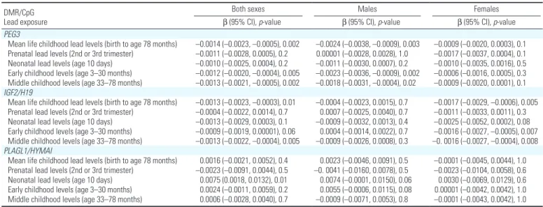
Table 2. Regression coefficients for the association between mean lead exposure and PEG3, IGF2/H19, and PLAGL1/HYMAI DMR methylation.a,b DMR/CpG
Lead exposure
Both sexes Males Females
β (95% CI), p-value β (95% CI), p-value β (95% CI), p-value PEG3
Mean life childhood lead levels (birth to age 78 months) –0.0014 (–0.0023, –0.0005), 0.002 –0.0024 (–0.0038, –0.0009), 0.003 –0.0009 (–0.0020, 0.0003), 0.1 Prenatal lead levels (2nd or 3rd trimester) –0.0011 (–0.0028, 0.0005), 0.2 0.00001 (–0.0028, 0.0028), 1.0 –0.0017 (–0.0037, 0.0004), 0.1 Neonatal lead levels (age 10 days) –0.0010 (–0.0025, 0.0004), 0.2 –0.0011 (–0.0030, 0.0007), 0.2 –0.0010 (–0.0035, 0.0016), 0.5 Early childhood levels (age 3–30 months) –0.0012 (–0.0020, –0.0004), 0.005 –0.0023 (–0.0036, –0.0009), 0.002 –0.0006 (–0.0016, 0.0005), 0.3 Middle childhood levels (age 33–78 months) –0.0013 (–0.0021, –0.0005), 0.002 –0.0018 (–0.0031, –0.0004), 0.02 –0.0009 (–0.0020, 0.0001), 0.1 IGF2/H19
Mean life childhood lead levels (birth to age 78 months) –0.0013 (–0.0023, –0.0003), 0.01 –0.0004 (–0.0023, 0.0015), 0.7 –0.0017 (–0.0029, –0.0006), 0.005 Prenatal lead levels (2nd or 3rd trimester) –0.0004 (–0.0022, 0.0014), 0.7 0.0007 (–0.0025, 0.0040), 0.7 –0.0011 (–0.0033, 0.0011), 0.3 Neonatal lead levels (age 10 days) –0.0013 (–0.0029, 0.0003), 0.1 –0.0009 (–0.0032, 0.0013), 0.4 –0.0025 (–0.0052, 0.0002), 0.08 Early childhood levels (age 3–30 months) –0.0009 (–0.0019, 0.00001), 0.06 0.0004 (–0.0014, 0.0022), 0.7 –0.0016 (–0.0027, –0.0005), 0.007 Middle childhood levels (age 33–78 months) –0.0013 (–0.0022, –0.0004), 0.005 –0.0009 (–0.0026, 0.0008), 0.3 –0. 0016 (–0.0027, –0.0004), 0.008 PLAGL1/HYMAI
Mean life childhood lead levels (birth to age 78 months) 0.0016 (–0.0021, 0.0052), 0.4 0.0023 (–0.0046, 0.0091), 0.5 –0.0001 (–0.0045, 0.0044), 1.0 Prenatal lead levels (2nd or 3rd trimester) –0.0023 (–0.0091, 0.0044), 0.5 –0. 0041 (–0.0160, 0.0078), 0.5 –0.0023 (–0.0104, 0.0058), 0.6 Neonatal lead levels (age 10 days) 0.0075 (0.0018, 0.0132), 0.01 0.0074 (–0.0001, 0.0150), 0.06 0.0030 (–0.0069, 0.0129), 0.6 Early childhood levels (age 3–30 months) 0.0024 (–0.0011, 0.0059), 0.2 0.0055 (–0.0006, 0.0115), 0.08 0.00001 (–0.0042, 0.0042), 1.0 Middle childhood levels (age 33–78 months) 0.0006 (–0.0028, 0.0040), 0.7 –0.0009 (–0.0071, 0.0053), 0.8 –0.0001 (–0.0043, 0.0042), 1.0
aUnstandardized regression coefficients. All models were adjusted for batch (first or second) and maternal cigarette smoking (none, one-half, 1 and 2 packs a day). Models of
combined estimates for males and females are also adjusted for sex. bMean for each developmental period (early childhood) was derived by summing up lead levels for the participant
Patterns of association observed in overall and sex-specific mean lead concen-trations were also evident when maximum lead concentrations were considered (see Supplemental Material, Table S5). The association between the maximum lead concentration and decreased PEG3 DMR methylation in adulthood (β = –0.0007; 95% CI: –0.0012, –0.0003, p = 0.003) was also more apparent in males (β = –0.0013; 95% CI: –0.0021, –0.0006, p-value = 0.001) than in females (β = –0.0004; 95% CI: –0.0010, 0.0002, p = 0.3). Maximum lead concentrations in the prenatal and neonatal ages were too low for meaningful analyses.
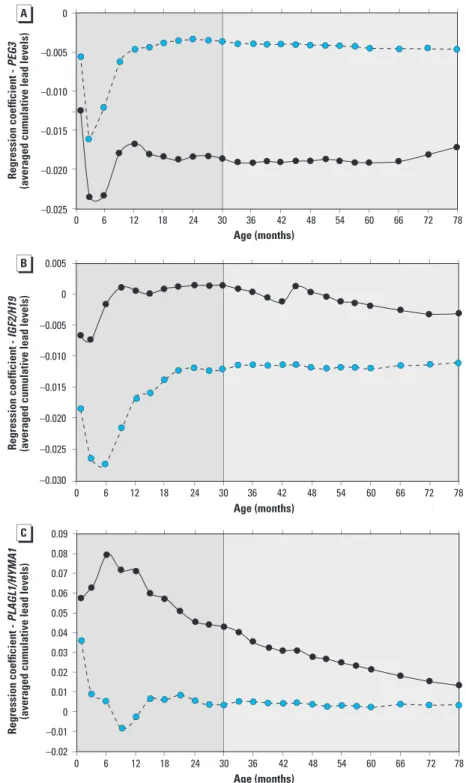
To refine potential windows of vulner-ability, nonstandardized regression coefficients were plotted for the associations between DMR methylation and averaged cumulative lead concentrations for each age at which lead was measured (Figure 3). Figure 3A confirms the association in Table 2 that higher lead exposure is associated with lower PEG3 DMR methyla-tion, and also shows that this association does not vary significantly after age 12–20 months to age 6.5 years, despite wide variation in lead concentrations during the observation period. These associations are male-specific.
Childhood lead levels and IGF2/H19 DMR methylation. Regression coefficients
and p-values for the association between mean lead concentration and IGF2/H19 DMR methylation in adulthood are also shown in Table 2. Mean childhood lead concentra-tion was significantly associated with adult
IGF2/H19 DMR methylation (β = –0.0013;
95% CI: –0.0023, –0.0003, p = 0.01). This association may be stronger in females (β = –0.0017; 95% CI: –0.0029, –0.0006,
p = 0.005) than in males (β = –0.0004;
95% CI: –0.0023, 0.0015, p = 0.7).
Associations for lead exposure and
IGF2/H19 DMR methylation were
also found in early (β = –0.0016; 95% CI: –0.0027, –0.0005, p = 0.007) and middle childhood (β = –0.0016; 95% CI: –0.0027, –0.0004, p = 0.008) in females. These associations were weaker and less consistent in males, for early (β = 0.0004; 95% CI: –0.0014, 0.0022,
p = 0.7) and middle childhood (β = –0.0009;
95% CI: –0.0026, 0.0008, p = 0.3). The
p-values for cross-product terms for early and
middle childhood lead levels and sex were 0.10 and 0.6, respectively. This age- and sex-specific pattern of association was also noted when maximum blood lead concen-trations were evaluated (see Supplemental Material, Table S5). As with the PEG3 DMR methylation, associations with prenatal and neonatal exposure were weaker although in the same direction.
Further exploration of cumulative lead concentrations suggests a female-specific
association between lead exposure and lower methylation at the IGF2/H19 DMR. The magnitude of the association between cumulative lead concentration and IGF2/H19 DMR methylation was the same after 12–20 months (Figure 3B). As with PEG3 DMR, this similarity persists for the entire observation period.
Childhood lead levels and PLAGL1/ HYMAI DMR methylation. Unlike IGF2/H19 and PEG3, which showed
no evidence for association between neonatal lead exposure and DMR tion, PLAGL1/HYMAI DMR methyla-tion was positively associated with lead levels during this period (β = 0.0075;
Figure 3. Unstandardized regression coefficients for associations between DMR methylation for PEG3 (A),
IGF2/H19 (B), and HYMA/PLAGL1 (C) and the average cumulative lead level (obtained by summing up blood
level measurements, up to and including the value measured at that age, divided by the number summed, in males (blue circles) and females (black circles). The darker gray area indicates early childhood; the lighter gray area indicates middle childhood.
0 6 12 18 24 30 36 42 48 54 60 66 72 78
0 6 12 18 24 30 36 42 48 54 60 66 72 78
0 6 12 18 24 42 48 54
Age (months) Age (months) Age (months)
60 66 72 78
30 36
0
–0.005
–0.010
–0.015
–0.020
–0.025
0.005
0
–0.005
–0.010
–0.015
–0.020
–0.025
–0.030
0.09 0.08 0.07 0.06 0.05 0.04 0.03 0.02 0.01 0 –0.01 –0.02
Regression coefficient -
PEG3
(averaged cumulative lead levels
)
Regression coefficient -
IGF2/H19
(averaged cumulative lead levels)
Regression coefficient -
PLAGL1/HYMA1
95% CI: 0.0018, 0.0132, p = 0.01). The wide confidence intervals suggest that this association varied little between males (β = 0.0074; 95% CI: –0.0001, 0.0150,
p = 0.06) and females (β = 0.0030;
95% CI: –0.0069, 0.0129, p = 0.6). No significant association was found between lead exposure and PLAGL1/HYMAI DMR methylation at any other age. Refined age-dependent and sex-specific analyses suggest that the magnitude of associations remained unaltered after ~ 12–20 months to the end of the observation period (Figure 3C).
Discussion
The DMRs regulating monoallelic expression of imprinted genes are proposed to function as epigenetic archives of early exposure to environmental factors (Hoyo et al. 2009). Nevertheless, until now, no empirical data have demonstrated associations between early exposure to lead and adult CpG methylation at DMRs controlling the parent-of-origin silencing of imprinted genes. Environmentally induced DNA methylation changes at imprint DMRs are usually stable once established (Heijmans et al. 2008), and have been asso-ciated with common chronic diseases and conditions, including neurological disorders, obesity, type 2 diabetes, and some cancers (Azzi et al. 2013; Feinberg 2007; Hoyo et al. 2012; Ishida and Moore 2013).
We have undertaken an analysis of 22 DMRs regulating human imprinted genes, and evaluated relationships between DNA methylation in adulthood and lead exposure spanning from the first or early second trimester to age 6.5 years. Our key findings were that childhood lead exposure was associ-ated with significantly lower DNA methyla-tion levels at the DMR regulating PEG3. We also found modest but consistent asso-ciations between average lead concentration and decreased methylation of the IGF2/H19 DMR, and higher DNA methylation levels at the PLAGL1/HYMAI DMR in relation to neonatal exposure. These data further indi-cated that although childhood lead exposure was associated with differences in PEG3 DMR methylation in males and the IGF2/H19 DMR methylation in females, the associa-tion between neonatal lead concentraassocia-tions and
PLAGL1/HYMAI DMR methylation may
not be sex-specific. Notably, lead associations with DNA methylation of imprint regulatory elements at these three loci were found in lead measured before age 30 months, regardless of sex or DMR. These data support the conten-tion that environmentally driven perturbaconten-tions at these DMRs occur early. Furthermore, developmental differences between the sexes may dictate the patterns of gene regulation that ensue in response to early challenges with this heavy metal.
PEG3 DMR methylation and early lead exposure. Although childhood lead exposure
has been associated with increased risk of neurodevelopmental disorders (Dietrich 2010), the mechanisms underlying these pathological conditions are poorly under-stood. PEG3 plays a critical role in brain development, with expression mainly in the mesencephalon and pituitary gland; in the adult brain PEG3 is found primarily in the hypothalamus and the pituitary gland (Li et al. 1999). In mouse models, Peg3 also plays an important role in social and maternal nurturing behaviors, and paternal transmission of disrupted Peg3 also leads to restricted growth (Chiavegatto et al. 2012; Li et al. 1999). In humans, hypermethyl-ation at this locus has been associated with decreased gene expression of this tumor suppressor gene in cervical (Nye et al. 2013) and ovarian (Feng et al. 2008) cancers. In primary neuronal cell cultures derived from wild-type, p53-deficient, or Bax-deficient mice, overexpression of Peg3 led to decreased neuronal viability via p53 and Bax dependent pathways (Johnson et al. 2002). It is there-fore possible that the male-specific reduced brain volume recently observed in these study participants (Cecil et al. 2008) may result, in part, from the dysregulation of PEG3 during early development.
Interestingly, early-childhood but not prenatal or neonatal lead levels were associated with adulthood PEG3 DMR hypomethyl-ation, an association that may be specific to males. Because these DNA methylation marks are established early, it is possible that meth-ylation differences observed were attributable to lead accumulated in utero and mobilized from soft tissue and bone after birth together with concurrent exposure. Alternatively, the reduced DNA methylation of the PEG3 DMR marks may have been established post-natally (Loke et al. 2013). The latter possi-bility is consistent with human developmental studies suggesting that the first 1,000 days can dictate lifetime risk of common diseases (Victora et al. 2008). Discriminating between these possibilities requires larger studies with long-term follow-up.
IGF2/H19 imprinted domain and early lead exposure. The paternally expressed insulin-like growth factor-2 (IGF2) is a commonly
studied imprinted gene, and is frequently shown to be altered epigenetically by in utero environmental perturbations, and in cancer (Cruz-Correa et al. 2004, 2009; Cui et al. 2003; Heijmans et al. 2008; Hoyo et al. 2011, 2012; Murphy et al. 2012a). Dysregulation of the IGF2/H19 domain was initially associ-ated with Beckwith–Wiedemann syndrome (BWS) (Engel et al. 2000). Decreased DNA methylation at the IGF2/H19 DMR has been associated with reduced IGF2 expression in
bladder cancer (Takai et al. 2001). This occurs when enhanced binding of the CTCF insu-lator protein to the normally unbound paternal allele (Nakagawa et al. 2001) blocks promoter interactions with downstream enhancers, thereby reducing gene expression (Hark et al. 2000; Kanduri et al. 2000). Igf2 over-expression results in animal overgrowth (Sun et al. 1997), whereas gene repression results in restricted growth (DeChiara et al. 1990). IGF2 is also required for memory formation (Chen et al. 2011).
PLAGL1/HYMA1 imprinted domain and early lead exposure. A higher-order regu-lation of imprinted gene clusters is thought to exist and occur through epigenetic marks present at imprinting centers (Lewis and Reik 2006). Our finding that neonatal lead exposure is associated with increased meth-ylation at the PLAGL1/HYMAI (ZAC) DMR regardless of sex is potentially of biological importance. In animals, microarray analysis shows that knockout of the mouse homolog,
Zac1 (Plagl1), disrupts a network of
coor-dinately regulated genes containing a large number that are also imprinted (Varrault et al. 2006). In vitro studies show induction of imprinted Igf2, Cdkn1c, H19, Dlk1, and
Mest when Zac1 is overexpressed (Varrault
et al. 2006). Conversely, loss of Zac1 expres-sion in null mice results in inhibition of Igf2,
Cdkn1c, H19, and Dlk1 expression. Another
imprinted gene network was identified by analyzing chromatin domains in other regions of the genome that interact with the Igf2/H19 domain, in vitro (Zhao et al. 2006). The maternally expressed long noncoding H19 RNA and the methyl-CpG–binding protein Mbd1 form a complex that regulates multiple imprinted genes by interacting with histone lysine methyltransferases. In mice, paternally expressed Plagl1 is implicated in transient neonatal diabetes when overexpressed (Ma et al. 2004). In ovarian cell lines, PLAGL1 was found to regulate CDKN1C (p57KIP2) expression and cell growth by inducing LIT1 transcription in a methylation-dependent manner (Arima et al. 2005). Overexpression of PLAGL1 induced IGF2, H19, and
CDKN1C expression in a prostate cancer cell
line (Ribarska et al. 2014). Together, these studies support a set of imprinted genes func-tioning in a network, coordinated in part by
Zac (Finkielstain et al. 2009; Lui et al. 2008).
mitotically heritable epigenetic alterations in DMRs controlling imprinted gene expression.
A cautious interpretation of our findings is warranted. Although lead is known to target multiple organs, DNA methylation was measured using unfractionated periph-eral blood collected in adulthood—the only accessible cell type—raising concerns about potential confounding by cell type, and other exposures during the life course that could not be evaluated. Another limitation of this study is the relatively small sample size, which reduced the precision of associa-tions found. Assay limitaassocia-tions also precluded the measurement of DNA methylation for ~ 50% of CpGs within CpG-containing fragments. Because methylation values for CpG-containing fragments were averaged from individual CpG sites with similar methylation values and are cis-acting, such missing data should not alter our findings. The small amount of peripheral blood leukocyte DNA available for methylation analysis also precluded the determination of altered gene expression via other epigenetic mechanisms (e.g., histone modifications and chromatin structure changes); however, similar meth-ylation changes at both the IGF2/H19 and
PEG3 DMRs have been associated previously
with altered gene expression in human cancers (Cui et al. 2003; Nye et al. 2013). Thus, our findings add preliminary support to accu-mulating evidence indicating that early lead exposure and gene-specific, epigenetic dysreg-ulation of some imprinted gene DMRs may contribute to developmental abnormalities (Ishida and Moore 2013).
Our study also has major strengths. They include the determination of lead levels ~ 30 years before quantification of DNA methylation levels at imprinted gene DMRs. The numerous measurements of lead concen-tration during early development also facili-tate estimating developmental windows in which lead exposure may exert its effects on regulatory DMRs. Furthermore, blood lead concentrations reflect both short- and longer-term exposure, including lead mobilized from physiological deposits.
To our knowledge, our findings repre-sent the first attempt in humans to quantify associations between early lead exposure and DNA methylation alterations in adulthood at imprinted loci that are known experimentally to result in developmental and neurological disorders if perturbed early in development. Because lead exposure disproportionately affects those in the lower socioeconomic strata (Emerson 2012; Rai et al. 2012; Wright et al. 2008), our findings, if repli-cated in larger studies, may offer a potential explanation for observed DNA methylation differences among socioeconomic strata (Szyf 2012, 2013).
Conclusions
Preventing lead exposure during vulner-able developmental windows remains sound policy. Nevertheless, effective therapeutic and public health strategies will depend on a better understanding of mechanisms underpinning the associations between lead exposure and the genesis of neurodevelopmental disorders and other poor health outcomes. Improved under-standing should also guide policy regarding the highest tolerable limits in humans, a value currently unknown. Although the small sample size limits inference, this study provides preliminary evidence for significant associations between early lead exposure and DNA methylation at the regulatory regions of PEG3, H19/IGF2, and PLAGL1/HYMAI. Because these changes in the epigenome are acquired early, resultant shifts in the regula-tion of imprinted genes may contribute to increased risk of poor health outcomes (Ishida and Moore 2013; Murphy and Jirtle 2003). It remains unknown whether lead exposure previously associated with decreased gray matter volume (Cecil et al. 2008) and delin-quent behavior (Dietrich et al. 2001) reported in this study population is mediated in part by the epigenetic alterations in imprinted gene regulatory elements, but this intriguing possibility needs to be investigated.
RefeRences
Arima T, Kamikihara T, Hayashida T, Kato K, Inoue T, Shirayoshi Y, et al. 2005. ZAC, LIT1 (KCNQ1OT1) and
p57KIP2 (CDKN1C) are in an imprinted gene network
that may play a role in Beckwith-Wiedemann syndrome. Nucleic Acids Res 33:2650–2660. Azzi S, Sas TC, Koudou Y, Le Bouc Y, Souberbielle JC,
Dargent-Molina P, et al. 2013. Degree of methyla-tion of ZAC1 (PLAGL1) is associated with prenatal and post-natal growth in healthy infants of the EDEN mother child cohort. Epigenetics 9:338–345. Barlow DP. 2011. Genomic imprinting: a mammalian
epigenetic discovery model. Annu Rev Genet 45:379–403.
Bellinger D, Leviton A, Needleman HL, Waternaux C, Rabinowitz M. 1986. Low-level lead exposure and infant development in the first year. Neurobehav Toxicol Teratol 8:151–161.
Bellinger D, Leviton A, Waternaux C, Needleman H, Rabinowitz M. 1987. Longitudinal analyses of prenatal and postnatal lead exposure and early cognitive development. N Engl J Med 316:1037–1043. Bernal AJ, Dolinoy DC, Huang D, Skaar DA,
Weinhouse C, Jirtle RL. 2013. Adaptive radiation-induced epigenetic alterations mitigated by anti-oxidants. FASEB J 27:665–671.
Binns HJ, Campbell C, Brown MJ, Centers for Disease Control and Prevention Advisory Committee on Childhood Lead Poisoning Prevention. 2007. Interpreting and managing blood lead levels of less than 10 μg/dL in children and reducing child-hood exposure to lead: recommendations of the Centers for Disease Control and Prevention Advisory Committee on Childhood Lead Poisoning Prevention. Pediatrics 120:e1285–e1298.
Bolin CM, Basha R, Cox D, Zawia NH, Maloney B, Lahiri DK, et al. 2006. Exposure to lead and the
developmental origin of oxidative DNA damage in the aging brain. FASEB J 20:788–790.
Cecil KM, Brubaker CJ, Adler CM, Dietrich KN, Altaye M, Egelhoff JC, et al. 2008. Decreased brain volume in adults with childhood lead exposure. PLoS Med 5:e112; doi:10.1371/journal.pmed.0050112. Chen DY, Stern SA, Garcia-Osta A, Saunier-Rebori B,
Pollonini G, Bambah-Mukku D, et al. 2011. A critical role for IGF-II in memory consolidation and enhancement. Nature 469:491–497.
Chiavegatto S, Sauce B, Ambar G, Cheverud JM, Peripato AC. 2012. Hypothalamic expression of
Peg3 gene is associated with maternal care differ-ences between SM/J and LG/J mouse strains. Brain Behav 2:365–376.
Cruz-Correa M, Cui H, Giardiello FM, Powe NR, Hylind L, Robinson A, et al. 2004. Loss of imprinting of insulin growth factor II gene: a potential heri-table biomarker for colon neoplasia predisposition. Gastroenterology 126:964–970.
Cruz-Correa M, Zhao R, Oviedo M, Bernabe RD, Lacourt M, Cardona A, et al. 2009. Temporal stability and age-related prevalence of loss of imprinting of the insulin-like growth factor-2 gene. Epigenetics 4:114–118.
Cui H, Cruz-Correa M, Giardiello FM, Hutcheon DF, Kafonek DR, Brandenburg S, et al. 2003. Loss of
IGF2 imprinting: a potential marker of colorectal cancer risk. Science 299:1753–1755.
DeChiara TM, Efstratiadis A, Robertson EJ. 1990. A growth-deficiency phenotype in heterozygous mice carrying an insulin-like growth factor II gene disrupted by targeting. Nature 345:78–80. Dietrich KN. 2010. Environmental toxicants. In: Pediatric
Neuropsychology: Research, Theory, and Practice (Yeates KO, Ris MD, Taylor HG, Pennington BF, eds). 2nd ed. New York:Guilford Press, 211–264. Dietrich KN, Berger OG, Succop PA. 1993. Lead
exposure and the motor developmental status of urban six-year-old children in the Cincinnati Prospective Study. Pediatrics 91:301–307. Dietrich KN, Krafft KM, Bornschein RL, Hammond PB,
Berger O, Succop PA, et al. 1987. Low-level fetal lead exposure effect on neurobehavioral develop-ment in early infancy. Pediatrics 80:721–730. Dietrich KN, Ris MD, Succop PA, Berger OG,
Bornschein RL. 2001. Early exposure to lead and juvenile delinquency. Neurotoxicol Teratol 23:511–518. Dolinoy DC, Huang D, Jirtle RL. 2007. Maternal nutrient
supplementation counteracts bisphenol A-induced DNA hypomethylation in early development. Proc Natl Acad Sci USA 104:13056–13061.
Dolinoy DC, Weidman JR, Waterland RA, Jirtle RL. 2006. Maternal genistein alters coat color and protects
Avy mouse offspring from obesity by modifying
the fetal epigenome. Environ Health Perspect 114:567–572; doi:10.1289/ehp.8700.
Edwards CA, Rens W, Clarke O, Mungall AJ, Hore T, Graves JA, et al. 2007. The evolution of imprinting: chromosomal mapping of orthologues of mammalian imprinted domains in monotreme and marsupial mammals. BMC Evol Biol 7:157; doi:10.1186/1471-2148-7-157.
Emerson E. 2012. Deprivation, ethnicity and the preva-lence of intellectual and developmental disabili-ties. J Epidemiol Community Health 66:218–224. Engel JR, Smallwood A, Harper A, Higgins MJ,
Oshimura M, Reik W, et al. 2000. Epigenotype-phenotype correlations in Beckwith-Wiedemann syndrome. J Med Genet 37:921–926.
Feinberg AP. 2007. Phenotypic plasticity and the epigenetics of human disease. Nature 447:433–440. Feng, W, Marquez RT, Lu Z, Liu, J, Lu KH, Issa JP, et al.
2008. Imprinted tumor suppressor genes ARHI and
PEG3 are the most frequently down-regulated in human ovarian cancers by loss of heterozygosity and promoter methylation. Cancer 112:1489–1502. Finkelstein Y, Markowitz ME, Rosen JF. 1998. Low-level
lead-induced neurotoxicity in children: an update on central nervous system effects. Brain Res Brain Res Rev 27:168–176.
Finkielstain GP, Forcinito P, Lui JC, Barnes KM, Marino R, Makaroun S, et al. 2009. An extensive genetic program occurring during postnatal growth in multiple tissues. Endocrinology 150:1791–1800. Hark AT, Schoenherr CJ, Katz DJ, Ingram RS,
Levorse JM, Tilghman SM. 2000. CTCF mediates methylation-sensitive enhancer-blocking activity at the H19/Igf2 locus. Nature 405:486–489. Heijmans BT, Tobi EW, Stein AD, Putter H, Blauw GJ,
Susser ES, et al. 2008. Persistent epigenetic differences associated with prenatal exposure to famine in humans. Proc Natl Acad Sci USA 105:17046–17049.
Hoyo C, Fortner K, Murtha AP, Schildkraut JM, Soubry A, Demark-Wahnefried W, et al. 2012. Association of cord blood methylation fractions at imprinted insulin-like growth factor 2 (IGF2), plasma IGF2, and birth weight. Cancer Causes Control 23:635–645.
Hoyo C, Murphy SK, Jirtle RL. 2009. Imprint regulatory elements as epigenetic biosensors of exposure in epidemiological studies. J Epidemiol Community Health 63:683–684.
Hoyo C, Murtha AP, Schildkraut JM, Jirtle RL, Demark-Wahnefried W, Forman MR, et al. 2011. Methylation variation at IGF2 differentially methyl-ated regions and maternal folic acid use before and during pregnancy. Epigenetics 6:928–936. Ishida M, Moore GE. 2013. The role of imprinted genes
in humans. Mol Aspects Med 34:826–840. Johnson MD, Wu X, Aithmitti N, Morrison RS. 2002.
Peg3/Pw1 is a mediator between p53 and Bax in DNA damage-induced neuronal death. J Biol Chem 277:23000–23007.
Kanduri C, Pant V, Loukinov D, Pugacheva E, Qi CF, Wolffe A, et al. 2000. Functional association of CTCF with the insulator upstream of the H19 gene is parent of origin-specific and methylation-sensitive. Curr Biol 10:853–856.
Lewis A, Reik W. 2006. How imprinting centres work. Cytogenet Genome Res 113:81–89.
Li L, Keverne EB, Aparicio SA, Ishino F, Barton SC, Surani MA. 1999. Regulation of maternal behavior and offspring growth by paternally expressed
Peg3. Science 284:330–333.
Loke YJ, Galati JC, Morley R, Joo EJ, Novakovic B, Li X, et al. 2013. Association of maternal and nutrient supply line factors with DNA methylation at the imprinted IGF2/H19 locus in multiple tissues of newborn twins. Epigenetics 8:1069–1079. Lui JC, Finkielstain GP, Barnes KM, Baron J. 2008. An
imprinted gene network that controls mammalian somatic growth is down-regulated during post-natal growth deceleration in multiple organs. Am J Physiol Regul Integr Comp Physiol 295:R189–R196. Luo M, Xu Y, Cai R, Tang Y, Ge MM, Liu ZH, et al.
2014. Epigenetic histone modification regulates
developmental lead exposure induced hyper-activity in rats. Toxicol Lett 225:78–85.
Ma D, Shield JP, Dean W, Leclerc I, Knauf C, Burcelin RR, et al. 2004. Impaired glucose homeo-stasis in transgenic mice expressing the human transient neonatal diabetes mellitus locus, TNDM. J Clin Invest 114:339–348.
Murphy SK, Adigun A, Huang Z, Overcash F, Wang F, Jirtle RL, et al. 2012a. Gender-specific methylation differences in relation to prenatal exposure to cigarette smoke. Gene 494:36–43.
Murphy SK, Huang Z, Hoyo C. 2012b. Differentially methylated regions of imprinted genes in prenatal, perinatal and postnatal human tissues. PLoS One 7:e40924; doi:10.1371/journal.pone.0040924. Murphy SK, Jirtle RL. 2003. Imprinting evolution and
the price of silence. Bioessays 25:577–588. Nakagawa H, Chadwick RB, Peltomaki P, Plass C,
Nakamura Y, de La Chapelle A. 2001. Loss of imprinting of the insulin-like growth factor II gene occurs by biallelic methylation in a core region of
H19-associated CTCF-binding sites in colorectal cancer. Proc Natl Acad Sci USA 98:591–596. Nye MD, Hoyo C, Huang Z, Vidal AC, Wang F, Overcash F,
et al. 2013. Associations between methylation of
paternally expressed gene 3 (PEG3), cervical
intraep-ithelial neoplasia and invasive cervical cancer. PLoS One 8:e56325; doi:10.1371/journal.pone.0056325. Pilsner JR, Hu H, Ettinger A, Sánchez BN, Wright RO,
Cantonwine D, et al. 2009. Influence of prenatal lead exposure on genomic methylation of cord blood DNA. Environ Health Perspect 117:1466–1471; doi:10.1289/ehp.0800497.
Rai D, Lewis G, Lundberg M, Araya R, Svensson A, Dalman C, et al. 2012. Parental socioeconomic status and risk of offspring autism spectrum disor-ders in a Swedish population-based study. J Am Acad Child Adolesc Psychiatry 51:467–476.e6; doi:10.1016/j.jaac.2012.02.012.
R Core Team. 2013. R: A Language and Environment for Statistical Computing. Vienna, Austria:R Foundation for Statistical Computing. Available: http://www.R-project.org/ [accessed 3 December 2014]. Reichard JF, Schnekenburger M, Puga A. 2007. Long
term low-dose arsenic exposure induces loss of DNA methylation. Biochem Biophys Res Commun 352:188–192.
Reik W, Walter J. 2001. Genomic imprinting: parental influence on the genome. Nat Rev Genet 2:21–32. Ribarska T, Goering W, Droop J, Bastian KM,
Ingenwerth M, Schulz WA. 2014. Deregulation of an imprinted gene network in prostate cancer. Epigenetics 9:704–717.
Roda SM, Greenland RD, Bornschein RL, Hammond PB. 1988. Anodic stripping voltammetry procedure modified for improved accuracy of blood lead analysis. Clin Chem 34:563–567.
Sanders T, Liu Y, Buchner V, Tchounwou PB. 2009. Neurotoxic effects and biomarkers of lead exposure: a review. Rev Environ Health 24:15–45. Skaar DA, Li Y, Bernal AJ, Hoyo C, Murphy SK,
Jirtle RL. 2012. The human imprintome: regulatory mechanisms, methods of ascertainment, and roles in disease susceptibility. ILAR J 53:341–358. Sun FL, Dean WL, Kelsey G, Allen ND, Reik W.
1997. Transactivation of Igf2 in a mouse model of Beckwith–Wiedemann syndrome. Nature 389:809–815.
Szyf M. 2012. The early-life social environment and DNA methylation. Clin Genet 81:341–349. Szyf M. 2013. The genome- and system-wide response
of DNA methylation to early life adversity and its implication on mental health. Can J Psychiatry 58:697–704.
Takai D, Gonzales FA, Tsai YC, Thayer MJ, Jones PA. 2001. Large scale mapping of methylcytosines in CTCF-binding sites in the human H19 promoter and aberrant hypomethylation in human bladder cancer. Hum Mol Genet 10:2619–2626.
Takiguchi M, Achanzar WE, Qu W, Li G, Waalkes MP. 2003. Effects of cadmium on DNA-(Cytosine-5) methyltransferase activity and DNA methylation status during cadmium-induced cellular transfor-mation. Exp Cell Res 286:355–365.
Varrault A, Gueydan C, Delalbre A, Bellmann A, Houssami S, Aknin C, et al. 2006. Zac1 regulates an imprinted gene network critically involved in the control of embryonic growth. Dev Cell 11:711–722. Victora CG, Adair L, Fall C, Hallal PC, Martorell R,
Richter L, et al. 2008. Maternal and child under-nutrition: consequences for adult health and human capital. Lancet 371:340–357.
Waterland RA, Jirtle RL. 2003. Transposable elements: targets for early nutritional effects on epigenetic gene regulation. Mol Cell Biol 23:5293–5300. Waterland RA, Kellermayer R, Laritsky E,
Rayco-Solon P, Harris RA, Travisano M, et al. 2010. Season of conception in rural Gambia affects DNA methylation at putative human metastable epial-leles. PLoS Genet 6:e1001252; doi:10.1371/journal. pgen.1001252.
Wilson AS, Power BE, Molloy PL. 2007. DNA hypo-methylation and human diseases. Biochim Biophys Acta 1775:138–162.
Winneke G, Krämer U, Brockhaus A, Ewers U, Kujanek G, Lechner H, et al. 1983. Neuropsychological studies in children with elevated tooth-lead concentrations. II. Extended study. Int Arch Occup Environ Health 51:231–252.
Woodfine K, Huddleston JE, Murrell A. 2011. Quantitative analysis of DNA methylation at all human imprinted regions reveals preservation of epigenetic stability in adult somatic tissue. Epigenetics Chromatin 4:1; doi:10.1186/1756-8935-4-1.
Wright JP, Dietrich KN, Ris MD, Hornung RW, Wessel SD, Lanphear BP, et al. 2008. Association of prenatal and childhood blood lead concentra-tions with criminal arrests in early adulthood. PLoS Med 5:e101; doi:10.1371/journal.pmed.0050101. Wright RO, Schwartz J, Wright RJ, Bollati V, Tarantini L,
Park SK, et al. 2010. Biomarkers of lead exposure and DNA methylation within retrotransposons. Environ Health Perspect 118:790–795; doi:10.1289/ ehp.0901429.
Zahran S, Mielke HW, Weiler S, Berry KJ, Gonzales C. 2009. Children’s blood lead and standardized test performance response as indicators of neuro-toxicity in metropolitan New Orleans elementary schools. Neurotoxicology 30:888–897.