ROLE OF METAVINCULIN IN ACTIN REORGANIZATION AND FORCE TRANSMISSION
Hyunna Theresa Lee
A dissertation submitted to the faculty at the University of North Carolina at Chapel Hill in partial fulfillment of the requirements for the degree of Doctor of Philosophy in the Department
of Biochemistry and Biophysics in the School of Medicine.
Chapel Hill 2019
Approved by: Sharon Campbell Keith Burridge Nikolay Dokholyan Qi Zhang
iii ABSTRACT
Hyunna Theresa Lee: Role of metavinculin in actin reorganization and force transmission (Under the directions of Sharon L. Campbell and Keith Burridge)
iv
differences between metavinculin and vinculin in cells, we additionally stably expressed either vinculin or metavinculin in vinculin-null mouse embryonic fibroblasts. While both metavinculin and vinculin were observed at focal adhesions (FA), metavinculin-expressing cells had larger but fewer FAs per cell compared to vinculin-expressing cells. Metavinculin-expressing cells
migrated faster and exhibited greater persistence compared to vinculin-expressing cells, even though vinculin-containing FAs assembled and disassembled faster. Magnetic tweezer
measurements on vinculin-expressing cells show a typical cell stiffening phenotype in response to externally applied force; however, this was absent in vinculin-null and
v
To my parents, my sister, and my close friends. I could not be where I am today without your unconditional love and support. To my advisors, Sharon Campbell and Keith Burridge. Thank you both for your trust and confidence in me as your student. Your support and mentoring cannot
vi
ACKNOWLEDGMENTS
This section is, in some ways, the most difficult section of the dissertation to write. I have received support and encouragement from numerous individuals throughout my journey, and I cannot express into words the gratitude I feel towards them. Without their intellectual
inspiration, mental and scientific support, and encouragement to go after my curiosities, I would not be where I am today.
I would like to start by acknowledging and thanking my mentors, Drs. Sharon Campbell and Keith Burridge, who have pushed me to be a better and critical scientist. First, I would like to thank Dr. Sharon Campbell for always allowing me to go after my scientific interests and supporting me in my experimental endeavors. Without her encouragement, I would not have been able to explore the cell biology/cytoskeleton field and discover a whole new field of interest. I immensely appreciate her trust and dedication in my training. Just as importantly, I would additionally like to thank Dr. Keith Burridge, who have always encouraged me to go after my curiosities as I pursued this project. He was always a source of motivation for me due to his positive outlook towards scientific results. However, he never sacrificed the quality of data he expected, which has trained me to be a better scientist. Perhaps I learned the most important life lessons from both of my mentors. From Sharon, I learned to enjoy the challenges that come my way, and from Keith, I learned that the only thing we have to fear is fear itself.
vii
The amazing people in these labs have carried me through the darkest of times and also
celebrated all the happy moments alongside me as if they were my family. It is with them that I celebrated every little personal and scientific growth. I will dearly miss the outside lunches and coffee runs in the morning.
Next, I would like to thank my committee members. I would first like to thank Dr.
Stephanie Gupton for her numerous experimental mentorship and career discussions. I love these discussions with Dr. Gupton as she seems to always know the answer to my questions. Reaching out to ask about focal adhesion experiments was probably one of the best things I did while I was here. I would also like to thank Dr. Joan Taylor. She has provided me with tremendous support, especially toward the beginning of my thesis project. When I was having problems with
expressing metavinculin in cells, it was because of her scientific advice that I was able to
continuously look for solutions. For this, I will always be grateful. Subsequently, I would like to thank Dr. Qi Zhang, who has been one of the best mentors in my program. Dr. Zhang’s
commitment to teaching and mentoring students is inspirational. Finally, I’d like to thank Dr. Nikolay Dokholyan, as discussions with his group about the vinculin project always provided me with a more interesting perspective.
viii
I also want to acknowledge my close friends, as hanging out with them made my life so much more fun. Your unconditional love and encouragement have brought me where I am today. You have been my inspiration.
Lastly, I would like to thank my parents and my sister. Without their encouragement and confidence, I would not have been able to finish. My family have been nothing but patient and supportive as I pursued my thesis, and at times, reminded me that it was okay to go slow. It was through them that I learned that it is most important to hold the values that are key to me, and never compare myself with others. My parents have always stressed the importance of education and encouraged me to test my limits. They have also instilled the value of hard work and
persistence as I grew up, which is one of reasons that I am where I am today. Their dedication to my personal and professional growth keeps me motivated to become better.
ix PREFACE
Parts of this work were done in collaboration with other talented scientists. Chapter 2 represents work that was done in collaboration with the labs of Dr. Greg Alushin, Dr. Nikolay Dokholyan, and Dr. Jack Griffith. My contribution focused on the protein purification for vinculin and metavinculin samples, site-directed mutagenesis for generating the necessary constructs, and performing actin co-sedimentation assays to determine actin binding and bundling levels. Figures 4-7 were generated by the Alushin lab. I contributed to the Figures generated in 8, 11, and 12 with Dr. Muzaddid Sarker from the Campbell lab while Figures 9 was generated by Dr. Sarker in collaboration with the Griffith lab. Figures 10 and 13 were generated by the Alushin lab, and Figure 14 was generated by the Dokholyan lab based on their discrete molecular dynamics (DMD) models. This work resulted in two publications that I co-authored. The papers were published previous to writing this thesis with the following citations:
Kim LY, Thompson PM, Lee HT, Pershad M, Campbell SL, Alushin GM. The Structural Basis of Actin Organization by Vinculin and Metavinculin. J Mol Biol. 2016;428(1):10-25. PMCID: 4738167.
Sarker M, Lee HT, Mei L, Krokhotin A, de Los Reyes SE, Yen L, et al. Cardiomyopathy Mutations in Metavinculin Disrupt Regulation of Vinculin-Induced F-Actin Assemblies. J Mol Biol. 2019;431(8):1604-18.
Permission to include the articles in a PhD dissertation was retained from Elsevier (publisher of JMB) as explained at https://www.elsevier.com/about/policies/copyright.
x
including Dr. Stephanie Gupton and Dr. Richard Superfine. I am the lead author on the paper, but Fabio Urbina from the Gupton lab has helped me to generate Figure 23. Additionally, all the 3DFM-related data in Figures 25 and 26 could not have been generated without the help of Dr. Timothy O’Brien in the Superfine lab. Finally, Minh Hyunh from the Campbell/Der lab has provided me with the constructs that were necessary for the generation of stable cell lines. This paper is currently in revision:
Lee, H.T., Sharek, L., O’Brien, E.T., Fabio L. Urbina, Gupton, S.L., Superfine, R., Burridge, K., and Campbell, S.L. (2019) Vinculin and Metavinculin Exhibit Distinct Effects on Focal Adhesion Properties, Cell migration, and Mechanotransduction. PLoS One, in revision.
xi
TABLE OF CONTENTS
LIST OF TABLES ... xiii
LIST OF FIGURES ... xiv
LIST OF ABBREVIATIONS ... xvi
CHAPTER 1 - INTRODUCTION ... 1
Focal adhesion architecture and signaling ... 1
The cell adhesion protein vinculin ... 8
Metavinculin is a splice isoform of vinculin expressed in muscle cells ... 11
Differences between vinculin and metavinculin structure and function ... 14
The role of vinculin-mediated actin-binding and actin-bundling ... 17
CHAPTER 2 – The Strutural Basis of Actin Organization by Vinculin, Metavinculin, and Metavinculin Cardiomyopathy-associated Mutants ... 21
Introduction ... 21
Materials and Methods ... 25
Results ... 31
Discussion ... 51
CHAPTER 3 – Vinculin and Metavinculin Exhibit Distinct Effects on Focal Adhesion Properties, Cell migration, and Mechanotransduction ... 56
Introduction ... 56
Materials and Methods ... 59
Results ... 66
xii
CHAPTER 4 – Conclusions and Future Directions ... 89
Overview ... 89
Review of Current Findings ... 89
Significance of this dissertation ... 92
Future directions ... 95
Speculations on metavinculin ... 99
xiii
LIST OF TABLES
xiv
LIST OF FIGURES
Figure 1. Nanoscale architecture of focal adhesions. ... 2
Figure 2. Vinculin is an autoinhibitory scaffolding protein. ... 10
Figure 3. Sequence and structural differences between Vcn and MVcn. ... 16
Figure 4. Cryo-EM reconstruction of Vt: F-actin and MVt:F-actin complex... 18
Figure 5. Sub-nanometer-resolution reconstruction of the Vt-actin interface. ... 33
Figure 6. A steric mechanism promotes H1 release to bundle actin. ... 34
Figure 7. MDFF model of the Vt-actin surface. ... 36
Figure 8. MVt WT and CM mutants exhibit similar actin binding but not crosslinking. ... 38
Figure 9. MVt exhibits reduced F-actin bundling (crosslinking) compared to Vt. ... 40
Figure 10. MVt CM mutants promote disordered, mesh-like F-actin assemblies. ... 42
Figure 11. MVt WT inhibits Vt-mediated actin bundling. ... 45
Figure 12. MVt CM mutants fail to inhibit Vt-induced F-actin bundling. ... 47
Figure 13. MVt CM mutants aggregate Vt-induced actin bundles. ... 48
Figure 14. Actin binding to MVt may induce a formation of protruding structure... 50
Figure 15. Model of vinculin activation and tension reinforcing actin engagement. ... 52
Figure 16. Model for how MVt WT and CM mutants affectVt-induced actin bundle. ... 55
Figure 17. MVcn-expressing cells have larger but fewer FAs compared to Vcn-expressing cells. ... 67
Figure 18. mEmerald-Vcn and mRFP-MVcn cells were sorted for expression using flow activated cell sorting (FACS). ... 68
Figure 19. MVcn rescues decreased cell area in Vcn-null MEFs. ... 72
xv
Figure 21. Random cell migration analysis shows enhanced
migration velocity and higher persistence of migration for cells expressing MVcn compared to Vcn-expressing cells. ... 75 Figure 22. Focal adhesion assembly and disassembly rates are higher
for Vcn-expressing cells compared to MVcn-expressing cells... 77 Figure 23. Average and representative assembly and disassembly curves at FA. ... 78 Figure 24. Focal adhesion assembly and disassembly rates display
consistent results with the same tagged fluorophore. ... 80 Figure 25. 3D-Force microscopy (3DFM) shows reduced ability of
xvi
LIST OF ABBREVIATIONS
3DFM Three-dimensional force microscopy
∆C5 Vinculin C-terminal hairpin deletion (lacks residues 1061-1066)
AJ Adherens junction
AF Aggregation factor
ARP 2/3 Actin-related protein 2/3
BME β-mercaptoethanol
BSA Bovine serum albumin
CM Cardiomyopathy
cryo-EM cryo-electron microscopy
DCM Dilated cardiomyopathy
DMD Discrete molecular dynamics DMEM Dulbecco Modified Eagle Medium
DMSO Dimethylsulfoxide
DTT Dithiothreitol
ECM Extracellular matrix
EM Electron microscopy
FA Focal adhesion
FACS Fluorescent activated cell sorting F-actin Filamentous actin
FAK Focal adhesion kinase
xvii
FN Fibronectin
FRNK Focal adhesion kinase-related nonkinase G-actin Monomeric actin
IPTG β-D-1-thiogalactopyranoside HCM Hypertrophic cardiomyopathy IRM Interference reflection microscopy
Kd Dissociation constant
KO Knock-out
LB Lysogeny broth
MEF Mouse embryonic fibroblast μm/nm Micrometer/nanometer MVt Metavinculin tail domain NMR Nuclear magnetic resonance PBS Phosphate buffered saline PCR Polymerase chain reaction
PDB Protein data bank
PI3K Phophoinositide 3-kinase
PIP2 Phosphatidylinositol 4,5-bisphosphate
PIPKIγ
Phosphatidylinositol phosphate kinase type 1 gamma
ROI Region of interest
RT Real-time
xviii SDS Sodium dodecyl sulfate
TEV Tobacco etch virus
VASP Vasodilator-stimulated phosphoprotein
Vh Vinculin head domain
Vt Vinculin tail domain
1
CHAPTER 1 - INTRODUCTION Focal adhesion architecture and signaling
When many cells migrate in culture dishes, structures called focal adhesions (FAs) assemble and disassemble, allowing cells to regulate points of adhesion and force. Cell adhesion and mechanotransduction regulation contributes to normal physiological control of cell motility, morphology, and survival. FAs are macromolecular structures that consist of ~150 different proteins that form adhesion plaques between cell membrane and the extra cellular matrix (ECM). FAs physically link actin cytoskeleton inside the cell to the outside environment, and they can be largely divided into three layers: integrin signalling layer, force transduction layer, and actin regulatory layer (1) (Fig. 1).
Historically, FAs were initially observed by Abercrombie et al. in 1971, who used electron microscopy (EM) to examine lamelliopodia in cultured migrating fibroblasts
2 Figure 1. Nanoscale architecture of focal adhesions.
3
“focal adhesions” (4). Couchman and Rees subsequently used this term upon studying cardiac fibroblasts migrating out of heart explants (5). Interestingly, these authors observed that during the initial phase of rapid cells migration, the fibroblasts barely showed any FAs but as these cells slowed down, FAs developed. It is important to note that FAs are not necessary for cell
migration as many cells have been shown to migrate without them. While integrin-mediated adhesion to the ECM still forms in cells that do not develop FAs, these adhesions do not form stable clusters of integrins. Nobes and Hall introduced the term “focal complex” to refer to smaller, more transient adhesions than FAs (6). In general, the term nascent adhesions has been widely adopted in the field to describe the initial small adhesions, which eventually develop into focal complexes and ultimately into FAs as the adhesion matures and stabilizes. Burridge
provides an excellent review on a historical perspective of the focal adhesion field (7). With the development of immunofluorescence microscopy in the mid-1970s and the discovery of vinculin by Geiger in 1979 (8), there was a breakthrough in the field of focal adhesion and cytoskeleton research. A number of FA components have been discovered since that time. Notably, several key components have been identified at FA, including integrin, talin, vinculin, and α-actinin, which will be discussed in more detail below. These FA components reside distinctly throughout the three layers that comprise the FA structure.
4
that cells use to both bind and respond to the ECM ligands, including collagen, fibronectin, and laminin. An integrin molecule consists of two non-covalently associated transmembrane subunits called α and β determines its affinity for specific ECM ligands. Integrins exist in 3 states: closed (inactive), bent, and open (active) conformations (9). Activation of integrins occur through either “outside-in” or “inside-out” signaling, where both the environmental cues and the intracellular signals regulate how FAs function (10, 11). Outside-in signaling initiates as the integrin extracellular domain binds to ECM ligand, driving a conformational change that exposes their cytoplasmic tails, which recruits specific proteins that form and mature FAs. In the context of FAs, integrins have been found to bind to other FA components such as talin (12) and α-actinin (13), providing a physical link between plasma membrane and FA. Force has been speculated to play a role in integrin activation as well, although it is still being determined. Some studies suggest that external mechanical loading is necessary for integrin activation (14). Inside-out signaling is also hypothesized to occur through actomyosin-driven force generation and regulated at the integrin signaling layer since by altering the activation state of integrins, intracellular signals can modulate the bindng affinity between integrins and ECM ligands (15). Furthermore, integrin signaling layer may be important for regulating feedback between inside-out and inside-outside-in signaling as well. In this layer, integrin activation has been shown to
5
(21-24). C-terminal domain of FAK is also a site of many protein-protein interactions, and overexpression of focal adhesion kinase-related nonkinase (FRNK), an autonamously expressed C-terminus of FAK, has been shown to inhibit the rate of cell spreading and migration rates in response to chemotactic and haptotactic cues (24-26). Therefore, the integrin signaling layer of FA is important for both responding to external forces and the generation of intracellular forces.
With respect to the cell membrane surface, the force transduction layer lies on top of the integrin signaling layer, and it consists various scaffolding and signaling proteins. Of note, talin and vinculin (though vinculin has been found in all FA layers) have been shown to play
important roles in mechanotransduction (1, 27). Talin can bind both integrins and filamentous actin (F-actin) and reinforce this connection to F-actin by further recruiting vinculin (12, 28, 29). Application of force on talin has been shown to expose the cryptic vinculin-binding sites (VBS) by unfurling talin, which then recruits vinculin to talin (30). This force is thought to occur as myosin-generated forces are transmitted across FAs or from forces resulting from actin
6
talin and/or vinculin can overcome this stiffness threshold depends on the stiffness of the cell’s environment, as the cell’s ability to sense mechanical cues and transduce force into biochemical signals depends on the rigidity of the environment that the cell is in. Coupled with the molecular clutch model is the role of actin retrograde flow in cells. Actin retrograde flow refers to actin that flows back toward the body of the cell as the cell migrates, resulting from the actin
polymerization against the cell membrane at the leading edge (36). It has been proposed that FAs at the cell’s leading edge act as “clutches” for the flowing actin that impede actin’s retrograde movement. By clutching onto the retrograde flow of actin, FAs can transduce the force from this actin flow to the ECM, creating the traction needed to push the cell forward (36). Hence, the speed of actin retrograde flow should be inversely related to the traction force at FAs: the more FAs clutch onto the flowing actin to create more traction against ECM, the slower the actin retrograde flow should be. Talin and vinculin act as molecular clutches that bind to F-actin at the force transduction layer of FAs, playing important roles for how FAs transmit force into
intracellular biochemical signals and vice versa.
7
and was found to be concentrated at FAs in non-muscle cells (39). α-actinin is a protein that can bundle actin in an anti-parallel manner and lines the stress fibers as it bundles the actin filaments there. Aside from binding to actin, α-actinin can also associate with a number of cytoskeletal and signaling molecules, including the cytoplasmic tail of integrin (13), which renders it an important structural and regulatory protein in cytoskeletal organization (40). Through the proteins that comprise the actin regulatory layer at FAs, this layer plays a significant role in transmitting signals to actin cytoskeleton from force transduction and integrin signaling layers. On the other hand, the actin regulatory layer can also participate in inside-out signaling by transmitting intracellular cues to force transduction and integrin signaling layers.
All three layers of FAs work together to link extracellular ECM to the intracellular actin cytoskeleton, where each layer plays a distinct role in sensing and responding to environmental cues or intracellular signals. Various force-sensitive mechanisms are at play in FAs and
8
The cell adhesion protein vinculin
Vinculin is an essential, ubiquitously expressed cytoskeletal protein that acts as a scaffold to link actin cytoskeleton to transmembrane receptors at FAs (vinculin binds to F-actin and talin, which binds to integrin transmembrane receptor) and at adherens junctions (AJs) (vinculin binds to F-actin and α-catenin, which binds to cadherin transmembrane receptor). Vinculin was
initially discovered in chicken gizzards in 1979 (42), and since then much about this protein has been studied at both the structural and cellular level. At the sites of adhesion, vinculin’s role as a physical link between actin and transmembrane receptor plays a crucial part in cell adhesion, motility, and force transmission. Without vinculin, mouse embryos do not survive past E10, and they exhibit defects in cardiac and neural tube development (43). Furthermore, lack of vinculin in mouse embryonic fibroblasts (MEFs) leads to rounded morphology, increased motility, and resistance to apoptosis and anoikis (43-45). Vinculin regulates these processes by binding to multiple biomolecules at specific times and at specific domains, thereby playing a key role in signaling at adhesion sites and creating physical linkages between proteins.
Vinculin is a 117 kDa protein that functions as a molecular scaffold. It is comprised of a large ~90 kDa head domain (Vh), flexible proline-rich linker, and the tail domain (Vt) (46) (Fig. 2). Vh interacts with talin at FAs, α-catenin at AJs, and α-actinin at both cellular locations (47-49) (Fig. 2). The proline-rich linker that connects Vh to Vt can bind to VASP, vinexin,
CAP/ponsin, and Arp2/3 complex (50-53). Vt directly binds to filamentous actin (F-actin) (54), phosphatidylinositol (4,5) biphosphate (PIP2) (55), and raver 1 (56-58) (Fig. 2). In my
9
formation (59), exocytosis (60), and phagocytosis (61). In the case of FAs, PIP2 is generated by phosphatidylinositol phosphate kinase type 1 gamma (PIPKIγ) (62), which is recruited to focal adhesions by talin (63, 64). Talin recruits vinculin, meaning that vinculin colocalizes with and PIP2, which regulates the interaction of vinculin with talin (28, 47). PIPKIγ regulates FA dynamics (65, 66), is required for FA formation (67), and is thought to be involved in
10
Figure 2. Vinculin is an autoinhibitory scaffolding protein.
11
Metavinculin is a splice isoform of vinculin expressed in muscle cells
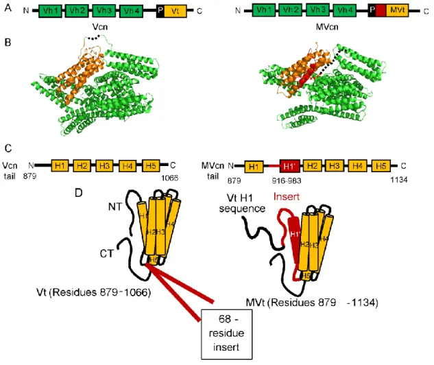
While vinculin is ubuitously expressed in all cell types, vinculin has one alternative splice isoform, termed metavinculin, that is co-expressed with vinculin in diverse muscle tissues and at low levels in platelets (42, 79-81). The metavinculin transcript contains an extra exon (exon 19) compared to the vinculin transcript, and this exon translates into an additional 68-residue insert in the tail domain (82). Metavinculin is expressed at sub-stoichiometric levels relative to vinculin (9-42%), and its expression correlates with the elevated contractile needs of these muscle cells (83, 84). It is currently believed that metavinculin plays a special role in mechanotransduction, as its expression levels positively correlate with the force exerted on cells (84). Metavinculin was initially discovered in chicken gizzards (42). Since then, it has been shown that the expression level of metavinculin in various tissues and cultured cells differ (83-88). Studies by Glukhova et al. showed that in adult human tissues, metavinculin was found in cardiac and smooth muscle of aorta and uterus, respectively (85); subsequent studies by Belkin et al. showed that metavinculin expression level in these tissues positiviely correlated with muscle contractility (83-85). Since this initial characterization of metavinculin, many groups investigated the (1) association of metavinculin with human cardiomyopathy patients at the tissue level, (2) the effect of disrupting the vinculin gene at the organismal level (mostly using mice), and (3) the characterization of the molecular structure and function of metavinculin.
In 1997, Maeda et al. found an association between deficiency of metavinculin expression and human dilated cardiomyopathy (DCM) patients (89). This observation led to several studies looking at the relationship between human cardiomyopathy patients and
metavinulin in these patients’ tissues (90, 91). Olson et al. found that in human DCM patients, point mutations/single amino acid deletions in metavinculin were located at residues within the
12
68 amino acid insert (A934V, ∆L954, and R975W). Of the three variants, R975W-associated patient showed the most pronounced defect in the organization of intercalated discs (90). Following this study, Vasile et al. also found that R975W is additionally associated with both hypertrophic cardiomyopathy (HCM) and DCM in human patients and that the reduced level of both vinculin and metavinculin at the intercalated disc is associated with HCM (91, 92).
Metavinculin and its association with cardiomyopathies was also studied at the
organismal level using mice by Zemljic-Harpf et al (93, 94). Zemljic-Harpf et al. observed that mice that have heterozygous inactivation of the vinculin gene are predisposed to stress-induced cardiomyopathy (94). These mice not only showed reduced expression levels of both vinculin (58% reduction) and metavinculin (63% reduction), but also abnormal myocardial ultrastructure and increased mortality rate (94). Furthermore, they found that the cardiac-myocyte-specific excision of the vinculin gene in mice led to disruptions in cellular junctions, leading to sudden death of DCM in these mice (93). One limitation of these studies, however, was that disruption of vinculin genes led to the deletion of both vinculin and metavinculin variants. Therefore, these studies were not able to tease apart individual functions due to vinculin or metavinculin.
13
metavinculin function (90). Additionally, Olson et al. had found that MVt A934V, ∆L954, and R975W have all led to an actin bundling phenotype in vitro, although MVt does not typically bundle actin filaments (90). Further structural studies will need to be done to elucidate how the structure of MVt contributes to DCM and HCM, as well as to clarify how A934V, ∆L954, and R975W mutations disrupt metavinculin function.
14
Differences between vinculin and metavinculin structure and function
Metavinculin and vinculin structurally share the same head domains (95, 96); however, their tail domains differ (Fig. 3). Vinculin tail domain possesses an N-terminal strap followed by a 5-helix bundle and C-terminal hairpin (46), while the metavinculin tail domain contains an additional exon that encodes a 68 amino acid insert (79) (Fig. 3). This extra exon codes for the residues between helices 1 and 2 in the vinculin tail domain and confers unique functions to the metavinculin tail domain (82). Structurally, while metavincuiln tail has a 5-helix bundle fold similar to vinculin tail, the sequence that makes up the helix 1 (H1) and strap of vinculin tail is displaced in the metavinculin tail by homologous sequences, which we term H1’, contained within this insert (95) (Fig. 3). Specifically, these new residues replace the vinculin residues 879-915, which translate to the N-terminal strap and the H1 in vinculin tail. The original vinculin tail sequence 879-915, in the context of metavinculin, is no longer observable in the existing crystal structures (95), which indicates that this region is either disorganized or dynamic. The altered tail domain structure in metavinculin increases the affinity for raver 1 (54) and decrease the affinity for PIP2 (97) compared to vinculin. This difference in raver 1 and PIP2 affinity with either Vt or MVt is especially interesting as vinculin and metavinculin differ structurally at the tail domain (95). Interestingly, raver 1 binds to not only both vinculin and metavinculin, but also to vinculin mRNA (56-58). Additionally, structural studies show that raver 1 can bind to full-length
15
16
Figure 3. Sequence and structural differences between Vcn and MVcn.
17
The role of vinculin-mediated actin-binding and actin-bundling Similar to Vt, MVt directly binds F-actin (95, 98, 99). However, one interesting difference is in their ability to crosslink F-actin. Vt has been shown to bundle F-actin in vitro; however, unlike Vt, MVt does not bundle filamentous actin into higher order structures in vitro (90, 98-100). We and others have previously shown that binding of F-actin to Vt causes a
conformational change in Vt that promotes dimerization and actin filament bundling (101, 102). However, the structure of the actin-induced Vt dimer is currently unknown. The susceptibility of H1 to proteolysis, combined with the lack of electron density observed for H1 in our cryo-EM reconstruction of the Vt-actin complex, suggests that H1 partitions away from the helix bundle upon engagement with filamentous actin to promote vinculin dimerization (99) (Fig. 4). Furthermore, the C-terminal tail of Vt plays a significant role in the formation of this actin-induced Vt dimer, as the deletion of the last five residues abrogates the ability of Vt to bundle actin (101). Expression of vinculin∆C5 mutant in vinculin-null MEFs lead to fewer and larger FAs and deficiency in cells to respond to external force (101). Similarly to Vt, H1’ in
metavinculin is not observable in the cryo-EM reconstruction of the metavinculin-actin complex (99), and the MVt binding site on F-actin is similar to that of Vt (99). The presence of H1 appears to interfere with the ability of metavinculin to bundle F-actin, as its deletion promotes actin filament bundling (95).
18
19
traditionally thought that metavinculin is more likely have a supporting role for vinculin in actin bundling. However, more recent studies seem to suggest that metavinculin may have a role in negatively regulating the actin bundling properties of vinculin physiologically. We and others have previously observed that the presence of metavinculin at sub-stoichiometric ratios impairs vinculin mediated F-actin bundling (99, 103), suggesting that MVt may negatively regulate Vt-mediated actin bundling. Furthermore, studies by Janssen et al. also observed actin filament fragments in the presence of MVt alone (98), and they attributed this observation to a potential ability of MVt to sever actin filaments. Additionally, studies by Durer et al. not only showed that MVt increases the proteolysis of F-actin by itself but also suggested that MVt may “tune” the flexibility and the architecture of vinculin-induced actin bundling (103). Durer et al. suggested this potential role of MVt as a negative-regulator of Vt-induced actin bundling as they found that MVt decreases the density and the thickness of actin filament bundles generated by Vt (103). However, unlike the studies by Janssen et al., Durer et al. found negligible actin severing activity by MVt (103). Though further studies are necessary to understand the molecular basis for the ability of MVt to negatively regulate actin bundling by Vt, several observations associated with metavinculin-associated diseases seem to support this perspective. Metavinculin mutants associated with DCM and HCM are able to form higher order actin assemblies in vitro (90), suggesting that the disturbance of metavinculin’s ability to disrupt vinculin-mediated actin bundling leads to cardiomyopathies. Similarly, Zemlijic-Harp et al. found decreased level of metavinculin expression in mice that’s developed DCM (93). Further molecular studies of how disease-associated MVt mutants lead to higher-order actin bundling will be helpful in
20
In addition to the molecular characterization of actin bundling by metavinculin, there has been a significant lack of cellular characterization of metavinculin. Although some studies on metavinculin have been done at the tissue and organismal level, there has yet to be a study that’s been done at the cellular level. Our lab has previously shown that the expression of actin
21
CHAPTER 2 – The Strutural Basis of Actin Organization by Vinculin, Metavinculin, and Metavinculin Cardiomyopathy-associated Mutants
Introduction
Vinculin functions as an important scaffolding protein and can engage at least 19 direct binding partners at both cell-cell junctions and cell-matrix adhesions. Included in these direct binding partners are several actin-binding proteins (104). These interactions alter cytoskeletal organization, which in turn play a role in cell morphology, motility, and force transduction. However, for the purposes of this study, we focused on direct interactions between Vt and F-actin. Vinculin also strengthens the physical connection between membrane receptors and actin cytoskeleton (105), and this mechanical reinforcement is primarily through vinculin’s
interactions with talin (47, 105, 106). Talin is a critical adhesion protein that binds to integrin (12) within its N-terminus and also to actin through its C-terminus end (1). Like vinculin, talin is a scaffolding protein that exists in an autoinhibited state (107). Once tension is applied across talin molecule, talin unfurls, revealing 11 binding sites for vinculin (30) and actin (108), though the actin-binding affinity is relatively weak (108). It is thought that the recruitment of vinculin by talin stabilizes the actin linkage to integrins by keeping talin in an active, unfurled state and also additionally increasing the linkage to actin through vinculin’s interactions with actin (109).
22
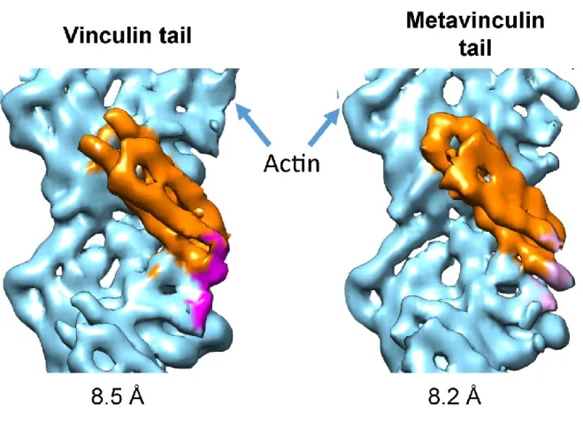
mechanotransduction (32, 101, 111, 112). While the vinculin tail domain in monomeric in solution, engagement to F-actin promotes a conformational change in Vt that facilitates Vt dimerization and actin bundling, as observed by cross-linking and electron tomography studies (101, 102, 110, 113). Unlike Vt, MVt cannot bundle actin filaments but can bind to actin failments (76, 82, 92, 100). The ability of vinculin, but not metavinculin, to reorganize actin filaments into bundles indicates that metavinculin and vinculin have distinct functions.
23
The cryo-EM reconstruction of Vt-actin and MVt-actin complexes provided us with a valuable insight into how Vt or MVt undergoes a conformational change upon binding to actin. However, as metavinculin is co-expressed with vinculin in muscle tissues (42, 79, 84), it is likely that vinculin and metavinculin coordinately regulate actin filament rearrangement in contractile cells. In fact, we found that the presence of MVt can suppress the actin bundling properties mediated by Vt in our actin cosedimentation assays, providing us with a hypothesis that MVt may negatively regulate the actin bundling properties of Vt. To better understand the role of metavinculin in actin filament assembly, we conducted a series of actin co-sedimentation and negative stain EM experiments in the absence and presence of vinculin. Consistent with our previous findings (99), MVt is unable to induce actin bundling, and the presence of
sub-stoichiometric amounts MVt relative to Vt inhibits the assembly of actin filaments into parallel bundles. Furthermore, we investigated whether cardiomyopathy (CM)-associated MVt mutants induce actin bundling either in the presence or absence of Vt. In contrast to wild type MVt, MVt CM mutants (which are all within the MVt insert) induce moderate actin assemblies but not defined parallel actin bundles induced by Vt. To better understand the molecular basis for the ability of MVt to negatively regulate Vt-mediated actin bundling, we performed DMD
simulations. Actin binding to vinculin promotes release of H1 from the tail domain helix bundle, exposing an interface in vinculin that promotes dimerization. However, our computational analyses indicates that in the case of metavinculin, the insert forms a higher order structure with H1’ that is released from the helix bundle upon actin binding, which occludes actin assembly into parallel F-actin bundles. Our MD simulations also indicate that cardiomyopathy mutations within the metavinulin tail domain, destabilize formation of this higher order structure.
24
25
Materials and Methods
Cloning and generation of expression constructs
Construct for chicken Vt residues 879-1066 was cloned into pQlinkH vector (Addgene, Cambridge, MA), and the C-terminally GFP-tagged construct Vt∆C5-GFP (Vt residues 879-1061 with C-terminal GFP fusion linked by the sequence “GIGSGSNGSSGS”) was generated using ligation-independent cloning in the H6-msfGFP vector (Addgene #29725). Vt∆C5-GFP construct encodes an N-terminal TEV (tobacco etch virus) cleavable hexa-histidine tag, the linker, and a C-terminal EGFP tag. The N-terminally tagged construct was generated using sequence- and ligation-independent cloning, inserting the open reading frame in-frame after the GFP (GFP-E892-Vt∆C5, Vt residues 892-1061 with an N-terminal GFP fusion and no linker sequence). The codon-optimized sequence of human MVt (residues 858-1134) for bacterial expression was synthesized (Geneart), and MVt and MVt∆C5 (residues 858-1129) were sub-cloned into the 2HR-T vector (#29718), which encodes an N-terminal TEV cleavable hexa-histidine tag. MVtp (residues 879-1134) was also generated in 2HR-T vector, and this construct was designed to lack the proline-rich linker region. Plasmids for MVt cardiomyopathy (CM) mutants, A934V, ∆L954, and R975W, were generated using QuikChange site-directed
mutagenesis kit (Stratagene) and verified by DNA sequencing (Genewiz). All of the Vt and MVt vectors contain TEV cleagable hexa-histidine tag.
Protein expression and purification
26
buffer [20mM Tris, 150 mM NaCl, 5mM imidazole, 2mM β-mercaptoethanol (pH 7.5) for Vt; 50 mM Tris, 200 mM NaCl, 10mM imidazole, 2mM β-mercaptoethanol (pH 8.0) for MVt]. Cells
were then lysed by sonication, and the proteins (either Vt or MVt) that remained in the soluble fractions were separated by centrifugation at 15,000 rpm for 45 min. Proteins were purified by using Ni-NTA agarose beads (Qiagen) as they bound to the beads through His-tag. Wash buffer [20 mM Tris, 150 mM NaCl, 60 mM imidazole, 2mM β-mercaptoethanol (pH 7.5) for Vt; 50
mM Tris, 200 mM NaCl, 25 mM imidazole, 2mM β-mercaptoethanol (pH 8.0) for MVt] was run
through the column to wash away any impurities bound to the column. Finally, the proteins were eluted using elution buffer [20 mM Tris, 150 mM NaCl, 500 mM imidazole, 2mM β
-mercaptoethanol (pH 7.5) for Vt; 50 mM Tris, 150 mM NaCl, 250 mM imidazole, 2mM β
-mercaptoethanol (pH 8.0) for MVt] from the column. His-tags from all proteins were removed by dialyzing the eluted volume into TEV cleavage buffer [20 mM Tris, 150 mM NaCl, 50 mM imidazole, 2mM β-mercaptoethanol (pH 7.5) for Vt; 50 mM Tris, 200 mM NaCl, 20 mM
imidazole, 2mM β-mercaptoethanol (pH 8.0) for MVt] overnight at 4 ºC in presence of TEV
protease. Vt and MVt proteins were then collected by running the dialyzed/TEV-cleaved volume over the Ni-NTA beads again. Size exclusion chromatography was used to purify these proteins further in gel filtration buffer [10 mM Tris, 200 mM KCl, 10 mM imidazole, 2.5 mM MgCl2, 1
mM EGTA, and 2mM DTT (pH 7.5)]. Purifed proteins were concentrated between 200-500 µM by centrifugation and used for experiments.
Actin co-sedimentation assays
27
from rabbit muscle acetonepowder (Pel-Freez Biologicals, Rogers, AR), was stored at − 80 °C in storage buffer [50 mM imidazole, 100 mM NaCl, 10 mM MgCl2, 10 mM EGTA, 0.5 mM DTT, 0.2 mM ATP (pH 7.0)]. Polymerization to filamentous actin (F-actin) was done by diluting and incubating G-actin at 100 μM concentration in actin polymerization buffer [10 mM Tris, 200 mM KCl, 10 mM imidazole, 2.5 mM MgCl2, 1 mM EGTA, 2 mM DTT (pH 7.5)] at room temperature for 30 min. The actin concentrations reported in this work were based on G-actin concentration, since the heterogeneity of F-actin polymers made it difficult to quantify F-actin concentrations. Vt and MVt variants were also diluted by actin polymerization buffer to prepare 100 μM stocks. To assess actin binding, 100 μl samples were prepared containing 10 μM Vt/MVt variants and 10 or 20 μM actin. The samples were incubated at room temperature for 1 h and then centrifuged at 100,000 RCF for 30 min. To assess actin bundling, 100 μl samples were prepared containing 10– 20 μM Vt/MVtvariants and 10 μM actin. The samples were incubated at room temperature for 1 h and then centrifuged at 12,000 RCF for 15 min. For both binding and bundling
co-sedimentation, the supernatant and pellet were separated, resuspended to equal volumes, and analyzed by 15% SDS-PAGE. Actin binding properties were calculated by determining the fractions of Vt/MVtvariants present in pellets using the densities of the pellet and supernatant bands. Actin bundling properties were calculated by determining the fractions of actin present in pellets using the densities of the pellet and supernatant bands. Densitometry was performed using ImageJ (115). Statistical significances (p values) of the measurements were determined using the Microsoft Excel t-Test function.
Negative-stain transmission electron microscopy
An aliquot of actin (1 μM) without or with Vt or MVt (10 μM) was incubated in actin
28
EGTA, 2 mM DTT (pH 7.5)] for 15 min and absorbed directly onto glow-discharged
carbon-coated 400 mesh copper grids for 3 min, and then stained with 2% (w/v) uranyl acetate in water.
TEM images were obtained using a FEI Tecnai 12 electron microscope at 80 kV and captured on
a Gatan First Light CCD camera using Gatan Digital Micrograph software (Gatan, Pleasanton,
CA). F-actin and the indicated Vt ± MVt constructs were mixed in KMEI [50 mM KCl, 1 mM
MgCl2, 1 mM ethylene glycol bis(b-aminoethyl ether) N,N′-tetraacetic acid (EGTA), 10 mM imidazole, 1 mM dithiothreitol (DTT), pH 7.0)] and incubated at room temperature for 15 min.
Sample (4 μl) was then applied to a glow-discharged continuous carbon grid (Ted Pella) and
incubated for 60 s. After incubation, the grid was washed with three 100 μl drops of 1% uranyl
acetate, then blotted to dryness. Images were acquired with the SerialEM package (116) on a
Tecnai F20 operating at 120 kV with a Gatan Ultrascan 4000 CCD camera. Tiled images with
20% overlap were acquired at 7800 × magnification, 3 μm underfocus, and 4-fold camera
binning, corresponding to a calibrated pixel size of 5.7 nm at the specimen level. Stitched images
were assembled with the “blendmont” program from the IMOD software package (117). This
work was done by Muzaddid Sarker, former lab member from Campbell lab, in collaboration
with Lindsey M. Constantini fromJack Griffith’s lab.
F-actin assembly quantification
Images were thresholded and binarized using ImageJ (115), then segmented into contiguous
regions of pixels using the built-in “Analyze Particles” plugin, including regions 100–500,000
pixels in size and with a circularity of 0–0.3. This procedure does not always capture every
region that an expert user would designate to contain F-actin in every image. However, we find
29
decisions on region boundaries and the minimum size of regions, as well as a sliding-box
quantification (a measure of local density), which is extremely sensitive to noise introduced by
slight differences in thresholding (data not shown).
Size measurements of regions were pooled from all images for a given condition, then divided
into 10,000 equally sized bins per data set and plotted via a normalized cumulative histogram.
Data were binned and cumulative sums calculated with a python script (available
at www.github.com/alushinlab/FactinAssemblyQuant) using the function “binned_statistic” implemented in SciPy (www.scipy.org). Plots were generated and statistical tests were conducted with GraphPad Prism. This work was done by Lin Mei and Santiago Espinosade los Reyes from
Greg Alushin’s lab.
Molecular dynamics simulation
Modeling was performed using a DMD package (118-120). The initial structure was obtained by
extending missing N- and C-termini of MVt (PDB ID: 3JBK) (99) with PYMOL built-in tool to
include residues 896–1134. The initial structure was relaxed at temperature T = 0.5 with high heat exchange coefficient Cex = 10 for 10,000 steps. The temperature unit is kcal/(mol kB).The relaxation was followed by replica exchange simulations with 10 replicas (T = 0.330, 0.360, 0.390, 0.420, 0.450, 0.480, 0.510, 0.540, 0.570, 0.600; Cex = 0.1 for 2 million steps). Replicas were exchanged every 1000 steps. To preserve contacts between MVt and actin, we applied
harmonic constraints to the N, CA, and C backbone atoms of selected residues (R1044, I1045,
N1048, R1055, T1058, I1059, Q1062, I1065, Q1086, E1089, M1090, H1093, N1094, E1104,
R1107, E1108, A1111, I1114). These constraints restrict atoms to move within 2A around initial
30
lowest-energy structures were selected and clustered based on pairwise root mean square
distance between structures. Two clusters were identified.
Structures representing the centroids of the two clusters were subject to DMD simulations at two
constant temperatures T1 = 0.5 kcal/(mol kB) and T2 = 0.55 kcal/(mol kB). For each temperature and for each structure, 5 independent simulations were run for 1 million steps with Cex = 0.1. To preserve contacts between MVt and actin, we applied harmonic constraints to N, CA, and C
backbone atoms of selected MVt residues (as described above). All atoms within actin were
considered static. This work was done by Andrey Krokhotin, a former lab member from Nikolay
31 Results
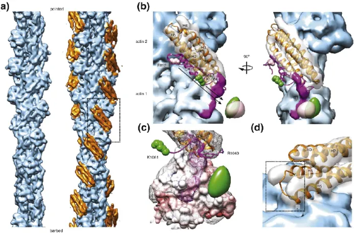
Cryo-EM reconstruction of Vt-actin and MVt-actin complex
In collaboration with the Alushin lab, we were able to obtain cryo-EM reonstructions of of both Vt and MVt in complex with F-actin complex. To circumvent the heterogeneity
associated with actin bundles formed by Vt, we used a C-terminal truncation mutant (from now on referred to as Vt∆C5) that preserves actin binding but disrupts actin bundling (101). I expressed and purified the Vt∆C5 and MVt∆C5 proteins used by the Alushin lab for
reconstructing the cryo-EM structures of Vt-actin and MVt-actin complexes. Both Vt and MVt have relatively low affinity for F-actin (~0.5 µM) (103, 121), making it technically challenging to fully decorate actin filaments. However, Alushin lab adapted a multi-reference iterative helical real-space reconstruction (IHRSR) (122) scheme that they developed for the study of
32
Vt undergoes a structural rearrangement upon actin binding by activating H1-mediated
bundling via steric mechanism
In Vt-actin complex reconstruction, we observed density for only four out of the five helices within Vt (Fig. 5), possibly due to disengagement of one helix from the bundle upon actin binding. Rigid-body docking of the isolated Vt crystal structure (PDB: 1QKR) (127)
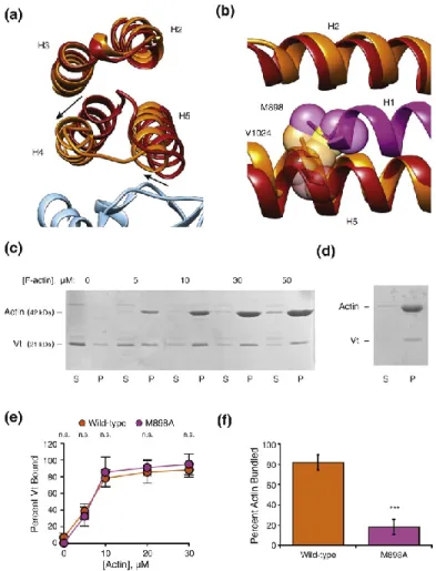
quantitatively supported the interaction pose produced using a DMD model our lab previously generated (112), in which helices 4 and 5 constitute the primary actin binding interface. Furthermore, we have performed actin binding and bundling co-sedimentation assays with Vt M898A, a mutant within H1 that is buried in the pre-bound state, reasoning that it would
sterically promote H1 release or mediate binding interactions upon being exposed. We found that Vt M898A did not affect actin binding but abrogated actin bundling, suggesting that H1 release upon actin binding is an important second step to in vinculin activation that mediates
Vt-mediated actin bundling (Fig. 6). This model suggests that helix 1 disengages from the helical bundle upon actin binding, which is further supported by our mutagenesis studies and that this region is susceptible to proteolytic cleavage upon actin binding by Vt (103).
In addition to the helix 1 displacement, the rigid-body fit of helices 2-5 showed
33
Figure 5. Sub-nanometer-resolution reconstruction of the Vt-actin interface.
34
which fits the density poorly. Asterisk highlights a clash with the actin surface.Generated by Alushin lab.
Figure 6. A steric mechanism promotes H1 release to bundle actin.
35
36 Figure 7. MDFF model of the Vt-actin surface.
37
to the remainder of the bundle (Fig. 6) upon actin binding. We hypothesized that H1 displacement is necessary for this structural transition to occur as this would generate steric clashes between large inward-facing hydrophobic residues in H1 and the rearranged hydrophobic core of Vt. Thus, we propose a model in which the need to relieve multiple clashes allosterically couples actin binding to H1 release.
MVt undergoes a similar structural mechanism
We also obtained an 8.2 Å-resolution reconstruction for MVt∆C -actin complex (Fig. 4). At sub-nanometer resolution, this reconstruction is indistinguishable from the Vt∆C5-bound reconstruction. In contrast to a previous negative-stain reconstruction where extra density was observed protruding from MVt when compared to Vt (98), rigid-body docking of the MVt crystal structure into our sub-nanometer-resolution density map showed that H1’ is displaced from the helical bundle when MVt binds actin. This observation is in agreement with the similar proteolysis susceptibility reported for MVt H1’ and Vt H1 upon actin binding (103).
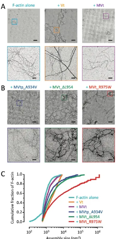
MVt WT does not induce F-actin bundling
With this H1’ displacement model, we next investigated the effects of MVt CM mutants. However, we first validated our WT Vt and MVt constructs to recapitulate previously reported binding and bundling activities (90, 98, 99, 103). I acquired actin co-sedimentation data with Dr. Muzaddid Sarker for these studies, while the negative-stain EM images were acquired by Dr. Muzaddid Sarker in collaboration with Griffith lab. We first confirmed that there were no significant differences between Vt and MVt in actin binding (Fig. 8), and also reproduced bundling differences between Vt and MVt proteins. We also determined whether the presence of the proline-rich link, that lies adjacent to the tail domain, contributes to higher order actin
38
Figure 8. MVt WT and CM mutants exhibit similar actin binding but not crosslinking.
(A) Representative SDS-PAGE results from high speed F-actin co-sedimentation assays in the presence of Vt, MVt, or MVtp WT and CM proteins (S, supernatant; P, pellet). (B)
39
containing MVt as MVtp from here on out. While the actin-alone sample showed single, linear actin filaments, addition of Vt to F-actin induced crosslinking of filaments into parallel bundles as expected, resulting in the formation of thick fibers (Fig. 9). When MVt or MVtp was added to F-actin instead of Vt, F-actin bundling was dramatically reduced, with few observable thick fibers. This is consistent with previous reports by our group and others that MVt does not induce large linear actin bundles like Vt (90, 98, 99, 103), indicating that the MVt insert region prohibits actin-induced MVt dimerization. In addition, inclusion of the proline-rich linker (MVtp) has a minor, slightly decreased actin bundling effect in vitro.
MVt CM mutants form higher-order, mesh-like actin assemblies
We next employed F-actin co-sedimentation assays to examine F-actin binding and aggregation activities of MVtp CM mutants, including A934V, ∆L954, and R975W. First, we compared the F-actin binding of MVtp WT and CM mutants relative to Vt. Samples containing actin (10 or 20 µM) and either Vt or MVt variants (10 µM) were subjected to high-speed
centrifugation to determine actin binding properties. Under these conditions, the supernatant (S) contains unbound Vt or MVt variant while the pellet (P) contains F-actin and bound protein. The percent of protein bound to F-actin were determined by SDS-PAGE (Fig. 8) and were quantified by ImageJ (115). All MVtp CM mutants, as well as the WT Vt, MVt, and MVtp, showed similar binding affinity to F-actin (Fig. 8). From these data, we demonstrate that MVt mutations do not impair F-actin binding, consistent with the distal location of the the mutations/deletions from the actin binding site.
40
Figure 9. MVt exhibits reduced F-actin bundling (crosslinking) compared to Vt.
Negative-stain EM images of actin filaments. Micrographs are acquired at the same
41
mutants are altered compared to WT Vt, MVt, and MVtp. With the low-speed centrifugation assay, only large cross-linked F-actin and bound proteins are pelleted, while individual actin filaments remain in the supernatant. The fraction of actin present in the pellet was quantified to determine the amount of higher-order assemblies in these mixtures (Fig. 8). With F-actin alone, only ~5% was found in the pellet. However, when Vt was added, almost all of the F-actin (~95%) was found in the pellet (Fig. 8). The amount of actin found in the pellet dramatically reduced when MVt was added (~26%) and reduced even further when MVtp was added (~13%) (Fig. 8). Significantly, there was an increase in the amounts of pelleted actin when MVtp CM mutants were added. MVtp A934V increased the amount of pelleted actin to ~29%, ∆L954 to ~41%, and R975W to 37% (Fig. 8). Even though the low-centrifugation assay is typically used to assess F-actin bundling activity, pellets from this assay may contain both thick bundled actin fibers but also other large disordered actin structures. Because low-speed centrifugation assay is not sufficient to visualize the type of actin assemblies that pellet in the presence of MVt CM mutants, we additionally employed negative-stain EM in parallel. The negative-stain EM data were found to be consistent with the actin co-sedimentation data (Fig. 10). We observed a significant increase in assemblies in the presence of CM mutants MVtp A934V, MVt ∆L954, and MVt R975W, with R975W having the most dramatic effect, in accordance with the severity of disease caused by this mutation in patients. Examination of the images shows primarily an irregular, mesh-like organization of actin filaments, unlike the majority species present as linear bundles formed in the presence of Vt.
42
Figure 10. MVt CM mutants promote disordered, mesh-like F-actin assemblies.
43
shown (bars = 1 μm). (C) Cumulative plots of F-actin assemblies from the indicated conditions. Pairwise comparisons show all distributions to be significantly different (KS test, *p < 0.01). N ≥ 10 fields and n ≥ 764 regions were quantified for each condition. F-actin, 0.5 μM;
44
MVt WT inhibits Vt-mediated actin bundling
As mentioned above, while Vt can bundle actin in vitro, MVt lacks the ability to bundle actin (90, 98, 99, 103). As metavinculin is co-expressed with vinculin at sub-stoichiometric levels under physiological settings, we next investigated higher order actin network organization in the presence of both WT Vt and MVt. Our observations that MVt H1′ and Vt H1 are released upon actin binding and that H1 mediates Vt's actin bundling activity suggest the following: MVt H1′, which differs in sequence from Vt H1, fails to promote MVt dimerization upon actin engagement. Interestingly, the H1 sequence is nevertheless present in our MVt construct, suggesting that the presence of H1′ inhibits the ability of released H1 to mediate MVt
45
Figure 11. MVt WT inhibits Vt-mediated actin bundling.
46
MVt CM mutants fail to inhibit Vt-mediated actin bundling
Given our finding that addition of WT MVtp at sub-stoichiometric levels relative to Vt inhibits Vt-mediated actin bundling, we next examined the effect of MVt or MVtp CM mutants on Vt-induced actin bundling. We employed low-speed actin pelleting assays to probe the effects of MVt or MVtp CM mutants in comparison to MVt WT, on Vt-induced F-actin assemblies. Three sets of actin co-sedimentation data were acquired, with 20 µM actin and Vt:MVtp at 5:5, 10:10, and 10:5 µM (Fig. 12). In the presence of WT MVt, we observed a proportionate
reduction of Vt-induced F-actin assemblies as expected. We found ~47-52% F-actin in the pellet for Vt:MVt at 1:1 and ~74% F-actin in the pellet for Vt:MVt at 2:1, as opposed to 95% F-actin in the pellet for Vt alone. Interestingly, for all 3 MVt or MVtp CM mutatns, almost all of F-actin was found in the pellet fractions at both 1:1 and 1:2 ratios. We observed ~87-95% F-actin for A934V, ~81-90% for ∆L954, and ~84-89% for R975W. These results indicate that unlike WT MVt, MVt or MVtp CM mutants fail to negatively regulate higher-order actin assemblies in the presence of Vt.
47
Figure 12. MVt CM mutants fail to inhibit Vt-induced F-actin bundling.
(A–C) Representative SDS-PAGE analysis of low-speed F-actin co-sedimentation assays incubated with Vt in the presence of MVtp WT protein (labeled as MVtp) or MVtp CM mutants at indicated concentrations (S, supernatant; P, pellet). (D) Quantification of the actin present in pellets representing higher-order F-actin assemblies that include F-actin bundles in case of Vt. Error bars represent standard deviation (SD) (n = 2, 5 replicates for each n). Statistical
48
Figure 13. MVt CM mutants aggregate Vt-induced actin bundles.
49
DMD suggests that MVt-specific insert region forms an additional sub-domain upon actin
binding
To gain further structural insight into rearrangements associated with MVt-actin interaction, we worked in collaboration with the Dokholyan lab. Using a single MVt including residues 896-1134 bound to an actin homodimer (F-actin) as the starting point (PDB: 3JBK) (99), DMD simulations were performed using replica exchange for 2 million steps. One hundred minimal energy structures were selected and clustered. Through this method, two clusters were identified (Fig. 14), with different N- and C-termini conformations. While structures from the first cluster have tightly intertwined N- and C- termini (Fig. 14), structures from the second cluster show the C-terminus interacting with the surface of F-actin (Fig. 14). Both clusters form a new additional structural sub-domain protruding outwards from F-actin. The structures were subjected to DMD simulations and appeared to be stable throughout the simulations, further supporting the protruding MVt sub-domain formation upon actin engagement. Therefore, we hypothesize that this sub-domain mediates unique biological functions of MVt relative to Vt, such as the inability to produce F-actin bundles and the ability to suppress Vt- mediated F-actin bundling. As the MVt CM mutants fail to inhibit Vt-induced actin bundling, we hypothesize that the CM mutations within the insert impair formation of the protruding structure. Further
50
51 Discussion
Using cryo-EM, molecular modeling, and complimentary biochemical techniques, we have produced detailed models of the critical interactions between F-actin, vinculin, and metavinculin. First, our cryo-EM reconstructions of Vt-actin and MVt-actin show that both H1 and H1’, respectively, are unfurled upon binding to F-actin. This finding adds an additional layer to the vinculin activation mechanism. After the interaction between Vh and Vt is disrupted, H1 must also be disengaged to license the Vt actin interaction. Although our data do not discriminate between H1 released followed by actin binding or vice versa, the steric incompatibility between the H1-docked state and the actin-bound state and NMR data suggesting that H1 undergoes conformational exchange in the isolated Vt (129) support the former model. Furthermore, vinculin sustains substantial tensile forces in adhesions in vivo (77), and a previous study demonstrated that vinculin is, on average, oriented along the dorsal-ventral axis of an adherent cell with the Vh domain closer to the ventral surface (27). These data suggest that a vinculin molecule bound to both talin via Vh and actin by Vt will experience tensile forces in geometry that will favor the undocking of H1 from the Vt bundle. Based on this geometry, we hypothesize a mechanism where vinculin can reinforce adhesion in response to force. We speculate that if H1 is in equilibrium between the docked and undocked states, the presence of tension will favor H1 undocking and by extension, actin binding and bundling, further increasing the
adhesion-cytoskeleton linkage (Fig. 15).
52
53
and metavinculin expression increases corresponding to the contractile load on the tissue (83, 84). Cardiomyocytes especially undergo rapid contraction and expansion as the heart beats. Based on these observations, we suggest that the presence of vinculin alone may cause the heart muscle to become stiff due to a large network of thick F-actin fibers, preventing the necessary contractile properties. Co-expression of metavinculin may be therefore necessary to regulate the vinculin-actin bundling so that cardiac cells remain flexible and functional. This hypothesis is further supported by our finding that MVt CM mutants are dysfunctional in suppressing the actin bundling by Vt.
We speculate that Vt H1 could mediate bundling contacts between Vt molecules after actin binding and further suggest that released H1’ and the upstream disordered sequence in MVt are important for MVt’s inhibitory activity by unknown mechanisms. As MVt CM-associated point mutants are located within the insert, either within H1’ or very close to the N-terminus of H1’, but distal from the direct actin binding regions of H2-H4, we hypothesize that these
mutations would compromise MVt’s regulation of Vt-mediated actin bundling without disrupting actin binding. Our results are in support of this model and accordingly, through actin
54
important subject for future studies, electron tomographic studies of Vt-induced 2D F-actin arrays on lipid monolayers suggested that filaments are very tightly apposed when cross-linked by Vt (110). We therefore propose that MVt sub-domain acts as a steric block that prevents another actin-bound Vt from coming in closer range, which would then prevent Vt-mediated actin bundling (Fig. 16).
Further in support of this model, our computational studies demonstrate that MVt CM mutants destabilize the MVt sub-domain formation, which would remove the steric block to actin-bound Vt and promote actin bundle formation (loss of function). However, it is important to note that the MVt sub-domain steric block model can only partially explain the effect MVt CM mutants have on actin network reorganization. Though not obvious through actin
co-sedimentation assay, negative-stain EM results revealed that all MVt CM mutants had a gain-of-function effect where they induced an increase in the formation of higher-order F-actin
assemblies (Fig. 10), with a disordered, mesh-like morphology. Also, our studies suggest that the MVt CM mutants additionally drive the coalescence of Vt-induced bundles into aberrantly large assemblies (Fig. 13). We hypothesize that MVt CM mutants stimulate aggregation of actin through the MVt insert region that would be exposed due to defects in sub-domain folding (Fig. 16).
55
Figure 16. Model for how MVt WT and CM mutants affectVt-induced actin bundle. Model for inhibition of Vt-induced F-actin bundle by MVt WT but failure of that by MVt CM mutants. (A) Release of H1 upon F-actin engagement enables Vt dimerization, thus resulting in parallel F-actin bundle formation. (B) An additional protruding structural sub-domain formed by the insert and displaced H1 at the N-terminus of MVt WT blocks homo- or hetero-dimer
formation with Vt, thus preventing F-actin bundling. (C) The protruding sub-domain is
56
CHAPTER 3 – Vinculin and Metavinculin Exhibit Distinct Effects on Focal Adhesion Properties, Cell migration, and Mechanotransduction
Introduction
57
mechanical tension (75-78), promote Vcn activation and scaffolding function by exposing multiple ligand binding sites.
Metavinculin (MVcn) is a larger splice isoform of Vcn that is selectively expressed in smooth and cardiac muscle cells and at low levels in platelets (42, 79, 81). MVcn is expressed at sub-stoichiometric levels relative to Vcn (9-42%), and its expression correlates with the elevated contractile needs of these muscle cells (83, 84). Complete knockout or heterozygous inactivation of the Vcn gene is associated with dilated cardiomyopathy in mice (93, 94), while reduced MVcn expression is also associated with dilated cardiomyopathy (DCM) and disorganized intercalated disc structures in humans (89). Point mutations in MVcn have also been identified in patients with DCM and hypertrophic cardiomyopathy (HCM) (89-91). While A934V and ∆L954 MVcn mutations are associated with DCM (90), an R975W mutation has been identified in patients with both DCM and HCM (91). Both DCM and HCM are diseases of the myocardium that diminish blood flow within the heart due to reduced force transmission.
58
regulate actin filament organization. In fact, we and others have previously observed that the presence of MVcn tail at sub-stoichiometric ratios impairs Vcn tail-mediated F-actin bundling (99, 103), suggesting that MVcn tail may negatively regulate Vcn tail-mediated actin bundling.
59
Materials and Methods Cell Culture
WT MEFs and Vcn-null MEFs were a gift from Dr. Brent Hoffman (Duke University), originally from Drs. Ben Fabry and Wolfgang Goldmann of the Erlangen Biophysics Group at the University of Erlangen-Nuremberg in Germany (134). Human embryonic kidney (HEK) 293T cells were a gift from Dr. Channing Der at UNC. All cells were cultured in Dulbecco’s modified Eagle’s medium (DMEM; Invitrogen) supplemented with 10% fetal bovine serum (Sigma) and antibiotic-antimycotic solution (Sigma). They were grown in a 37°C incubator with 5% CO2.
DNA Constructs and Generation of Stable Cell Lines
mEmerald-Vinculin-23 was a gift from Michael Davidson (Addgene plasmid #54302; http://n2t.net/addgene:54302; RRID: Addgene_54302). mRFP-C1 was a gift from Robert
Campbell & Michael Davidson & Roger Tsien (135) (Addgene plasmid #54764; http://n2t.net/addgene:54764; RRID: Addgene_54764). Human MVcn gene, a generous gift from
60
After 48 hours, the viruses were harvested and used to infect Vcn-null MEFs using 8 µg/mL polybrene. Vcn-null MEFs were infected for 24-48 hours and those expressing either mEmerald-Vcn or mRFP-MmEmerald-Vcn proteins were selected with 7.5 µg/ml puromycin for a week. After the cells were kept under the selection pressure at 5 µg/ml puromycin for about 3 weeks, they were sorted for expression by flow cytometry. Expression levels of both mEmerald-Vcn and mRFP-MVcn were verified by Western blot analysis using anti-mouse vinculin antibody (Sigma), which recognizes both Vcn and MVcn, and HRP-conjugated anti-mouse IgG (Jackson). Actin bands were detected using a mouse anti-actin monoclonal antibody (Millipore).
Flow Cytometry
61
Quantification of Focal Adhesion Area and Number per Cell