PREVOTELLA MELANINOGENICA, AN ORAL ANAEROBIC BACTERIUM, PREVALENT IN CYSTIC FIBROSIS CHRONIC LUNG INFECTION
Sarah Elizabeth Council
A dissertation submitted to the faculty of the University of North Carolina at Chapel Hill in partial fulfillment of the requirements for the degree of Doctor of Philosophy in the
Curriculum in Oral Biology
Chapel Hill 2013
Approved By:
ABSTRACT
SARAH ELIZABETH COUNCIL: Prevotella melaninogenica, an oral anaerobic bacterium, prevalent in cystic fibrosis chronic lung infection.
(Under the direction of Dr. Matthew Wolfgang)
Prevotella melaninogenica, an anaerobic Gram-negative bacterium, is a member
of the normal oral flora and is one of the most abundant anaerobic species found in respiratory specimens from individuals with cystic fibrosis (CF). Because of P.
melaninogenica’s designation as a commensal, its role in CF disease pathogenesis and host immune response has been largely ignored.
In our study of 61 CF patients at UNC hospitals, P. melaninogenica was cultured from 61% of adults and 57% of pediatric CF patients, and represented the most
abundant strict anaerobe in both groups. Lung function did not correlate with the presence or abundance of P. melaninogenica but there was an increased antibody response against P. melaninogenica in both adult and pediatric CF patients compared to non-diseased controls. To explore innate host response, we characterized the
From the tongue to the lung, P. melaninogenica must acquire nutrients to sustain life. The lung environment within chronically infected CF patients contains high levels of host iron proteins and pockets of anaerobic space. In vitro growth experiments
demonstrated that heme or hemoglobin were sufficient iron sources for P.
melaninogenica growth. To identify the first step of acquisition, we sequenced the P. melaninogenica genome and searched for homologues of known hemoglobin receptors.
We identified a comprehensive list of putative P. melaninogenica hemoglobin receptors. Together these studies characterize the prevalence of P. melaninogenica in CF infection, evaluate P. melaninogenica’s impact on the host and determine nutritional requirements, which will lead to a better understanding about the role of P.
melaninogenica in CF. Continued research into anaerobic pathogens, in particular P.
ACKNOWLEDGEMENTS
I first must thank my Mom and Dad who have always been behind me, pushing me towards my full potential. Thank you for letting me grow my own sugar crystals in the foyer, allowing me to tumble rocks in the basement and helping me with my science projects involving egg shells, coke and toothpaste. My family has been such an integral part of my journey. This includes many encouraging talks with Grandma and hanging out with my cousins Carrie and Kristan, who have become my sisters. For Grandpa and Mama, who passed away during this time, I will remember and cherish the time I spent with you.
The daily grind of graduate school would have been too much without my
roommates, Brittany and Jen, whose constant support and smiles after a bad day in lab were indispensable. I am blessed to have had so many supportive friends through the years from high school, college, NIH, church, and in my home away from home, lab. Ami, Gulshan, Marya and Emily have always had my back since high school. The same is true for Beth, Jon, Jes, Jared, Bhavna, Julie, Stephanie, Virginia, Chris, Brian, Amy, Emily, Josh, Marylois, John, Ted, Barbara and Hank in more recent years. I have to thank my lab mates Nan, Kim, Erin, Erich, Joe, Jeremiah and Cindy. Along with those whose lab I was only unofficially a part of even though I was a regular presence -- Phil, Donnie, Susan, Isabelle, Bill, Neelima, Debbie, Pete, Sharon, Jason and Cheryl. A special thank you to Dr. Flood, Cindy and students of the Oral Biology program who lovingly let me take part in the dynamic research community of the Dental school.
to thank Marcia, who was and is supportive and always pushing me to do better. I have truly benefitted from her guidance and mentorship. And thank you to Matt for giving me a cool project, allowing me to combine my love of everything related to oral ecology and systemic disease.
Finally, I’d like to recognize Joy, who gave my project life, literally!!! She has been an inspiration to me as I’ve watched her overcome the rigors of living with cystic fibrosis. She is someone who I can always turn to for a sarcastic joke.
Table of Contents
List of Tables ... viii
List of Figures ...ix
List of Abbreviations ...xi
Chapter 1 :Introduction ... 1
P. melaninogenica Classification and Epidemiology ... 1
Host Immune Response ... 4
Nutritional Requirements of P. melaninogenica ... 11
CF Lung Infection ... 16
References ... 24
Chapter 2:Prevotella melaninogenica, an oral anaerobe, in chronic cystic fibrosis lung disease ... 40
Abstract ... 40
Introduction ... 41
Material and Methods: ... 43
Results: ... 48
Discussion... 53
Figures ... 55
References ... 68
Chapter 3:Structure and innate immune response to lipopolysaccharide lipid A of Prevotella melaninogenica ... 72
Abstract: ... 72
Introduction: ... 73
Materials and Methods: ... 76
Results: ... 80
Discussion... 83
Figures: ... 87
Chapter 4: Prevotella melaninogenica iron requirements and
hemoglobin receptor characterization. ... 99
Abstract ... 99
Introduction: ... 100
Materials and Methods: ... 103
Results: ... 106
Discussion... 113
Figures: ... 117
References ... 129
Chapter 5:Conclusion ... 135
List of Tables
List of Figures
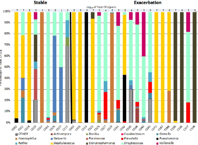
Figure 2.1. Microbiome Composition of Sputum
and BALF Samples ... 56
Figure 2.2. Most frequently cultured species in CF samples ... 59
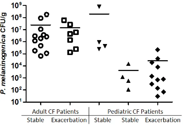
Figure 2.3. P. melaninogenica CFU/g in CF patient samples ... 61
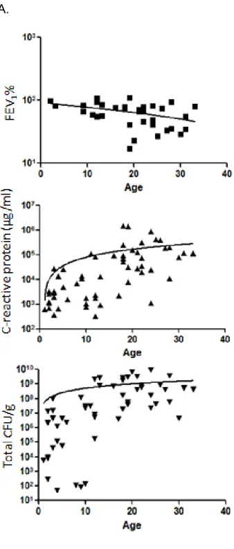
Figure 2.4. Comparisons of age to clinical measurements ... 62
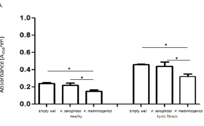
Figure 2.5. Absorbance experiments testing specificity of P. melaninogenica ELISA ... 64
Figure 2.6. P. melaninogenica reactive antibody titer in CF adults, CF children and controls ... 66
Figure 2.7. P. melaninogenica and P. aeruginosa reactive antibody comparisons ... 67
Figure 3.1. Structure of P. melaninogenica endotoxin ... 88
Figure 3.2. P. melaninogenica LPS stimulates THP1 cells significantly less than P. aeruginosa LPS. ... 89
Figure 3.3. P. melaninogenica stimulates NFĸB and IL8 production in a TLR4 independent mechanism ... 90
Figure 3.4. P. melaninogenica LPS NFĸB and IL-8 production response with TLR2 antibodies... 91
Figure 4.1. P. melaninogenica requires heme or hemoglobin to grow in ATSB. ... 119
Figure 4.2. P. melaninogenica iron and heme-iron acquisition system. ... 121
Figure 4.3. P. melaninogenica proteins homologous to characterized hemoglobin receptors. ... 122
Figure 4.4. Outline of experiments with E. coli K12 hemA system. ... 123
Figure 4.5. Functional assessment of P. melaninogenica proteins using hemoglobin agarose. ... 124
List of Abbreviations
ABC ATP-binding cassette
ATP Adenosine-5'-triphosphate
ATSB Anaerobic Tryptic Soy Broth
BALF Bronchoalveolar lavage fluid
BLAST Basic Local Alignment Search Tool
CF Cystic fibrosis
CFTR Cystic fibrosis transmembrane conductance regulator
CFU Colony forming units
CHO Chinese hamster ovary
COPD Chronic obstructive pulmonary disease
CRP C-reactive protein
Cyclic AMP Cyclic adenosine monophosphate
ELISA Enzyme-linked immunosorbent assay
ENaC Epithelial sodium channel
Fe2+ Ferrous iron
Fe3+ Ferric iron
FEV Forced expiratory volume
FEV1% Forced expiratory volume in 1 second compared to patients of similar characteristics (height, age, sex, and weight)
Ig Immunoglobulin
IL Interleukin
LB Luria broth
LPS Lipopolysaccharide
m/z Mass-to-charge ratio
MAP3K Mitogen-Activated Protein Kinase Kinase Kinase
mmHg Millimeter of mercury
MyD88 Myeloid differentiation primary response gene 88
NF-κB Nuclear Factor-Kappa B
O2 Dioxygen
OMP Outer membrane protein
ORF Open reading frame
PAMP Pathogen-associated molecular pattern
PMN Polymorphonuclear
PPIX Protoporphyrin IX
qPCR Quantitative polymerase chain reaction
SMG Streptococcus milleri group
THP-1 Human acute monocytic leukemia cell line
TLR Toll-like receptor
TNFα Tumor necrosis factor-alpha
Chapter 1
Introduction
P. melaninogenica Classification and Epidemiology
Prevotella melaninogenica, previously Bacteroides melaninogenicus (subspecies melaninogenicus), is an anaerobic, black pigmented, Gram-negative bacterium
belonging to the family Prevotellaceae (1). P. melaninogenica is a non-motile, catalase negative, saccharolytic bacterium that can produce an uncharacterized capsule (2, 3). P. melaninogenica is considered to be a member of the normal human oral flora and
can be cultivated from the tongue, gingival crevice, saliva and plaque of healthy individuals (4-7). Initial reports (1950-90s) showed that P. melaninogenica only colonized the mouth following tooth eruption (8, 9), but more recently, P.
melaninogenica has been isolated from the oral cavity of infants as young as two months of age (10, 11).
P. melaninogenica infections
P. melaninogenica has been described as a ‘potential pathogen’ because of its
the sole infectious agent in ‘extra-oral’ abscesses such as vertebral osteomyelitis, pyomyositis, peritonsillar abscesses and vaginal mesh infections (15-19). However, closely related oral bacteria, such as Porphyromonas gingivalis and Prevotella intermedia have received more attention due to their established association with
systemic diseases such as atherosclerosis, pneumonia, preeclampsia, cardiovascular disease, stroke, heart disease, and diabetes mellitus (20-22).
In addition to single species infection, P. melaninogenica is frequently cultured in the context of polymicrobial disease, including brain abscesses, pleuropulmonary
infections, endocarditis, illicit drug injection sites, intra-abdominal infections, wound infections, necrotizing fasciitis, pyogenic infections, decubitus and diabetic ulcers (16, 23-29). P. melaninogenica is also one of the most prevalent and abundant anaerobic species found in respiratory specimens from individuals with cystic fibrosis (CF) (12, 30-37).
Bacterial Synergism
The presence of P. melaninogenica within complex bacterial populations raises the possibility that its growth at different body sites requires bacterial synergism, where the presence of other bacterial species could improve the likelihood of colonization by this otherwise fastidious species. Pathogenic bacterial synergism occurs when a
polymicrobial community is more destructive to the host than any single member of the community. This type of synergism is often the result of microbial interactions such as nutritional sharing or interspecies quorum sensing, that can alter bacterial gene
In its simplest form, bacterial synergism has been studied in two species model systems. For example, it has been shown that virulence genes of the opportunistic pathogen Pseudomonas aeruginosa are upregulated in the presence of oropharyngeal isolates of either Streptococcus or Staphylococcus species. This modulation of
virulence factors was found to be partially due to an increase in autoinducer-2 signaling and competition for iron (38). Furthermore, experiments using an abscess model showed a greater host inflammatory response to mixed species infection compared to single species infection (39, 40). Additional studies have shown that there are growth benefits to Bacteroides species (Bacteroides fragilis and Bacteroides asaccharolyticus) when present in a polymicrobial abscess model of infection (41). It is believed that Bacteroides species specifically benefit from nutrients produced by the community.
Similarly, it has been shown that P. melaninogenica, can acquire vitamin K, an essential growth factor, when grown in the presence of Staphylococcus aureus (42). Because of its ability to survive and grow in polymicrobial infections, P. melaninogenica likely contributes to the pathogenic potential of these communities. Ultimately, bacterial virulence potential in the context of polymicrobial conditions depends on the virulence characteristics of the individual species, the host response, and environmental
conditions within the infected niche (20, 43, 44).
Virulence Characteristics of P. melaninogenica
against host detection and antimicrobial factors, P. melaninogenica isolates can
produce both IgA and IgG proteases (47-49), a β-lactamase (24), and a polysaccharide capsule. To further modulate the immune system, P. melaninogenica produces a
neuraminidase, which has been shown to cleave sialic acid from host surface
glycoproteins and leukocytes to disrupt immune recognition and trigger degradation of host proteins (50). Once colonization is established, P. melaninogenica can further damage host tissue through the production of collagenase (11, 51) and a lipase, both of which contribute to abscess formation (11).
Host Immune Response
P. melaninogenica is primarily regarded as a member of the oral commensal flora; consequently its role in disease pathogenesis and host immune response has been largely ignored. However, with the use of molecular-based detection methods, P. melaninogenica is frequently identified in abscesses and infected tissues throughout the body. Despite its association with a wide-variety of infections, little is known about its contribution to disease progression.
Innate Immunity
dendritic cells. LPS is composed of three distinct components: lipid A, core
polysaccharide, and O antigen. The covalently bound lipid component of LPS, lipid A, constitutes the hydrophobic outer leaflet of the Gram-negative outer membrane and is responsible for LPS toxicity. It is traditionally composed of a glucosamine disaccharide backbone with acyl chains of varying length and number and the presence or absence of phosphate groups. Length of the fatty acid chains and the addition of phosphate groups can greatly impact the toxicity of lipid A. The core polysaccharide, attached to lipid A, is composed of heptose and 2-keto-deoxyoctulosonic acid (KDO). The O antigen component, which is the outermost portion and hydrophilic part of the LPS, is made up of repeating subunits of 3 to 5 sugars that vary between species and even strains. The presence of O antigen is responsible for the ‘smoothness’ characteristics of the bacterial colonies and helps to confer resistance to phagocytosis. LPS, which is released during growth and death of a bacterium, has been shown to induce
macrophages to produce reactive oxygen species and to activate antigen presentation and cytokine response pathways. Traditionally, LPS activates the alternative
complement pathway.
Historically, P. melaninogenica was a member of a group of species collectively designated as Bacteroides melaninogenicus. Other members of this closely related group included Porphyromonas gingivalis, Prevotella intermedia and Prevotella
shown to exhibit weak pro-inflammatory properties (endotoxicity) (53). Most reports have shown that P. gingivalis LPS is significantly less inflammatory than E. coli LPS (54), while some studies suggest P. gingivalis LPS is equal or even more
proinflammatory than that of E. coli (55, 56). It has been suggested that the differences reported in these studies are due to alteration in LPS structure that result from different growth conditions and extraction techniques (57). P. gingivalis LPS has been shown to stimulate IL-1β, IL-6, IL-8, IL-10, IL-12, TNFα, IGF1 and NFĸB expression and
production in THP1 cells (58, 59) as well as, IL-1β, TNFα and IL-8 in polymorphonuclear neutrophils (54, 60, 61). Because controversy still remains about the magnitude of P. gingivalis LPS cytokine response, it is difficult to predict the impact of P. melaninogenica LPS on the host inflammatory response. Foundational studies need to be completed to thoroughly characterize the structure and inflammatory effect of P. melaninogenica.
Toll like receptors:
An important aspect of the inflammatory cytokine response to bacterial infection is due to a cascade of signaling pathways initiated by Toll like receptors (TLRs), which are transmembrane receptors that respond to PAMPS and have a primary role in innate immunity initiation (62). The TLR response to bacterial products occurs by both
stem from cytosolic pattern receptors that stimulate inflammasome components that in turn activate caspase 1 to cleave IL-1β and IL-18 into their active forms (58).
Traditionally, bacterial LPS binds to an accessory protein, lipopolysaccharide-binding protein, which then triggers the CD14-MD-2-TLR 4 complex and activates TLR 4 LPS dependent responses (62, 64, 65). Other TLRs, specifically TLR 2, are known to recognize bacterial products such as peptidoglycan, lipoteichoic acid and lipoproteins. Number and length of lipid A acyl chains in addition to their branched structure can lead to differential TLR signaling. Confusion in the field of LPS dependent TLR signaling has been clouded by lipoprotein contamination of LPS preparation causing TLR 2 activity leading researchers to conclude TLR 2 dependence (66).
Bacteroides species, such as P. gingivalis, demonstrate unique structure
changes dependent on environmental cues (57, 67-69). In particular, environmental heme concentration modifies the activity of lipid A 1-dephosphorylatases that leads to an alteration in phosphate group attachment to the glucosamine portion of lipid A. This change in structure modulates the ability of P. gingivalis to stimulate TLR 4 (53, 57, 62, 69). In these reports, P. gingivalis LPS signals through TLR 4 either acting as an
agonist in low heme (1 mg/ml) or an antagonist in high heme (10 µg/ml) (68-70). P. gingivalis LPS response is controversial due to the questions that still remain about the variability of P. gingivalis cytokine response compared to other more
traditional agonists (i.e. E. coli LPS) and the inconsistent reports deciphering P.
gingivalis TLR signaling pathways. Despite the similarities and phylogenetic relationship
gingivalis LPS structure and properties make it difficult to make predictions about P.
melaninogenica TLR signaling.
P. melaninogenica innate immune response
Characterization of host immune response to P. melaninogenica has been limited in the past because of its inclusion under the grouping of B. melaninogenicus, which is now recognized to have encompassed multiple species including P. gingivalis.
Consequently, reports prior to 1990 have been considered in this review only if a specific P. melaninogenica strain was clearly specified.
P. melaninogenica LPS studies are incomplete; and have only focused on O antigen and the hemagglutination properties of total LPS preparations. P.
melaninogenica O antigen appears to have similarities to O antigen from Prevotella levi (71) which is involved in evasion of phagocytosis. Total LPS preparations from P. melaninogenica have been shown to have less hemagglutination activity compared to
other Prevotella species such as P. intermedia and P. denticola (52). Because of the role of LPS in initiating bacterial infection and host immune recognition a more thorough characterization of P. melaninogenica LPS is needed.
melaninogenica could be a TLR 4 antagonist. In a study designed to model the
polymicrobial environment of COPD, P. melaninogenica lysates dampened
Haemophilus influenzae TLR 4 signaling in dendritic cells leading to a decrease in IL-12 response (74). Additionally, experiments investigating the effect of ‘normal flora’ to pathogenic bacteria immune evasion found that P. melaninogenica supernatants,
representing normal flora, impaired the phagocytosis of the pathogen, Proteus mirabilis, by polymorphonuclear leukocytes (75). These studies represent the effect of a
complex mixture on cellular response, but do show how P. melaninogenica could have a potential impact on innate immune response and the immune stimulation by other pathogens.
Adaptive Immunity
Among the five classes of immunoglobulins, immunoglobulin G (IgG) makes up the majority of serum antibodies and is commonly used as a predictive marker for infection (76). IgG aids phagocytosis through opsonization and complement activation. In the case of bacterial infections, the development of a specific acquired immune response is characteristic of pathogen exposure and bacterial burden (77-79). Elevated antibody titer, (specifically IgG titer) for specific pathogens, has been used to diagnose infections such as syphilis (80), and human papilloma virus (81).
including the reduction of bacterial load, correlate with a reduction of serum IgG titer to specific pathogens (76).
IgG levels to P. melaninogenica have been measured in a small number of studies. P. melaninogenica IgG titer has been shown to be increased in rheumatoid arthritis patients compared to healthy controls (2). Also, P. melaninogenica IgM (an early response antibody) is increased in ventilator-associated pneumonia patients compared to controls (78). Based on these findings, immunoglobulin titer can be an effective tool in evaluating infection by Prevotella species (78).
Immune response to commensal bacteria
The presence of commensal bacteria in the body is a constant stimulus to the immune system and requires the host to produce an immune response, both cytokine and antibody, to keep the commensal contained. The host response against
endogenous bacteria becomes increased when the bacteria spread to non-traditional locations in the body.
In the oral cavity, commensal bacteria stimulate low-level inflammation that contributes to oral health. Specifically, low levels of IL-8 induce the chemotaxis of neutrophils into the gingivalis crevice to patrol for bacterial pathogens (13). In other parts of the body, the low level stimulation by commensal bacteria aids in the
Nutritional Requirements of P. melaninogenica
Iron
Iron, commonly observed in one of two states (Fe2+, Fe3+), has a extensive redox potential, making it a critical enzyme cofactor in the metabolism of amino acid and biosynthesis of nucleotides, vital processes in all organisms (87, 88). For aerobic bacteria, iron is critical for respiration, where it serves as a cofactor for cytochromes involved in electron transport (89). In contrast, anaerobes use iron-sulfur compounds and fumarase, in addition to other iron containing molecules such as catalase and peroxidase, for protection against superoxide and in the production of ATP through pyruvate aided fermentation (90, 91).
Iron in the host:
Iron is a necessary nutrient for basic cellular metabolism; however, because of its reactivity it can be harmful if not complexed. Under reducing (anaerobic) conditions or at low pH, ferrous iron (Fe2+) is the dominant iron form. Ferrous iron is more soluble than ferric iron (Fe3+) making it more toxic and able to pass through semi permeable
membranes. Iron can catalyze the Fenton Reaction which leads to the production of reactive oxygen species that damage cellular components (92). Because of the
transferrin (bodily fluid such as blood). The majority of iron in the human body is sequestered in hemoglobin in the form of heme (94).
Bacterial Iron Regulation
To survive in the host where iron is scarce, bacteria have developed tightly controlled mechanisms to alter the expression of iron acquisition proteins such as siderophores, degradative enzymes, hemolysins, and hemagglutination (46, 89, 95-97) in response to iron abundance and scarcity. In many bacteria, iron acquisition is
regulated by the ferric uptake regulator (Fur), which represses transcription of iron transport and scavenging genes when intracellular iron concentrations are sufficient. Available intracellular iron binds to Fur, facilitating the formation of Fur dimers. The dimer complex binds to the promoter region of iron regulated gene through recognition of specific (Fur box) sequences (98-100). The DNA bound complex hinders access of RNA polymerase. Under iron limiting conditions, Fur controlled genes involved in iron acquisition and storage become derepressed (94, 99, 101-104).
Porphyromonas and Prevotella heme requirements
In bacteria, the heme molecule is essential for electron transport and the activity of several metabolic enzymes (100, 105). In P. gingivalis, heme is a cofactor of the cytochrome b subunit of fumarate reductase, which plays a role in metabolic energy production (94, 102, 106, 107). Also a heme derivative, µ-oxo bishaem, is stored on the outer surface of many Bacteroides species and produces the characteristic black
pigment associated with P. gingivalis (108) and other black pigmented Bacteroides such as P. melaninogenica and P. intermedia when exposed to oxygen. The surface
localized heme derivative is used for protection against oxygen radicals, and is thought to promote local environmental anaerobiosis (108-110).
Many microbes can produce heme through a complex in vivo heme biosynthesis pathway; however, all bacteria in the genera Bacteroides lack the enzymes to
synthesize their own heme. Specifically, P. gingivalis lacks genes encoding
5-aminolevulinic acid synthase and porphobilinogen deaminase (111) and must rely on exogenous sources of this molecule.
P. gingivalis can acquire heme from host heme binding proteins such as
P. melaninogenica, like other Bacteroides species including P. intermedia,
requires heme for growth (42). P. intermedia and P. gingivalis represent closely related species that have differing abilities to use iron and heme based sources. Further
research is necessary to determine the iron requirements for P. melaninogenica and its capacity to utilize host-based iron sources.
In vivo heme acquisition
The majority of useable iron in the human body comes in the form of heme, complexed inside of hemoglobin, myoglobin or haptoglobin. Bacteria have successfully overcome this limitation by using two mechanisms that remove heme from host heme-containing proteins: 1) production of hemophores, which are secreted proteins that bind heme and are subsequently recognized by a cognate bacterial surface receptor, and 2) direct extraction of heme via high affinity heme- or hemoglobin-binding bacterial surface proteins (116). Some bacteria encode only high affinity hemoglobin receptors whereas others use a combination of receptors and hemophores. In addition, there are
Heme acquisition by Gram-negative bacteria is an orchestrated process that involves heme binding to an outer membrane receptor (in some cases aided by hemophore) followed by a series of steps that transfer heme through the outer membrane and periplasm and into the cytoplasm where it is used as a cofactor itself (e.g. in cytochromes or catalases) or is broken down for its iron component (123). Bacterial hemoglobin receptors form a beta barrel confirmation in the outer membrane and have characteristic domains (FRAP and NPNL signatures) and specific histidine residues that aid in the removal of heme from hemoglobin (116, 124-127). Once bound, heme is transported through the beta barrel channel of the receptor by the energy
derived from the proton motive force associated with the binding of the TonB complex to the receptor. Binding of the two proteins is mediated through a ‘TonB box’, which is a conserved sequence present in the N-terminal periplasmic portion of the outer
membrane receptor (128, 129). Once heme enters the periplasm, it is bound and transported by a heme permease to the inner membrane, where an ABC transporter can then transport it to the cytoplasm (130).
been characterized but a key to its in vivo growth could be its ability to promote
aggregation and lysis of red blood cells (46), which suggests that it is capable of freeing hemoglobin for bacterial binding and eventual heme uptake. The proteins involved in the subsequent steps in heme acquisition have yet to be identified.
CF Lung Infection
Mechanism
The most common fatal genetic disease in the Caucasian population is cystic fibrosis (CF). CF is inherited in an autosomal recessive pattern and occurs in
with mucin hyper-secretion by goblet cells leads to thickened mucus in the conducting airways of CF patients.
Inefficient clearance of mucus in the CF lung provides an optimal colonization niche for a diverse assembly of bacteria (146-148). From infancy to adulthood, CF patients experience a decline in lung function caused by persistent bacterial infection and unrelenting pulmonary inflammation (149). Over a period of years, an accumulation of bacterial products and cellular debris produces irreversible airway damage and inflammation that ultimately leads to respiratory failure and death, with an average life expectancy of 37 years.
CF Treatment:
Treatment of CF starts early in life through therapeutic bronchodilators, anti-inflammation treatments and antibiotics. Antibiotic treatment is based on aerobic culture of bronchoalveolar lavage fluid (BALF) and sputum. BALF is captured through an
invasive procedure where sterile saline is released into the conducting airways through a bronchoscope then collected. Induced and spontaneous sputum samples are
clearing bacterial infection (142, 151, 152). For example, there is no significant change in aerobic bacterial load comparing episodes of disease exacerbation and subsequent recovery periods (38) or testing pre and post antibiotic treatment (153). These
discrepancies suggest that aerobic culture does not provide the full picture of CF
pathogenesis. There is now growing evidence that pulmonary infections in CF should be treated as a polymicrobial infection with aerobic, anaerobic and fungal components (146).
Key aerobic bacteria in the CF microbiome
By as early as 3 months, nearly 40% of infants diagnosed through neonatal CF screening have a lower respiratory bacterial infection (154). Aerobic culture based techniques and quantitative PCR (qPCR) for the highly conserved bacterial 16S rRNA gene from CF samples (BALF, induced sputum, spontaneous sputum) illustrate that CF affected children are colonized by Staphylococcus aureus, Streptococcus spp,
within mucus plugs, allowing the bacteria to be highly resistant to phagocytosis, antibodies and antibiotic treatment (143, 151, 163). There is direct evidence of P. aeruginosa enmeshed in alginate biofilm aggregates in CF lung samples (147). In adults, the bacterial community within the biofilm may also include other aerobes such as Streptococcus milleri species (164), Burkholderia cepaci, Stenotrophomonas
maltophilia and multiple fungal species (165). As CF lung disease progresses the
successful growth of P. aeruginosa is aided by the ability of P. aeruginosa to adapt to other bacteria and environmental changes including the reduction of oxygen in CF mucus plugs (166).
CF anaerobic niche
Direct measurements of the oxygen gradient in CF mucus plugs range from 180 mmHg outside to 2.5 mmHg inside the plug, demonstrating a significant drop in oxygen and near anaerobic conditions within the airway mucus (166). The exact mechanism of oxygen depletion within CF mucus plugs has not been confirmed but there are multiple hypotheses involving accelerated O2 consumption either by the lung epithelium,
immune cell respiratory bursts or elevated bacterial respiration (141). P. aeruginosa and other facultative anaerobes that are present in the lungs of CF patients can live
anaerobically in the presence of an appropriate terminal electron acceptor, such as arginine (163, 166-170).
samples processed using anaerobic culture techniques and other unbiased molecular methods such as 16s rRNA gene profiling by microarray, pyrosequencing and reverse transcription terminal restriction fragments length polymorphisms (TRFLP), have
revealed the presence of strict anaerobes (29-32, 34-37, 148, 156, 164, 165, 171-173). Because of this, many have hypothesized that anaerobes may play a role in the
pathogenesis of CF airway disease (12, 31, 37, 38, 142, 148, 171). Tunney et al. (2008) noted that the most prevalent strict anaerobes isolated from sputum were Prevotella species, including P. melaninogenica. Anaerobic bacteria in this study were isolated in high numbers (104-109 CFU/g sputum) in 64% of adults and, in some cases, were present in higher numbers than P. aeruginosa (37). The prominence of Prevotella is consistent with past studies involving anaerobic culture techniques and molecular based approaches (12, 29-32, 34-37, 148, 156, 164, 165, 172-175). It has been suggested that anaerobes, in particular Bacteroides spp. (now Prevotella spp., Porphyromonas spp. etc), could be of clinical importance in CF (148) and potentially pathogenic (30, 32, 37). Of the Prevotella species enumerated in CF samples, P. melaninogenica is the most common anaerobe reported using culture independent and anaerobic culture
techniques (37), and it is frequently present in high numbers (12, 30-32, 34-36). Despite the cumulative evidence for the presence of anaerobic bacteria in the CF lung based on analysis of sputum and BALF, two studies of explanted lung specimens suggested that oral anaerobes are not present in lower airway but instead are found in upper airway specimens such as the trachea due to ‘oral contaminants’ (174, 176).
In CF, when bacteria colonize the lung, the host responds with an influx of
polymorphonuclear leukocytes followed by cytokine and antibody production (138, 140, 141, 146, 149, 177-179). Studies have shown that this CF characteristic response of PMNs is not a CFTR dependent response as some had speculated (177, 180). The immune response of CF patients is defined by ineffective killing of bacteria colonizing the lung, and the development of chronic inflammation that leads to lung dysfunction and respiratory failure. These processes are responsible for the majority of CF deaths (37, 141, 169). The exact initiation events leading to CF airway inflammation are the subject of debate, in part, because of the complexity of CF pathogenesis and
inconsistent experimental results. Further studies investigating cytokine response shows that there is significantly more IL-8 and neutrophils in BALF from CF children than children with other non-CF respiratory disease (179). Additionally, studies using sputum and BALF samples from CF adults show an increased number of neutrophils, increased levels of the pro-inflammatory cytokines TNF α, IL-1β, IL-6, IL-8, and reduced levels of the anti-inflammatory cytokine IL-10 (181-183); this response is thought to be mediated by TLR-dependent pathways (137, 184).
Iron in the CF lung
The lung environment within chronically infected CF patients contains higher levels of iron compared to the lungs of healthy patients (89, 150, 158). Significant amounts of iron have been detected in sputum from CF patients in the form of ferritin, lactoferrin, transferrin and small amounts of hemoglobin (89). In an unpublished study, there was more than twice the amount of hemoglobin and heme in CF BALF than in asthmatic patients; ten times more than healthy individuals (188). The underlying cause of elevated iron in the CF lung is thought to be from micro-hemorrhage, inflammation (189), transferrin/lactoferrin proteolysis and release of intracellular iron stores from CF airway epithelial cells (93, 190). Additionally, the iron present in the CF lung is predicted to be more soluble (and therefore better available for bacterial uptake) because the pH of the lung is more acidic (5.8) in CF patients compared to healthy controls (6.1) (191).
The role of P. melaninogenica in CF lung infection
Despite evidence that anaerobic bacteria are part of the polymicrobial community in the CF lung, the question of their clinical relevance remains unanswered. The role of anaerobes in CF pathogenesis can be thought of in two ways: 1) the anaerobes
P. aeruginosa and anaerobes in several cases (28, 39, 41). Bacteroides species
including Prevotella are known to produce quorum-sensing signaling molecules and thus have the potential to affect the virulence of P. aeruginosa. Further work needs to be done in this area comparing P. aeruginosa to prominent anaerobic species.
Here, we will explore the contribution of P. melaninogenica, which is the most commonly cultured anaerobic bacterium in the CF lung, to CF pathogenesis. In chapter 2, we investigate the prevalence and abundance of P. melaninogenica in a cohort of UNC hospital CF patents and test P. melaninogenica reactive antibody response as a measurement of bacterial burden and exposure. This study is aimed at determining whether host response to a nontraditional CF associated bacterium is different in CF patients compared to non CF individuals. In chapter 3, we determine the structure and inflammatory properties of P. melaninogenica LPS, the most toxic part of gram-negative outer membrane. In this chapter, we will examine the effect of P. melaninogenica LPS on a human monocytic cell line and determine TLR signaling pathways. Chapter 4 focuses on how P. melaninogenica survives in the CF lung, specifically determining its ability to acquire iron from host sources and the mechanism for heme acquisition.
References
1. Shah HN, Collins DM. Notes: Prevotella, a new genus to include Bacteroides
melaninogenicus and related species formerly classified in the genus Bacteroides. Int J Syst Bacteriol. 1990;40(2):205-8.
2. Ogrendik M, Kokino S, Ozdemir F, Bird PS, Hamlet S. Serum antibodies to oral anaerobic bacteria in patients with rheumatoid arthritis. Medscape General Medicine. 2005;7(2):2.
3. Wu CC, Johnson J, Moore W, Moore L. Emended descriptions of Prevotella
denticola, Prevotella loescheii, Prevotella veroralis, and Prevotella melaninogenica. Int J Syst Bacteriol. 1992;42(4):536-41.
4. Duerden B. The isolation and identification of Bacteroides spp. from the normal human gingival flora. J Med Microbiol. 1980;13(1):89-101.
5. Bik EM, Long CD, Armitage GC, Loomer P, Emerson J, Mongodin EF, et al. Bacterial diversity in the oral cavity of 10 healthy individuals. The ISME journal. 2010;4(8):962-74. 6. Papaioannou W, Gizani S, Haffajee A, Quirynen M, Mamai‐Homata E,
Papagiannoulis L. The microbiota on different oral surfaces in healthy children. Oral Microbiol Immunol. 2009;24(3):183-9.
7. Könönen E. Pigmented Prevotella species in the periodontally healthy oral cavity. FEMS Immunol Med Microbiol. 1993;6(2):201-5.
8. Bailit H, Baldwin D, Hunt E. The increasing prevalence of gingival Bacteroides melaninogenicus with age in children. Arch Oral Biol. 1964;9(4):435-8.
9. Hentges DJ. The anaerobic microflora of the human body. Clinical infectious diseases. 1993;16(Supplement 4):S175-80.
10. Könönen E, Kanervo A, Takala A, Asikainen S, Jousimies-Somer H. Establishment of oral anaerobes during the first year of life. J Dent Res. 1999;78(10):1634-9.
11. Alauzet C, Marchandin H, Lozniewski A. New insights into Prevotella diversity and medical microbiology. Future microbiology. 2010;5(11):1695-718.
13. Robert R, Grollier G, Frat JP, Godet C, Adoun M, Fauchère JL, et al. Colonization of lower respiratory tract with anaerobic bacteria in mechanically ventilated patients.
Intensive Care Med. 2003;29(7):1062-8.
14. Falagas ME, Siakavellas E. Bacteroides, Prevotella, and Porphyromonas species: A review of antibiotic resistance and therapeutic options. Int J Antimicrob Agents.
2000;15(1):1-9.
15. Mukhopadhyay S, Rose F, Frechette V. Vertebral osteomyelitis caused by Prevotella (Bacteroides) melaninogenicus. South Med J. 2005;98(2):226.
16. Bowler P, Duerden B, Armstrong D. Wound microbiology and associated approaches to wound management. Clin Microbiol Rev. 2001;14(2):244-69.
17. Odeh M, Oliven A, Potasman I, Solomon H, Srugo I. Pyomyositis of the thigh due to Prevotella melaninogenica. Infection. 2000;28(1):49-50.
18. Athanasiou S, Matthaiou DK, Falagas ME. Vaginal mesh infection due to
Bacteroides melaninogenicus: A case report of another emerging foreign body related infection. Scand J Infect Dis. 2006;38(11-12):1108-10.
19. Jousimies-Somer H, Savolainen S, Mäkitie A, Ylikoski J. Bacteriologic findings in peritonsillar abscesses in young adults. Clinical infectious diseases.
1993;16(Supplement 4):S292-8.
20. Li X, Kolltveit KM, Tronstad L, Olsen I. Systemic diseases caused by oral infection. Clin Microbiol Rev. 2000;13(4):547-58.
21. Moutsopoulos NM, Madianos PN. Low‐Grade inflammation in chronic infectious diseases. Ann N Y Acad Sci. 2006;1088(1):251-64.
22. Paju S, Scannapieco F. Oral biofilms, periodontitis, and pulmonary infections. Oral Dis. 2007;13(6):508-12.
23. Hsiao WWL, Li KL, Liu Z, Jones C, Fraser-Liggett CM, Fouad AF. Microbial transformation from normal oral microbiota to acute endodontic infections. BMC Genomics. 2012;13(1):345.
24. Talan DA, Abrahamian FM, Moran GJ, Citron DM, Tan JO, Goldstein EJC. Clinical presentation and bacteriologic analysis of infected human bites in patients presenting to emergency departments. Clinical infectious diseases. 2003;37(11):1481-9.
26. Harding G, Sutter V, Finegold S, Bricknell K. Characterization of Bacteroides melaninogenicus.. J Clin Microbiol. 1976;4(4):354-9.
27. De A, Varaiya A, Mathur M. Anaerobes in pleuropulmonary infections. Indian Journal of Medical Microbiology. 2002;20(3):150.
28. Brook I. Prevotella and Porphyromonas infections in children. J Med Microbiol. 1995;42(5):340-7.
29. Sibley CD, Grinwis ME, Field TR, Eshaghurshan CS, Faria MM, Dowd SE, et al. Culture enriched molecular profiling of the cystic fibrosis airway microbiome. PloS one. 2011;6(7):e22702.
30. Field TR, Sibley CD, Parkins MD, Rabin HR, Surette MG. The genus Prevotella in cystic fibrosis airways. Anaerobe. 2010;16(4):337-44.
31. Worlitzsch D, Rintelen C, Böhm K, Wollschläger B, Merkel N, Borneff‐Lipp M, et al. Antibiotic‐resistant obligate anaerobes during exacerbations of cystic fibrosis patients. Clinical Microbiology and Infection. 2009;15(5):454-60.
32. Ulrich M, Beer I, Braitmaier P, Dierkes M, Kummer F, Krismer B, et al. Relative contribution of Prevotella intermedia and Pseudomonas aeruginosa to lung pathology in airways of patients with cystic fibrosis. Thorax. 2010;65(11):978-84.
33. Bittar F, Richet H, Dubus JC, Reynaud-Gaubert M, Stremler N, Sarles J, et al. Molecular detection of multiple emerging pathogens in sputa from cystic fibrosis patients. PLoS One. 2008;3(8):e2908.
34. Stressmann FA, Rogers GB, Klem ER, Lilley AK, Donaldson SH, Daniels TW, et al. Analysis of the bacterial communities present in lungs of patients with cystic fibrosis from American and British centers. J Clin Microbiol. 2011;49(1):281-9.
35. Harris JK, De Groote MA, Sagel SD, Zemanick ET, Kapsner R, Penvari C, et al. Molecular identification of bacteria in bronchoalveolar lavage fluid from children with cystic fibrosis. Proceedings of the National Academy of Sciences. 2007;104(51):20529-33.
36. van der Gast CJ, Walker AW, Stressmann FA, Rogers GB, Scott P, Daniels TW, et al. Partitioning core and satellite taxa from within cystic fibrosis lung bacterial
communities. The ISME journal. 2010;5(5):780-91.
38. Duan K, Dammel C, Stein J, Rabin H, Surette MG. Modulation of Pseudomonas aeruginosa gene expression by host microflora through interspecies communication. Mol Microbiol. 2003;50(5):1477-91.
39. Brook I. Pathogenicity of capsulate and non-capsulate members of Bacteroides fragilis and B. melaninogenicus groups in mixed infection with Escherichia coli and Streptococcus pyogenes. J Med Microbiol. 1988;27(3):191-8.
40. Brook I. Encapsulated anaerobic bacteria in synergistic infections. Microbiol Rev. 1986;50(4):452.
41. Brook I. Enhancement of growth of aerobic and facultative bacteria in mixed infections with Bacteroides species. Infect Immun. 1985;50(3):929-31.
42. Gibbons RJ, Macdonald JB. Hemin and vitamin K compounds as required factors for the cultivation of certain strains of Bacteroides melaninogenicus. J Bacteriol. 1960;80(2):164-70.
43. Dalwai F, Spratt D, Pratten J. Modeling shifts in microbial populations associated with health or disease. Appl Environ Microbiol. 2006;72(5):3678-84.
44. Peters BM, Jabra-Rizk MA, Graeme A, Costerton JW, Shirtliff ME. Polymicrobial interactions: Impact on pathogenesis and human disease. Clin Microbiol Rev.
2012;25(1):193-21.
45. Haraldsson G, Meurman JH, Könönen E, Holbrook WP. Properties of hemagglutination by Prevotella melaninogenica. Anaerobe. 2005;11(5):285-9.
46. Allison HE, Hillman JD. Cloning and characterization of a Prevotella melaninogenica hemolysin. Infect Immun. 1997;65(7):2765-71.
47. Kilian M, Thomsen B, Petersen T, Bleeg H. Occurrence and nature of bacterial IgA proteases. Ann N Y Acad Sci. 2006;409(1):612-24.
48. Botta GA, Arzese A, Minisini R, Trani G. Role of structural and extracellular virulence factors in gram-negative anaerobic bacteria. Clinical infectious diseases. 1994;18(Supplement 4):S260-4.
49. Grenier D, Mayrand D, McBride BC. Further studies on the degradation of immunoglobulins by black‐pigmented Bacteroides. Oral Microbiol Immunol. 1989;4(1):12-8.
51. Jin KC, Barua PK, Zambon J, Neiders ME. Proteolytic activity in black-pigmented Bacteroides species. J Endod. 1989;15(10):463-7.
52. Okuda K, Kato T. Hemagglutinating activity of lipopolysaccharides from subgingival plaque bacteria. Infect Immun. 1987;55(12):3192-6.
53. Berezow AB, Ernst RK, Coats SR, Braham PH, Karimi-Naser LM, Darveau RP. The structurally similar, penta-acylated lipopolysaccharides of Porphyromonas gingivalis and Bacteroides elicit strikingly different innate immune responses. Microb Pathog.
2009;47(2):68-77.
54. Yoshimura A, Hara Y, Kaneko T, Kato I. Secretion of IL‐1β, TNF‐α, IL‐8 and IL‐1ra by human polymorphonuclear leukocytes in response to lipopolysaccharides from periodontopathic bacteria. J Periodont Res. 2006;32(3):279-86.
55. Koga T, Nishihara T, Fujiwara T, Nisizawa T, Okahashi N, Noguchi T, et al. Biochemical and immunobiological properties of lipopolysaccharide (LPS) from
Bacteroides gingivalis and comparison with LPS from Escherichia coli.. Infect Immun. 1985;47(3):638-47.
56. Zhang D, Chen L, Li S, Gu Z, Yan J. Lipopolysaccharide (LPS) of Porphyromonas gingivalis induces IL-1β, TNF-α and IL-6 production by THP-1 cells in a way different from that of Escherichia coli LPS. Innate immunity. 2008;14(2):99-107.
57. Al-Qutub MN, Braham PH, Karimi-Naser LM, Liu X, Genco CA, Darveau RP. Hemin-dependent modulation of the lipid A structure of Porphyromonas gingivalis lipopolysaccharide. Infect Immun. 2006;74(8):4474-85.
58. Taxman DJ, Zhang J, Champagne C, Bergstralh DT, Iocca HA, Lich JD, et al. Cutting edge: ASC mediates the induction of multiple cytokines by Porphyromonas gingivalis via caspase-1-dependent and-independent pathways. The Journal of Immunology. 2006;177(7):4252-6.
59. Park SY, Park DJ, Kim YH, Kim YH, Choi YW, Lee SJ. Schisandra chinensis α-iso-cubebenol induces heme oxygenase-1 expression through PI3K/akt and Nrf2 signaling and has anti-inflammatory activity in Porphyromonas gingivalis lipopolysaccharide-stimulated macrophages. Int Immunopharmacol. 2011;11(11):1907-15.
60. Ogawa T, Uchida H, Amino K. Immunobiological activities of chemically defined lipid A from lipopolysaccharides of Porphyromonas gingivalis. Microbiology.
1994;140(5):1209-16.
62. Jain S, Darveau RP. Contribution of Porphyromonas gingivalis lipopolysaccharide to periodontitis. Periodontol 2000. 2010;54(1):53-70.
63. Banerjee A, Gerondakis S. Coordinating TLR-activated signaling pathways in cells of the immune system. Immunol Cell Biol. 2007;85(6):420-4.
64. Dziarski R, Wang Q, Miyake K, Kirschning CJ, Gupta D. MD-2 enables toll-like receptor 2 (TLR2)-mediated responses to lipopolysaccharide and enhances TLR2-mediated responses to gram-positive and gram-negative bacteria and their cell wall components. The Journal of Immunology. 2001;166(3):1938-44.
65. Lu YC, Yeh WC, Ohashi PS. LPS/TLR4 signal transduction pathway. Cytokine. 2008;42(2):145-51.
66. Zähringer U, Lindner B, Inamura S, Heine H, Alexander C. TLR2–promiscuous or specific? A critical re-evaluation of a receptor expressing apparent broad specificity. Immunobiology. 2008;213(3):205-24.
67. Cutler CW, Eke PI, Genco CA, Van Dyke TE, Arnold RR. Hemin-induced modifications of the antigenicity and hemin-binding capacity of Porphyromonas gingivalis lipopolysaccharide. Infect Immun. 1996;64(6):2282-7.
68. Darveau RP, Arbabi S, Garcia I, Bainbridge B, Maier RV. Porphyromonas gingivalis lipopolysaccharide is both agonist and antagonist for p38 mitogen-activated protein kinase activation. Infect Immun. 2002;70(4):1867-73.
69. Coats SR, Jones JW, Do CT, Braham PH, Bainbridge BW, To TT, et al. Human toll‐ like receptor 4 responses to P. gingivalis are regulated by lipid A 1‐and 4′‐phosphatase activities. Cell Microbiol. 2009;11(11):1587-99.
70. Coats SR, Reife RA, Bainbridge BW, Pham TTT, Darveau RP. Porphyromonas gingivalis lipopolysaccharide antagonizes Escherichia coli lipopolysaccharide at toll-like receptor 4 in human endothelial cells. Infect Immun. 2003;71(12):6799-807.
71. Firoozkoohi J, Zandi H, Olsen I. Comparison of lipopolysaccharides from
Bacteroides, Porphyromonas, Prevotella, Campylobacter and Wolinella spp. by Tricine‐ SDS‐PAGE. Dental Traumatology. 1997;13(1):13-8.
72. Rossano F, Rizzo A, Sanges MR, Cipollaro de L’Ero G, Tufano MA. Human
monocytes and gingival fibroblasts release tumor necrosis factor-α, interleukin-1α and interleukin-6 in response to particulate and soluble fractions of Prevotella
melaninogenica and Fusobacterium nucleatum. Int J Clin Lab Res. 1993;23(1):165-8.
commensal bacteria preferentially stimulating toll-like receptor 4. J Gen Virol. 2010;91(11):2804-13.
74. Larsen JM, Steen-Jensen DB, Laursen JM, Søndergaard JN, Musavian HS, Butt TM, et al. Divergent pro-inflammatory profile of human dendritic cells in response to commensal and pathogenic bacteria associated with the airway microbiota. PLoS One. 2012;7(2):e31976.
75. Jones G, Gemmell C. Impairment by Bacteroides species of opsonisation and phagocytosis of enterobacteria. J Med Microbiol. 1982;15(3):351-6.
76. Sugi N, Naruishi K, Kudo C, Hisaeda‐Kako A, Kono T, Maeda H, et al. Prognosis of periodontitis recurrence after intensive periodontal treatment using examination of serum IgG antibody titer against periodontal bacteria. J Clin Lab Anal. 2011;25(1):25-32.
77. Cole LE, Toffer KL, Fulcher RA, San Mateo LR, Orndorff PE, Kawula TH. A humoral immune response confers protection against Haemophilus ducreyi infection. Infect Immun. 2003;71(12):6971-7.
78. Grollier G, Doré P, Robert R, Ingrand P, Gréjon C, Fauchere JL. Antibody response to Prevotella spp. in patients with ventilator-associated pneumonia. Clin Diagn Lab Immunol. 1996;3(1):61-5.
79. West Seh, Zeng L, Lee BL, Kosorok MR, Laxova A, Rock MJ, et al. Respiratory infections with Pseudomonas aeruginosain children with cystic fibrosis. JAMA: the journal of the American Medical Association. 2002;287(22):2958-67.
80. Tsang RSW, Martin IE, Lau A, Sawatzky P. Serological diagnosis of syphilis: Comparison of the Trep‐Chek IgG enzyme immunoassay with other screening and confirmatory tests. FEMS Immunology & Medical Microbiology. 2007;51(1):118-24. 81. Lundstig A, Dillner J. Serological diagnosis of human polyomavirus infection. Polyomaviruses and Human Diseases. 2006:96-101.
82. Colhoun H, Slaney J, Rubens M, Fuller J, Sheiham A, Curtis M. Antibodies to periodontal pathogens and coronary artery calcification in type 1 diabetic and nondiabetic subjects. J Periodont Res. 2008;43(1):103-10.
83. Dye BA, Herrera-Abreu M, Lerche-Sehm J, Vlachojannis C, Pikdoken L, Pretzl B, et al. Serum antibodies to periodontal bacteria as diagnostic markers of periodontitis. J Periodontol. 2009;80(4):634-47.
85. van Belkum A, Melles DC, Nouwen J, van Leeuwen WB, van Wamel W, Vos MC, et al. Co-evolutionary aspects of human colonisation and infection by Staphylococcus aureus. Infection, Genetics and Evolution. 2009;9(1):32-47.
86. Gómez MI, Prince A. Opportunistic infections in lung disease: Pseudomonas infections in cystic fibrosis. Current opinion in pharmacology. 2007;7(3):244-51. 87. Butler A. Iron acquisition: Straight up and on the rocks? Nature Structural & Molecular Biology. 2003;10(4):240-1.
88. Andrews SC, Robinson AK, Rodríguez‐Quiñones F. Bacterial iron homeostasis. FEMS Microbiol Rev. 2006;27(2‐3):215-37.
89. Lamont IL, Konings AF, Reid DW. Iron acquisition by Pseudomonas aeruginosa in the lungs of patients with cystic fibrosis. Biometals. 2009;22(1):53-60.
90. Green J, Crack JC, Thomson AJ, LeBrun NE. Bacterial sensors of oxygen. Curr Opin Microbiol. 2009;12(2):145-51.
91. Nakano MM, Zuber PA. Strict and facultative anaerobes: Medical and environmental aspects. CRC Press; 2004.
92. Kell D. Iron behaving badly: Inappropriate iron chelation as a major contributor to the aetiology of vascular and other progressive inflammatory and degenerative diseases. BMC medical genomics. 2009;2(1):2.
93. Reid DW, Anderson GJ, Lamont IL. Role of lung iron in determining the bacterial and host struggle in cystic fibrosis. American Journal of Physiology-Lung Cellular and Molecular Physiology. 2009;297(5):L795-802.
94. Lewis JP. Metal uptake in host–pathogen interactions: Role of iron in Porphyromonas gingivalis interactions with host organisms. Periodontol 2000. 2009;52(1):94-116.
95. Vasil ML, Ochsner UA. The response of Pseudomonas aeruginosa to iron: Genetics, biochemistry and virulence. Mol Microbiol. 2002;34(3):399-413.
96. Genco C. Regulation of hemin and iron transport in Porphyromonas gingivalis. Adv Dent Res. 1995;9(1):41-7.
97. Kim EJ, Sabra W, Zeng AP. Iron deficiency leads to inhibition of oxygen transfer and enhanced formation of virulence factors in cultures of Pseudomonas aeruginosa PAO1. Microbiology. 2003;149(9):2627-34.
99. Carpenter BM, Whitmire JM, Merrell DS. This is not your mother's repressor: The complex role of fur in pathogenesis. Infect Immun. 2009;77(7):2590-601.
100. Otto B, Verweij-van Vught A, MacLaren D. Transferrins and heme-compounds as iron sources for pathogenic bacteria. Crit Rev Microbiol. 1992;18(3):217-33.
101. Mills M, Payne SM. Genetics and regulation of heme iron transport in Shigella dysenteriae and detection of an analogous system in Escherichia coliO157: H7. J Bacteriol. 1995;177(11):3004-9.
102. Simpson W, Olczak T, Genco CA. Characterization and expression of HmuR, a TonB-dependent hemoglobin receptor of Porphyromonas gingivalis. J Bacteriol. 2000;182(20):5737-48.
103. Bagg A, Neilands J. Ferric uptake regulation protein acts as a repressor, employing iron (II) as a cofactor to bind the operator of an iron transport operon in Escherichia coli. Biochemistry (N Y ). 1987;26(17):5471-7.
104. McHugh JP, Rodríguez-Quiñones F, Abdul-Tehrani H, Svistunenko DA, Poole RK, Cooper CE, et al. Global iron-dependent gene regulation in Escherichia coli. J Biol Chem. 2003;278(32):29478-86.
105. Loew G. Structure, spectra, and function of heme sites. International Journal of Quantum Chemistry. 2000;77(1):54-70.
106. Meuric V, Rouillon A, Chandad F, Bonnaure-Mallet M. Putative respiratory chain of Porphyromonas gingivalis. Future Microbiology. 2010;5(5):717-34.
107. Rizza V, Sinclair PR, White DC, Cuorant PR. Electron transport system of the protoheme-requiring anaerobe Bacteroides melaninogenicus. J Bacteriol.
1968;96(3):665-71.
108. Smalley JW, Birss AJ, Silver J. The periodontal pathogen Porphyromonas gingivalis harnesses the chemistry of the μ‐oxo bishaem of iron protoporphyrin IX to protect against hydrogen peroxide. FEMS Microbiol Lett. 2000;183(1):159-64.
109. Smalley JW, Silver J, Birss AJ, Withnall R, Titler PJ. The haem pigment of the oral anaerobes Prevotella nigrescens and Prevotella intermediais composed of iron (III) protoporphyrin IX in the monomeric form. Microbiology. 2003;149(7):1711-8.
111. Roper JM, Raux E, Brindley AA, Schubert HL, Gharbia SE, Shah HN, et al. The enigma of cobalamin (vitamin B12) biosynthesis in Porphyromonas gingivalis. J Biol Chem. 2000;275(51):40316-23.
112. Paramaesvaran M, Nguyen KA, Caldon E, McDonald JA, Najdi S, Gonzaga G, et al. Porphyrin-mediated cell surface heme capture from hemoglobin by Porphyromonas gingivalis. J Bacteriol. 2003;185(8):2528-37.
113. Bramanti TE, Holt SC. Roles of porphyrins and host iron transport proteins in regulation of growth of Porphyromonas gingivalis W50. J Bacteriol. 1991;173(22):7330-9.
114. Leung KP, Folk SP. Effects of porphyrins and inorganic iron on the growth of Prevotella intermedia. FEMS Microbiol Lett. 2002;209(1):15-21.
115. Leung KP, Subramaniam P, Okamoto M, Fukushima H, Lai CH. The binding and utilization of hemoglobin by Prevotella intermedia. FEMS Microbiol Lett.
2006;162(2):227-33.
116. Elkins C. Identification and purification of a conserved heme-regulated
hemoglobin-binding outer membrane protein from Haemophilus ducreyi. Infect Immun. 1995;63(4):1241-5.
117. Weaver VB, Kolter R. Burkholderia spp. alter Pseudomonas aeruginosa physiology through iron sequestration. J Bacteriol. 2004;186(8):2376-84.
118. Mashburn LM, Jett AM, Akins DR, Whiteley M. Staphylococcus aureus serves as an iron source for pseudomonas aeruginosa duringin vivo coculture. J Bacteriol. 2005;187(2):554-66.
119. Hansen SK, Rainey PB, Haagensen JAJ, Molin S. Evolution of species interactions in a biofilm community. Nature. 2007;445(7127):533-6.
120. Ramsey MM, Rumbaugh KP, Whiteley M. Metabolite cross-feeding enhances virulence in a model polymicrobial infection. PLoS Pathogens. 2011;7(3):e1002012. 121. Afonina G, Leduc I, Nepluev I, Jeter C, Routh P, Almond G, et al. Immunization with the Haemophilus ducreyi hemoglobin receptor HgbA protects against infection in the swine model of chancroid. Infect Immun. 2006;74(4):2224-32.
122. Alteri CJ, Hagan EC, Sivick KE, Smith SN, Mobley HLT. Mucosal immunization with iron receptor antigens protects against urinary tract infection. PLoS pathogens. 2009;5(9):e1000586.
124. Burkhard KA, Wilks A. Characterization of the outer membrane receptor ShuA from the heme uptake system of Shigella dysenteriae substrate specificty and identification of the heme proteins ligands. J Biol Chem. 2007;282(20):15126-3.
125. Cobessi D, Meksem A, Brillet K. Structure of the heme/hemoglobin outer
membrane receptor ShuA from Shigella dysenteriae: Heme binding by an induced fit mechanism. Proteins: Structure, Function, and Bioinformatics. 2009;78(2):286-94. 126. Gaudin CFM, Grigg JC, Arrieta AL, Murphy MEP. Unique heme-iron coordination by the hemoglobin receptor IsdB of Staphylococcus aureus. Biochemistry (N Y ). 2011;50(24):5443-52.
127. Nepluev I, Afonina G, Fusco WG, Leduc I, Olsen B, Temple B, et al. An immunogenic, surface-exposed domain of Haemophilus ducreyi outer membrane protein HgbA is involved in hemoglobin binding. Infect Immun. 2009;77(7):3065-74. 128. Postle K. TonB system, in vivo Assays and characterization. Meth Enzymol. 2007;422:245-69.
129. Gumbart J, Wiener MC, Tajkhorshid E. Mechanics of force propagation in TonB-dependent outer membrane transport. Biophys J. 2007;93(2):496-504.
130. Létoffé S, Delepelaire P, Wandersman C. The housekeeping dipeptide permease is the Escherichia coli heme transporter and functions with two optional peptide binding proteins. Proceedings of the National Academy of Sciences. 2006;103(34):12891-6. 131. Yu F, Anaya C, Lewis JP. Outer membrane proteome of Prevotella intermedia17: Identification of thioredoxin and iron‐repressible hemin uptake loci. Proteomics.
2007;7(3):403-12.
132. Guan SM, Nagata H, Maeda K, Kuboniwa M, Minamino N, Shizukuishi S. Purification and characterization of a hemoglobin‐binding outer membrane protein of Prevotella intermedia. FEMS Microbiol Lett. 2004;235(2):333-9.
133. Karunakaran T, Madden T, Kuramitsu H. Isolation and characterization of a hemin-regulated gene,hemR, from Porphyromonas gingivalis.. J Bacteriol. 1997;179(6):1898-90.
134. Aduse-Opoku J, Slaney JM, Rangarajan M, Muir J, Young KA, Curtis MA. The tla protein of Porphyromonas gingivalis W50: A homolog of the RI protease precursor (PrpRI) is an outer membrane receptor required for growth on low levels of hemin. J Bacteriol. 1997;179(15):4778-8.
136. Ratjen F, Döring G. Cystic fibrosis. The Lancet. 2003 2/22;361(9358):681-9. 137. Greene CM, Carroll TP, Smith SGJ, Taggart CC, Devaney J, Griffin S, et al. TLR-induced inflammation in cystic fibrosis and non-cystic fibrosis airway epithelial cells. The Journal of Immunology. 2005;174(3):1638-46.
138. Jacquot J, Tabary O, Le Rouzic P, Clement A. Airway epithelial cell inflammatory signalling in cystic fibrosis. Int J Biochem Cell Biol. 2008;40(9):1703-15.
139. Pier GB, Grout M, Zaidi TS, Olsen JC, Johnson LG, Yankaskas JR, et al. Role of mutant CFTR in hypersusceptibility of cystic fibrosis patients to lung infections. Science. 1996;271(5245):64-7.
140. Banner KH, De Jonge H, Elborn S, Growcott E, Gulbins E, Konstan M, et al. Highlights of a workshop to discuss targeting inflammation in cystic fibrosis. Journal of Cystic Fibrosis. 2009;8(1):1-8.
141. Boucher R. New concepts of the pathogenesis of cystic fibrosis lung disease. European Respiratory Journal. 2004;23(1):146-58.
142. Davies JC, Bilton D. Bugs, biofilms, and resistance in cystic fibrosis. Respir Care. 2009;54(5):628-40.
143. Costerton JW, Stewart PS, Greenberg EP. Bacterial biofilms: A common cause of persistent infections. Science. 1999;284(5418):1318-22.
144. Trout L, King M, Feng W, Inglis SK, Ballard ST. Inhibition of airway liquid secretion and its effect on the physical properties of airway mucus. American Journal of
Physiology-Lung Cellular and Molecular Physiology. 1998;274(2):L258-63.
145. Matsui H, Wagner VE, Hill DB, Schwab UE, Rogers TD, Button B, et al. A physical linkage between cystic fibrosis airway surface dehydration and Pseudomonas
aeruginosa biofilms. Proceedings of the National Academy of Sciences. 2006;103(48):18131-6.
146. Sibley CD, Rabin H, Surette MG. Cystic fibrosis: A polymicrobial infectious disease. Future microbiology. 2006;1(1):53-61.
147. Bjarnsholt T, Jensen PØ, Fiandaca MJ, Pedersen J, Hansen CR, Andersen CB, et al. Pseudomonas aeruginosa biofilms in the respiratory tract of cystic fibrosis patients. Pediatr Pulmonol. 2009;44(6):547-58.
149. De Rose V. Mechanisms and markers of airway inflammation in cystic fibrosis. European Respiratory Journal. 2002;19(2):333-40.
150. Alexis NE, Richards JH, Carter JD, Ghio AJ. Iron-binding and storage proteins in sputum. Inhal Toxicol. 2002;14(4):387-400.
151. Landry RM, An D, Hupp JT, Singh PK, Parsek MR. Mucin–Pseudomonas aeruginosa interactions promote biofilm formation and antibiotic resistance. Mol Microbiol. 2005;59(1):142-51.
152. Smith AL, Fiel SB, Mayer-Hamblett N, Ramsey B, Burns JL. Susceptibility testing of Pseudomonas aeruginosa isolates and clinical response to parenteral antibiotic administration lack of association in cystic fibrosis. CHEST Journal. 2003;123(5):1495-502.
153. Fodor AA, Klem ER, Gilpin DF, Elborn JS, Boucher RC, Tunney MM, et al. The adult cystic fibrosis airway microbiota is stable over time and infection type, and highly resilient to antibiotic treatment of exacerbations. PLOS ONE. 2012;7(9):e45001. 154. Armstrong DS, Grimwood K, Carzino R, Carlin JB, Olinsky A, Phelan PD. Lower respiratory infection and inflammation in infants with newly diagnosed cystic fibrosis. BMJ. 1995;310(6994):1571-2.
155. Razvi S, Quittell L, Sewall A, Quinton H, Marshall B, Saiman L. Respiratory microbiology of patients with cystic fibrosis in the United States, 1995 to 2005. CHEST Journal. 2009;136(6):1554-60.
156. Madan J, Koestler D, Stanton B, Davidson L, Moulton L, Housman M, et al. Serial analysis of the gut and respiratory microbiome in cystic fibrosis in infancy: Interaction between intestinal and respiratory tracts and impact of nutritional exposures. mBio. 2012;3(4).
157. Foweraker J. Recent advances in the microbiology of respiratory tract infection in cystic fibrosis. Br Med Bull. 2009;89(1):93-110.
158. Reid D, Carroll V, O'May C, Champion A, Kirov S. Increased airway iron as a potential factor in the persistence of Pseudomonas aeruginosa infection in cystic fibrosis. European Respiratory Journal. 2007;30(2):286-92.
159. Hoffman LR, Kulasekara HD, Emerson J, Houston LS, Burns JL, Ramsey BW, et al. Pseudomonas aeruginosa lasR mutants are associated with cystic fibrosis lung disease progression. Journal of Cystic Fibrosis. 2009;8(1):66-70.
161. Stapper AP, Narasimhan G, Ohman DE, Barakat J, Hentzer M, Molin S, et al. Alginate production affects Pseudomonas aeruginosa biofilm development and
architecture, but is not essential for biofilm formation. J Med Microbiol. 2004;53(7):679-90.
162. Klepac‐Ceraj V, Lemon KP, Martin TR, Allgaier M, Kembel SW, Knapp AA, et al. Relationship between cystic fibrosis respiratory tract bacterial communities and age, genotype, antibiotics and Pseudomonas aeruginosa. Environ Microbiol.
2010;12(5):1293-30.
163. Yoon SS, Hennigan RF, Hilliard GM, Ochsner UA, Parvatiyar K, Kamani MC, et al. Pseudomonas aeruginosa anaerobic respiration in biofilms: Relationships to cystic fibrosis pathogenesis. Developmental cell. 2002;3(4):593-60.
164. Filkins L, Hampton T, Gifford A, Gross M, Hogan D, Sogin M, et al. Prevalence of Streptococci and increased polymicrobial diversity associated with cystic fibrosis patient stability. J Bacteriol. 2012;194(17):4709-17.
165. Delhaes L, Monchy S, Fréalle E, Hubans C, Salleron J, Leroy S, et al. The airway microbiota in cystic fibrosis: A complex fungal and bacterial community—implications for therapeutic management. PloS one. 2012;7(4):e36313.
166. Worlitzsch D, Tarran R, Ulrich M, Schwab U, Cekici A, Meyer KC, et al. Effects of reduced mucus oxygen concentration in airway Pseudomonas infections of cystic fibrosis patients. J Clin Invest. 2002;109(3):317-25.
167. Hassett DJ. Anaerobic production of alginate by Pseudomonas aeruginosa: Alginate restricts diffusion of oxygen. J Bacteriol. 1996;178(24):7322-5.
168. Hassett DJ, Sutton MD, Schurr MJ, Herr AB, Caldwell CC, Matu JO. Pseudomonas aeruginosa hypoxic or anaerobic biofilm infections within cystic fibrosis airways. Trends Microbiol. 2009;17(3):130-8.
169. Platt MD, Schurr MJ, Sauer K, Vazquez G, Kukavica-Ibrulj I, Potvin E, et al. Proteomic, microarray, and signature-tagged mutagenesis analyses of anaerobic Pseudomonas aeruginosa at pH 6.5, likely representing chronic, late-stage cystic fibrosis airway conditions. J Bacteriol. 2008;190(8):2739-58.
170. Filiatrault MJ, Picardo KF, Ngai H, Passador L, Iglewski BH. Identification of Pseudomonas aeruginosa genes involved in virulence and anaerobic growth. Infect Immun. 2006;74(7):4237-45.
172. Rogers G, Hart C, Mason J, Hughes M, Walshaw M, Bruce K. Bacterial diversity in cases of lung infection in cystic fibrosis patients: 16S ribosomal DNA (rDNA) length heterogeneity PCR and 16S rDNA terminal restriction fragment length polymorphism profiling. J Clin Microbiol. 2003;41(8):3548-5.
173. Rogers GB, Skelton S, Serisier DJ, Van Der Gast CJ, Bruce KD. Determining cystic fibrosis-affected lung microbiology: Comparison of spontaneous and serially induced sputum samples by use of terminal restriction fragment length polymorphism profiling. J Clin Microbiol. 2010;48(1):78-86.
174. Willner D, Haynes MR, Furlan M, Schmieder R, Lim YW, Rainey PB, et al. Spatial distribution of microbial communities in the cystic fibrosis lung. The ISME Journal. 2011. 175. Thomassen M, Klinger J, Badger S, Van Heeckeren D, Stern R. Cultures of
thoracotomy specimens confirm usefulness of sputum cultures in cystic fibrosis. J Pediatr. 1984;104(3):352-6.
176. Goddard AF, Staudinger BJ, Dowd SE, Joshi-Datar A, Wolcott RD, Aitken ML, et al. Direct sampling of cystic fibrosis lungs indicates that DNA-based analyses of upper-airway specimens can misrepresent lung microbiota. Proceedings of the National Academy of Sciences. 2012;109(34):13769-74.
177. Aldallal N, McNaughton EE, Manzel LJ, Richards AM, Zabner J, Ferkol TW, et al. Inflammatory response in airway epithelial cells isolated from patients with cystic fibrosis. American journal of respiratory and critical care medicine. 2002;166(9):1248-56.
178. Terheggen-Lagro S, Rijkers G, Van der Ent C. The role of airway epithelium and blood neutrophils in the inflammatory response in cystic fibrosis. Journal of Cystic Fibrosis. 2005;4:15-23.
179. Muhlebach MS, Stewart PW, Leigh MW, Noah TL. Quantitation of inflammatory responses to bacteria in young cystic fibrosis and control patients. American journal of respiratory and critical care medicine. 1999;160(1):186-91.
180. Black HR, Yankaskas JR, Johnson LG, Noah TL. Interleukin-8 production by cystic fibrosis nasal epithelial cells after tumor necrosis factor-alpha and respiratory syncytial virus stimulation. Am J Respir Cell Mol Biol. 1998 Aug;19(2):210-5.
181. Bonfield TL, Konstan MW, Berger M. Altered respiratory epithelial cell cytokine production in cystic fibrosis. J Allergy Clin Immunol. 1999;104(1):72-8.