ROLE OF THE VENTRAL SUBICULUM-TO-NUCLEUS ACCUMBENS CIRCUIT IN REINFORCEMENT AND CHOICE OF DELAYED OUTCOMES
Domenic Hayden Cerri
A dissertation submitted to the faculty of the University of North Carolina at Chapel Hill in partial fulfillment of the requirements for the degree of Doctor of Philosophy in the Department
of Psychology (Behavioral Neuroscience)
Chapel Hill 2016
ii © 2016
iii ABSTRACT
Domenic Hayden Cerri: Role of the Ventral Subiculum-to-Nucleus Accumbens Circuit in Reinforcement and Choice of Delayed Outcomes
(Under the direction of Regina M. Carelli)
In order for organisms to survive in changing environments with limited resources, it is essential that they form and maintain associations between their actions, environmental stimuli, and biologically salient outcomes. Moreover, these associations motivate organisms to continue beneficial activities, and are used to guide decision making between actions for different
potential outcomes. Half a century of research has identified a distributed brain reward network centered around the nucleus accumbens (NAc) that is thought to mediate these behavioral mechanisms, however with classic techniques such as electrical stimulation and pharmacology it was impossible to delineate the function of specific inputs to the NAc. The experiments
described here take advantage of recently developed optogenetic techniques to selectively stimulate the strong glutamatergic projection from the ventral subiculum (vSUB) to the shell subregion of the NAc (NAcSh) in order to characterize the functionality of this pathway in motivated behavior. First, this procedure was used to determine the general reinforcing
iv
v
ACKNOWLEDGEMENTS
vi PREFACE
This dissertation was prepared within the guidelines set forth by the University of North Carolina at Chapel Hill Graduate School. The reader should note that this dissertation is
vii
TABLE OF CONTENTS
LIST OF TABLES ... xii
LIST OF FIGURES ... xiii
LIST OF ABBREVIATIONS ...xv
CHAPTER I: GENERAL INTRODUCTION ...1
Operant conditioning ...2
The NAc in operant conditioning ...3
The NAc in the acquisition of operant behaviors ...3
The NAc in the expression of operant behaviors ...4
Anatomy and physiology of the NAc ...5
NAc cellular composition ...5
NAc afferent and efferent projections ...7
NAc subregions ...8
The vSUB in operant conditioning ...9
Anatomy and physiology of the vSUB ...10
vSUB cellular composition and physiology ...10
vSUB afferent and efferent projections ...11
The interconnectivity of brain-reward circuitry ...13
Pathway-specific modulation of neural activity with optogenetics ...14
viii
Intracranial self-stimulation and neural substrates of reward ...16
Brain-stimulation escape and neural substrates of aversion ...17
Delayed reinforcement ...19
The NAc in delayed reinforcement ...21
The vSUB in delayed reinforcement ...23
Goals of this dissertation ...25
Specific aims ...27
To characterize the reinforcing properties of vSUB and vSUB-NAcSh pathway stimulation in rats ...27
To determine the relative importance of the vSUB versus vSUB-NAcSh pathway in the use of cued representations to guide choices between delayed and immediate rewards ...28
To evaluate the relative importance of the vSUB versus vSUB-NAcSh pathway in maintaining reward representations over a delay to guide choices between delayed and immediate rewards ...29
CHAPTER II: CHARACTERIZATION OF THE REINFORCING PROPERTIES OF VSUB AND VSUB-NACSH PATHWAY STIMULATION IN RATS ...30
Introduction ...30
Methods ...33
Animals ...33
Surgical procedures ...33
Apparatus ...34
Self-stimulation behavioral procedures ...35
Stimulation escape behavioral procedures ...36
Data analysis ...37
ix
Fluorescence quantification ...38
Results ...40
vSUB self-stimulation behavior ...40
vSUB-NAcSh pathway self-stimulation behavior ...41
vSUB stimulation escape behavior ...42
vSUB-NAcSh pathway stimulation escape behavior ...43
Histology and fluorescence quantification ...44
Discussion ...54
CHAPTER III: ROLE OF THE VSUB AND VSUB-NACSH PATHWAY IN THE USE OF CUED REPRESENTATIONS TO GUIDE CHOICE OF DELAYED OUTCOMES ...59
Introduction ...59
Methods ...62
Animals and surgical procedures ...62
Apparatus ...62
Behavioral training ...62
Behavioral testing ...64
Data analysis ...65
Histology and fluorescence quantification ...66
Results ...68
Baseline behavioral performance and preference ...68
Force-choice trial performance with vSUB stimulation during cues ...70
Free-choice trial preference with vSUB stimulation during cues ...72
x
Free-choice trial preference with vSUB-NAcSh stimulation during cues ...76
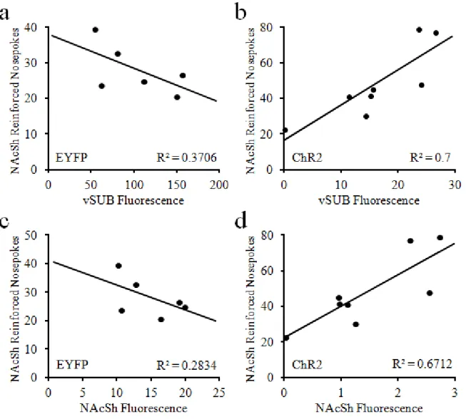
Correlations between virus expression and behavioral measures ...77
Discussion ...86
CHAPTER IV: ROLE OF THE VSUB AND VSUB-NACSH PATHWAY IN MAINTAINING REPRESENTATIONS TO GUIDE CHOICE OF DELAYED OUTCOMES ...92
Introduction ...92
Methods ...95
Animals and surgical procedures ...95
Apparatus and behavioral training ...95
Behavioral testing ...95
Data analysis ...96
Histology and fluorescence quantification ...97
Results ...99
Baseline behavioral performance and preference ...99
Forced-choice trial performance with post-response vSUB stimulation ...101
Free-choice trial preference with post-response vSUB stimulation ...103
Forced-choice trial performance with post-response vSUB-NAcSh stimulation 105 Free-choice trial preference with post-response vSUB-NAcSh stimulation ...107
Correlations between virus expression and behavioral measures ...108
Discussion ...120
CHAPTER V: GENERAL DISCUSSION ...124
Summary of experiments ...125
xi
The role of the vSUB and vSUB-NAcSh pathway in cued outcome
representations for delay-based decision making ...125
The role of the vSUB and vSUB-NAcSh pathway in behavior-outcome representations for delay-based decision making ...126
General discussion and relevance of findings ...127
The delineation of brain reward circuitry into pathway-specific components ...127
The vSUB-NAcSh pathway in decision making behavior ...130
Implications for drug addiction ...133
Future directions ...135
The reinforcing properties of the BLA and mPFC projections to the NAc ...135
The role of the vSUB-NAcSh pathway in delay discounting ...136
The role of the vSUB-NAcSh pathway in delay-based memory tasks ...137
The role of BLA and mPFC projections to the NAc in delay-based decision making ...139
Concluding remarks ...140
xii
LIST OF TABLES
Table
3.1 Correlation coefficients for the relationships between vSUB
fluorescence and cue stimulation test session behavior ...84 3.2 Correlation coefficients for the relationships between NAcSh
fluorescence and cue stimulation test session behavior ...85 4.1 Correlation coefficients for the relationships between vSUB
fluorescence and post-response stimulation test session behavior ...115 4.2 Correlation coefficients for the relationships between NAcSh
xiii
LIST OF FIGURES
Figure
2.1 Surgical preparation for optogenetic stimulation of vSUB
cell bodies and vSUB-NAcSh pathway ...39
2.2 Mean (± SEM) reinforced nosepokes for vSUB self-stimulation ...47
2.3 Nosepoking behavior for vSUB self-stimulation ...47
2.4 Mean (± SEM) reinforced nosepokes for vSUB-NAcSh pathway self-stimulation ...48
2.5 Nosepoking behavior for vSUB-NAcSh self-stimulation ...48
2.6 Mean (± SEM) vSUB stimulation duration between escape responses ...49
2.7 Temporal pattern of vSUB stimulation escape behavior ...49
2.8 Mean (± SEM) duration of vSUB-NAcSh pathway stimulation between escape responses ...50
2.9 Temporal pattern of vSUB-NAcSh stimulation escape behavior ...50
2.10 Quantified virus expression near optical fibers in the vSUB and NAcSh ...51
2.11 Correlations between vSUB-NAcSh self-stimulation behavior and virus expression in the vSUB and NAcSh ...52
2.12 Correlations between vSUB-NAcSh stimulation escape behavior and virus expression in the vSUB and NAcSh ...53
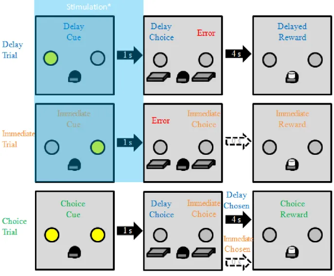
3.1 Behavioral task during retraining and cue stimulation test sessions ...67
3.2 Average delay choice task behavior across retraining sessions before cue stimulation test sessions ...79
3.3 Test session delay trial performance with vSUB stimulation during forced-choice cues ...80
3.4 Test session immediate trial performance with vSUB stimulation during forced-choice cues ...80
xiv
3.6 Test session free-choice response latency with vSUB
stimulation during forced-choice cues ...81 3.7 Test session delay trial performance with vSUB-NAcSh
pathway stimulation during forced-choice cues ...82 3.8 Test session immediate trial performance with vSUB-NAcSh
stimulation during forced-choice cues ...82 3.9 Test session free-choice outcome preference with vSUB-NAcSh
stimulation during forced-choice cues ...83 3.10 Test session free-choice response latency with vSUB-NAcSh
stimulation during forced-choice cues ...83 4.1 Behavioral task during retraining and post-response stimulation test sessions ....98 4.2 Average delay choice task behavior across retraining sessions
before post-response stimulation test sessions ...110 4.3 Test session delay trial performance with post-response vSUB stimulation ...111 4.4 Test session immediate trial performance with
post-response vSUB stimulation ...111 4.5 Test session free-choice outcome preference with
post-response vSUB stimulation ...112 4.6 Test session free-choice response latency with
post-response vSUB stimulation ...112 4.7 Test session delay trial performance with post-response
vSUB-NAcSh pathway stimulation ...113 4.8 Test session immediate trial performance with post-response
vSUB-NAcSh pathway stimulation ...113 4.9 Test session free-choice outcome preference with
post-response vSUB-NAcSh stimulation ...114 4.10 Test session free-choice response latency with post-response
xv
LIST OF ABBREVIATIONS
AAV Adeno-associated virus ACC Anterior cingulate cortex ACh Acetylcholine
ANOVA Analysis of variance BLA Basolateral amygdala
Camk2α Calmodulin-dependent protein kinase II alpha ChR2 Channelrhodopsin-2
CS Conditioned stimulus
DA Dopamine
DNMS Delayed-nonmatch-to-sample EYFP Enhanced yellow fluorescent protein IACUC Institutional animal care and use committee ICSS Intracranial self-stimulation
ITI Inter-trial interval LH Lateral hypothalamus MFB Medial forebrain bundle
MH Medial hypothalamus
xvi PB Phosphate buffer
PIT Pavlovian-to-instrumental transfer PFC Prefrontal cortex
mPFC Medial prefrontal cortex SEM Standard error of the mean vSUB Ventral subiculum
1 CHAPTER 1 INTRODUCTION
Decades of research has focused on the role of the nucleus accumbens (NAc) and the ventral subiculum (vSUB) in reward-related behavior, including operant conditioning and
delayed reinforcement (Balleine and Dickinson, 1998; Cardinal, 2006; Cooper et al., 2006; Abela and Chudasama, 2013; Laurent, 2013). However, the vSUB is one of many glutamatergic inputs to the NAc (Mogenson et al., 1980; Zahm, 1999), and these share dense reciprocal connections (Ishikawa and Nakamura, 2003; Sah et al., 2003; Vouimba and Maroun, 2011; Esmaeili and Grace, 2013), which suggests a functional difference between the interconnected vSUB and its direct pathway to the NAc in reward-related behavior. Indeed, only Britt and colleagues (2012) have examined the vSUB-NAc pathway in reward-related behavior in mice, thus much more research on this topic is needed. Further, while the vSUB is classically important for the processing of delayed rewards (Cheung and Cardinal, 2005; Bangasser et al., 2006; Deadwyler and Hampson, 2006; Abela and Chudasama, 2013), the vSUB-NAc pathway has not been examined in this context. The experiments described in this dissertation seek to expand upon the work of Britt et al. (2012) and then extend examination of the vSUB-NAc pathway to decisions regarding delayed rewards. Therefore, this chapter will review the literature on the role of the NAc and vSUB in operant conditioning and delayed reinforcement. First, this review will cover the importance and mechanism of operant conditioning, and the anatomy of NAc reward
2
of broader reward circuitry and how optogenetics can be used to isolate effects of the vSUB-NAc pathway. Finally, the importance, mechanism, and neural systems specific to delayed
reinforcement will be reviewed.
Operant conditioning
In operant conditioning (also known as instrumental conditioning or reinforcement learning), animals infer a causal link between biologically salient outcomes (reinforcers; e.g. food, water, predators, mates, drugs of abuse) and the behaviors which preceded them, resulting in the increased or decreased frequency of those behaviors (Thorndike, 1933; Skinner, 1938, 1981). In addition, stimuli in an animal's environment (discriminative stimuli) can signal the appropriate time to engage in conditioned behaviors (Colwill and Rescorla, 1990), and evoke representations of reinforcers which activate the behavior that brought them about in the past (Colwill and Rescorla, 1988). Operant learning mechanisms are fundamental for survival in that they allow organisms to adapt their behavior in order to find resources and escape threats in changing environments, and refine existing behaviors to more efficiently obtain limited resources (Staddon, 1975). However, with extended experience operant behaviors become habit as animals begin to associate their behavior with environmental cues instead of reinforcers, thereby losing the behavioral flexibility to efficiently deal with sudden challenges (Adams, 1982; Dickinson and Balleine, 1994).
3
and with a higher frequency for larger reinforcers, and this effect is amplified when known outcomes suddenly increase in value (Zeaman, 1949). In addition, the frequency of behavior follows the direction of the contingency between behaviors and outcomes. As such (for positive reinforcers), behavior will increase with a positive contingency, decrease with a negative contingency, or remain unchanged when there is zero contingency (Hammond, 1980). In addition, the shorter the delay between the behavior and consequence, the stronger the learning and the faster the acquisition of the target behavior (Wilkenfield et al., 1992). Finally,
the schedule of reinforcement also has substantial effects on conditioned behaviors. Under partial reinforcement, where every response does not bring about a reinforcer, behavior persists longer under periods of nonreinforcement, and responses over time will become patterned
depending on the type of schedule imposed (Schoenfeld et al., 1956; Ferster and Skinner, 1957).
The NAc in operant conditioning
The NAc in the acquisition of operant behaviors
4
The dynamics in NAc cell firing and neurotransmitter release have not been thoroughly studied during the acquisition of operant conditioning tasks, however a few reports suggest the NAc is encoding task-specific information. In support, one group has shown that NAc neurons phasically change their firing rate during the operant behavior and reinforcer initially, and also display phasic activity for discriminate stimuli as learning progresses (Wood and Rebec, 2009). Further, microdialysis during acquisition of operant tasks reveals an increase in NAc DA release during early learning, increasing further with additional training (Cheng and Feenstra, 2006; Ahn and Phillips, 2007; Segovia et al., 2011). While limited, these lines of evidence suggest a role for the NAc in the acquisition of operant behaviors.
The NAc in the expression of operant behaviors
5
Temporally acute physiological measures taken during the performance of operant conditioning tasks suggest that a broad scope of task-related information is encoded in the NAc. Several studies from our lab and others using in vivo electrophysiology have shown phasic changes in cell firing in the NAc to cocaine and natural reinforcers, lever presses to obtain those reinforcers, and reinforcer-paired stimuli (Carelli and Deadwyler, 1994; Carelli et al., 1999; Carelli et al., 2000; Nicola and Deadwyler, 2000; Peoples et al., 2004; Jones et al., 2010). In addition, rapid voltametric measurements of DA release in the NAc during operant conditioning reveal rapid increases in DA just before animals press a lever for cocaine, and again for cocaine-associated cues (Phillips et al., 2003; Cheer et al., 2007a; Day et al., 2010; Sugam et al., 2012; Saddoris et al., 2015b). Similar findings were observed during operant responding for
intracranial self-stimulation (Young and Michael, 1993; Cheer et al., 2005; Owesson-White et al., 2008; Beyene et al., 2010), and natural rewards (Roitman et al., 2004; Day et al., 2007; Roitman et al., 2008; Brown et al., 2011).
Anatomy and physiology of the NAc NAc cellular composition
6
Different classifications of MSNs have been ascertained by the presence of unique
immunohistochemical markers, including substance P, dynorphin, enkephalin, and neurotensin, and these markers are strong predictors of the neuron's projection target (Meredith, 1999). An interesting characteristic of MSNs is that they have a bistable membrane potential. In "down states", MSNs are hyperpolarized at ~ -85 mV, and in "up states" they are close to action potential threshold at ~ -60 mV (Wilson and Kawaguchi, 1996). Critically, it has been shown that MSNs cannot generate spike trains in the down state, and synaptic input is necessary to transition them to the up state (Nicola et al., 2000; O'Donnell, 2003). As such, MSNs typically fire at an irregular low rate (1-3 Hz) (Wilson and Groves, 1981; Yim and Mogenson, 1982; Koós and Tepper, 1999; Berke et al., 2004), but are capable of short bursts of activity up to 20 Hz (Chang et al., 1994; Plenz and Kitai, 1998; Carelli et al., 2000; Jones et al., 2010; Day et al., 2011; Cerri et al., 2014).
The remaining NAc cell types are interneurons (< 10% of all neurons), which exert inhibitory control of MSNs (Koós and Tepper, 1999). Approximately 5% are cholinergic interneurons (Groves, 1983; Kawaguchi et al., 1995; Berlanga, 2006). These neurons are relatively large (20-50 um diameter cell bodies), with short myelinated axons and radial dendrites (Kawaguchi, 1993; Kawaguchi et al., 1995). Cholinergic interneurons are
distinguishable from MSNs in that they typically fire at 8-15 Hz (Yim and Mogenson, 1982; Koós and Tepper, 1999). There are also GABAergic interneurons in the NAc, which account for less than 5% of striatal cells and approximately half of all interneurons (Kawaguchi et al., 1995). Like MSNs, GABAergic interneurons can be subdivided based upon unique
7
Berke, 2008, 2011). GABAergic interneurons can also be identified electrophysiologically by their relatively high firing rates (>20 Hz) (Yim and Mogenson, 1982; Koós and Tepper, 1999; Berke, 2011).
NAc afferent and efferent projections
It has been suggested that excitations among NAc neurons may originate from glutamatergic inputs from cortical and limbic structures that compete for access to motor
8
continued representation of future reward value (Schoenbaum et al., 1998; Winstanley et al., 2004; Cardinal, 2006).
In addition to the aforementioned glutamatergic inputs, the NAc also receives a dense dopaminergic input from the ventral tegmental area (VTA) (Zahm and Brog, 1992). Dopamine projections from the VTA to the NAc function as a neuromodulator and are believed to serve as a learning signal (Schultz et al., 1997; Fiorillo et al., 2003; Tobler et al., 2005; Day et al., 2007), and aid in behavioral selection (Roesch et al., 2007; Day et al., 2010; Sugam et al., 2012;
Saddoris et al., 2015b). The NAc, in turn, impacts behavior through projections to motor-related areas such as the substantia nigra, lateral hypothalamus (LH), and ventral pallidum, (Zahm, 1999). Given this anatomical arrangement, it has been postulated that the NAc integrates
information about memory, drive and motivation and influences behavior through its projections to motor-related neural regions thereby serving as a ‘limbic motor interface’ (Mogenson et al., 1980).
NAc subregions
The NAc can be delineated into core (NAcc) and shell (NAcSh) subterritories (Parkinson et al., 1999). Physically, there are differences in efferent and afferent projections between
regions (Groenewegen et al., 1987; Zahm and Brog, 1992; Brog et al., 1993; Zahm and Heimer, 1993; Heimer et al., 1997; Zahm, 1999). Functionally, the NAcSh classically plays a larger role in integrating emotional information whereas the core is instrumental for selecting and
generating reward-oriented behaviors (Stratford and Kelley, 1997; Kalivas and Nakamura, 1999; Parkinson et al., 1999; Corbit et al., 2001; Saddoris et al., 2013; Saddoris et al., 2015a).
9
identified, suggesting that these subregions do not function independently of one another, but are part of an interacting neural network (Van Dongen et al., 2005).
There are several notable differences between core and shell projection targets of
glutamatergic inputs to the NAc. In the PFC, the orbitofrontal, posterior piriform, and infralimbic cortices project predominantly to the NAcSh, whereas the prelimbic and anterior cingulate cortices project to the NAcc (Brog et al., 1993; Montaron et al., 1996). In addition, the vSUB's projection to the NAc primarily terminates in the dorsomedial region of the NAcSh
(Groenewegen et al., 1987; Brog et al., 1993). By contrast, the BLA projects to both subregions of the NAc, albeit with a heterogeneous distribution (Brog et al., 1993; Sah et al., 2003). VTA DAergic input to the NAc also differs by subregion, with projections from the medial VTA terminating in the medial NAcSh, and more-lateral VTA projections terminating in the NAcc and lateral NAcSh (Ikemoto, 2007). Likewise, there are differences in the projection profiles between MSNs in the NAcc and NAcSh. For example, the NAcc has outputs to the dorsolateral ventral pallidum, substantia nigra, and subthalamic nucleus, whereas the NAcSh has outputs to
ventromedial ventral pallidum, LH, and VTA (Zahm and Brog, 1992; Zahm and Heimer, 1993; Zahm, 1999).
The vSUB in operant conditioning
It is generally agreed that output of the hippocampus via the vSUB is not critical for operant conditioning. In one study, animals with pretraining bilateral lesions of the hippocampus (including vSUB) only had a slight impairment in their ability to acquire operant conditioning, and post-training lesions produced no deficit in the expression of basic operant
10
had no effect on the acquisition or performance of operant conditioning (Corbit and Balleine, 2000; Corbit et al., 2002). However, there is some evidence that rats with lesions of the vSUB are unable to use behavior-outcome representations to guide operant behavior, and instead respond habitually (Corbit et al., 2002). Thus, if the hippocampus and vSUB is at all involved in operant conditioning, it is in the formation of action-outcome representations, but not for
forming or using stimulus-response habits which are sufficient for behavior. These reports are intriguing considering the known projection from the vSUB to the NAcSh. The NAc, while controversially involved in the acquisition of operant conditioning, clearly encodes task-related information, and is critical for the performance of operant conditioning when discriminative stimuli are present, yet the vSUB does not seem important for task acquisition or performance. This comparison is not surprising, as the vSUB experiments did not include discriminative stimuli; thus, like the NAc, vSUB involvement in operant conditioning may require discrete stimuli to be involved in the task.
Anatomy and physiology of the vSUB vSUB cellular composition and physiology
The subiculum (including the vSUB and dorsal subiculum) has 3 layers, a molecular layer adjacent to the hippocampal CA1 area, an enlarged pyramidal cell layer where subicular cell bodies reside, and a polymorphic layer (O'Mara, 2005). The pyramidal cell layer contains the principal cells of the subiculum, large glutamatergic pyramidal cells of consistent shape and size (~20 x 40 um; (Menendez de la Prida et al., 2003; Vulović et al., 2012)), with apical dendrites extending in the molecular layer and basal dendrites further in the pyramidal layer (O'Mara, 2005). Smaller interneurons are also located in the pyramidal cell layer (~15 um
11
Cells in the subiculum can be further classified based upon their electrophysiological properties. While pyramidal cells in the subiculum typically have a resting firing rate of ~2 Hz, they can produce phasic activity to behaviorally relevant events at > 6 Hz (Hampson and
Deadwyler, 2003). Further, pyramidal cells are classified as either "bursting" units, which fire 2-6 fast action potentials with ~5-ms interspike intervals (Sharp and Green, 1994; Anderson and O'Mara, 2003; O'Mara, 2005), or as "regular" spiking cells, which fire a single action potential every 60 to 160 ms (Sharp and Green, 1994; Anderson and O'Mara, 2003; O'Mara, 2005). At any given time, approximately 74% of cells are bursting, and 26% regular (Mason, 1993; Taube, 1993). However, individual cells can transition between bursting and regular spiking by voltage-gated sodium channel activation kinetics, such that with prolonged activity bursting cells will become regular spiking cells (Cooper et al., 2005); however, hyperpolarisation of regular cells will not convert them to bursting cells (Stewart and Wong, 1993; O'Mara et al., 2001). As such, bursting cells will typically have a resting membrane potential of ~ -67.5 mV(Taube, 1993), whereas tonic firing will replace bursting at ~ -55 mV (Mason, 1993; Mattia et al., 1997). Finally, in the subiculum there are also "fast-spiking" units which have been morphologically identified as interneurons (Menendez de la Prida et al., 2003; O'Mara, 2005).
vSUB afferent and efferent projections
All layers of the subiculum receive the primary projections of the hippocampal CA1 area, designating the subiculum as the major output of the hippocampus (O'Mara, 2005). The
12
al., 2001; Britt et al., 2012). Further, as compared to the other glutamatergic inputs, the vSUB contains the majority of NAcSh-projecting cell bodies, and activation of vSUB terminals evoke the strongest EPSCs in the NAcSh, suggesting that the vSUB is the strongest glutamatergic input to the NAcSh (Britt et al., 2012). In addition to the vSUB-NAc pathway, the subiculum has several other inputs and outputs that can differ between dorsal subiculum and vSUB, although functional differences between subregions have not been well established (O'Mara, 2005).
Within the parahippocampal region, the subiculum as a whole receives and sends a strong projection to the neighboring entorhinal cortex (Naber et al., 2001), as well as a reciprocal
connection with the perirhinal cortex (Kosel et al., 1983; Köhler, 1985). The vSUB tends to share connectivity with frontal cortical and subcortial, limbic structures, including other
glutamatergic projection areas to the NAc. For instance, the vSUB projects to and receives input from subcortices of the mPFC (White et al., 1990; Finch, 1993). The vSUB also projects to, and receives input from the BLA (Witter and Groenewegen, 1990; Canteras and Swanson, 1992; Pitkanen et al., 2000). In addition, the vSUB also has a dense projection to the ventromedial hypothalamus, with a reciprocal connection from the ventral premammillary nucleus (Köhler, 1990; Witter and Groenewegen, 1990; Canteras and Swanson, 1992). In contrast, the dorsal subiculum instead projects to and receives connections from both lateral and medial mammillary nuclei (Witter et al., 1990). Further, both the dorsal subiculum and vSUB have reciprocal
connections with the lateral septum (Namura et al., 1994). Finally, while only the dorsal
subiculum projects to the anterior thalamus (Witter and Groenewegen, 1990; Witter et al., 1990;
Risold et al., 1997), there is a light projection from the thalamus to a diffuse area of the
13 The interconnectivity of brain-reward circuitry
Beyond the direct connections between glutamatergic and dopaminergic projections and the NAc, there are many more connections between the inputs themselves. Connectivity is shared both within the NAc and between structures. Within the NAc, MSNs share inputs, and as a result vSUB inputs can gate mPFC inputs (O'Donnell and Grace, 1995; French and Totterdell, 2002), and may gate BLA inputs as well (Mulder et al., 1998; French and Totterdell, 2003). Further, BLA inputs have been shown to gate and modulate mPFC inputs (Goto and O'Donnell, 2002; McGinty and Grace, 2008). In addition, DAergic projections from the VTA converge on the same MSNs as projections from the vSUB(Totterdell and Smith, 1988; Sesack and Pickel, 1990).
Several NAc inputs also interact within the mPFC. As such, DAergic inputs from the VTA can gate vSUB activity in the mPFC (Floresco and Grace, 2003). Likewise, vSUB and BLA projections to the mPFC converge and interact to drive mPFC activity (Gabbott et al., 2003; Ishikawa and Nakamura, 2003; Esmaeili and Grace, 2013). In the BLA, VTA DAergic projections modulate other inputs (de Oliveira et al., 2011), and mPFC projections can cause inhibitions of projection neurons (Rosenkranz and Grace, 2002; Vouimba and Maroun, 2011). The vSUB also projects to BLA (Sah et al., 2003), and the two structures often display
synchronized activity (Ikegaya et al., 1996; Pitkanen et al., 2000). Finally, as previously noted, the vSUB also shares reciprocal connections with other NAc inputs. Projections from the BLA can alter synaptic plasticity (Ikegaya et al., 1995), and the VTA(Gasbarri et al., 1994), as well as the mPFC also project to the vSUB (White et al., 1990; Finch, 1993).
14
motor resources (Pennartz et al., 1994). However, the same reasoning suggests that perturbations to one NAc input should shift the balance of behavioral control for all inputs. Indeed, this has likely been true for the bulk of literature on NAc afferent function, where functional profiles were constructed using pharmacology, electrophysiology, or electrical stimulation on cell bodies within each region of interest. Therefore, an investigation of pathway-specific function on behavior, where cell bodies and interconnections are undisturbed, could yield different results than previous studies on region-specific function.
Pathway-specific modulation of neural activity with optogenetics
While specific neural pathways have been traced with anterograde or retrograde vectors for decades, until recently there have not been means to manipulate those pathways without perturbing afferent or efferent cell bodies. However, the development of optogenetic tools has largely solved that problem and enables investigators to selectively activate (or inactivate) discrete neural pathways. For example, a recent study by Stuber and colleagues (2011) used optogenetics to specifically stimulate BLA inputs in the NAc and found the BLA-NAc pathway sufficient to reinforce motivated responding, whereas optogenetic inhibition of the same pathway could disrupt goal-seeking behavior to reward-predictive cues. Indeed, classic studies of BLA lesions or NAc lesions have shown no effects on the expression of simple goal-seeking behavior (Parkinson et al., 1999; Parkinson et al., 2000; Balleine and Killcross, 2006), suggesting a functional difference between pathway-specific and cell-body manipulations. Thus, applying optogenetics to study the vSUB-NAcSh pathway should be insightful.
15
modulate neural function in vivo or ex vivo (Boyden et al., 2005; Aravanis et al., 2007; Zhang et al., 2010; Bernstein and Boyden, 2011; Boyden, 2011; Stuber et al., 2012). There are several commonly used opsins. The blue-light-gated cation channel, channelrhodopsin-2 (ChR2) has been well characterized for optical stimulation, and is capable of producing rapid and reversible depolarizations of transfected cells and processes at physiologically relevant frequencies (Nagel et al., 2003; Zhang et al., 2006). In addition, there are two major optical silencers capable of fast and sustained hyperpolarization of neural targets: the light-activated electrogenic inward chloride pump, halorhodopsin (Gradinaru et al., 2008; Zhao et al., 2008), and the light-driven outward proton pump, archaerhodopsin (Chow et al., 2010; Chow et al., 2012). Importantly, studies have reported no major differences in basic neuronal characteristics including resting membrane properties and morphology in neurons expressing common opsins versus uninfected control neurons (Zhang et al., 2006; Gradinaru et al., 2008; Chow et al., 2010).
Several anterograde and retrograde viral vectors with specific promoters are available for precise neural targeting of optogenetic manipulations (Bernstein and Boyden, 2011; Boyden, 2011; Stuber et al., 2012). Further specificity can be achieved with precise targeting of optical fibers and calibration of light intensity to limit opsin activation to cell bodies or terminal regions of interest (Aravanis et al., 2007; Sparta et al., 2012). For example, to target the vSUB-NAcSh pathway for stimulation, one could simply infuse ChR2 packaged in an anterograde viral vector, such as adeno-associated virus (AAV) with the ubiquitous calmodulin-dependent protein kinase II alpha (Camk2α) promoter, into the vSUB, and place optical fibers in the NAcSh to deliver light to vSUB terminals once ChR2 is fully expressed. Indeed, Britt and colleagues (2012) used this technique to demonstrate that mice found stimulation of the vSUB-NAcSh pathway
16
techniques and published experiments have largely been focused on mice, whether the same manipulations and effects can be reproduced in rats and other species has yet to be thoroughly investigated.
Operant conditioning for brain stimulation reward Intracranial self-stimulation and neural substrates of reward
Intracranial self-stimulation (ICSS, or simply self-stimulation) is an operant conditioning task where animals perform a target behavior for electrical brain stimulation (known as brain stimulation reward) delivered via electrodes in the brain (Carlezon and Chartoff, 2007). The ICSS paradigm has been used extensively for over fifty years, as there has been considerable interest in mapping out the brain structures capable of supporting self-stimulation (Olds and Milner, 1954; Olds, 1958, 1962). Today, there is a general consensus that the reinforcing properties of natural rewards and drugs of abuse arise from several well characterized brain regions, which comprise the brain-reward pathway (Wise, 1996). Some of the brain areas
17
(Andén et al., 1964; Andén et al., 1965; Ungerstedt, 1971; Nauta et al., 1978), and DA is released in the NAc during ICSS (Young and Michael, 1993; Rada et al., 1998). Indeed, DA is associated with the ability of food and other rewards to evoke behavior (Wise et al., 1978b; Wise et al., 1978a; Gallistel et al., 1982; Wise et al., 1992). However, DA is not necessary for all reinforcing effects, as NAc DA alone is not reinforcing (Adamantidis et al., 2011), and is coordinated with glutamate in the NAc to drive the activity of MSNs during ICSS (Cheer et al., 2005; Cheer et al., 2007a). Further, ICSS can occur without activation of DAergic axons (Murray and Shizgal, 1994), without continuous DA release in the NAc(Garris et al., 1999; Hernandez et al., 2006), and is only reduced but not eliminated by DAergic lesions of the NAc (Phillips and Fibiger, 1978). This evidence suggests MFB stimulation may cause an interaction between several neurotransmitter systems, perhaps transynapically, to produce reward-driven behavior (Bielajew and Shizgal, 1986; Yeomans et al., 2000).
Beyond NAc DA release, ICSS may produce reward-related behaviors in response to NAc glutamate. General lesions of the NAc have profound effects on ICSS behavior(Carlezon Jr et al., 1996; Todtenkopf et al., 2006), and AMPA receptors in the NAc have been shown to regulate brain-stimulation reward (Todtenkopf et al., 2006). In addition, some DAergic terminals are known to corelease glutamate in the NAc (Stuber et al., 2010). The reinforcing effects of NAc glutamate release are further supported by a recent report demonstrating that mice will perform ICSS for activation of NAc MSNs, or terminal stimulation of glutamatergic inputs from the PFC, vSUB, or BLA in the NAc (Britt et al., 2012).
Brain-stimulation escape and neural substrates of aversion
18
other regions, the medial hypothalamus (MH) (Kiser and Lebovitz, 1975; Jenck et al., 1983; Rada et al., 1998), mesencephalic central gray (Jenck et al., 1983), dorsal midbrain tegmentum (Olds and Milner, 1954; Kiser and Lebovitz, 1975), medial geniculate bodies (Olds and Milner, 1954), and medial lemniscus (Olds and Milner, 1954). Stimulation of the MH also decreases DA in the NAc, providing further evidence that the sensation is aversive (Rada et al., 1998).
Surprisingly, animals will also work to terminate continuous stimulation of the same brain areas they find rewarding. As such, animals learn stimulation-escape for ongoing stimulation of the LH(Steiner et al., 1973; Kornblith and Hoebel, 1976; Bielajew and Shizgal, 1980), hippocampus (Olds and Olds, 1963), NAc (Olds and Olds, 1963), VTA (Bielajew and Shizgal, 1980), MFB (Steiner et al., 1969), dorsal raphe (Steiner et al., 1973), and locus coeruleus (Steiner et al., 1973). Indeed, animals even learn to escape brain stimulation delivered in the exact pattern they had previously self-administered (Steiner et al., 1969), and NAc DA increases during escape responding, suggesting the behavior is rewarding (Rada et al., 1998).
19
increases NAc ACh and the feeling of satiety to aversive levels, animals will promptly work to escape said stimulation.
However, with electrical stimulation alone, the analysis of neural circuits involved in self-stimulation and stimulation-escape behaviors can only be attributed to specific anatomical regions. It follows that, the role of glutamatergic inputs to the NAc in ICSS has not been
thoroughly investigated; this and other studies of pathway-specific function may yield intriguing results. As described previously, using optogenetics, Britt and colleagues (2012) demonstrated that mice would self-stimulate the vSUB-NAcSh pathway and other glutamatergic pathways to the NAc. Therefore, it would be informative to determine whether rats will also work for optogenetic self-stimulation of the vSUB-NAcSh pathway, and whether these effects and optogenetic stimulation in general can become aversive with automatic stimulation.
Delayed reinforcement
In many contexts, the consequence of a behavior is often not produced until some time has passed. While animals can learn operant behaviors with delayed reinforcement, nearly a century of research has revealed that learning is progressively slower with increasing delay between behaviors and outcomes (Skinner, 1938; Grice, 1948; Harker, 1956; Renner, 1964; Lattal and Gleeson, 1990; Dickinson et al., 1992; Wilkenfield et al., 1992; Cardinal, 2006). Nonetheless, rats can eventually learn operant responses with behavior-outcome delays of up to 32 s, albeit the asymptotic level of responding is also reduced as a function of delay (Dickinson et al., 1992).
20
to maintain some representation of their behavior until the outcome actually arrives. Assuming the behavioral representation degrades as time passes (due to the animal performing other behaviors or focusing on different aspects of their environment), then animals will be less likely to associate outcomes with the correct behavior (Hull, 1932; Thorndike, 1965; Revusky and Garcia, 1970; Mackintosh, 1974; Killeen and Fetterman, 1988). Similarly, if delay is sufficiently long such than an animal fails to attribute an outcome to its actions, it may instead associate the outcome with the environmental context, since the context can be a cue that is temporally closer to the outcome than the correct behavior (Dickinson et al., 1992; Dickinson, 1996; Cardinal, 2006). In support of the "contextual competition" hypothesis, delayed reinforcement is enhanced when rats are pre-exposed to the context sufficiently to learn its irrelevance to the task before training, and delayed reinforcement is disrupted when the context is made more salient with noncontingent presentations of reward before conditioning (Dickinson et al., 1992; Dickinson and Balleine, 1994; Dickinson, 1996). It follows that animals can overcome some of the difficulties of delayed reinforcement if a signal is presented between behavior and outcome. Signals or cues present during the delay can bridge the gap between behaviors and outcomes, providing immediate feedback for the correct response and serving as a better predictor of the outcome than the environment (Skinner, 1938; Bolles and Popp, 1964; Lattal, 1987; Mazur, 1997; Cardinal, 2006).
21
phenomenon is known as delay discounting (Rachlin, 1992; Green and Myerson, 2004; Rachlin, 2006). Notably, in order to assess the value of delayed outcomes in practice, there must be other outcomes available for comparison. As such, when animals are given the opportunity to work for immediate and delayed outcomes concurrently, they work more for the immediate outcome, allocating their rate of responding between the outcomes to match the relative delays to reinforcement (Herrnstein, 1970; Ainslie, 1975; Herrnstein and Loveland, 1975; Davison and McCarthy, 1988). Further, when given a discrete choice between an immediate or delayed outcome, animals will almost always choose the immediate option (Green and Myerson, 2004; Cardinal, 2006; Day et al., 2011).
Surprisingly, there is little consensus over the psychological reasons for delay
discounting. One explanation is that increasing delays may be related to increasing uncertainty over reward delivery (Rachlin et al., 1991; Green and Myerson, 1996; Mazur, 1997). While this certainly may be true during the acquisition of delayed reinforcement, this theory does not explain why animals still choose immediate outcomes over delayed outcomes of the same objective value after animals have established that reward delivery is guaranteed (Cardinal, 2006). Indeed, more recent reports suggest that delay and uncertainty are separate processes (Ho et al., 1999; Mitchell and Rosenthal, 2003; Green and Myerson, 2004). Nonetheless, it appears that the act of waiting for a desirable outcome is sufficient to induce a negative emotional state (Mischel et al., 1972; Wheeler et al., 2008; Casey et al., 2011; Wheeler et al., 2011).
The NAc in delayed reinforcement
22
contextual competition. In support, NAcc lesions impair Pavlovian conditioning to discrete cues but enhance conditioning to contextual cues (Parkinson et al., 1999). Intriguingly, whether the NAcSh is also necessary for delayed reinforcement learning or expression has not been directly investigated. Even so, lesions of the NAcSh do not have effects on delay discounting tasks with choices between immediate or delayed rewards, suggesting that NAcSh lesioned animals can both acquire and use information about delayed outcomes to guide behavior (Pothuizen et al., 2005). By contrast, NAcc lesions greatly alter delay discounting, shifting preference towards immediate rewards even when delayed rewards are larger in magnitude(Cardinal et al., 2001; Cardinal et al., 2003; Pothuizen et al., 2005). Critically, despite the concurrent manipulation of outcome size in these studies, NAcc lesioned animals are not impaired in their ability to discriminate reward magnitude, suggesting a specific detriment in the representation of delays (Acheson et al., 2006; Cardinal, 2006; Bezzina et al., 2007).
Primate and rodent electrophysiology studies alike have shown that NAc activity during periods of reward anticipation increases as animals wait for rewards (Hollerman et al., 1998; Schultz et al., 2000; Day et al., 2011). Specifically, Day (Day et al., 2011) found that in a delay task where the objective value of the immediate and delayed outcome were identical, a subset of cells displayed prolonged changes in activity from the period before responses were made, and lasting until reward delivery. These prolonged changes may represent the response
23
further investigations of the functional nature of NAcSh encoding in delay choice tasks are warranted. Lastly, differences in NAc phasic activity for cues signaling immediate and delayed outcomes have been reported (Roesch et al., 2009), supporting the growing idea that NAc neuron cue responses signal the subjective value of upcoming outcomes (Hassani et al., 2001; Cromwell et al., 2005; Samejima et al., 2005; Wilson and Bowman, 2005; Kable and Glimcher, 2007; Saddoris et al., 2015b).
The role of NAc DA release during delayed reinforcement or delay choice tasks has only recently begun to gather attention. So far, it has been determined that NAc dopamine depletion alone has no effect on delay discounting, but NAc DA is necessary for serotonin agonists to bias animals towards immediate outcomes (Winstanley et al., 2005). Likewise, systemic DA
injections reduce preference for delayed outcomes (Wade et al., 2000; Denk et al., 2005), indicating the existence of a locus of action for DA in delay choice tasks potentially outside of the NAc. By contrast, phasic DA release in the NAc measured by fast-scan cyclic voltammetry shows a small degree of preferential encoding of cues for immediate over delayed outcomes (Day et al., 2010). Furthermore, direct manipulation of NAc DA via optogenetics can change responding for delayed versus immediate rewards, indicating a causal link between DA and the choice of delayed outcomes (Saddoris et al., 2015b). Taken together, it is apparent that while the NAc is active and critical in the acquisition and use of representations of delayed outcomes to guide behavior, the functionality of NAc DA is still under debate.
The vSUB in delayed reinforcement
24
learning of operant conditioning with delays (Cheung and Cardinal, 2005). It follows that, whereas lesions of the NAcc are thought to enhance contextual competition, vSUB lesions likely improve delayed reinforcement by reducing contextual competition. In support, lesions of the hippocampus have been shown to impair Pavlovian conditioning to contextual cues, but not discrete cues in a number of paradigms (Hirsh, 1974; Selden et al., 1991; Honey and Good, 1993; Jarrard, 1993; Phillips and LeDoux, 1995; Chen et al., 1996; Maren et al., 1997; Anagnostaras et al., 2001; Rudy et al., 2002). Curiously, despite enhancing the acquisition of delayed reinforcement, ventral hippocampal lesions, like NAcc lesions, bias animals toward immediate smaller options over larger delayed rewards (Cheung and Cardinal, 2005; McHugh et al., 2008; Abela and Chudasama, 2013). Further, this effect seems specific to delay
representations, as hippocampal lesioned animals are not impaired in their ability to discriminate reward magnitude (Kesner and Williams, 1995; Gilbert and Kesner, 2002; Cheung and Cardinal, 2005). Furthermore, evidence of activity in the dorsal and ventral hippocampus in the form of nicotinic acetylcholine receptor binding has been associated with choice of delayed rewards (Mendez et al., 2013), and stimulation of vSUB activity via noradrenergic a2a receptor agonists can also bias animals toward delayed rewards (Abela and Chudasama, 2014).
The mechanism by which lesions of the hippocampus reduce choice of delayed outcomes is undetermined. However, one explanation may be that the hippocampus, like NAc, can hold representations of behaviors and upcoming outcomes across a delay (Bangasser et al., 2006; McHugh et al., 2008; Laurent, 2013). It follows that, while electrophysiological assessments of vSUB function have only been conducted in working memory tasks to date, vSUB neurons are highly activated during delay periods preceding reward (Deadwyler and Hampson, 2003;
25
different aspects of the vSUB are involved in the acquisition of delayed reinforcement and delay choice. Intriguingly, despite the strength of vSUB-NAcSh connectivity (Britt et al., 2012), it is instead the vSUB and NAcc that are predominantly involved in delay tasks (Cheung and
Cardinal, 2005; Pothuizen et al., 2005), not the NAcSh, and these regions have contrasting roles during delayed reinforcement (Parkinson et al., 1999; Cheung and Cardinal, 2005). However, this inconsistency may be due to indirect effects from larger changes in reward circuitry when these regions are perturbed (O'Donnell and Grace, 1995; Pitkanen et al., 2000; Esmaeili and Grace, 2013), or from cross talk between NAcc and NAcSh subregions (Van Dongen et al., 2005). These open questions prompt further investigation of the vSUB-NAcSh pathway in delayed reinforcement and delay-choice tasks.
Goals of this dissertation
Research has long placed the NAc at the center of reinforcement-driven learning and behavior (Mogenson et al., 1980; Wise et al., 1992; Carelli, 2002; Yin et al., 2008; Saddoris et al., 2013). However, there is a considerable amount of interconnectivity among inputs to the NAc (O'Donnell and Grace, 1995; Pitkanen et al., 2000; Esmaeili and Grace, 2013), and it is thought that these inputs compete for behavioral control (Pennartz et al., 1994), thus with
traditional techniques (e.g. pharmacology, electrical stimulation) it is challenging to separate the function of specific NAc inputs from circuit-level effects. Therefore, behavioral investigations with pathway-specific manipulations of NAc inputs via optogenetics (Bernstein and Boyden, 2011; Boyden, 2011; Stuber et al., 2012), where cell bodies and interconnections are
unperturbed, could yield novel and exciting results.
26
(Groenewegen et al., 1987; Britt et al., 2012). One potential application of vSUB-NAcSh manipulation is in operant conditioning for brain-reward stimulation. Earlier studies have revealed that animals will learn to perform behaviors for electrical stimulation of the NAc, parts of the hippocampus, and other inputs to the NAc (Olds and Milner, 1954; Olds, 1962; Olds and Olds, 1963; Ursin et al., 1966), and disruptions of NAc function alter this type of
behavior(Phillips and Fibiger, 1978; Carlezon Jr et al., 1996; Todtenkopf et al., 2006). Further, animals can become sated on electrical stimulation, at which point it becomes aversive (Hoebel and Thompson, 1969; Rada and Hoebel, 2001). Yet, it was only recently that an optogenetics study revealed that mice will self-stimulate the vSUB-NAcSh pathway and other direct
glutamatergic inputs to the NAc (Britt et al., 2012). Still, whether vSUB-NAcSh stimulation is also reinforcing in rats and subject to satiation has yet to be investigated.
Another promising application of vSUB-NAcSh pathway manipulation is in operant conditioning for delayed reinforcement. Lesions of the vSUB bias animals away from delayed outcomes (Cheung and Cardinal, 2005; Abela and Chudasama, 2013), while pharmacological activation of the vSUB increases preference for delayed outcomes (Abela and Chudasama, 2014). By contrast, despite input from the vSUB and evidence that neurons in the NAcSh encode information about delay (Day et al., 2011), lesions of the NAcSh do not bias animals away from delayed outcomes (Cardinal and Cheung, 2005; Pothuizen et al., 2005); Instead, this function is attributed to the NAcc subregion (Cardinal et al., 2001; Cardinal and Cheung, 2005; Pothuizen et al., 2005). This inconsistency could be due to larger changes in reward circuitry from the
27
vSUB-NAcSh pathway as compared to vSUB cell bodies in operant conditioning for brain stimulation reward and in delayed reinforcement.
Specific aims:
1. To characterize the reinforcing properties of vSUB and vSUB-NAcSh pathway stimulation in rats.
In ICSS paradigms animals will quickly learn to self-administer electrical stimulation to a number of brain regions, including the NAc, hippocampus, and other inputs to the NAc (Olds and Milner, 1954; Olds, 1962; Olds and Olds, 1963; Ursin et al., 1966). Electrical brain
stimulation is thought to be rewarding much in the same way as natural reinforcers such as food and water, in that the NAc must be intact (Phillips and Fibiger, 1978; Carlezon Jr et al., 1996; Todtenkopf et al., 2006), and NAc DA release and glutamatergic transmission is correlated with stimulation (Wise et al., 1992; Young and Michael, 1993; Pennartz et al., 1994; Rada et al., 1998; Todtenkopf et al., 2006; Cheer et al., 2007a). As such, animals can also become sated when electrical stimulation is delivered automatically and will instead work to terminate said stimulation (Hoebel and Thompson, 1969; Rada and Hoebel, 2001). Recently Britt and colleagues (2012) demonstrated that mice learn ICSS for optogenetic pathway-specific
stimulation of the vSUB-NAcSh and other direct glutamatergic pathways to the NAc. First, this aim seeks to replicate these findings by determining whether rats will also learn to self-stimulate the vSUB-NAcSh pathway via optogenetics. Further, this aim builds upon the ICSS literature by comparing pathway-specific optogenetic stimulation to more traditional vSUB cell body
28
2. To determine the relative importance of the vSUB versus vSUB-NAcSh pathway in the use of cued representations to guide choices between delayed and immediate rewards.
In addition to the role of the vSUB and NAc in ICSS, both regions have been heavily implicated in decision making with delayed reinforcement. Animals will discount the subjective value of delayed outcomes (Rachlin, 1992; Green and Myerson, 2004; Rachlin, 2006), such that when given a discrete choice between an immediate or delayed outcome of equal objective value, animals will almost always choose the immediate option (Green and Myerson, 2004; Cardinal, 2006; Day et al., 2011). Lesions of both the NAc and vSUB bias animals further towards immediate outcomes, even when delayed rewards are of larger magnitude (Cardinal et al., 2001; Cheung and Cardinal, 2005; Pothuizen et al., 2005; Abela and Chudasama, 2013). Further, pharmacological potentiation of vSUB activity increases preference for delayed
outcomes (Abela and Chudasama, 2014). However, despite the fact that the vSUB projects to the NAcSh (Groenewegen et al., 1987; Britt et al., 2012), only lesions of the NAcc but not NAcSh subregion change animals' preferences for delayed rewards (Pothuizen et al., 2005). Thus, this aim and the next seek to explain the functional disconnection between vSUB and NAcSh in delayed reinforcement by using optogenetics to isolate the effects of the vSUB-NAcSh pathway from the potentially confounding effects of other vSUB or NAcSh connections. Further, in both the NAcc and NacSh it has been observed that MSNs and DA differentially encode cues
29
outcome to guide behavior, and this influence is compared to that of circuit effects arising from vSUB cell bodies.
3. To evaluate the relative importance of the vSUB versus vSUB-NAcSh pathway in maintaining reward representations over a delay to guide choices between delayed and immediate rewards.
Both the vSUB and NAc are known to be involved in decision making involving delayed outcomes. Lesions of the vSUB bias animals away from delayed outcomes and pharmacological activation of the vSUB increases tolerance to delayed choices. However, despite strong
connectivity between the vSUB and NAcSh, only lesions of the NAcc bias animals away from choices for delayed rewards. Notably, previous reports suggest that neurons in both the NAcc and NAcSh as well as vSUB are modulated during delays until reward delivery (Hollerman et al., 1998; Schultz et al., 2000; Deadwyler and Hampson, 2003; Hampson and Deadwyler, 2003; Deadwyler and Hampson, 2006; Day et al., 2011), and this activity may reflect the maintenance of operant representations critical for learning and performing delayed reinforcement tasks (Bangasser et al., 2006; Cardinal, 2006; McHugh et al., 2008; Day et al., 2011; Laurent, 2013). Here, I incorporate optogenetic tools to examine the functional role of the vSUB-NAcSh
30 CHAPTER 2
CHARACTERIZATION OF THE REINFORCING PROPERTIES OF VSUB AND VSUB-NACSH PATHWAY STIMULATION IN RATS
INTRODUCTION
In order to survive, organisms must learn to adapt their behavior to find resources and escape threats in their environment (Staddon, 1975). Animals are capable of such behavioral flexibility through the mechanisms of operant conditioning, wherein causal inferences are formed between biologically salient outcomes and the behaviors which preceded them, resulting in the increased or decreased occurrence of those behaviors in the future (Thorndike, 1933; Skinner, 1938, 1981). A particularly interesting form of operant conditioning is ICSS, wherein animals learn to electrically self-stimulate areas of their brain as reinforcement (Carlezon and Chartoff, 2007), and will work to escape the same stimulation delivered automatically once a point of satiety has been reached, much like with natural reinforcers such as food and water (Hoebel and Thompson, 1969; Steiner et al., 1969; Rada and Hoebel, 2001). Indeed, ICSS was used more than 50 years ago to identify the neural substrates of reward (Olds and Milner, 1954; Olds, 1958, 1962).
At the center of the brain-reward pathway is the NAc, a region that integrates
31
2008; Saddoris et al., 2013). In support, evidence suggests that ICSS is driven by an interaction of activity from several neurotransmitter systems within the NAc including, DA, glutamate, ACh, and MSN cell firing (Hoebel and Thompson, 1969; Bielajew and Shizgal, 1986; Young and Michael, 1993; Carlezon Jr et al., 1996; Rada et al., 1998; Yeomans et al., 2000; Rada and Hoebel, 2001; Todtenkopf et al., 2006; Cheer et al., 2007a). However, there is substantial interconnectivity among inputs to the NAc both directly between regions and between terminals within the NAc (White et al., 1990; Gasbarri et al., 1994; O'Donnell and Grace, 1995; Pitkanen et al., 2000; Floresco and Grace, 2003; de Oliveira et al., 2011; Vouimba and Maroun, 2011; Esmaeili and Grace, 2013). As such, the function of brain regions targeted by nonspecific manipulations such as electrical self-stimulation and stimulation-escape behaviors cannot be disentangled from circuit-level effects.
32
whether this type of stimulation, like electrical stimulation, is subject to satiation, and how pathway-specific stimulation compares to more traditional cell-body stimulation.
To address these questions, this aim seeks to replicate and then expand upon the findings of Britt et al. (2012). Here, rats were prepared to nosepoke for optogenetic stimulation of the vSUB-NAcSh pathway. Notably, 3 different intensities of stimulation were used to better characterize the parameters of optogenetic ICSS in rats. Further, rats were also given the
33 METHODS
Animals
Fourteen male Sprague-Dawley rats (Harlan Laboratories, Indianapolis, IN) aged 90−120 d and weighing 300-400 g before surgery were used. Animals were individually housed on a 12:12 reversed dark:light schedule, and experiments were conducted between 12:00 and 8:00 pm. Rats were maintained at a stable body-weight (300-450 g) with light food restriction (14−18 g of Harlan Laboratories Teklad 2920X laboratory chow each day) for the duration of behavioral testing. Food was given ad libitum before surgery and during the post-operative recovery period. Animal procedures were conducted in accordance with the National Institutes of Health
Guidelines for the Care and Use of Laboratory Animals, and were approved by the University of North Carolina at Chapel Hill Institutional Animal Care and Use Committee (IACUC).
Surgical procedures
34
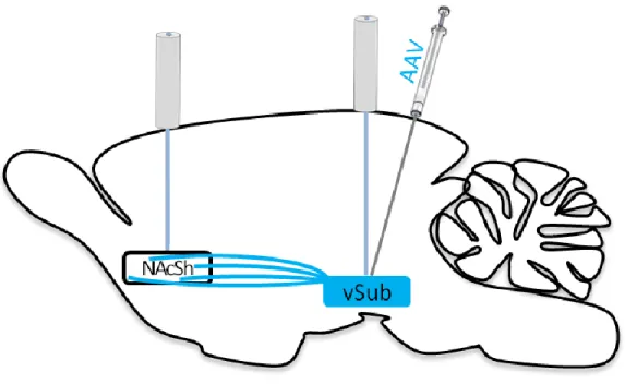
10 mm length; Thorlabs, Sussex County, NJ) were implanted bilaterally over the vSUB (AP: -5.75mm, ML: +/- 4.6mm, DV: -8.5mm from skull at bregma) and NAcSh (AP: +1.3mm, ML: +/- 2.2mm, DV: -6mm from skull at bregma, angled in the ML plane towards midline 10°). Optical fibers were permanently attached with dental acrylic to 6 screws embedded in the skull surface. Animals were given an oral dose of 1.0 mg/kg meloxicam (Metacam, Boehringer Ingelheim Vetmedica, St Joseph, MO) as an analgesic the day of, and for 2 d post surgery. Animals were given ample time to recover and for vSUB cell body and NAcSh terminal expression of virus before behavioral testing (>4 wks for vSUB and >10 wks for vSUB-NAcSh projections) (Witten et al., 2011). Figure 2.1 shows a schematic diagram of the surgical procedure.
The AAV viruses were packaged as serotype 5 by the University of North Carolina vector core with a titer range of 1-4 x 10² molecules/ml. Animals were divided into experimental groups based upon the virus received, 8 "ChR2" animals received a construct of
channelrhodopsin with enhanced yellow fluorescent protein (AAV-CaMK2α-hChR2(H134R)-EYFP). As a control, 6 "EYFP" animals received a construct of enhanced yellow fluorescent protein (AAV-CaMK2α-EYFP). Importantly, 1 animal prepared with ChR2 had no evidence of virus expression in either hemisphere and was included in the EYFP group for behavioral analyses, and in the ChR2 group for fluorescence-based analyses (see below).
Apparatus
35
chamber. A speaker located 18 cm above the floor was configured to produce a tone (65 dB, 2900 Hz).
During behavioral sessions, rats were connected to opaque fiber optic patch cables (200 µm core, 0.22 NA, Thorlabs). These cables terminated with ferrule connectors that were secured to the rat's optical fiber implants with fitted ceramic sleeves (Precision Fiber Products, Milpitas, CA), and were attached at the other end to an optical commutator (Doric Lenses, Ville de Quebec, QC, Canada). The commutator allowed for bilateral stimulation of vSUB cell bodies or terminals in the NAcSh and provided unrestrained movement for the animal. The commutator was connected via a second optical patch cable to a 150 mW DPSS 473 nm laser (Shanghai Laser & Optics Century Co., Shanghai, China). Optical stimulation was pulsed at 20 Hz via TTL pulses from an Arduino Duemilanove microcontroller board. Optogenetic stimulation and other external stimuli were produced and synchronized with behavioral events by a computer running Med PC IV (Med Associates) software.
Self-stimulation behavioral procedures
36
lasted 30 min, while baited sessions lasted for 30 min after the 10th nosepoke (end of baited responses).
Animals began self-stimulation of vSUB cell-bodies approximately 4 weeks post-surgery. Following completion of the delay choice task (Aims 2 and 3; see Chapters 3 and 4) at
approximately 11 weeks, animals resumed the same behavioral procedures for self-stimulation of the vSUB-NAcSh pathway with laser directed at vSUB axon terminals in the NAcSh. Self-stimulation of each target lasted for 6 days, and these were interspersed with 6 days of the stimulation escape behavioral procedure (see below).
Stimulation escape behavioral procedures
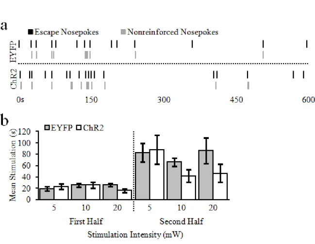
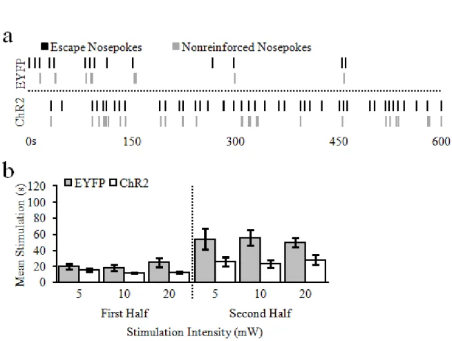
For each day of the stimulation escape procedure, bilateral optogenetic stimulation (20Hz, 5 ms pulse width) was on by default. Rats could make a single nosepoke into the central food-cup to terminate (i.e., escape) the stimulation for 5 s. While the stimulation was off, a tone was played to signify that additional nosepokes made during that period would not be reinforced. Both nosepokes that terminated the stimulation, and nonreinforced nosepokes were recorded. Like the self-stimulation procedure, animals received 2 days each of 5, 10, and 20mW stimulation, and were baited for the first 10 escape nosepokes on the first day at each power. Escape sessions were only 10 min long to minimize the risk of seizure or tissue damage (Milner, 1991; Cardin et al., 2010); baited sessions lasted for 10 min after the 10th escape nosepoke (end of baited responses).
As noted above, stimulation escape of each target lasted for 6 days, and these were
37 Data analysis
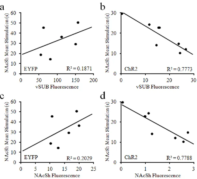
The primary dependent variable for self-stimulation behavioral procedures was overall reinforced nosepokes, and for stimulation escape procedures the mean duration of stimulation between escape nosepokes. Data were averaged between the two sessions of each power (excluding baited trials) for each animal. Separate analyses for each task were conducted for vSUB and vSUB-NAcSh stimulation targets. All statistical analyses were conducted using STATISTICA version 13 (Dell, Round Rock, TX). Repeated measures Analysis of Variance (ANOVA) were used to compare reinforced nosepokes or mean stimulation duration for all animals. In one type of analyses, group (ChR2 or EYFP) was used as a between-subjects factor to examine the general reinforcing properties of stimulation, and power (5, 10, and 20mW) was used as a within-subjects factor to determine whether the reinforcing properties and intensity of stimulation are related. In another analyses, reinforced nosepokes or mean stimulation duration data was divided for the first and second half of each session and compared with the addition of epoch (first or last half of session) as a within-subjects factor. Finally, linear regressions were calculated to determine whether reinforced nosepokes or mean stimulation duration data were significantly correlated to the quantified level of AAV expression in the vSUB or NAcSh of each animal (see below). For fluorescence analyses, behavioral data was averaged across all
stimulation powers. Descriptive statistics were reported as mean and standard error of the mean. All analyses were considered significant at α < .05.
Histology
38
placement with a light microscope and the other half placed in 0.1M phosphate buffer (PB) for immunohistochemistry to verify virus expression. Free floating sections were washed in Triton-X (0.5% solution in phosphate-buffered saline) and 0.1M phosphate-buffered saline. Sections then were incubated for 60 minutes in 2% NeuroTrace (530/615 nm, Invitrogen LifeTech, Carlsbad, CA), then washed and mounted onto microscope slides in phosphate-buffered water and coverslipped with Vectashield mounting medium (Vector Laboratories, Burlingame, CA). Sections were visualized on a confocal microscope to evaluate virus expression as well as optical fiber placement.
Fluorescence quantification
The amount of AAV expression in the vSUB or NAcSh was quantified using open source NIH Image-J analysis software (Schneider et al., 2012; Jensen, 2013). Measurements were focused on single representative images closest to fiber placement within each hemisphere from each animal. Images of the vSUB were 2355 x 2355 pixels and images of the NAcSh were 1895 x 1895 pixels in size at 1.25 x 1.25 um per pixel resolution. Within each image, measurements of average pixel intensity were taken from 3 areas determined by one of the following
characteristics: 1) with distinct fluorescence near the fiber tract within each region of interest (ROI; raw fluorescence), 2) without tissue (black), and 3) with autofluorescence only.
39
40 RESULTS
vSUB self-stimulation behavior
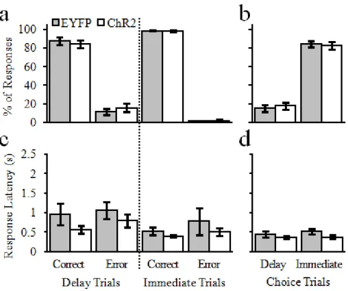
Rats (EYFP, n = 7; ChR2, n = 7) were given the opportunity to nosepoke for different intensities of optogenetic stimulation of the vSUB. Unlike electrical stimulation of various parts of the hippocampus (Olds and Milner, 1954; Ursin et al., 1966), optogenetic stimulation of cell bodies in the vSUB was not sufficient to reinforce operant behavior (Fig. 2.2). A 2-way repeated-measures ANOVA that examined reinforced nosepokes for vSUB stimulation as a function of group (EYFP, ChR2) and stimulation intensity (5, 10, & 20 mW) revealed no main effect of group, F(1,12) = 2.02, p = .18, no main effect of stimulation intensity, F(2,24) = 1.78, p = .19, and no significant interaction, F(2,24) = 1.90, p = .17.
41
reported above (p > .05 for remaining within-epoch, and p < .001 for between-epoch
comparisons). Finally, there was no epoch by stimulation intensity by group interaction, F(2,24) = 0.66, p = .52. All other main effects (group, stimulation intensity) and interactions are as noted above.
vSUB-NAcSh pathway self-stimulation behavior
While rats would not nosepoke for vSUB cell body stimulation I next examined if rats would nosepoke for different intensities of vSUB-NacSh pathway optogenetic stimulation. Indeed, pathway-specific stimulation was capable of supporting operant behavior in ChR2 animals as compared to EYFP controls (Fig. 2.4). Specifically, a 2-way repeated-measures ANOVA revealed a main effect of group, F(1,12) = 10.76, p = .007, no main effect of stimulation intensity, F(2,24) = 0.03, p = .97, and no significant interaction between the two, F(2,24) = 0.25, p = 0.78. These findings indicate that while ChR2 rats nose poked significantly more than EYFP for optical vSub-NAcSh pathway stimulation (ChR2,52.14 ± 7.11; EYFP, 27.43 ± 2.49), responses did not differ within or across groups as a function of stimulation intensity.