October 2019; 18(5):543-553.
Anti-varicella Zoster Virus IgG and hsCRP Levels Correlate with Progression
of Coronary Artery Atherosclerosis
Hamed Fouladseresht1, Atefe Ghamar Talepoor1, Shirin Farjadian1, Shahdad Khosropanah2, and Mehrnoosh Doroudchi1
1
Department of Immunology, School of Medicine, Shiraz University of Medical Sciences, Shiraz, Iran
2
Department of Cardiology, School of Medicine, Shiraz University of Medical Sciences, Shiraz, Iran
Received: 12 June 2019; Received in revised form: 9 July 2019; Accepted: 17 July 2019
ABSTRACT
The relationship between high levels of anti-Varicella Zoster Virus (VZV) IgG in cerebrospinal fluid (CSF) and cerebrovascular atherosclerosis commends a possible similar association in other vessels. We aimed to investigate the association of VZV-seropositivity with coronary artery atherosclerosis.
We recruited 88 newly diagnosed patients with more than 50% stenosis in at least one of the main coronary arteries. As the control group, 99 age-matched individuals with normal/insignificant coronary artery findings were included. Clinical, paraclinical, and demographical data were gathered at the time of sampling. High‐sensitivity C‐reactive
protein (hsCRP) levels were measured by nephelometry. VZV-seropositivity was determined by measuring of anti-VZV IgG level in plasma. Multivariable logistic regression was used to evaluate the correlation of data with coronary vascular atherosclerosis.
The frequency of VZV-seropositivity was significantly higher in the atherosclerosis group compared to the controls (OR=1.88; 95%CI=1.03-3.44). The plasma levels of anti-VZV IgG were significantly higher in patients with atherosclerosis (Median=2.70, IQR=1.53-4.30 AU/mL) than in the controls (Median=2.10, IQR=1.70-3.10 AU/mL, p=0.034). The hsCRP levels in patients and controls were 5.19±2.00 and 1.51±1.07 mg/L, respectively. The correlation between hsCRP and anti-VZV IgG level in plasma was observed (r=0.40, p<0.001). The levels of hsCRP and anti-VZV IgG increased based on the number of diseased vessels but only the difference in hsCRP levels reached a significant level (p<0.001 and p=0.168, respectively).
Our data suggest that VZV-seropositivity and hsCRP elevation jointly increase the risk of atherosclerosis. The multifactorial nature of atherosclerosis; however, leaves more options for the inflammatory milieu to be generated.
Keywords: Atherosclerosis; C-reactive protein; Immunoglobulin G; Varicella-zoster virus
Corresponding Author: Mehrnoosh Doroudchi, PhD; Department of Immunology, School of Medicine, Shiraz University
INTRODUCTION
Varicella-Zoster Virus (VZV) is a member of herpes simplex virus family which can survive in the host body after the primary infection as a latent virus. VZV is responsible for chickenpox (primary infection) in the childhood and herpes zoster (re-exposure or latent reactivity) in adulthood.1,2 Different molecular and serological studies have shown that VZV is one of the common infectious agents worldwide with an extensive range of clinical symptoms in the respiratory, skin, nervous, and vascular systems.3 In addition, several studies have shown the association between VZV and cerebrovascular atherosclerosis (AS).4,5 Atherosclerosis is a chronic inflammatory disease initiated by activation of endothelial cells (ECs), expression of adhesion molecules, secretion of pro-inflammatory chemokines and cytokines, migration of immune cells, and eventually foam cells formation, smooth muscle cells (SMCs) proliferation, and plaque formation.6,7 Atherosclerotic lesion growth and rupture over time can cause obstruction or thrombosis in the lumen of the artery that reduces blood flow to the tissues. Depending on the affected arteries and tissues, this may lead to the cerebrovascular, coronary artery, and kidney artery diseases.8,9
Several studies showed the role of VZV in cerebrovascular atherosclerosis; using molecular, histological, seroepidemiological, and experimental examinations.10,11 Primarily, by PCR method, VZV DNA in cerebrospinal fluid (CSF) and/or atherosclerotic plaques of patients with stroke was detected which suggested a correlation between the VZV and atherosclerosis. Afterward, the investigation of VZV or its antigens was used by histological methods to confirm the role of VZV in atherosclerosis.3 Furthermore, seroepidemiological methods revealed high levels of anti-VZV IgG in the CSF of 93% of patients with cerebrovascular atherosclerosis.12 On the other hand, several studies have suggested an increased risk of cardiovascular events following herpes zoster infection.9,13 Contrarily, another study showed that VZV infection in childhood is negatively associated with coronary heart disease (CHD).14 Accordingly, VZV infection in childhood was related to a 1.5 fold decreased the risk of CHD in adulthood. Most of the previous studies addressed VZV vasculopathy in cerebrovascular disease and information on the effect of VZV infection in cardiac
vasculopathy is scarce.15
C-reactive protein (CRP) is an acute inflammatory protein that is produced by liver hepatocytes, macrophages, lymphocytes, ECs, SMCs, and adipocytes in inflammation.16 Age, gender, obesity, hypertension, dyslipidemia, diabetes mellitus, smoking state, and acute and chronic inflammatory diseases (such as infections and atherosclerosis) are factors that can alter CRP levels in individuals.17,18 CRP is a direct risk factor for the induction and progression of atherosclerotic plaques.19,20 High sensitive CRP (hsCRP) levels have clinical value for the detection of inflammation and predictive value for asymptomatic atherosclerosis in the presence of other risk factors. In this regard, hsCRP levels of 1-3 mg/L have intermediate and hsCRP levels greater than 3 mg/L have high-risk values for atherosclerosis.16,21 CRP acts through induction of complement activation, phagocytosis, nitric oxide (NO) release, and the secretion of cytokines, such as interleukin (IL)-1, -6, and tumor necrosis factor (TNF)-α.16 Mean levels of CRP are elevated in infectious diseases, but previous studies reported no association between CRP levels and anti-HIV or anti-H.pylori IgG in atherosclerosis.22,23
It is shown that plasma levels of CRP are associated with clinical manifestations of VZV infection,24 however, a percentage of patients with VZV infection do not develop high levels of CRP.25 Therefore, low and high-grade inflammation attributed to different levels of infection activity (acute, latent or chronic phases) may have different effects in terms of inflammatory diseases progression such as coronary artery disease (CAD). The immune response to VZV infection develops over time and is boosted by intermittent activation of a latent virus which may rather induce a low grade chronic inflammation. Therefore, we asked if there is a correlation between anti-VZV IgG as a correlate of the immune response to VZV, hsCRP levels, and CAD.
PATIENTS AND METHODS
Patients and Controls
atherosclerosis were defined with more than 50% stenosis in at least one of the main coronary arteries. The distribution of patients according to the number of diseased vessels was as follows: 15 patients had a single-vessel disease (SVD), 24 patients had the two-vessel disease (2VD), and 49 patients had the three-vessel disease (3VD). Whereas controls were individuals who had insignificant/normal coronary vascular findings confirmed by coronary angiography. All subjects were selected from individuals who were referred to hospitals affiliated to the Shiraz University of Medical Sciences, Shiraz, Iran between January and September 2018 for diagnostic angiography. Subjects were older than 48 years and were all admitted as new cases. Demographic characteristics, medical history, medication, disease history, smoking habits, and physical signs were collected during admission. The exclusion criteria were the positive history of previous coronary vascular disease, peripheral vascular disease, cancer, allergy, smoking history, a family history of cardiac disease, lipid-lowering therapy, and inflammatory or infectious diseases in the last 3 months (CRP>10 mg/L). The control group consisted of individuals who were suspected of coronary artery problems and were referred to the angiography ward for diagnostic procedures by physicians. All individuals who participated in this research consented to enter the study. All procedures were reviewed and approval was obtained from the Ethics Committee of Shiraz University of Medical Sciences, Shiraz, Iran. The code of ethical approval of this project is IR.SUMS.REC.1396.S652
Sample Size Calculation
Based on the CAD prevalence of 38% in middle aged adults in Iran26, power=90%, Zα/2 =1.96, Zβ90%
=1.28, p1=38% and p2=62%, and the formula of
n=(Zα/2+Zβ) 2
×(p1(1-p1)+p2(1-p2))/(p1-p2) 2
, the sample size was calculated as n=86 for each group.
Serological Analysis
Peripheral blood samples were obtained from all participants during angiography, plasma was separated, and kept frozen at –80°C until the experiment. Each specimen was labeled with a number, thus the investigators were blind to all personal data of subjects at the time of the experiment until statistical analysis of the results. Anti-VZV IgG plasma levels were measured; using a commercial ELISA assay (DIESSE
Diagnostica Senese, Italy) according to the manufacturer’s instructions. The titer was expressed in arbitrary Units/Milliliter (AU/mL) and IgG concentrations ≥2.1 AU/mL were considered as seropositive (anti-VZV+). The sensitivity and specificity of the assay were 89% and 100%, respectively. The minimum detection limit of the assay was ~ 0.1 AU/mL. The hs-CRP levels were measured by a high sensitive nephelometry assay (Biorex, UK).
Statistical Analyses
Chi-square test () was used for comparison of
qualitative variables such as distributions of demographic, clinical, and laboratory data between patients and controls. A simple (crude) logistic regression was performed on all known risk factors that were recorded at the time of sampling. Then, for those variables with a p-value less than 0.2 and one of the major risk factors (i.e. BMI), multivariable logistic regression (Enter method) was performed. Variables considered in the multivariate analysis included sex, age, obesity (BMI ≥30 kg/m), hypertension,
RESULTS
Demographic, Clinical, and Laboratory Data of Patients with Coronary Artery Atherosclerosis and Controls
The distribution of the patients with coronary vascular atherosclerosis and the controls according to demographic, clinical, and laboratory data are demonstrated in Table 1.
Whereas, there was no significant difference in the frequencies of males and females between patients and controls (test, p=0.089), men had a significantly
higher odds for coronary artery atherosclerosis compared to women after adjusting for all potential confounding variables (OR=1.95; 95%CI=1.10-3.44). In addition, individuals above the age of 65 years had increased odds of atherosclerosis (OR=2.00; 95%CI=0.86-4.63). The frequencies of hypertension (test, p<0.001), hyperlipidemia (test, p=0.061),
and diabetes mellitus (test, p=0.006) were higher in
patients than controls. After adjusting for various confounding factors, the existence of hypertension
(OR=5.58; 95%CI=2.52-12.34), hyperlipidemia (OR=2.19; 95%CI=0.99-4.80), and diabetes mellitus (OR=2.29; 95%CI=1.09-4.80) were also correlated with a higher risk for atherosclerosis (Table 2).
The Plasma Levels of Anti-VZV IgG
Of the 88 patients in the atherosclerosis group, 61 (69.3%) had titers equal to or higher than 2.1 AU/mL (positive titer) and 27 (30.7%) had titers lower than 2.1 AU/mL (negative titers) for anti-VZV IgG antibody. In the control group, 50 individuals (50.5%) had positive titers and 49 (49.5%) had negative titers. The frequencies of individuals with positive titers for anti-VZV IgG antibody were significantly higher in patients with atherosclerosis than the control group (test,
p=0.009; Table 2). After adjusting for all confounding variables, this difference remained significant (OR=1.88; 95%CI=1.03-3.44; Table 2).
Among the quantitative variables, the mean of systolic blood pressure on admission was significantly higher in atherosclerosis group than controls (Student t -test, p=0.001; Table 1). The plasma levels of blood
Table 1. Demographic, clinical, and laboratory data of patients with coronary artery atherosclerosis and controls
Variable Atherosclerosis
Group (n=88)
Control Group (n=99)
Statistical Test
p-value
Demography data Gender, na (%) Female Male
37 (42) 51 (58)
54 (54.5) 45 (45.5)
A 0.089
Age (years) 61.75±9.05 59.67±6.70 B 0.073
BMIb 25.20±3.50 25.32±3.59 B 0.825
Clinical data
Blood pressure (mmHg)c Systolic
Diastolic
125.67±15.36 80.00±10.77
119.02±11.06 77.71±7.71
B B
0.001* 0.093 Laboratory data
FBSd (mg/dL) 108.89±39.44 103.12±24.86 B 0.228 TGe (mg/dL) 204.82±73.04 180.55±64.84 B 0.074 Cholesterol (mg/dL) 186.60±42.39 171.73±45.69 B 0.082 LDLf (mg/dL) 126.83±19.68 119.02±13.11 B 0.019* HDLg (mg/dL) 38.83±9.78 40.45±9.54 B 0.391 hsCRPk (mg/L) 5.19±2.00 1.51±1.07 B <0.001* Anti-VZV IgG Ab (AU/mL) 2.27 (1.53-4.30) 2.1 (1.70-3.10) C 0.034* a
n, number; b
BMI: body mass index; c
mmHg: millimeter of mercury; d
FBS: fasting blood sugar; e
TG: triglycerides; f
LDL: low-density lipoprotein; g
HDL: high-density lipoprotein; k
hsCRP: high sensitive c-reactive protein; *
Bold text indicates statistical significance (p<0.05).
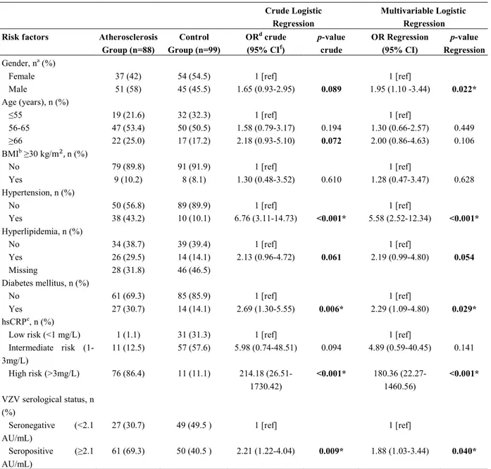
Table 2. Association of different risk factors and VZV-seropositivity with atherosclerosis in the patient group compared to the control group based on single and multivariable logistic regression.
Crude Logistic Regression
Multivariable Logistic Regression Risk factors Atherosclerosis
Group (n=88)
Control Group (n=99)
ORd crude (95% CIf)
p-value crude OR Regression (95% CI) p-value Regression Gender, na (%)
Female Male 37 (42) 51 (58) 54 (54.5) 45 (45.5) 1 [ref]
1.65 (0.93-2.95) 0.089
1 [ref]
1.95 (1.10 -3.44) 0.022* Age (years), n (%)
≤55 56-65 ≥66 19 (21.6) 47 (53.4) 22 (25.0) 32 (32.3) 50 (50.5) 17 (17.2) 1 [ref] 1.58 (0.79-3.17) 2.18 (0.93-5.10) 0.194 0.072 1 [ref] 1.30 (0.66-2.57) 2.00 (0.86-4.63) 0.449 0.106 BMIb ≥30 kg/m
, n (%) No Yes 79 (89.8) 9 (10.2) 91 (91.9) 8 (8.1) 1 [ref]
1.30 (0.48-3.52) 0.610
1 [ref]
1.28 (0.47-3.47) 0.628 Hypertension, n (%)
No Yes 50 (56.8) 38 (43.2) 89 (89.9) 10 (10.1) 1 [ref]
6.76 (3.11-14.73) ˂0.001*
1 [ref]
5.58 (2.52-12.34) <0.001* Hyperlipidemia, n (%)
No Yes Missing 34 (38.7) 26 (29.5) 28 (31.8) 39 (39.4) 14 (14.1) 46 (46.5) 1 [ref]
2.13 (0.96-4.72) 0.061
1 [ref]
2.19 (0.99-4.80) 0.054
Diabetes mellitus, n (%) No Yes 61 (69.3) 27 (30.7) 85 (85.9) 14 (14.1) 1 [ref]
2.69 (1.30-5.55) 0.006*
1 [ref]
2.29 (1.09-4.80) 0.029* hsCRPc, n (%)
Low risk (<1 mg/L) Intermediate risk (1-3mg/L)
High risk (>3mg/L)
1 (1.1) 11 (12.5) 76 (86.4) 31 (31.3) 57 (57.6) 11 (11.1) 1 [ref] 5.98 (0.74-48.51) 214.18 (26.51-1730.42) 0.094 <0.001* 1 [ref] 4.89 (0.59-40.45) 180.36 (22.27-1460.56) 0.141 <0.001*
VZV serological status, n (%)
Seronegative (<2.1 AU/mL)
Seropositive (≥2.1 AU/mL)
27 (30.7)
61 (69.3)
49 (49.5 )
50 (40.5 )
1 [ref]
2.21 (1.22-4.04) 0.009*
1 [ref]
1.88 (1.03-3.44) 0.040*
a
n, number; b
BMI: body mass index; c
hsCRP: high sensitive c-reactive protein; d
OR, odds ratio; f
CI, confidence interval; *
Bold text indicates
statistical significance (p<0.05). The logistic regression test was used.
LDL, hsCRP and anti-VZV IgG concentrations in patients with atherosclerosis (Mean±SD= 126.83±19.68 mg/dL; Mean±SD= 5.19±2.00 mg/L and Median (IQR)= 2.70 (1.53-4.30) AU/mL, respectively) were significantly higher than controls (Mean±SD = 119.02±13.11 mg/dL; Mean±SD 1.51±1.07 mg/L and Median (IQR)=2.10 (1.70-3.10) AU/mL, Student t-test,
p=0.019, Student t-test, p<0.001 and Mann-Whitney U
test, p=0.034, respectively; Table 1).
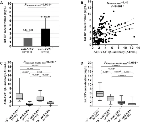
were VZV-seronegative (Student t-test, p<0.001; Figure 1A). We also found a statistically significant correlation between hsCRP concentration and plasma level of anti-VZV IgG in (Spearman test, r=0.40;
p<0.001; Figure 1B). The highest level of anti-VZV IgG antibody was seen in “AS+ anti-VZV+” group followed by “AS- anti-VZV+” group (Figure 1C). Interestingly, the plasma levels of hsCRP were significantly higher in “AS+ anti-VZV+”, “AS+ anti-VZV-” and “AS- anti-VZV+” groups as compared to the “AS- anti-VZV-” group (Dunn's multiple comparisons test, p<0.001, p<0.001 and p=0.006, respectively; Figure 1D).
In patients, the plasma levels of anti-VZV IgG
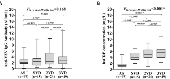
increased with advancing of the disease, however, the difference in the plasma levels of anti-VZV IgG between groups based on the number of diseased vessels was not statistically significant (p=0.168; Figure 2A). No significant difference regarding the plasma levels of anti-VZV IgG was observed in comparisons between groups (Dunn's multiple comparisons test, p>0.05; Figure 2A).
Kruskal-Wallis test showed a significant difference in the plasma levels of hsCRP between groups based on the number of diseased vessels (p<0.001; Figure 2B). In patients, the plasma levels of hsCRP increased with advancing of the disease, i.e. a significant difference in the plasma levels of hsCRP between groups based on
Figure 1. Comparison of the plasma levels of anti-VZV IgG and hsCRP levels. A. hsCRP levels were higher in
individuals who were anti-VZV+. B. There was a correlation between hsCRP and anti-VZV IgG antibody levels
in all subjects. C. The highest level of anti-VZV IgG was seen in “AS+ anti-VZV+” group. D. The highest level of
hsCRP was seen in “AS+ anti-VZV+” group and decreased gradually in subjects based on their status of atherosclerosis and seropositivity for anti-VZV.
B A
the number of diseased vessels was seen. The blood levels of hsCRP in patients having SVD, 2VD, 3VD were significantly higher as compared with the control group (Dunn's multiple comparisons test, p<0.001,
p<0.001 and p<0.001, respectively; Figure 2B).
The Relationship between Anti-VZV IgG and Risk Factors
To determine the relationship between risk factors and anti-VZV IgG in the induction of atherosclerosis, patients were divided into VZV-seronegative and seropositive subgroups. Our results showed that patients with VZV-seropositivity exhibited a higher prevalence of diabetes mellitus than patients with
VZV-seronegativity (test, p=0.039). This difference
remained significant after adjusting for all confounding variables. Multivariable logistic regression indicated that the presence of VZV-seropositivity was associated with an increased risk for coronary artery atherosclerosis (OR=3.33; 95%CI=1.02-10.88; Table 3). Interestingly, “AS+ anti-VZV+” patients were less likely to have BMI higher than 30 kg/m compared to
“AS+ anti-VZV-” patients (test, p=0.022), which
after adjusting for all potential confounding variables remained significant (OR=0.17; 95%CI=0.04-0.76). Individuals between ages 56-65 years had higher odds of being anti-VZV seropositive (OR=2.86; 95%CI=0.93-8.84; Table 3).
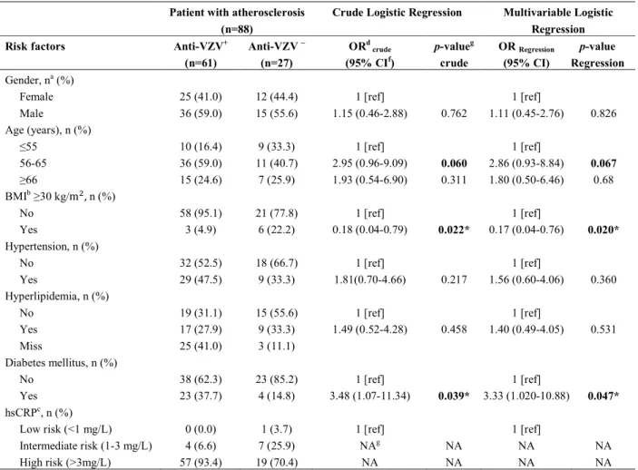
Table 3. Association of different risk factors of atherosclerosis with VZV-seropositivity in the patient group based on single and multivariable logistic regression
Patient with atherosclerosis (n=88)
Crude Logistic Regression Multivariable Logistic Regression
Risk factors Anti-VZV+
(n=61)
Anti-VZV – (n=27)
ORd crude (95% CIf)
p-valueg crude
OR Regression (95% CI)
p-value Regression
Gender, na (%) Female Male 25 (41.0) 36 (59.0) 12 (44.4) 15 (55.6) 1 [ref]
1.15 (0.46-2.88) 0.762
1 [ref]
1.11 (0.45-2.76) 0.826 Age (years), n (%)
≤55 56-65 ≥66 10 (16.4) 36 (59.0) 15 (24.6) 9 (33.3) 11 (40.7) 7 (25.9) 1 [ref] 2.95 (0.96-9.09) 1.93 (0.54-6.90) 0.060 0.311 1 [ref] 2.86 (0.93-8.84) 1.80 (0.50-6.46) 0.067 0.68 BMIb ≥30 kg/m
, n (%) No Yes 58 (95.1) 3 (4.9) 21 (77.8) 6 (22.2) 1 [ref]
0.18 (0.04-0.79) 0.022*
1 [ref]
0.17 (0.04-0.76) 0.020* Hypertension, n (%)
No Yes 32 (52.5) 29 (47.5) 18 (66.7) 9 (33.3) 1 [ref]
1.81(0.70-4.66) 0.217
1 [ref]
1.56 (0.60-4.06) 0.360 Hyperlipidemia, n (%)
No Yes Miss 19 (31.1) 17 (27.9) 25 (41.0) 15 (55.6) 9 (33.3) 3 (11.1) 1 [ref]
1.49 (0.52-4.28) 0.458
1 [ref]
1.40 (0.49-4.05) 0.531
Diabetes mellitus, n (%) No Yes 38 (62.3) 23 (37.7) 23 (85.2) 4 (14.8) 1 [ref]
3.48 (1.07-11.34) 0.039*
1 [ref]
3.33 (1.020-10.88) 0.047* hsCRPc, n (%)
Low risk (<1 mg/L) Intermediate risk (1-3 mg/L) High risk (>3mg/L)
0 (0.0) 4 (6.6) 57 (93.4) 1 (3.7) 7 (25.9) 19 (70.4) 1 [ref] NAg NA NA NA 1 [ref] NA NA NA NA a
n, number; b
BMI: body mass index; c
hsCRP: high sensitive c-reactive protein; d
OR, odds ratio; f
CI, confidence interval; g
NA, not applicable because of the presence of a zero in the denominator; *
Figure 2. Comparison of anti-VZV IgG and CRP levels in individuals based on the number of diseased vessels. A. The level of anti-IgG antibodies non-significantly increased along with the number of involved vessels. B The level of hsCRP significantly
increased along with the number of involved vessels.
DISCUSSION
Our study demonstrated that VZV-seropositivity is associated with higher odds of atherosclerosis after controlling for sex, age, BMI, hypertension, hyperlipidemia, and diabetes mellitus. In support of these results, Wang et al. showed the relationship between VZV infection and CAD.13 The information on the correlation between VZV and coronary vascular atherosclerosis is scarce.27 Most studies, however, have examined the relationship between VZV infection and atherosclerosis in the peripheral and cerebral vascular system.15,28,29 Previously, higher anti-VZV IgGtiters in CSF of patients with cerebrovascular atherosclerosis was shown.12 In most of the recent studies, increased CSF levels and reduced serum/CSF ratio of anti-VZV IgG in patients with cerebrovascular atherosclerosis confirmed intrathecal production of anti-VZV IgG.12,30 Although, there was no previous study on the association of serum or plasma level of anti-VZV IgG and coronary vascular atherosclerosis, it is known that anti-VZV IgG increases in serum after primary or re-exposure with the virus (natural or vaccination), as well as at the time of reactivation of the latent virus.31,32 Uthman et al.33 and Massano et al34 suggested that anti-VZV IgG induce the formation of immune complexes (antigen-antibody) due to cross-reaction with different lipid types such as phospholipids. The sedimentation of immune complexes can induce systemic inflammatory damage in various vascular tissues.35 In addition, VZV
displays pro-atherogenic roles through direct infection of the arterial wall, as well as provocation of pro-inflammatory, and pro-thrombotic responses.36 On the other hand, it is shown that viruses induce secretion of chemokines and cytokines, recruitment of leukocytes, the formation of foam cells, the proliferation of VSMCs, and the progression of plaques in the atherosclerosis process.6,37 Also, in the early phase of VZV induced vasculopathy, the presence of different immune cells in adventitia leads to intima thickening and vascular remodeling.29
Our finding of the correlation of hsCRP concentration with atherosclerosis irrespective of other factors in not new.38 CRP is one of the main inflammatory factors produced by liver hepatocytes, SMCs, macrophages, ECs, lymphocytes, and adipocytes in the body.16 We also found that hsCRP levels were significantly higher in individuals with higher anti-VZV IgG. This indicates that humoral immune response to VZV is generally associated with inflammation and hsCRP production depicted in the correlation analysis (Figure 1B). Further analysis indicated that the highest hsCRP level in the blood was found in the presence of both atherosclerosis and anti-VZV. Either one of atherosclerosis or anti-VZV alone was associated with lower levels of hsCRP production. The above findings once again point to the importance of hsCRP in inflammatory responses and also suggest that VZV infection is not the sole mechanism for hsCRP elevation.
We also showed that the plasma levels of anti-VZV IgG and hsCRP increase with advancing of atherosclerosis, which is suggestive of the relation between VZV infection, inflammation, and progression of atherosclerosis and is in accordance with a recent meta-analysis study.15
Our findings that patients between 56-65 years of age were more likely to have anti-VZV IgG, shows that seropositivity against VZV is not solely dependent on older age, as our group with ages greater than 65 did not have the highest level of antibody. These finding rules out the effect that older age of patients compared to controls may have had on the antibody levels. On the other hand, the higher level of hsCRP and a higher frequency of atherosclerosis in individuals older than 65 years were notable. Accordingly, when we considered VZV-seropositivity along with age, older subjects had a 5.62 fold odds of increased hsCRP and 1.80 folds increased risk of coronary artery atherosclerosis after adjusting for all confounding variables. This implied that atherosclerosis in older people is more likely to be associated with previous VZV infection as well as inflammatory response. One question that may arise is the correlation of hsCRP, anti-VZV and viral load between AS+ patients with acute VZV reactivation and those without. Obviously this piece of informative data is missing in our study; however, due to the chronic nature of atherosclerosis progression, it is more likely that repeated reactivations in the past and even a combination of infection history may be more relevant to atherosclerosis progression than a single episode of reactivation. Although a more careful matching of age between cases and controls would add to the credibility of the results, previous data have shown that weakened immune responses in older individuals with hyperlipidemia and/or diabetes mellitus may increase their susceptibility to reactivation of VZV infection and increase the odds of atherosclerosis.39,40 Interestingly, studies that were conducted in the Taiwanese, Canadian, and English populations, showed that VZV infection in patients over the age of 65 years was associated with poor prognosis.13,41
In the original analysis, we found that atherosclerosis was more frequent in men (Table 2), which is in accordance with previous reports.42,43 However, since the frequency of VZV-seropositivity was higher among women, including the VZV factor in the comparison of atherosclerosis between males and
females counteracted the gender effect. This finding once again points to the multifactorial nature of atherosclerosis in general and also to the more cautious interpretation of gender differences in this disease. Previously, Gialloreti et al. showed that the most important risk factor for reactivation of VZV in female and older subjects is immune compromisation. Nevertheless, the role of gender is yet to be clarified.44
In conclusion, this study showed that VZV-seropositivity is prevalent among those who are at a higher risk of atherosclerosis. Thus, the higher titers of anti-VZV IgG antibody and hsCRP in plasma or serum may predict a higher risk of atherosclerosis and its progression. Higher titers of anti-VZV IgG may indicate a possible recent re-exposure or recent reactivation of a latent VZV infection, which may indirectly cause vascular change and remodeling through hsCRP generation. These results support the role of infection mediated immune inflammation in exacerbation of atherosclerosis.
ACKNOWLEDGEMENTS
This work was performed as a part of Hamed Fouladseresht (Ph.D. candidate) dissertation as a requirement for graduation and was supported by a grant (95-01-13588) from Shiraz University of Medical Sciences, Shiraz, Iran. We thank all the participants for their support. The code of ethical approval of this project is IR.SUMS.REC.1396.S652.
REFERENCES
1. Nagel MA, Gilden D. Neurological complications of VZV reactivation. Curr Opin Neurol 2014; 27(3):356-60. 2. Gershon AA, Breuer J, Cohen JI, Cohrs RJ, Gershon MD,
Gilden D, et al. Varicella zoster virus infection. Nat Rev Dis Primers 2015; 1:15016.
3. Yawn BP, Wollan PC, Nagel MA, Gilden D, editors. Risk of stroke and myocardial infarction after herpes zoster in older adults in a US community population. Mayo Clin Proc; 2016: Elsevier.
4. Sundström K, Weibull CE, Söderberg-Löfdal K, Bergström T, Sparén P, Arnheim-Dahlström L. Incidence of herpes zoster and associated events including stroke— a population-based cohort study. BMC Infect Dis 2015; 15(1):488.
study in the UK. Neurology 2014; 83(2):e27-e33. 6. Zhu Y, Xian X, Wang Z, Bi Y, Chen Q, Han X, et al.
Research Progress on the Relationship between Atherosclerosis and Inflammation. Biomolecules 2018; 8(3).
7. Shapiro MD, Fazio S. From lipids to inflammation: new approaches to reducing atherosclerotic risk. Circ Res 2016; 118(4):732-49.
8. Ridker PM. How common is residual inflammatory risk? Circ Res 2017; 120(4):617-9.
9. Minassian C, Thomas SL, Smeeth L, Douglas I, Brauer R, Langan SM. Acute cardiovascular events after herpes zoster: a self-controlled case series analysis in vaccinated and unvaccinated older residents of the United States. PLoS Med 2015; 12(12):e1001919.
10. Nagel M, Traktinskiy I, Azarkh Y, Kleinschmidt-DeMasters B, Hedley-Whyte T, Russman A, et al. Varicella zoster virus vasculopathy: analysis of virus-infected arteries. Neurology 2011; 77(4):364-70. 11. Elkind MS. The varicella zoster virus vasculopathies:
Clinical, CSF, imaging, and virologic features. Neurology 2009; 72(11):1028-30.
12. Nagel M, Forghani B, Mahalingam R, Wellish M, Cohrs R, Russman A, et al. The value of detecting anti-VZV IgG antibody in CSF to diagnose VZV vasculopathy. Neurology 2007; 68(13):1069-73.
13. Wang CC, Lin CL, Chang YJ, Wang GJ, Sung FC, Kao CH. Herpes zoster infection associated with acute coronary syndrome: a population‐based retrospective cohort study. Br J Dermatol 2014; 170(5):1122-9. 14. Pesonen E, Andsberg E, Öhlin H, Puolakkainen M,
Rautelin H, Sarna S, et al. Dual role of infections as risk factors for coronary heart disease. Atherosclerosis 2007; 192(2):370-5.
15. Erskine N, Tran H, Levin L, Ulbricht C, Fingeroth J, Kiefe C, et al. A systematic review and meta-analysis on herpes zoster and the risk of cardiac and cerebrovascular events. PLoS One 2017; 12(7):e0181565.
16. Sproston NR, Ashworth JJ. Role of C-reactive protein at sites of inflammation and infection. Front Immunol 2018; 9:754.
17. Hage FG, Szalai AJ. C-reactive protein gene polymorphisms, C-reactive protein blood levels, and cardiovascular disease risk. J Am Coll Cardiol 2007; 50(12):1115-22.
18. Ramamoorthy RD, Nallasamy V, Raghavendra Reddy NE, Maruthappan Y. A review of C-reactive protein: A diagnostic indicator in periodontal medicine. J Pharm Bioallied Sci 2012; 4(Suppl 2):S422.
19. Ridker PM, Stampfer MJ, Rifai N. Novel risk factors for systemic atherosclerosis: a comparison of C-reactive protein, fibrinogen, homocysteine, lipoprotein (a), and standard cholesterol screening as predictors of peripheral arterial disease. JAMA 2001; 285(19):2481-5.
20. Lowe GD, Yarnell JW, Rumley A, Bainton D, Sweetnam PM. C-reactive protein, fibrin D-dimer, and incident ischemic heart disease in the Speedwell study: are inflammation and fibrin turnover linked in pathogenesis? Arterioscler Thromb Vasc Biol 2001; 21(4):603-10. 21. Ridker PM, Rifai N, Rose L, Buring JE, Cook NR.
Comparison of C-reactive protein and low-density lipoprotein cholesterol levels in the prediction of first cardiovascular events. N Engl J Med 2002; 347(20):1557-65.
22. Danesh J, Whincup P, Walker M, Lennon L, Thomson A, Appleby P, et al. Low grade inflammation and coronary heart disease: prospective study and updated meta-analyses. BMJ 2000; 321(7255):199-204.
23. Roivainen M, Viik-Kajander M, Palosuo T, Toivanen P, Leinonen M, Saikku P, et al. Infections, inflammation, and the risk of coronary heart disease. Circulation 2000; 101(3):252-7.
24. Kim JY, Park G-H, Kim MJ, Sim HB, Lee WJ, Lee S-J, et al. Usefulness of Inflammatory Markers for the Prediction of Postherpetic Neuralgia in Patients with Acute Herpes Zoster. Ann Dermatol 2018; 30(2):158-63. 25. Skripuletz T, Pars K, Schulte A, Schwenkenbecher P,
Yildiz Ö, Ganzenmueller T, et al. Varicella zoster virus infections in neurological patients: a clinical study. BMC Infect Dis 2018; 18(1):238.
26. Maleki A, Ghanavati R, Montazeri M, Forughi S, Nabatchi B. Prevalence of Coronary Artery Disease and the Associated Risk Factors in the Adult Population of Borujerd City, Iran. J Tehran Heart Cent 2019; 14(1):1. 27. Esteban-Hernandez J, San JRM, Gil R, Anegón M, Gil A.
Association between herpetic burden and chronic ischemic heart disease: matched case-control study. Med Clin (Barc) 2011; 137(4):157-60.
28. Tezcan ME, Teksut TK, Oenal AB, Oeztuerk MA. Reactivated varicella zoster virus may cause peripheral arterial thrombosis. J Rheumatol 2010; 37(8):1785-6. 29. Nagel MA, Gilden D. Developments in varicella zoster
virus vasculopathy. Curr Neurol Neurosci Rep 2016; 16(2):12.
31. Smith-Norowitz TA, Saadia TA, Norowitz KB, Joks R, Durkin HG, Kohlhoff S. Negative IgG varicella zoster virus antibody status: immune responses pre and post re-immunization. Infect Dis Ther 2018; 7(1):175-81. 32. Ihara H, Miyachi M, Imafuku S. Relationship between
serum anti‐varicella zoster virus antibody titer and time from onset of herpes zoster. J Dermatol 2018; 45(2):189-93.
33. Uthman I, Taher A, Khalil I. Hughes syndrome associated with varicella infection. Rheumatol Int 2001; 20(4):167-8.
34. Massano J, Ferreira D, Toledo T, Mansilha A, Azevedo E, Carvalho M. Stroke and multiple peripheral thrombotic events in an adult with varicella. Eur J Neurol 2008; 15(10):e90-e1.
35. Blum A, Peleg A, Weinberg M. Anti-cytomegalovirus (CMV) IgG antibody titer in patients with risk factors to atherosclerosis. Clin Exp Med 2003; 3(3):157-60. 36. Nagel MA, Traktinskiy I, Stenmark KR, Frid MG, Choe
A, Gilden D. Varicella-zoster virus vasculopathy: immune characteristics of virus-infected arteries. Neurology 2013; 80(1):62-8.
37. Rosenfeld ME, Campbell LA. Pathogens and atherosclerosis: update on the potential contribution of multiple infectious organisms to the pathogenesis of atherosclerosis. Thromb Haemost 2011; 106(11):858-67. 38. Auer J, Berent R, Lassnig E, Eber B. C-reactive protein
and coronary artery disease. Jpn Heart J 2002; 43(6):607-19.
39. Guilford T, Morris D, Gray D, Venketaraman V. Atherosclerosis: pathogenesis and increased occurrence in individuals with HIV and Mycobacterium tuberculosis infection. HIV AIDS (Auckl) 2010; 2:211.
40. Okamoto S, Hata A, Sadaoka K, Yamanishi K, Mori Y. Comparison of varicella-zoster virus-specific immunity of patients with diabetes mellitus and healthy individuals. J Infect Dis 2009; 200(10):1606-10.
41. Brisson M, Edmunds W, Law B, Gay N, Walld R, Brownell M, et al. Epidemiology of varicella zoster virus infection in Canada and the United Kingdom– corrigendum. Epidemiol Infect 2015; 143(6):1332-. 42. Spence JD, Pilote L. Importance of sex and gender in
atherosclerosis and cardiovascular disease. atherosclerosis 2015; 241(1):208-10.
43. Thomas SL, Hall AJ. What does epidemiology tell us about risk factors for herpes zoster? Lancet Infect Dis 2004; 4(1):26-33.
44. Gialloreti LE, Merito M, Pezzotti P, Naldi L, Gatti A, Beillat M, et al. Epidemiology and economic burden of