Determining the fate of internalized CFTR after cigarette smoke exposure
By
Amanda J. Smith
Senior Honors Thesis Biology Department
University of North Carolina at Chapel Hill
April 2015
Approved:
Dr. Robert Tarran, Research Advisor
2
ABSTRACT
Chronic Obstructive Pulmonary Disease (COPD) is a global health problem with tobacco smoke exposure being the predominant cause. COPD has two subtypes: emphysema, the
destruction of the alveolar surfaces, and chronic bronchitis (CB), the accumulation of mucus in the airways. Patients with cystic fibrosis (CF), a disease caused by genetic mutations in the CF transmembrane conductance regulator (CFTR) gene, have similar symptoms to the CB form of COPD, such as mucus dehydration and failure in mucociliary clearance. Research has shown that these characteristics in CB may be caused, at least in part, by the rapid internalization and
aggregation of CFTR after cigarette smoke (CS) exposure. My research investigated the
colocalization, or proximity, of CFTR after CS-induced internalization with various endosomes of the endocytic pathway.
For my research, I have labeled early and late endosomes, and CFTR, with fluorescent proteins and expressed them in HEK293T cells, a human embryonic kidney cell line, exposed the cells to CS, and imaged the cells using confocal microscopy. My results suggest that internalized CFTR colocalized with, or relocated to, early and late endosomes at various time points after CS exposure. Understanding the mechanisms of how CFTR is internalized will help lead to therapies to treat CFTR dysfunction in COPD patients.
INTRODUCTION
Chronic obstructive pulmonary disease (COPD) is estimated to affect 24 million people in the United States according to the COPD Foundation and is the fourth leading cause of death in the world [1]. COPD is caused by long-term exposure to toxic gases and particles. In
3
factor [1]. COPD has two primary phenotypes: emphysema, or the abnormal permanent enlargement of air spaces distal to the terminal bronchioles with destruction of their walls, and chronic bronchitis (CB), clinically defined as chronic cough or mucus production for at least three months in at least two consecutive years [2].
Since chronic bronchitis and cystic fibrosis (CF) have similar symptoms, researchers have suggested dysfunction of the cystic fibrosis transmembrane conductance regulator (CFTR) protein may contribute to the development of cigarette smoke-induced chronic bronchitis [3]. CFTR is an important anion channel that secretes chloride and bicarbonate out of airway epithelial cells into the airway lumen and acts a regulator of other ion channels, such as the epithelial sodium channel (ENaC) or calcium-activated chloride channel (CACC) [4]. The regulation of chloride ions and other ion channels by CFTR maintains proper volume and hydration of airway surface liquid (ASL), which contains a mucus layer and a periciliary layer (PCL) [4]. A proper ASL volume helps cilia beat mucus-trapped pathogens out of the airways and prevents respiratory infections [4]. Therefore, a disruption of the balance of NaCl, and thus water transport into the ASL, can lead to impaired beating of cilia and an accumulation of mucus in the respiratory airways. This accumulation of mucus eventually leads to phenotypes of
4
compartments instead of lysosomes [3]. Rasmussen et al. discovered that an increase in intracellular calcium levels due to cigarette smoke acts as the signal to trigger the CFTR
internalization in airway epithelial cells, and that this increased release of calcium may be from lysosomes [6].
Further research needs to be completed to fully understand the mechanisms that cause cigarette smoke-induced CFTR internalization. Recent findings in our lab have shown that CFTR does not aggregate or change its conformation at the plasma membrane after cigarette smoke exposure; therefore, it was concluded that CFTR aggregates after internalization into the cell [7]. It was also discovered that cigarette smoke-induced internalization of CFTR was inhibited by endocytic inhibitors, such as hypertonic sucrose and Dynasore, which may suggest that cigarette smoke-induced CFTR internalization is dynamin-dependent [7]. Therefore, our lab hypothesized that CFTR was internalized via the clathrin-mediated endocytic pathway after CS exposure.
In the clathrin-mediated endocytic pathway, clathrin is recruited to the plasma membrane where it interacts with adaptor proteins to initiate endocytosis. The area of the plasma membrane coated with clathrin buds around the material being endocytosed and is then pinched off into a clathrin-coated vesicle by dynamin proteins [8]. After endocytosis, the vesicle loses its clathrin coating and fuses with the early endosome, which is important for sorting endocytosed material. Early endosomes can direct the material to late endosomes, and then to lysosomes for
5
In this study, we examined if CFTR was internalized after cigarette smoke exposure via the clathrin-mediated endocytic pathway. To study this aspect of CFTR trafficking, we
colocalized CFTR with known markers of different organelles, such as clathrin-coated vesicles, early endosomes, late endosomes, recycling endosomes, or lysosomes, after cigarette smoke exposure in HEK293T cells. We hypothesized that the greatest amount of internalized CFTR will first colocalize with clathrin-coated vesicles, then early endosomes, then late endosomes, then recycling endosomes, or finally lysosomes based on the time after exposure to cigarette smoke. Determining the different endocytic compartments that the internalized CFTR colocalizes with can help determine the mechanism that causes CFTR to be internalized after exposure to
cigarette smoke, and thus, can further help develop treatments for the chronic bronchitis form of COPD.
MATERIALS AND METHODS
Cell Culture and Transfection
6
needed. All constructs were ordered from Addgene and constructs were purified using a Maxiprep Kit (QIAGEN).
Cigarette Smoke Exposure
On the second or third day after transfection, the media was removed from transfected HEK293T cells in 6-well plates and was replaced with 1 mL of Pyruvate Ringers solution (120 mM NaCl, 12 mM NaHCO3, 24 mM HEPES, 1.2 mM MgCl2, 5.2 mM KCl, 1 mM sodium pyruvate, 1x NEAA, 10 mM glucose, 1.2 mM CaCl2 ·2H2O, 0.1% Albumin, pH 7.4). Cells were incubated at 37°C for 5 minutes to equilibrate the experimental conditions. After the 5 minute incubation, plates were tipped to remove most of the Ringers solution to leave a thin film to represent in vivo airway epithelial cells. Half of the HEK293T cells for each transfection were then exposed to the smoke from a whole cigarette (~14-15 x 30 mL puffs) over the course of about 10 minutes using an LM1 smoke engine (Borgwaldt, Hamburg, Germany) to mimic in vivo smoke exposure while the other half of the cells were only exposed to air during the same time to be the controls. After cigarette smoke exposure, the cells were left in these environments for 5 more minutes, the thin film of Ringers solution was then removed, and 1 mL of normal
HEK293T media was added to cells. The cells were then incubated at 37°C for 15, 30, 60 or 120 minutes in a CO2 incubator to allow endocytosis of CFTR in cigarette smoke-exposed cells. If the cells were to be imaged at 0 minutes after cigarette smoke exposure, the following steps were done immediately after the 5 minutes after cigarette smoke exposure.
Confocal Imaging and Analysis
7
were imaged using a Leica SP5 confocal microscope with a glycerol 63x objective using the Leica Application Suite: Advanced Fluorescence (LAS AF) software. About 5 or 6 different images were taken from each coverslip. Confocal image channels were merged using Adobe Photoshop CS2 to qualitatively assess the amount of colocalization and regions of interest (ROIs) were analyzed using the LAS AF Colocalization Tool to analyze them quantitatively. All data were recorded in Microsoft Excel 2007 (Microsoft Corporation, USA). All quantitative data are presented as mean percent colocalization ± S.E.M. Percent colocalization was calculated by colocalization area divided by area foreground of the ROIs, where area foreground was
calculated by area image divided by area background. These calculations were all performed automatically using the Colocalization Tool in the LAS AF software. Statistical significance was calculated using two-tailed non-paired t-tests and one-way ANOVA tests using GraphPad Instat 3 software, and P values of P < 0.05 were considered significant. Graphs of data were created using GraphPad Prism 6 software. LAMP1 and RFP-CFTR transfected cells and DsRed-Rab5A and GFP-CFTR transfected cells at 60 minutes after CS exposure experiments were performed twice, while the DsRed-Rab7 and GFP-CFTR transfected cells at 60 minutes after CS exposure experiment was performed only once. The time course experiments of DsRed-Rab5A and DsRed-Rab7 with GFP-CFTR after CS exposure were both performed three times.
RESULTS
8
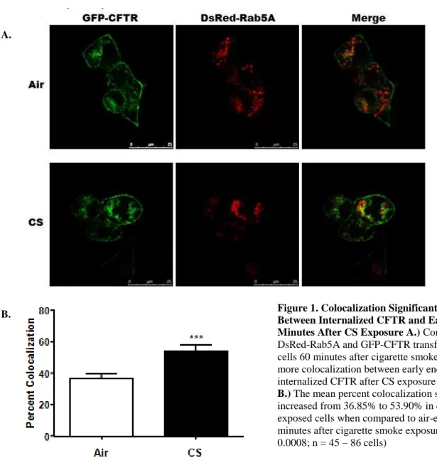
colocalization from 36.85 ± 2.82 in air-exposed cells to 53.90 ± 4.21 in cigarette smoke-exposed cells (Figure 1b). This increase in colocalization was considered highly significant with a P value of 0.0008 according to an unpaired two-tailed t-test where n = 45 – 86 cells per group. Therefore, the next experiment performed was a time course involving the colocalization of internalized CFTR and early endosomes.
The second experiment performed was a time course to determine the different amounts of colocalization of early endosomes and internalized CFTR in HEK293T cells 0, 15, 30, 60, and 120 minutes after exposure to cigarette smoke using DsRed-Rab5A and GFP-CFTR transfected
Figure 1. Colocalization Significantly Increased Between Internalized CFTR and Early Endosomes 60 Minutes After CS Exposure A.) Confocal images of DsRed-Rab5A and GFP-CFTR transfected HEK293T cells 60 minutes after cigarette smoke exposure showed more colocalization between early endosomes and internalized CFTR after CS exposure at this time point
B.) The mean percent colocalization significantly increased from 36.85% to 53.90% in cigarette smoke-exposed cells when compared to air-smoke-exposed cells at 60 minutes after cigarette smoke exposure. (P value = 0.0008; n = 45 – 86 cells)
A.
9
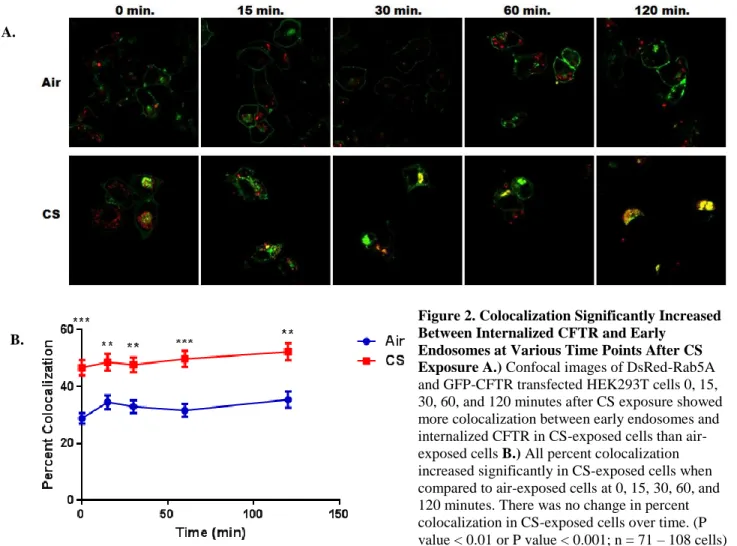
cells. This experiment was performed on three separate occasions. Qualitatively, the confocal images showed that at every time point there was greater colocalization between Rab5A (early endosomes) and internalized CFTR in cigarette smoke-exposed cells compared to air-exposed cells (Figure 2a). Quantitatively, the amount of colocalization between early endosomes and internalized CFTR in cigarette smoke-exposed cells significantly increased at each time point when compared to air-exposed cells (Figure 2b). The percent colocalization increased from 28.85 ± 1.85 to 48.71 ± 2.94 at t = 0, 34.90 ± 2.30 to 48.88 ± 2.93 at t = 15, 34.08 ± 2.44 to 47.69 ± 2.63 at t = 30, 31.61 ± 2.33 to 49.75 ± 2.57 at t = 60, and 35.07 ± 2.44 to 52.29 ± 3.03 at t = 120 with P values of less than 0.001, 0.01, 0.01, 0.001, and 0.01 respectively according to an one-way ANOVA test where n = 71 – 108 cells per group. When comparing the percent
10
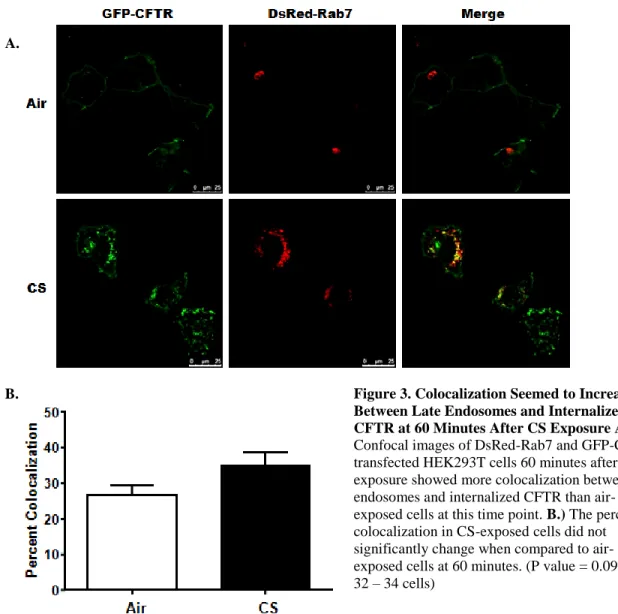
Similar experiments were performed to determine if late endosomes colocalized with internalized CFTR with time after exposure to cigarette smoke in HEK293T cells using DsRed-Rab7 and GFP-CFTR. The 60 minute experiment was performed on one occasion in triplicate to test the DsRed-Rab7 construct. Qualitatively, there was some colocalization seen in CS-exposed cells when compared to air-exposed cells (Figure 3a). Quantitatively, the mean percent
colocalization of CS-exposed cells did not significantly change when compared to air-exposed cells according to a two-tailed unpaired t-test (Figure 3b).
Figure 2. Colocalization Significantly Increased Between Internalized CFTR and Early
Endosomes at Various Time Points After CS Exposure A.) Confocal images of DsRed-Rab5A and GFP-CFTR transfected HEK293T cells 0, 15, 30, 60, and 120 minutes after CS exposure showed more colocalization between early endosomes and internalized CFTR in CS-exposed cells than air-exposed cells B.) All percent colocalization increased significantly in CS-exposed cells when compared to air-exposed cells at 0, 15, 30, 60, and 120 minutes. There was no change in percent colocalization in CS-exposed cells over time. (P value < 0.01 or P value < 0.001; n = 71 – 108 cells)
A.
11
An additional time course experiment was performed to examine colocalization between late endosomes and internalized CFTR at 0 minutes, 15 minutes, 30 minutes, 60 minutes, and 120 minutes after cigarette smoke-exposure using DsRed-Rab7 and GFP-CFTR transfected HEK293T cells. This experiment was performed on three separate occasions. Qualitatively, the confocal images seemed to show that there was greater colocalization between late endosomes and internalized CFTR in cigarette smoke-exposed cells compared to air-exposed cells (Figure 4a). Quantitatively, the amount of colocalization between late endosomes and internalized CFTR in cigarette smoke-exposed cells significantly increased only at 30 minute and 120 minute time
Figure 3. Colocalization Seemed to Increase Between Late Endosomes and Internalized CFTR at 60 Minutes After CS Exposure A.)
Confocal images of DsRed-Rab7 and GFP-CFTR transfected HEK293T cells 60 minutes after CS exposure showed more colocalization between late endosomes and internalized CFTR than air-exposed cells at this time point. B.) The percent colocalization in CS-exposed cells did not significantly change when compared to air-exposed cells at 60 minutes. (P value = 0.0983; n = 32 – 34 cells)
A.
12
points when compared to air-exposed cells with P values of less than 0.05 according to an one-way ANOVA test where n = 64 – 90 cells per group (Figure 4b). The percent colocalization increased from 23.43 ± 2.26 to 32.87 ± 2.07 at t = 30 and increased from 22.55 ± 1.70 to 31.51 ± 1.77 at t = 120. When comparing the colocalization rate in CS-exposed cells between each time point, there was no significant change over time.
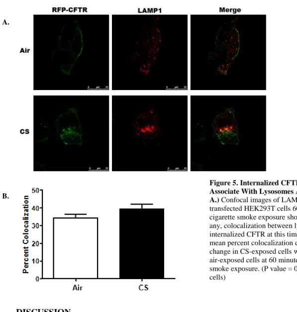
Another experiment that we performed examined whether internalized CFTR colocalized with lysosomes by transfecting HEK293T cells with LAMP1 and RFP-CFTR and imaging them at 60 minutes after smoke exposure. This experiment was performed twice in triplicate.
Figure 4. Colocalization Significantly Increased Between Late Endosomes and Internalized CFTR at 30 and 120 Minutes After CS Exposure A. Confocal images of DsRed-Rab7 and GFP-CFTR transfected HEK293T cells 0, 15, 30, 60, and 120 minutes after CS exposure showing more colocalization at 30 minutes and 120 minutes between late endosomes and internalized CFTR in CS-exposed cells than air-CS-exposed cells. B.) The percent colocalization significantly increased at 30 minutes and 120 minutes after CS exposure in CS-exposed cells when compared to air-exposed cells. There was not a significant change in percent colocalization in CS-exposed cells over time. (P value < 0.05;
n = 64 - 90 cells)
A.
13
Qualitatively, the confocal images did not show much, if any at all, colocalization between CS-induced internalized CFTR and lysosomes (Figure 5a). Quantitative analysis showed that the mean percent colocalization of CS-exposed cells did not significantly change when compared to air-exposed cells according to a two-tailed unpaired t-test (Figure 5b).
DISCUSSION
Through this study, we have explored the possibility that the clathrin-mediated endocytic pathway is used to internalize CFTR after cigarette smoke exposure. We have determined the biomarkers of the endocytic pathway with which the internalized CFTR colocalizes or
internalizes to. The most novel finding that resulted from this study is the discovery that
CS-Figure 5. Internalized CFTR Did Not
Associate With Lysosomes After CS Exposure A.) Confocal images of LAMP1 and RFP-CFTR transfected HEK293T cells 60 minutes after cigarette smoke exposure showed not much, if any, colocalization between lysosomes and internalized CFTR at this time point. B.) The mean percent colocalization did not significantly change in CS-exposed cells when compared to air-exposed cells at 60 minutes after cigarette smoke exposure. (P value = 0.1629; n = 74 - 106 cells)
A.
14
induced internalized CFTR colocalizes with endosomes, a result that has not been shown before. Colocalization between internalized CFTR and early endosomes was found to be significantly increased at 0 minutes, 15 minutes, 30 minutes, 60 minutes, and 120 minutes after cigarette smoke exposure compared to controls. When comparing the percent colocalization between the cigarette smoke-exposed cells between time points for early endosomes, there was no significant change over time. Colocalization between internalized CFTR and late endosomes was found to significantly increase at 30 minutes and 120 minutes compared to controls. When comparing the percent colocalization between the cigarette smoke-exposed cells between time points for late endosomes, there was not a significant change over time. Our results also suggested that after exposure to cigarette smoke, colocalization of lysosomes and internalized CFTR did not occur.
15
exposure to cigarette smoke [3], which is interesting since one would think that after locating to late endosomes, CFTR would then locate to lysosomes. However, Clunes et al. also discovered that CS-induced internalized CFTR relocates to a perinuclear compartment and closely
associates with vimentin, an intermediate filament protein that has been shown to be an indicator of aggresome formation [3]. Therefore, we speculate that internalized CFTR relocates to
aggresomes after CS-exposure, instead of relocating to lysosomes or proteasomes for degradation.
While we have found some promising data that supports our hypothesis, there are many other experiments that can be done to verify our findings. Other than repeating the above experiments, future experiments will show if the colocalization seen here is blocked when CS-exposed cells are also treated with Dynasore, an inhibitor of dynamin which will thus inhibit the endocytic pathway, to make sure that the data we have collected is actually endocytosed CFTR colocalizing with the different endocytic compartments. Other biomarkers of the endocytic pathway will also be examined if they colocalize with CS-induced internalized CFTR, such as continuing our studies involving lysosomes and starting experiments dealing with recycling endosomes, etc.
16
Acknowledgements:
I would like to thank the Tarran lab for giving me this wonderfulopportunity to work on exciting and innovative research these past couple years, even though some of that was not dealing with my thesis. I would like to especially thank Dr. Robert Tarran, Dr. John Sheridan, Dr. Jody White, and soon-to-be Dr. Abigail Marklew for directly advising me, training me, dealing with all my questions, and teaching me pretty much everything I know about conducting research over the years. I would also like to thank the UNC Biology Department and the UNC Biology Honors Program.
REFERENCES
1. Barnes, Peter. (2004) Small Airways in COPD. The New England Journal of Medicine 350.26: 2635 – 2637.
2. Kerstjens HA, Postma DS, and Hacken NT. (2008) COPD. Clinical Evidence: 1502. 3. Clunes LA, Davies CM, Coakley RD, Aleksandrov AA, Henderson AG, Zeman KL, Worthington EN, Gentzsch M, Kreda SM, Cholon D, Bennett WD, Riordan JR, Boucher RC, Tarran R. (2012) Cigarette smoke exposure induces CFTR internalization and insolubility, leading to airway surface liquid dehydration. The FASEB Journal 26.2: 533 – 545.
4. Tarran R. (2004) Regulation of airway surface liquid volume and mucus transport by active ion transport. Proceedings of the American Thoracic Society 1.1: 42 – 46.
5. Ramos, FL, Krahnke, JS, and Kim, V. (2014) Clinical issues of mucus accumulation in COPD. International Journal of Chronic Obstructive Pulmonary Disease 9: 139 – 150.
17
7. Marklew AJ, Gilmore RC, Gray MA, and Tarran R. (2014) Exploring the Mechanisms Behind Cigarette Smoke-induced Internalization of CFTR. Pediatric Pulmonology 49: 218.