PICTORIAL INTERLUDE
Experiences of using a single post-contrast CT
scan of the chest after biphasic contrast injection
P C Pretorius, FCRad (Diag) SA
Drs Visser, Erasmus, Vawda & Partners, Port Elizabeth
Corresponding author: P Pretorius (ppret@telkomsa.net)
Introduction
While experimenting with single post-contrast computed tomographic (CT) scans of the urinary tract after a triphasic contrast injection,1 I considered whether a similar technique could be used to cut down on unnecessary scan series in our patients referred for chest CT scans. A precontrast scan and then a biphasic contrast injection followed by a single scan of the thorax was therefore considered and implemented to test the reliability and quality of this technique.
Protocols for chest CT surveys for mediastinal pathology, lung masses and ‘rule out pathology’ chest scans traditionally dictate a post-contrast scan beginning 60 s after initiating an intravenous post-contrast injection. In numerous practices, 100 - 120 cc of contrast is traditionally used. This process usually results in adequate contrast throughout the vascular structures in the mediastinum and lungs and enhancement of pathologic masses or lymph nodes.
When there are clinical indications of vascular pathology (i.e. aortic or pulmonary arteries and their respective branch vessels), it is imperative to add a dedicated arterial phase scan before the 60 s scan series. If there are no clinical indications to include an arterial phase, a single 60 s post-contrast scan only should be performed.
Unfortunately, clinical information from referring physicians is not always clear and, even when clear, many radiologists feel insecure without including an arterial series. We felt that this biphasic injection followed by a single post-contrast scan technique improved the diagnostic information and also alleviated the insecurity of excluding the arterial series when performing chest CT scans for non-vascular indications.
After scanning a few test cases using a single post-contrast series after a biphasic contrast injection, we were pleasantly surprised with the results, and we have now introduced this technique in most of our practice, when a ‘general rule out’ or ‘mass lesion characterisation’ scan is requested. This requires radiologists to shift their comfort zones, to rely on a single post-contrast scan replacing either a post-contrast arterial, 60 s, or both arterial and 60 s scans.
I have included a pictorial representation of a variety of pathologies while using the newer technique, to share our experiences over a wide range of pathologies, which will assist our colleagues in making this shift of technique with more confidence, and will hopefully address some of the queries that may be raised.
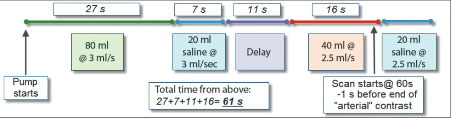
Injection plan
1. 80 cc contrast @ 3 cc/s (27 s) – contributes to ‘venous phase’ of the scan 2. 20 cc saline @ 3 cc/s (7 s) – pushes contrast into system.
3. Delay (11 s)
4. 40 cc contrast @ 2.5 cc/s (16 s) – contributes to the ‘arterial phase’ of the scan
5. Followed by 20 cc saline @ 2.5 cc/s
6. The scan is started at 60 s, which is 15 s into the arterial phase of the injection, 1 s before the end of the injection. Most scanners require a 4 - 5 s delay from the time the scan is initiated until the first ‘cuts’ are taken – see comments in the discussion below.
Computed tomographic (CT) chest investigations can be enhanced; in many cases, the arterial phase of a post-contrast arterial and delay (60 seconds) study can be omitted when planning the contrast injection and scanning technique carefully. A biphasic contrast injection was used before starting a single 60-second post-contrast scan.
S Afr J Rad 2012;16(2):56-60.
PICTORIAL INTERLUDE
Case studies
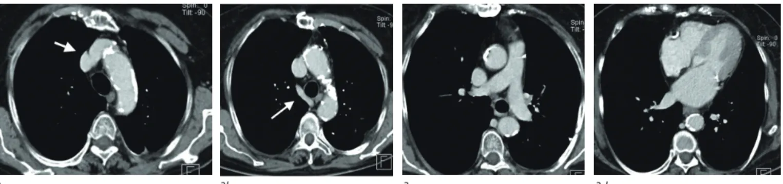
Normal arterial phase scan (Fig. 2)
Arterial phase-contrast scans of the chest show good contrast enhancement of pulmonary arteries and veins, heart chambers, the aorta and its branch vessels. There is good differentiation of contrast between the central venous and arterial structures (dense enhancement v. no enhancement). The returning systemic veins contralateral to the side injected, from the head and neck, from infradiaphragmatic veins (especially the azygos vein) and any mediastinal and hilar masses and lymph nodes and lung pathology are not enhanced at this early post-contrast phase. Consequently, there is often difficulty in differentiating the unenhanced dilated or anomalous venous structures from the unenhanced lymph nodes and masses in the chest.
Normal 60 s post-contrast scan (Fig. 3)
The contrast enhancement of the vascular structures, mass lesions and lymph nodes within the chest is usually adequate, but differentiation between central arterial and venous structures and returning systemic
venous structures relies on anatomical identification, as the contrast density of these structures is usually very similar. The contrast in the pulmonary arteries and branches is often mediocre, and incidental pulmonary arterial emboli may not be well seen.
Post biphasic contrast injection scan (Fig. 4)
After a biphasic contrast injection, there is dense enhancement of central pulmonary arteries and veins and the aorta and its branches. Returning systemic veins also show enhancement, but less dense than the central vessels, allowing further clarity to confirm the anatomical differentiation. Lung and mediastinal masses and lymph nodes will also enhance, usually to a lesser degree than systemic veins, and will show their enhancement patterns.
Lung carcinoma after biphasic contrast injection
(Fig. 5)
Despite the scan beginning at 60 s, there is dense enhancement of the pulmonary arteries, veins and aorta, without compromising enhancement and characterisation of the lung mass and lymph nodes.
Mycetoma after biphasic contrast injection (Fig. 6)
It is interesting to compare this case with the preceding lung carcinoma case (Fig. 5), where there was enhancement of the mass and the typical peripheral enhancement of infiltrated lymph nodes, unlike the non-enhancement of this mass and the uniform node non-enhancement.
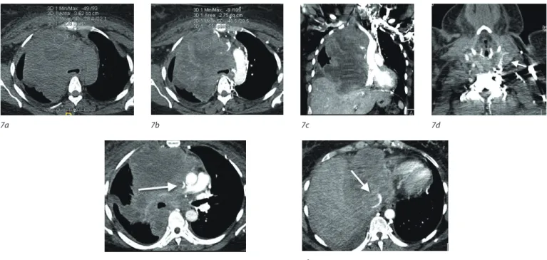
Lymphoma after biphasic contrast injection (Fig. 7)
Having dense contrast in all the vascular structures, with enhancement of the mass, all on one post-contrast series, helped to ‘unpack’ all the pathological processes going on in this case, which initially appeared quite complex because of the gross changes.
2a 2b
Fig. 2a. White arrows point to unenhanced left brachiocephalic vein anteriorly and unenhanced lymph nodes posteriorly. In Fig. 2b, the white arrow indicates the unenhanced azygos vein.
3a 3b 3c 3d
Fig. 3. Scan of the chest 60 s after contrast injection. Arrows in 3a and 3b point to the left brachiocephalic vein and azygos vein, respectively.
4a 4b 4c 4d
Fig. 4. Biphasic contrast injection single post-contrast injection series. The white arrow in 4a and 4b shows contrast-enhanced left brachiocephalic and azygos veins. The white arrow in Figs 4c and 4d show an enhancing ectopic thyroid nodule, with calcification, abutting on the enhanced left brachiocephalic vein.
PICTORIAL INTERLUDE
Goitre after biphasic contrast injection (Fig. 8)
Although not a particularly challenging diagnosis to make, following the enhanced thyroid from the neck, into the mediastinum, and differentiation from adjacent vascular structures all on one post-contrast series was made easier in this case.
Sarcoid after biphasic contrast injection (Fig. 9)
There is clear differentiation of the enhancing lymph nodes from the
adjacent, more densely enhancing vascular structures. Again, this is not a particularly challenging call to make – but compare with the next case below (Fig. 10).
Interstitial lung disease after biphasic contrast injection
(Fig. 10)
The lymphadenpathy was more subtle than the sarcoid case above, but can be confidently called because of the adjacent azygos vein and aortic enhancement.
5a 5b 5c 5d
Fig. 5. There is a carcinoma mass in the right lung (arrows in precontrast 5a and post-contrast 5b scans) with peripheral enhancement of the mass. Infiltrated lymph nodes show typical peripheral enhancement on the post-contrast series (arrowheads in 5b, 5c and 5d).
6a 6b 6c 6d
Fig. 6. A lung mass showing increased density of 52HU on the precontrast scan (6b) with no significant enhancement on the 60 s post-contrast scan (6c). There are inflammatory uniformly enhancing lymph nodes in the adjacent mediastinum – arrow in 6d.
7a 7b 7c 7d
7e 7f
Fig. 7. Mass in right chest with superior medistinal syndrome clinically. The mass seen on precontrast (7a) shows vague enhancement (20HU) on the 60 s scan (7b and 7c). There is tumour infiltration causing partial obstruction of SVC (7e), the IVC (7f) and the left internal jugular vein (7d) (white arrows).
PICTORIAL INTERLUDE
Opportunistic infection after biphasic contrast injection
(Fig. 11)
The pulmonary arterial emboli might not have been as clearly seen on a single standard post-contrast 60 s scan, and the splenic micro-abscesses might also have been missed on a standard post-arterial scan. These were all seen on the single post-contrast series.
Atelectasis after biphasic contrast injection (Fig. 12)
The collapsed lung is enhanced, with dense enhancement of vascular structures.
Discussion
The contrast load traditionally used for a survey chest CT is 100 - 120 cc, to gain adequate contrast density in vascular structures in the chest. A ‘rule
9a 9b 9c 9d
Fig. 9. Sarcoid infiltration of lung (arrow in 9a), with enlarged uniformly enhancing mediastinal and hilar lymph nodes (arrows in 9b - d). 8a 8b 8c
Fig. 8. Retrosternal extension of a goitre (arrow), with cystic change in left lobe. Clear separation and differentiation from adjacent aortic arch branches bilaterally and the enhanced left brachiocephalic vein anteriorly noted in 8b.
10a 10b
Fig. 10. Interstitial changes in the right lung. There is mediastinal lymphadenopathy (arrow in 10b) differentiated from the adjacent enhanced azygos vein (arrowhead in 10b).
11a 11b 11c 11d
Fig. 11. Opportunistic nodular lung infection in immunocompromised patient. Unsuspected pulmonary arterial emboli noted on the right (arrow in 11b and arrowhead in 11c). Lymph node enlargement noted (arrow in 11c). Micro-abscesses also identified in spleen (arrows in 11d) at the inferior range of the scan.
PICTORIAL INTERLUDE
of thumb’ is that, from the time the contrast injection into an antecubital vein begins, it takes 9 s to reach the heart, by 12 s it is through the lungs back to the heart, and by 15 s has reached the descending thoracic aorta.
Contrast injection rates vary between 2 - 5 cc/s, depending on the sequence chosen. A contrast injection of 100 cc at 4 cc/s. will ensure a 25 s bolus of contrast. Since modern multislice scanners can scan the whole thorax in 10 - 16 s and the resolution is so high, it is conceivable that, if one could get the timing perfect in every patient, one could theoretically perform a good arterial phase diagnostic scan of the chest with only 16 s x 2.5 cc/s = 40 cc of contrast.
Naturally, there are numerous variable factors in patients such as body morphology, cardiac function, degree of valsalva with breath hold, etc. ‘Perfect timing’ of the scan in every case is consequently not feasible but, as can be seen from the above description, a theoretical 60 - 80 cc of contrast (100 - 120 cc minus 40 cc) is non-contributory to the diagnostic information, if one were looking purely at the arterial filling of vessels. This ‘extra’ contrast can be used to enhance the contrast appearances in the other vessels by splitting the bolus delivery.
Using the above logic, we designed an injection sequence to try to maximise the benefit of this ‘non-contributory’ portion of contrast that we inject. The logic is as follows:
• The initial 80 cc starting 60 s before the scan allows adequate contrast filling of the returning systemic veins, particularly the brachiocephalic and azygos veins. The injection rate is kept similar to that of a ‘traditional’ chest survey CT scan. This contrast is ‘pushed’ by a saline chaser injection at the same rate, followed by a calculated delay. • A second contrast injection begins 15 s before the CT scan starts. This
will enhance the pulmonary arterial and pulmonary venous vessels and the aortic vessels in the chest. This timing allows the end of the second phase bolus of contrast to densely enhance the central vessels (pulmonary veins and arteries), while the lead portion of this second bolus will have reached the descending aorta in almost all patients. • In fit patients, this timing is invariably adequate.
• In patients with known poor cardiac function, the contrast passage may be a bit slower. In these cases, one may start the scan a little later – possibly at 65 s This is still usually adequate as the contrast bolus tends to ‘stretch out’ i.e. a tight bolus injected into a peripheral vein over 15 s. will have spread out to possibly over 20 s by the time it has gone through the heart, lungs and heart again, before entering the aorta.
• The 60 s initiation of the scan after the beginning of the contrast injection, means that the scan effectively starts at 64 s, because of the built-in delay for a scan to initiate. This is 3 s after completion of the contrast injection in the arm. I have found that this timing
is adequate (as the contrast bolus still has to travel up the arm and through the heart and lungs and into the aorta) and the dense arterial phase of contrast in the vessels from the superior vena cava, through the heart, pulmonary arteries and veins and aorta is captured regularly with this timing.
• A final saline chaser injection pushes the contrast towards the chest. This routine gave the following advantages:
• adequate enhancement of returning systemic veins, to help differentiate them from mediastinal and hilar masses and lymph nodes
• relatively dense contrast enhancement of pulmonary arteries, veins and thoracic aorta and its branches, which allowed clear differentiation between systemic returning veins and mediastinal and hilar masses and lymph nodes
• enhancement of masses and lymph nodes allowed their characterisation
• all the post-contrast information was seen on one set of images, negating the need to run both an arterial and a 60 s post-contrast series (2 series). This made interpretation quicker and in many cases easier (see case of lymphoma above – Fig. 7).
In my opinion, the information gained is increased when compared with a single 60 s post-contrast scan.
Radiation exposure is reduced by omitting the arterial phase scan in practices where both an arterial and 60 s post-contrast are run for those cases where the arterial series is not indicated, but is added for the radiologist’s comfort.
Other advantages from reducing unnecessary series of post-contrast CT scans have been discussed in a previous article using a single post-contrast CT scan of the urinary tract after triphasic post-contrast injection.1 These advantages include:
• more efficient reading of the scans owing to more information presented on a single post-contrast series
• ease of viewing of a single scan post-contrast series for the referring clinician
• reduction in radiation to the patient from unnecessary extra scan series • reduced wear and tear on scanner hardware
• reduction in the number of images needing electronic archiving. It must again be emphasised that this technique is used to replace chest CT scans where the clinical information dictates a pre-contrast and a single post-contrast scan at 60 s and should not to be used when a dedicated arterial series is indicated.
1. Pretorius PC. Experiences of using a single post-contrast CT scan of the urinary tract after triphasic contrast injection. SA Journal of Radiology 2011;15(4):140-145.
12a 12b
Fig. 12. Right pneumothorax (arrow in 12a) with atelectasis of underlying lung (arrow in 12b) and pleural fluid.