Circulating microRNAs as potential
biomarkers to detect transformation of
Barrett’s oesophagus to oesophageal
adenocarcinoma
Juntaro Matsuzaki,1,2 Hidekazu Suzuki3
To cite: Matsuzaki J, Suzuki H. Circulating microRNAs as potential biomarkers to detect transformation of Barrett’s oesophagus to oesophageal adenocarcinoma. BMJ Open Gastro
2017;4:e000160. doi:10.1136/ bmjgast-2017-000160
►Additional material for this paper are available online. To view please visit the journal (http:// dx. doi. org/ 10. 1136/ bmjgast- 2017- 000160).
Received 10 June 2017 Revised 5 July 2017 Accepted 7 July 2017
1Division of Molecular and
Cellular Medicine, National Cancer Center Research Institute, Tokyo, Japan 2Division of Gastroenterology
and Hepatology, Department of Internal Medicine, Keio University School of Medicine, Tokyo, Japan
3Medical Education Center, Keio
University School of Medicine, Tokyo, Japan
Correspondence to Dr Hidekazu Suzuki; hsuzuki@ a6. keio. jp
AbsTrACT
Objective Circulating microRNAs (miRNAs) are promising biomarkers for the early detection of cancers. This study aimed to address potential circulating miRNAs to monitor the progression from Barrett’s oesophagus (BO) to oesophageal adenocarcinoma (OAC).
Design We comprehensively analysed tissue and serum miRNA expression profiles of BO mice model (L2-interleukin-1β (IL-1β) mice) using microarray analysis. To validate the data from mice, a published dataset of human plasma miRNAs, consisting of eight patients with OAC, eight with BO and six healthy controls, was used (GSE51410).
results We identified 20 upregulated miRNAs and 44 downregulated miRNAs both in tissues and in sera of 46-week-old mice compared with 28-week-old mice. Two of the 20 miRNAs (miR-128-3 p and miR-328-3 p) were upregulated, and five of the 44 miRNAs (miR-143-3 p, miR-144-3 p, miR-15a-5p, miR-1-3 p and miR-133b) were downregulated in plasma of patients with OAC compared with plasma of patients with BO. Receiver operating characteristic curve analysis revealed that a prediction index calculated by the above-mentioned seven miRNAs could discriminate between patients with OAC and those without OAC with the area under the curve of 0.91, sensitivity of 1 and specificity of 0.75.
Conclusions Levels of the seven circulating miRNAs may represent the tissue miRNA levels and could be promising non-invasive biomarkers to evaluate the carcinogenic process of BO.
IntroductIon
Oesophageal adenocarcinoma (OAC) is one of the most dramatically increasing malig-nant diseases in Western countries. The inci-dence of OAC has increased around six times in the last four decades.1 Well-known causal
factors of OAC are the presence of gastro-oe-sophageal reflux diseases and obesity and the absence of Helicobacter pylori infection.2–5
Barrett’s oesophagus (BO), which is histo-logically characterised by the replacement of the normal stratified squamous epithelium of the oesophagus with a columnar epithe-lium, is considered to be a precursor lesion
of OAC.3 4 To improve patient survival and
reduce disease burden, early-stage detection or preventing the progression of OAC from BO constitute the best options. Although endoscopic surveillance for patients with BO has been usually conducted, an absolute annual risk for the development of OAC from BO is only 0.12%.6 Therefore, considering the
invasiveness, recommending conventional endoscopic screening procedure has been deemed controversial.7 Thus, identification
of non-invasive risk stratification biomarkers to determine the risk of progression from BO to OAC may improve disease outcome and render patient management more efficient.
MicroRNAs (miRNAs) are a class of small non-coding endogenous RNAs, which are 18–25 nucleotides in length. They regulate gene expression at the post-translational level by guiding the RNA-induced silencing
summary box
What is already known about this subject? ► Risk stratification and early detection of
oesophageal adenocarcinoma must improve the prognosis.
► Circulating microRNAs (miRNAs) are potential biomarkers for the early detection of cancers.
What are the new findings?
► Seven circulating miRNAs (miR-128-3 p, miR-328-3 p, miR-143-3 p, miR-144-3 p, miR-15a-5p, miR-1-3 p and miR-133b) were identified as promising biomarkers to evaluate the carcinogenesis from Barrett’s oesophagus to oesophageal adenocarcinoma using mouse and human circulating miRNA datasets.
How might it impact on clinical practice in the foreseeable future?
► Our results would assist the development of blood-based future diagnostic strategy for the detection of early-stage oesophageal adenocarcinoma.
copyright.
on May 30, 2020 by guest. Protected by
complex to miRNA target sites in the 3′-untranslated region of mRNA, which leads to mRNA degradation or the inhibition of translation.8 miRNAs participate in
many essential biological processes, including prolifera-tion, differentiaprolifera-tion, apoptosis, necrosis, autophagy and stress responses. miRNAs also play a crucial role in cancer pathogenesis through their functions as oncogenes or tumour suppressors depending on their gene targets.9–11
In fact, aberrant expression of various miRNAs has been reported during the transformation from BO to OAC.12–20
More importantly, recent evidence has emerged that miRNAs can be secreted from cells, are in circulation or can be taken up by other cells.21 22 Because unprotected
miRNAs are sensitive to degradation by RNases present in blood, stably existing circulating miRNAs are assumed to be bound to either RNA-binding proteins23 or
high-den-sity lipoproteins24 or are encapsulated within extracellular
vesicles.22 Circulating miRNAs are desirable candidates
as non-invasive biomarkers for diagnosis, prognosis and prediction in cancer management.25 26 Therefore,
circu-lating miRNA profiling could also improve the risk strat-ification for the progression of BO to OAC. However, because expression levels of circulating miRNAs are affected by various systemic conditions such as age, sex and obesity,27 28 it is difficult to clarify just by human
observational study whether OAC-specific circulating miRNA expression profiles are derived from the tissue expression profiles of OAC. Therefore, we investigated circulating miRNA levels in a mouse model of BO. Subse-quently, we validated the presence of circulating miRNA using a published human circulating miRNA dataset.
Methods Animals
L2-IL-1β mice were kindly provided by Dr Timothy C Wang of Columbia University Medical Center, New York.
L2-IL-1β mice that are 28 and 46 weeks old (n=2 for each) were sacrificed by cervical dislocation, their oesophagus and stomachs were dissected and sera were obtained. Animal experiments were performed under the approval of the Animal Research Committee of Keio University School of Medicine.
histological examination
Specimens of the oesophago-gastric junction in mice were fixed in 10% neutralised buffered formalin, embedded in paraffin and sectioned with a thickness of 5 µm. Sections were depleted of paraffin, and then rehy-drated in a graded series of ethanol solutions. Sections were stained with H&E and observed with a BZ-X700 microscope (Keyence, Tokyo, Japan).
total rnA extraction
Total RNA, including small RNA, was extracted using the mirVana miRNA isolation kit (Ambion, Austin, Texas, USA) for tissue samples, and miRNeasy Serum/Plasma Kit (Qiagen, Hilden, Germany) was used for serum samples in accordance with the manufacturer’s instructions. The
quality of total RNA was checked on a NanoDrop 1000 spectrophotometer (Thermo Scientific, Wilmington, Delaware, USA) and a 2100 Bioanalyzer (Agilent Tech-nologies, Santa Clara, California, USA). The amount of total RNA of tissue samples was evaluated using Nano-Drop 1000, whereas that of serum samples was evaluated using 2100 Bioanalyzer because the RNA concentration was low.
mirnA expression analysis
Comprehensive miRNA expression analysis was performed with 250 ng of total RNA from tissue samples and 40 ng of total RNA from serum samples using a 3D-Gene miRNA labelling kit and a 3D-Gene Mouse miRNA Oligo Chip (Toray Industries, Tokyo, Japan), which are designed to detect 1265 miRNA sequences registered in miRBase release 19 (http://www. mirbase. org/). A miRNA was considered to be present if the corre-sponding microarray signal was higher than (mean +2 × SD] the signal of the negative controls, of which the 5% of the top and bottom ranked were eliminated by signal intensity. Once a miRNA was considered present, the mean signal of the negative controls, of which the 5% of the top and bottom ranked were eliminated by signal intensity, was subtracted from the miRNA signal. To normalise the signals among microarrays tested, a global median normalisation method was applied.
statistical analysis
Hierarchical clustering analysis of miRNAs were performed using Partek Genomics Suite 6.6 (Partek, St. Louis, Missouri, USA). A prediction model using multiple miRNAs associated with the presence of OAC was constructed by logistic regression analysis. The area under the receiver operating characteristic curve (AUC), diagnostic sensitivity and specificity were calculated by receiver operating characteristic (ROC) curve analysis. Two-sided p values of <0.05 were considered statistically significant. Statistical analyses were performed using IBM SPSS Statistics V.22.
results
detecting circulating mirnAs that represent mirnA expression profiles of the progression of Bo to oAc in mice
We used human interleukin-1β (IL-1β) transgenic mice (L2-IL-1β mice),29 generated by targeting the expression
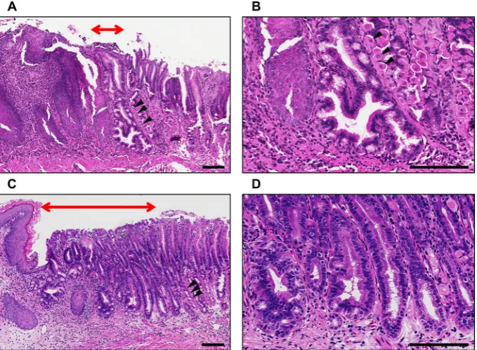
of human IL-1β to the oesophagus using the Epstein-Barr virus promoter as a model of BO. Histopathology and gene signatures of this model are reported to resemble human BO. We confirmed that metaplastic glands could be observed at the squamocolumnar junction in 28-week-old L2-IL-1β mice (figure 1A,B). In 46-week-old
L2-IL-1β mice, multiple dysplastic glands could be observed at the squamocolumnar junction, although OAC was not developed (figure 1C,D).
We comprehensively analysed tissue and serum miRNA expression profiles of 28-week-old and 46-week-old L2-IL-1β mice using microarray analysis
copyright.
on May 30, 2020 by guest. Protected by
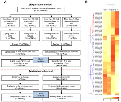
(online supplementary figure S1). Results of miRNA microarrays have been deposited in the National Center for Biotechnology Information Gene Expression Omnibus database with accession code GSE100390. By comparing between 28-week-old and 46-week-old mice, potentially deregulated miRNAs during the progression of BO were investigated (figure 2A). First, we selected all the deregulated miRNAs with fold change of >1.5 between the values of 28-week-old and 46-week-old mice either in tissues or in sera. Second, miRNAs with inversely altered levels in tissues and sera due to ageing were excluded. Finally, miRNAs with levels of <25 in sera after normalisa-tion were excluded as it limits the data reliability of lower signal levels in serum because the level of total RNA is relatively low in the serum. Using this strategy, we iden-tified 20 upregulated miRNAs and 44 downregulated miRNAs in both tissues and sera (figure 2B).
Validation of circulating mirnAs using published human dataset
To expand the mouse result to human, we excluded 39 miRNAs that are not evolutionally conserved in humans (figure 2A). Subsequently, we validated the data from mice using a published dataset of human plasma miRNAs, consisting of eight patients with OAC, eight with BO and six healthy controls (GSE51410).30 Our
primitive aim was to identify biomarkers for human early stage OAC, namely intramucosal carcinoma and high-grade dysplasia. However, circulating miRNA profiles in patients with high-grade dysplasia have not been publicly available yet. Therefore, we used dataset of OAC instead of high-grade dysplasia. Since it is necessary that the biomarker of high-grade dysplasia can also detect OAC,
validation using dataset of OAC is thought to be mean-ingful for the identification of biomarkers of high-grade dysplasia.
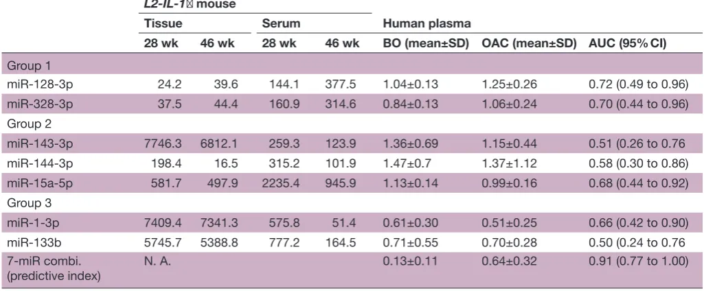
This dataset analysed plasma expression levels of 175 miRNAs that are present in human plasma samples using Serum/Plasma Focus miRNA PCR panels (Exiqon, Vedbaek, Denmark). Among 25 miRNAs that were iden-tified in mice and conserved in humans, seven miRNAs could not be evaluated because they were not included in the PCR panels. Therefore, plasma levels of 18 miRNA were compared between patients with OAC and those with BO. Of the eight miRNAs that were upregulated in 46-week-old mice compared with 28-week-old mice, two miRNAs were also upregulated in patients with OAC compared with those with BO. Of the 10 miRNAs that were downregulated in 46-week-old mice compared with 28-week-old mice, five miRNAs were also downreg-ulated in patients with OAC compared with those with BO. Thus, seven circulating miRNAs were considered as potential candidates of non-invasive biomarkers for the progression of BO to OAC (table 1).
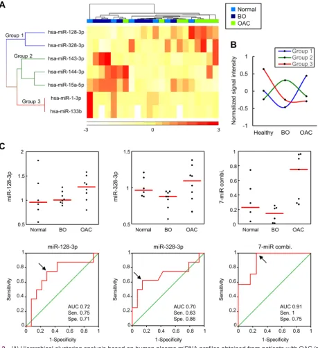
[image:3.595.129.469.55.304.2]The clustering heat map revealed more explicit segre-gation of OAC samples from healthy or BO samples (figure 3A). In addition, seven miRNAs could be cate-gorised into three groups, namely, miR-128-3 p and miR-328-3 p (group 1); miR-143-3 p, miR-144-3 p and miR-15a-5p (group 2); and miR-1-3 p and miR-133b (group 3), according to their expression profiles (figure 3A). Levels of miRNAs in group 1 were upregulated, whereas levels of miRNAs in groups 2 and 3 were downregulated during the progression from BO to OAC (figure 3B). Levels of miRNAs in group 2 were the highest in patients Figure 1 Representative histopathology at the squamocolumnar junction in L2-IL-1β mice; (A) histopathology in 28-week-old mice; (B) magnified image of A; (C) histopathology in 46-week-old mice; (D) magnified image of C. Red double-headed arrows, area of metaplastic or dysplastic glands; black arrow heads, gastric parietal cells; scale bars=100 µm. IL-1β, interleukin-1β.
copyright.
on May 30, 2020 by guest. Protected by
with BO, whereas levels of miRNAs in group 3 were the highest in healthy controls (figure 3B). We determined the accuracy of these seven miRNAs in discriminating patients with OAC from those without OAC (healthy and BO) using the ROC curve analysis (table 1). Levels of miRNAs in group 1 could achieve the AUC of more than 0.7 to discriminate patients with OAC from those without OAC, although the discriminating accuracy was not statis-tically significant. When we calculated a predictive index by the combination of seven miRNAs, this index could discriminate patients with OAC significantly. The respec-tive data and ROC curves of miRNAs in group 1 and a predictive index are shown in figure 3C. By using the prediction index, patients with OAC could be discrimi-nated accurately with the AUC of 0.91, sensitivity of 1 and specificity of 0.75.
dIscussIon
We identified seven miRNAs in mice tissue and serum samples with expression levels that were compatible with the levels in human plasma. Hierarchical clustering anal-ysis revealed that these seven miRNAs could be categorised
into three clusters: miR-128-3 p and miR-328-3 p (group 1); miR-143-3 p, miR-144-3 p and miR-15a-5p (group 2); and miR-1-3 p and miR-133b (group 3). Levels of miRNAs in group 1 were upregulated in OAC compared with healthy controls or patients with BO and could discrim-inate OAC from healthy controls or BO comparatively better than levels of miRNAs in groups 2 and 3. In addi-tion, by combining groups 1, 2 and 3, the discriminating accuracy could be enhanced, indicating that miRNAs in groups 2 and 3 need to be investigated further as poten-tial candidates of biomarkers.
Although the role of miRNAs has not been investigated in the present study, we could hypothesise its role based on previous studies. In line with our results, miR-128-3 p is upregulated in colorectal, pancreatic and gastric adeno-carcinoma tissues.31 32 However, in prostate cancer, glioma
and non-small cell lung cancer (NSCLC), miR-128-3 p is downregulated.33–35 Tumour-suppressive properties have been explained by the inhibition of Bmi-1.35 The
tran-scription factor Bmi-1 is essential for promoting self-re-newal of several types of normal and cancer stem cells.36
[image:4.595.91.508.54.405.2]Because Bmi-1 is overexpressed in gastric and pancreatic Figure 2 (A) Flow chart illustration of the experimental design; (B) hierarchical clustering analysis based on selected
tissue and serum miRNA profiles obtained from 28-week-old and 46-week-old L2-IL-1β mice. Twenty upregulated and 44 downregulated miRNAs in both tissues and sera in 46-week-old mice compared with 28-week-old mice were selected. miRNAs, microRNAs; T28, tissue miRNAs obtained from 28-week-old mice; T46, tissue miRNAs obtained from 46-week-old mice; S28, serum miRNAs obtained from 28-week-old mice; S46, serum miRNAs obtained from 46-week-old mice.
copyright.
on May 30, 2020 by guest. Protected by
cancer,37 miR-128-3 p may be compensatory upregulated to suppress Bmi-1 expression in adenocarcinoma tissues.
Tissue expression levels of miR-328-3 p have been reported to be higher in NSCLC with brain metastasis than in NSCLC without brain metastasis.38 Functional analysis showed that miR-328-3 p has a role in confer-ring migratory potential to NSCLC cells, working in part, through the upregulation of the protein kinase C alpha gene. Circulating miR-328 has also been reported as a biomarker to discriminate early stage NSCLC from healthy controls with an AUC of 0.82.39 However, levels of miR-328-3 p are downregulated in colorectal cancer, and low miR-328-3 p expression tends to correlate with increased number of cancer stem-like cells.40 CD44, a well-known cancer stem cell marker, is a direct target of miR-328-3 p.41–44 Thus, both miRNAs in group 1 play a tumour-suppressive role through the inhibition of stem cell-like properties of the cancer cells. Upregulation of these miRNAs may be a compensatory reaction during the carcinogenesis of BO.
miR-143-3 p, miR-144-3 p and miR-15a-5p in group 2 are assumed to be tumour suppressors. Tissue levels of miR-143-3 p have been reported to be downregulated in colorectal cancer.45 Overexpression on miR-143-3 p suppresses tumour growth through the direct inhibition of the KRAS and ERK5 genes.46 47 Downregulation of miR-144-3 p is associated with colorectal cancer progres-sion via activation of the mechanistic target of rapa-mycin signalling pathway.48 miR-15a-5p is a member of the miR-15 family, which includes miR-16-1, miR-16-2, miR-195 and miR-497. This cluster is known to suppress cell cycle through the inhibition of several oncogenes such as E2F1 and BCL2.49–51 Of note, circulating levels of group 2 miRNAs were elevated during the development of
BO compared with those in healthy controls (figure 3B). Cabibi et al52 also reported that tissue and circulating miR-143-3 p levels are elevated during metaplastic trans-formation of the oesophagus, suggesting that group 2 miRNA may be associated with intestinal differentiation.
miR-1-3 p and miR-133b, categorised in group 3, are known as muscle-specific miRNA and exhibit tumour-sup-pressive functions.53 54 In L2-IL-1β mice, human IL-1β
is overexpressed only in the squamous cell layer of the oesophagus. Downregulation of oesophageal tissue levels of miR-1-3 p and miR-133b in this mouse model indicated that IL-1β in mucosal layer may affect the muscle layer, leading to the deregulation of muscle-specific miRNAs. We have previously reported that miR-1-3 p, miR-133a and miR-133b levels are decreased by H. pylori infection in mice gastric tissues, leading to impaired gastric motility.55 Because IL-1 expression is enhanced in gastric mucosa by
H. pylori infection,56 57 the deregulation of muscle-specific miRNAs in these two animal models may be due to the same aetiology.
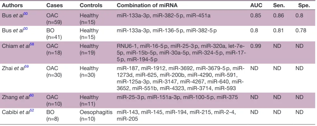
[image:5.595.41.554.77.289.2]So far, several other circulating miRNAs have been employed to detect OAC (table 2).30 52 58–60 In addi-tion, some miRNAs have been reported as prognostic biomarkers of OAC. Zhai et al59 reported that a combi-nation of two circulating miRNA (low miR-3935 expres-sion and high miR-4286 expresexpres-sion) risk score exhibits greater risk for worse overall survival (HR=2.22) than the independent levels of high miR-3935 expression or low miR-4286 expression. Odenthal et al61 reported that low miR-302c and high miR-222 expression is significantly correlated to worse overall survival (log-rank test, p<0.01). Moreover, we have previously reported that miR-222 expression levels are enhanced during exposure to bile acid in the oesophageal epithelial cells, which leads to the Table 1 Circulating microRNA candidates that represent the development of OAC from BO
L2-IL-1β mouse
Human plasma
Tissue Serum
28 wk 46 wk 28 wk 46 wk BO (mean±SD) OAC (mean±SD) AUC (95% CI)
Group 1
miR-128-3p 24.2 39.6 144.1 377.5 1.04±0.13 1.25±0.26 0.72 (0.49 to 0.96) miR-328-3p 37.5 44.4 160.9 314.6 0.84±0.13 1.06±0.24 0.70 (0.44 to 0.96) Group 2
miR-143-3p 7746.3 6812.1 259.3 123.9 1.36±0.69 1.15±0.44 0.51 (0.26 to 0.76 miR-144-3p 198.4 16.5 315.2 101.9 1.47±0.7 1.37±1.12 0.58 (0.30 to 0.86) miR-15a-5p 581.7 497.9 2235.4 945.9 1.13±0.14 0.99±0.16 0.68 (0.44 to 0.92) Group 3
miR-1-3p 7409.4 7341.3 575.8 51.4 0.61±0.30 0.51±0.25 0.66 (0.42 to 0.90) miR-133b 5745.7 5388.8 777.2 164.5 0.71±0.55 0.70±0.28 0.50 (0.24 to 0.76 7-miR combi.
(predictive index)
N. A. 0.13±0.11 0.64±0.32 0.91 (0.77 to 1.00)
Grouping was performed based on hierarchical clustering analysis shown in figure 3A. Bold value, statistically significant (p<0.05).
AUC, area under the receiver operating characteristic curve to discriminate patients with OAC from those without OAC; BO, Barrett’s oesophagus; IL-1β, interleukin-1β; N. A., not applicable; OAC, oesophageal adenocarcinoma; wk, week.
copyright.
on May 30, 2020 by guest. Protected by
proteasomal degradation of tumour-suppressive protein Cdx2.19 62 Therefore, it would be reasonable to conclude
that high levels of miR-222 are associated with poor prog-nosis. However, these reports are small-scale pilot studies, including our study; therefore, well-validated large-scale biomarker studies are warranted.
Since circulating miRNA status in patients with high-grade dysplasia has not been reported, it has been
unknown that our results also fit for the biomarker of high-grade dysplasia. According to reported tissue miRNA profiles, miRNA signatures of high-grade dysplasia were similar to those of OAC as compared with the signatures of non-dysplastic BO and normal squamous epithelium.63
[image:6.595.69.529.54.556.2]Interestingly, tissue miRNA profiles of BO adjacent to grade dysplasia were also similar to those of high-grade dysplasia and OAC. Therefore, we could expect Figure 3 (A) Hierarchical clustering analysis based on human plasma miRNA profiles obtained from patients with OAC (n=8), with BO (n=8) and without BO (n=6). (B) The average of normalised signal intensity used in the heat map. (C) Dot plots show that plasma levels of miR-128-3 p and miR-328 and a predictive index calculated from the levels of seven miRNAs (miR-128-3 p, miR-(miR-128-328-(miR-128-3 p, miR-14(miR-128-3-(miR-128-3 p, miR-144-(miR-128-3 p, miR-15a-5p, miR-1-(miR-128-3 p and miR-1(miR-128-3(miR-128-3b) were higher in patients with OAC (upper panels). ROC curves show the discrimination accuracy for discriminating patients with OAC from those without OAC using plasma levels of miR-128-3 p, miR-328 or a predictive index calculated from levels of seven miRNAs (lower panels). Red bar, median; black arrows, optimum cut-off points to calculate sensitivity and specificity. AUC, area under the receiver operating characteristic curve; BO, Barrett’s oesophagus; OAC, oesophageal adenocarcinoma; Sen., sensitivity; Spe., specificity.
copyright.
on May 30, 2020 by guest. Protected by
that circulating miRNA profiles of high-grade dysplasia might also be similar to those of OAC. To consider early-stage biomarkers for OAC, this issue must be intensively evaluated in future.
In conclusion, seven circulating miRNAs (miR-128-3 p, miR-328-3 p, miR-143-3 p, miR-144-3 p, miR-15a-5p, miR-1-3 p and miR-133b), consisting of three different characteristic miRNA groups, were identified as prom-ising non-invasive biomarkers to evaluate the carcinogen-esis from BO to OAC. However, our data were not based on enough number of samples. Although confirmation of data reproducibility and further prospective valida-tions are needed, accumulating knowledge about circu-lating miRNAs will change future medical practices.
Acknowledgements We would like to thank Dr Timothy C Wang for providing
L2-IL-1β mice and Hitoshi Tsugawa, Hideki Mori, Tsuyoshi Yamane and Yuki Kashiwazaki for technical supports and valuable advices.
Contributors Both authors conceived and designed the experiments. JM
performed the experiments and analysed the data. JM wrote the paper, and HS revised the paper.
Funding This study was supported by a Grant-in-Aid for Young Scientists (B)
(26860527; to JM), a Grant-in-Aid for Scientific Research B (16H05291; to HS) from the Japan Society for the Promotion of Science (JSPS) (https://www. jsps. go. jp/ english/), MEXT-Supported Program for the Strategic Research Foundation at Private Universities (S1411003; to HS) (http://www. mext. go. jp/ english/ pressrelease/ 1317526. htm), a grant from Takeda Science Foundation (to JM), the Princess Takamatsu Cancer Research grants (to HS) and the Medical School Faculty and Alumni Grant from Keio University Medical Science Fund (to JM and HS).
Disclaimer The funders have no roles in study design, data collection, data
analysis, data interpretation or drafting the manuscript. The corresponding author had full access to all the data in the study and had final responsibility for the decision to submit for publication.
Competing interests During the last 2 years, HS received scholarship funds for
the research from Daiichi-Sankyo Co., EA Pharma Co., Otsuka Pharmaceutical Co. Ltd and Tsumura Co. and received service honoraria from Astellas Pharma Inc., Astra-Zeneca K.K., EA Pharma Co., Otsuka Pharmaceutical Co. Ltd, Daiichi-Sankyo Co., Takeda Pharmaceutical Co. Ltd, Mylan EPD, Co., Tsumura Co. and Zeria Pharmaceutical Co. Ltd.
Provenance and peer review Commissioned; externally peer reviewed.
Data sharing statement All relevant data are within the paper and its supporting
materials.
Open Access This is an Open Access article distributed in accordance with the
Creative Commons Attribution Non Commercial (CC BY-NC 4.0) license, which permits others to distribute, remix, adapt, build upon this work non-commercially, and license their derivative works on different terms, provided the original work is properly cited and the use is non-commercial. See: http:// creativecommons. org/ licenses/ by- nc/ 4. 0/
© Article author(s) (or their employer(s) unless otherwise stated in the text of the article) 2017. All rights reserved. No commercial use is permitted unless otherwise expressly granted.
reFerenCes
1. Rubenstein JH, Shaheen NJ. Epidemiology, diagnosis, and management of esophageal adenocarcinoma. Gastroenterology
2015;149:302–17.
2. Matsuzaki J, Suzuki H, Kobayakawa M, et al. Association of visceral fat area, smoking, and alcohol consumption with reflux esophagitis and Barrett's esophagus in Japan. PLoS One
2015;10:e0133865.
3. Matsuzaki J, Suzuki H, Asakura K, et al. Etiological difference between ultrashort- and short-segment Barrett's esophagus.
J Gastroenterol 2011;46:332–8.
4. Matsuzaki J, Suzuki H, Asakura K, et al. Gallstones increase the prevalence of Barrett's esophagus. J Gastroenterol 2010;45:171–8. 5. Fischbach LA, Graham DY, Kramer JR, et al. Association between
Helicobacter pylori and Barrett's esophagus: a case-control study.
Am J Gastroenterol 2014;109:357–68.
6. Hvid-Jensen F, Pedersen L, Drewes AM, et al. Incidence of adenocarcinoma among patients with Barrett's esophagus.
N Engl J Med 2011;365:1375–83.
7. Corley DA, Mehtani K, Quesenberry C, et al. Impact of endoscopic surveillance on mortality from Barrett's esophagus-associated esophageal adenocarcinomas. Gastroenterology 2013;145:312–9. 8. Bartel DP. MicroRNAs: genomics, biogenesis, mechanism, and
function. Cell 2004;116:281–97.
9. Nishizawa T, Suzuki H. The role of microRNA in gastric malignancy.
Int J Mol Sci 2013;14:9487–96.
10. Saito Y, Suzuki H, Matsuura M, et al. MicroRNAs in Hepatobiliary and Pancreatic Cancers. Front Genet 2011;2:66.
11. Saito Y, Suzuki H, Hibi T. The role of microRNAs in gastrointestinal cancers. J Gastroenterol 2009;44(Suppl 19):18–22.
[image:7.595.43.556.71.275.2]12. Matsuzaki J, Suzuki H. MicroRNAs in Barrett's esophagus: future prospects. Front Genet 2014;5:69.
Table 2 Previously reported circulating microRNA (miRNA) for the diagnosis of OAC and/or BO
Authors Cases Controls Combination of miRNA AUC Sen. Spe.
Bus et al30 OAC
(n=59) Healthy(n=15) miR-133a-3p, miR-382-5 p, miR-451a 0.85 0.86 0.8 Bus et al30 BO
(n=41) Healthy(n=15) miR-133a-3p, miR-136-5 p, miR-382-5 p 0.8 0.81 0.78 Chiam et al58 OAC
(n=18) Healthy(n=19) RNU6-1, miR-16-5 p, miR-25-3 p, miR-320a, let-7e-5p, miR-15b-5p, miR-30a-5p, miR-324-5 p, miR-17-5 p, miR-194-miR-17-5 p
0.99 ND ND
Zhai et al59 OAC
(n=30) Healthy(n=30) miR-187, miR-1912, miR-3692, miR-3679-5 p, miR-1273d, miR-625, miR-200b, miR-4290, miR-591, 125a-3p, 3147, 4267, 640, miR-3652, miR-551b, miR-4323, miR-3714, miR-593
ND ND ND
Zhang et al60 OAC
(n=10) Healthy(n=11) miR-25-3 p, miR-151a-3p, miR-100-5 p, miR-375 ND ND ND Cabibi et al52 BO
(n=8)
Oesophagitis (n=10)
miR-143, miR-145, miR-194, miR-215, miR-2-4, miR-205
ND ND ND
AUC, area under the receiver operating characteristic curve; BO, Barrett’s oesophagus; ND, not described; OAC, oesophageal adenocarcinoma; Sen., sensitivity; Spe., specificity.
copyright.
on May 30, 2020 by guest. Protected by
13. Feber A, Xi L, Luketich JD, et al. MicroRNA expression profiles of esophageal cancer. J Thorac Cardiovasc Surg 2008;135:255–60. discussion 60.
14. Kan T, Sato F, Ito T, et al. The miR-106b-25 polycistron, activated by genomic amplification, functions as an oncogene by suppressing p21 and Bim. Gastroenterology 2009;136:1689–700.
15. Leidner RS, Ravi L, Leahy P, et al. The microRNAs, 31 and MiR-375, as candidate markers in Barrett's esophageal carcinogenesis.
Genes Chromosomes Cancer 2012;51:473–9.
16. Fassan M, Volinia S, Palatini J, et al. MicroRNA expression profiling in human Barrett's carcinogenesis. Int J Cancer 2011;129:1661–70. 17. Wu X, Ajani JA, Gu J, et al. MicroRNA expression signatures during
malignant progression from Barrett's esophagus to esophageal adenocarcinoma. Cancer Prev Res 2013;6:196–205.
18. Yang H, Gu J, Wang KK, et al. MicroRNA expression signatures in Barrett's esophagus and esophageal adenocarcinoma. Clin Cancer Res 2009;15:5744–52.
19. Matsuzaki J, Suzuki H, Tsugawa H, et al. Bile acids increase levels of microRNAs 221 and 222, leading to degradation of CDX2 during esophageal carcinogenesis. Gastroenterology 2013;145:1300–11. 20. Bansal A, Gupta V, Wang K. MicroRNA Expression Signatures During
Malignant Progression From Barrett's Esophagus. J Cell Biochem
2016;117:1288–95.
21. Kosaka N, Iguchi H, Yoshioka Y, et al. Secretory mechanisms and intercellular transfer of microRNAs in living cells. J Biol Chem
2010;285:17442–52.
22. Valadi H, Ekström K, Bossios A, et al. Exosome-mediated transfer of mRNAs and microRNAs is a novel mechanism of genetic exchange between cells. Nat Cell Biol 2007;9:654–9.
23. Arroyo JD, Chevillet JR, Kroh EM, et al. Argonaute2 complexes carry a population of circulating microRNAs independent of vesicles in human plasma. Proc Natl Acad Sci U S A
2011;108:5003–8.
24. Vickers KC, Palmisano BT, Shoucri BM, et al. MicroRNAs are transported in plasma and delivered to recipient cells by high-density lipoproteins. Nat Cell Biol 2011;13:423–33.
25. Matsuzaki J, Ochiya T. Circulating microRNAs and extracellular vesicles as potential cancer biomarkers: a systematic review. Int J Clin Oncol 2017;22:413–20.
26. Shimomura A, Shiino S, Kawauchi J, et al. Novel combination of serum microRNA for detecting breast cancer in the early stage.
Cancer Sci 2016;107:326–34.
27. Karolina DS, Tavintharan S, Armugam A, et al. Circulating miRNA profiles in patients with metabolic syndrome. J Clin Endocrinol Metab 2012;97:E2271–E2276.
28. Thomou T, Mori MA, Dreyfuss JM, et al. Adipose-derived circulating miRNAs regulate gene expression in other tissues. Nature
2017;542:450–5.
29. Quante M, Bhagat G, Abrams JA, et al. Bile acid and inflammation activate gastric cardia stem cells in a mouse model of Barrett-like metaplasia. Cancer Cell 2012;21:36–51.
30. Bus P, Kestens C, Ten Kate FJ, et al. Profiling of circulating microRNAs in patients with Barrett's esophagus and esophageal adenocarcinoma. J Gastroenterol 2016;51:560–70.
31. Volinia S, Calin GA, Liu CG, et al. A microRNA expression signature of human solid tumors defines cancer gene targets. Proc Natl Acad Sci U S A 2006;103:2257–61.
32. Katada T, Ishiguro H, Kuwabara Y, et al. microRNA expression profile in undifferentiated gastric cancer. Int J Oncol 2009;34:537–42. 33. Adlakha YK, Saini N. MicroRNA-128 downregulates Bax and
induces apoptosis in human embryonic kidney cells. Cell Mol Life Sci 2011;68:1415–28.
34. Khan AP, Poisson LM, Bhat VB, et al. Quantitative proteomic profiling of prostate cancer reveals a role for miR-128 in prostate cancer. Mol Cell Proteomics 2010;9:298–312.
35. Cui JG, Zhao Y, Sethi P, et al. Micro-RNA-128 (miRNA-128) down-regulation in glioblastoma targets ARP5 (ANGPTL6), Bmi-1 and E2F-3a, key regulators of brain cell proliferation. J Neurooncol
2010;98:297–304.
36. Fasano CA, Dimos JT, Ivanova NB, et al. shRNA knockdown of Bmi-1 reveals a critical role for p21-Rb pathway in NSC self-renewal during development. Cell Stem Cell 2007;1:87–99.
37. Wang MC, Li CL, Cui J, et al. BMI-1, a promising therapeutic target for human cancer. Oncol Lett 2015;10:583–8.
38. Arora S, Ranade AR, Tran NL, et al. MicroRNA-328 is associated with (non-small) cell lung cancer (NSCLC) brain metastasis and mediates NSCLC migration. Int J Cancer 2011;129:2621–31.
39. Ulivi P, Foschi G, Mengozzi M, et al. Peripheral blood miR-328 expression as a potential biomarker for the early diagnosis of NSCLC. Int J Mol Sci 2013;14:10332–42.
40. Xu XT, Xu Q, Tong JL, et al. MicroRNA expression profiling identifies miR-328 regulates cancer stem cell-like SP cells in colorectal cancer.
Br J Cancer 2012;106:1320–30.
41. Wang CH, Lee DY, Deng Z, et al. MicroRNA miR-328 regulates zonation morphogenesis by targeting CD44 expression. PLoS One
2008;3:e2420.
42. Ishimoto T, Sugihara H, Watanabe M, et al. Macrophage-derived reactive oxygen species suppress miR-328 targeting CD44 in cancer cells and promote redox adaptation. Carcinogenesis
2014;35:1003–11.
43. Hirata K, Suzuki H, Imaeda H, et al. CD44 variant 9 expression in primary early gastric cancer as a predictive marker for recurrence.
Br J Cancer 2013;109:379–86.
44. Tsugawa H, Suzuki H, Saya H, et al. Reactive oxygen species-induced autophagic degradation of Helicobacter pylori CagA is specifically suppressed in cancer stem-like cells. Cell Host Microbe
2012;12:764–77.
45. Slaby O, Svoboda M, Fabian P, et al. Altered expression of miR-21, miR-31, miR-143 and miR-145 is related to clinicopathologic features of colorectal cancer. Oncology 2007;72:397–402. 46. Chen X, Guo X, Zhang H, et al. Role of miR-143 targeting KRAS in
colorectal tumorigenesis. Oncogene 2009;28:1385–92.
47. Borralho PM, Simões AE, Gomes SE, et al. miR-143 overexpression impairs growth of human colon carcinoma xenografts in mice with induction of apoptosis and inhibition of proliferation. PLoS One
2011;6:e23787.
48. Iwaya T, Yokobori T, Nishida N, et al. Downregulation of miR-144 is associated with colorectal cancer progression via activation of mTOR signaling pathway. Carcinogenesis 2012;33:2391–7. 49. Ofir M, Hacohen D, Ginsberg D. MiR-15 and miR-16 are direct
transcriptional targets of E2F1 that limit E2F-induced proliferation by targeting cyclin E. Mol Cancer Res 2011;9:440–7.
50. Bonci D, Coppola V, Musumeci M, et al. The miR-15a-miR-16-1 cluster controls prostate cancer by targeting multiple oncogenic activities. Nat Med 2008;14:1271–7.
51. Xia L, Zhang D, Du R, et al. miR-15b and miR-16 modulate multidrug resistance by targeting BCL2 in human gastric cancer cells. Int J Cancer 2008;123:372–9.
52. Cabibi D, Caruso S, Bazan V, et al. Analysis of tissue and circulating microRNA expression during metaplastic transformation of the esophagus. Oncotarget 2016;7:47821–30.
53. Kojima S, Chiyomaru T, Kawakami K, et al. Tumour suppressors miR-1 and miR-133a target the oncogenic function of purine nucleoside phosphorylase (PNP) in prostate cancer. Br J Cancer
2012;106:405–13.
54. Kano M, Seki N, Kikkawa N, et al. 145, 133a and miR-133b: Tumor-suppressive miRNAs target FSCN1 in esophageal squamous cell carcinoma. Int J Cancer 2010;127:2804–14. 55. Saito Y, Suzuki H, Tsugawa H, et al. Dysfunctional gastric emptying
with down-regulation of muscle-specific microRNAs in Helicobacter pylori-infected mice. Gastroenterology 2011;140:189–98.
56. Hwang IR, Kodama T, Kikuchi S, et al. Effect of interleukin 1 polymorphisms on gastric mucosal interleukin 1beta production in Helicobacter pylori infection. Gastroenterology 2002;123:1793–803. 57. El-Omar EM. The importance of interleukin 1beta in Helicobacter
pylori associated disease. Gut 2001;48:743–7.
58. Chiam K, Wang T, Watson DI, et al. Circulating serum exosomal miRNAs as potential biomarkers for esophageal adenocarcinoma.
J Gastrointest Surg 2015;19:1208–15.
59. Zhai R, Wei Y, Su L, et al. Whole-miRNome profiling identifies prognostic serum miRNAs in esophageal adenocarcinoma: the influence of Helicobacter pylori infection status. Carcinogenesis
2015;36:87–93.
60. Zhang K, Wu X, Wang J, et al. Circulating miRNA profile in esophageal adenocarcinoma. Am J Cancer Res 2016;6:2713–21. 61. Odenthal M, Hee J, Gockel I, et al. Serum microRNA profiles as
prognostic/predictive markers in the multimodality therapy of locally advanced adenocarcinomas of the gastroesophageal junction.
Int J Cancer 2015;137:230–7.
62. Matsuzaki J, Suzuki H. Role of MicroRNAs-221/222 in Digestive Systems. J Clin Med 2015;4:1566–77.
63. Saad R, Chen Z, Zhu S, et al. Deciphering the unique microRNA signature in human esophageal adenocarcinoma. PLoS One
2013;8:e64463.
copyright.
on May 30, 2020 by guest. Protected by