INTRODUCTION
The skin is a bi-compartmental organ. The outer layer is maintained by stem cells and comprises a stratified epithelium, the interfollicular epidermis, with associated hair follicles, sebaceous glands and sweat glands (Fuchs and Horsley, 2008; Watt et al., 2006). The sub-epidermal compartment comprises dermal fibroblasts, peripheral nerves, blood vessels, muscle and fat. Subpopulations of dermal cells, located in the dermal papilla and dermal sheath, regulate epidermal stem cell properties, most notably by controlling the hair growth cycle (Schneider et al., 2009; Waters et al., 2007). A number of key developmental signaling pathways, including those that involve Notch, BMP, Wnt and FGF, mediate epidermal-dermal communication (Estrach et al., 2006; Kawano et al., 2005; Kishimoto et al., 2000; Lowell et al., 2000; Rendl et al., 2008; Zhang et al., 2008).
Notch signalling is activated when ligand binding initiates cleavage of the Notch receptor, which releases the Notch intracellular domain (NICD) from the plasma membrane (Kopan and Ilagan, 2009). The NICD translocates to the nucleus and interacts with its binding partners RBP-J (Rbpj – Mouse Genome Informatics) and mastermind 1 to activate transcription of downstream targets, including members of the Hes and Hey families of transcriptional repressors. Notch signalling is
important in local cell-to-cell communication, as both ligands and receptors are tethered to the cell membrane. Nevertheless, recent studies demonstrate that the cleaved domain of jagged 1 can be secreted and can modulate Notch signaling over a longer range (Aho, 2004; Duarte et al., 2008; Nikopoulos et al., 2007; Urs et al., 2008).
The importance of the Notch pathway in skin is well established (Watt et al., 2008). High expression of the Notch ligand delta-like 1 (Dll1) is a marker of human epidermal stem cells and plays a dual role in promoting stem cell cohesion and stimulating differentiation of neighbouring epidermal cells in culture (Estrach et al., 2007; Lowell et al., 2000; Lowell and Watt, 2001). Consistent with this, Notch acts as an epidermal tumour suppressor (Estrach et al., 2008; Lefort et al., 2007; Nicolas et al., 2003). Disrupting Notch signaling in embryonic and neonatal mouse epidermis though genetic ablation of RBP-J, Hes1, or Notch1 and Notch2 leads to failure to maintain the hair follicles, abnormal keratinocyte differentiation, barrier disruption and neonatal lethality (Blanpain et al., 2006; Moriyama et al., 2008; Pan et al., 2004). Dll1 regulates differentiation of the interfollicular epidermis, while jagged 1, a -catenin target gene, is required for hair follicle maintenance (Estrach et al., 2006; Estrach et al., 2008). Conversely, forced Notch activation in developing interfollicular epidermis leads to loss of hemidesmosomes and promotion of keratinocyte differentiation (Blanpain et al., 2006).
Although recent studies have tended to focus on the epidermal consequences of modulating Notch, there are clear indications that Notch signalling controls other aspects of skin function. Epidermal deletion of Notch leads to increased epidermal production of thymic stromal lymphopoietin (TSLP), which triggers a B lymphoproliferative disorder with massive dermal accumulation of B cells (Demehri et al., 2008). Notch signalling in melanoblasts is required for their survival; when signalling is blocked, the hair Development 137, 3569-3579 (2010) doi:10.1242/dev.050310
© 2010. Published by The Company of Biologists Ltd
1Cancer Research UK, Cambridge Research Institute, Li Ka Shing Centre, Cambridge CB2 0RE, UK. 2School of Biological and Biomedical Sciences and NorthEast England Stem Cell Institute, Durham University, South Road, Durham DH1 3LE, UK. 3Wellcome Trust Centre for Stem Cell Research, University of Cambridge, Tennis Court Road, Cambridge CB2 1QR, UK.
*Authors for correspondence (c.a.ambler@durham.ac.uk; fiona.watt@cancer.org.uk)
Accepted 27 August 2010 SUMMARY
Notch signalling regulates epidermal differentiation and tumour formation via non-cell autonomous mechanisms that are incompletely understood. This study shows that epidermal Notch activation via a 4-hydroxy-tamoxifen-inducible transgene caused epidermal thickening, focal detachment from the underlying dermis and hair clumping. In addition, there was dermal
accumulation of T lymphocytes and stromal cells, some of which localised to the blisters at the epidermal-dermal boundary. The T cell infiltrate was responsible for hair clumping but not for other Notch phenotypes. Notch-induced stromal cells were
heterogeneous, expressing markers of neural crest, melanocytes, smooth muscle and peripheral nerve. Although Slug1 expression was expanded in the epidermis, the stromal cells did not arise through epithelial-mesenchymal transition. Epidermal Notch activation resulted in upregulation of jagged 1 in both epidermis and dermis. When Notch was activated in the absence of epidermal jagged 1, jagged 1 was not upregulated in the dermis, and epidermal thickening, blister formation, accumulation of T cells and stromal cells were inhibited. Gene expression profiling revealed that epidermal Notch activation resulted in
upregulation of several growth factors and cytokines, including TNF, the expression of which was dependent on epidermal jagged 1. We conclude that jagged 1 is a key mediator of non-cell autonomous Notch signalling in skin.
KEY WORDS: Notch, Epidermis, Neural crest, Jagged 1, Mouse
Adult epidermal Notch activity induces dermal accumulation
of T cells and neural crest derivatives through upregulation
of jagged 1
Carrie A. Ambler1,2,* and Fiona M. Watt1,3,*
D
E
V
E
LO
P
M
E
N
follicles become depigmented (Moriyama et al., 2008). In addition, the tumour suppressive function of Notch is not exclusively cell autonomous (Demehri et al., 2009).
We previously generated transgenic mice in which 4-hydroxy-tamoxifen (4OHT)-inducible expression of NICD is under the control of the keratin 14 promoter (K14NICDER transgenics) (Estrach et al., 2006). In addition to hair clumping and thickening of the interfollicular epidermis, NICD activation causes changes in the cellularity of the dermis. We now present evidence that those changes result from signalling between the epidermis and dermis that is mediated by jagged 1.
MATERIALS AND METHODS
Mice
Experimental procedures were performed under a UK government Home Office licence. K14NICDER (also known as K14NICD⌬OPER), K14CreER, Jag1flox/flox, K14⌬NcateninER and CAG-CAT-EGFP mice have been
described previously (Brooker et al., 2006; Estrach et al., 2006; Jensen et al., 2009; Kawamoto et al., 2000; Lo Celso et al., 2004). To activate the NICDER and CreER transgenes, 7-week-old mice were treated topically with 2 mg 4OHT (Sigma) dissolved in acetone (Estrach et al., 2006).
Some mice were injected with 4 mg/kg dexamethasone (or saline, as a control) into the abdominal subcutaneous space, starting 3 days prior to the initial 4OHT treatment. Thereafter, mice received daily injections of dexamethasone or saline, and 4OHT or acetone was applied topically three times per week for 21 days.
Assay for epidermal barrier integrity
Pieces of back skin (2 cm2) were fixed overnight in 4% paraformaldehyde
in PBS and then dehydrated and rehydrated through a graded methanol series. Skin was attached, dermal side down, to a Petri dish containing petroleum jelly (Vaseline), leaving only the epidermis exposed. Toluidine Blue solution (1%) was added for 2 minutes, and then the epidermis was destained in PBS for 5-10 minutes (Byrne and Hardman, 2005). Frozen sections (25 mm) were imaged using a Leica MZ9.5 dissecting microscope.
Histology, immunostaining and in situ hybridisation
Tissue was collected and processed as previously described (Braun et al., 2003; Estrach et al., 2006). The following antibodies (dilutions in brackets) were used: K14 (1:1000, Covance), K10 (1:1000, Covance), Ki67 (1:400, NeoMarkers), K17 (1:1000, a gift from P. Coulombe, Johns Hopkins University School of Medicine) (McGowan and Coulombe, 2000), CD4 (1:100, BD Biosciences), CD8 (1:100, BD Biosciences), Hes1 (1:1000, a gift from N. Brown, Cincinnati Children’s Hospital Medical Centre), 6 integrin subunit (1:100, BD Biosciences), Laminin 5 (rabbit, 1:500, a gift from P. Marinkovich, Stanford School of Medicine), nestin (rabbit, 1:1000, a gift from R. McKay, NIH; mouse monoclonal, 1:100, Cell Signaling Technologies), Crabp1 (1:400, Sigma), Desmin (1:100, Abcam), Sm22
(1:200, Abcam), jagged 1 (1:100, Santa Cruz Biotechnology), Slug1 (1:1000, Abcam), E-cadherin, (ECCD-2, 1:600, Calbiochem), p75 (1:500, Abcam), phosphorylated p65 (1:100, Abcam) and GFP (1:1000, Abcam). To visualise alkaline phosphatase activity, frozen sections were fixed in 0.4% paraformaldehyde for 15 seconds, washed in phosphate-buffered saline and then reacted with 4.5 ml of 75 mg/ml Nitro Blue Toluidine (Roche) and 3.5 ml of 5 mg/ml 5-bromo-4-chloro-3-indolyl phosphate substrate (Roche). Sections were post-fixed in 10% neutral buffered formalin, counterstained with Fast Red and mounted using Permount (Fisher Scientific).
Tissues were imaged using a Zeiss 510 confocal, Leica Tandem SP5 confocal or Nikon 90i brightfield microscope. Brightness of images was adjusted using Adobe Photoshop CS3 software.
Quantitation was performed using Adobe Photoshop CS3 and ImageJ software. To determine proliferation index, the number of Ki67-positive basal cells and the total number of nuclei were counted per 200 mm length of interfollicular epidermis; at least 40 lengths were scored. The percentage of each 200 mm length of epidermis that was K10 positive (at least 20 lengths were scored) was also determined. The percentage of disrupted
basement membrane was determined per 200 mm length of epidermis (at least 20 lengths were scored) stained for laminin 5. The percentage of Crabp1-, nestin- or Sm22-expressing cells was determined by counting the number of antibody-stained cells relative to the total number of DAPI positive nuclei in 200 mm (length) ⫻50 mm (depth) dermis adjacent to the basement membrane; at least 15 areas were scored.
In situ hybridisation was performed as previously described (Ambler and Watt, 2007) and photographed using bright- and darkfield illumination on an Olympus Darkfield Microscope.
Electron microscopy
Adult mouse dorsal skin was fixed in 2.5% glutaraldehyde and 4% paraformaldehyde in Sorensen’s buffer (pH 7.4), then embedded in araldite resin. Sections (100 nm) were cut on a Reichert ultracut S ultramicrotome and stained with 1.5% uranyl acetate and lead citrate. Specimens were analysed on a JEOL 1010 electron microscope equipped with a US1000 camera (Gatan) using Digital Micrograph software (Gatan).
Dermal dissociation and sphere culture
The procedures have been described previously (Wong et al., 2006). Back skin (60 mm2) was scraped free of fat and muscle and then diced into 2-5
mm2 pieces. These pieces were digested in 1 mg/ml crude Type I
Collagenase (C-9891, Sigma) in 1:1 DMEM/F12 culture medium (31331-028, Invitrogen) containing antibiotics at 37°C for 1 hour, dissociated mechanically and filtered through a 70 mm cell strainer.
Cells were plated in quadruplicate into 48-well plates at a density of 5⫻104cells/ml in DMEM/F12 medium containing 2% B27 supplement
(Invitrogen) and antibiotics. Seven days later, each well was photographed using a Nikon TE1000 microscope with a motorised stage and Plan Apo 4⫻objective. Using Nikon NIS-Elements automated imaging software, sphere number was counted in the same five areas of each well. Sphere-forming capacity was compared for wild-type (n6 per time point) and K14NICDER (n6) transgenic littermate mice treated with 4OHT for 10 or 14 days. Wild-type sphere formation was designated 100%.
RNA isolation and microarray analysis
Seven-week old female mice were treated with 4OHT for 14 days. The back skin was collected, bisected, immersed directly in RNAlater (Qiagen) and stored overnight at 4°C. One piece of the bisected skin was subsequently heated at 60°C for 10 seconds and then scraped gently to separate epidermis from dermis. Total RNA was isolated from epidermis, dermis and whole skin using the RNeasy Mini Kit with on-column DNase digestion (Qiagen) and hybridised to Affymetrix Mouse 430_2 gene chips (Patterson Microarray Facility, Manchester, UK). The average MAS5-calculated signal intensity of replicate samples (n3) of epidermis, dermis and whole skin from wild-type (n3) and transgenic mice (n3) was determined. Fold change represents the average difference in transgenic versus wild-type signal intensity.
qPCR
Total RNA was reverse transcribed using a Superscript III first-strand synthesis kit (Invitrogen) and quantitative PCR was performed under standard conditions with an ABI 7500 fast real-time PCR machine. Samples were run in triplicate for each probe and quantification was based on ⌬⌬CT calculations. Samples were normalised to -actin and GAPDH as loading controls and calibrated to wild-type levels. Pre-designed TAQman probes were purchased from Applied Biosystems.
Western blotting
Pieces of back skin (0.5-1 cm2) were snap-frozen and stored in liquid
nitrogen. Frozen tissue was homogenised in RIPA buffer [150 mM NaCl, 50 mM Tris-HCl (pH 7.5), 1% Nonidet P-40, 0.25% sodium deoxycholate with protease inhibitors]. Lysates were run on a 4-12% gradient polyacrylamide gel (Invitrogen), transferred to PVDF membrane, blocked with 3% cold water fish skin gelatin (Sigma)/0.2% Tween-20/PBS and hybridised with goat polyclonal antibodies to jagged 1 (C-terminal, c-20 1:100; Santa Cruz Biotechnologies). Blots were rinsed in 0.2% Tween-20/PBS, incubated with HRP-conjugated anti-goat secondary antibody (Sigma) and visualised with ECL Western Blotting Substrate (Pierce).
D
E
V
E
LO
P
M
E
N
RESULTS
Epidermal Notch activation leads to changes in skin architecture
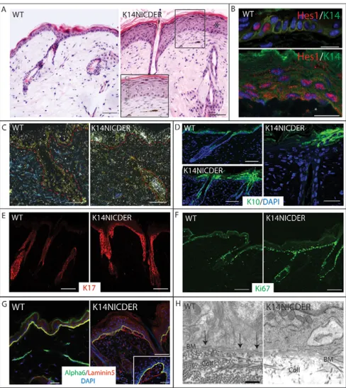
We activated Notch in basal keratinocytes by applying 4OHT to skin of 7-week-old K14NICDER transgenic mice and their wild-type littermates for 21 days (Estrach et al., 2006). K14NICDER transgenic mice had a thickened epidermis (Fig. 1A) (Estrach et al., 2006). In addition, the dermal cells immediately adjacent to the interfollicular epidermis were more numerous and had an elongated morphology compared with the surrounding dermal cells (Fig. 1A). One readout of Notch activity is upregulation of Hes and Hey genes. In wild-type back skin, Hes1 was detected in rare suprabasal epidermal cells (Fig. 1B). In K14NICDER transgenic skin treated with 4OHT for 15 days, Hes1 protein was detected in basal and suprabasal epidermal cells, and occasional dermal cells (Fig. 1B). Upregulation of Hes1 mRNA was confirmed by in situ hybridisation (Fig. 1C). In addition, HeyL mRNA was strongly and selectively upregulated in the dermis (see Fig. S1 in the supplementary material). Thus, epidermal Notch activation leads to upregulation of the Notch signalling pathway in both epidermis and dermis.
Notch activation resulted in an increase in the number of keratin 14-positive epidermal layers (Fig. 1B). This was accompanied by patchy loss of keratin 10 (Fig. 1D). The remaining keratin 10-positive cells were located mainly in the interfollicular epidermis adjacent to hair follicles (Fig. 1D; see Fig. S2A in the supplementary material). Reduced keratin 10 expression correlated with epidermal hyperproliferation: keratin 17 (McGowan and Coulombe, 2000) was expressed in the interfollicular epidermis and most basal layer cells were Ki67 positive (Fig. 1E,F; see Fig. S2B in the supplementary material).
As loss of Notch perturbs the epidermal barrier (Demehri et al., 2009), we investigated the effect of increased Notch activity. After 21 days of 4OHT treatment, the skin of K14NICDER transgenic mice was permeable to Toluidine Blue (Byrne and Hardman, 2005), whereas that of wild-type littermates was not (see Fig. S3 in the supplementary material), indicating that sustained Notch activation led to disruption of the epidermal barrier. Nevertheless, epidermal cells still underwent complete terminal differentiation, as evidenced by the presence of spinous, granular and cornified layers (Fig. 1A).
In wild-type skin, the hemidesmosome component 64 integrin and its extracellular matrix ligand laminin 5 colocalised at the basal surface of keratinocytes (Fig. 1G). By contrast, colocalisation of these markers was disrupted in 4OHT-treated K14NICDER skin (Fig. 1G; see Fig. S2C in the supplementary material). Small blisters formed through separation of epidermis from the underlying dermis. As a result, the 6 integrin subunit and laminin 5 were still detected on the basal surface of epidermal keratinocytes, but laminin 5 was also detected on the exposed dermis underlying detached epidermal cells. The blisters contained nucleated cells (Fig. 1G). Electron microscopy revealed a marked reduction in hemidesmosomes in 4OHT-treated K14NICDER transgenic compared with wild-type skin (Fig. 1H) (Blanpain et al., 2006).
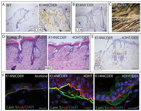
Ectopic Notch activity induces a dermal inflammatory infiltrate
To investigate whether the increase in dermal cell density on epidermal Notch activation was due to an inflammatory infiltrate, we stained skin sections with antibodies to the T-lymphocyte markers CD3, CD4 and CD8 (Fig. 2A and data not shown). In
4OHT-treated K14NICDER back skin, there was a massive infiltrate of CD3-positive T cells (data not shown). These comprised CD4-positive cells, with few CD8-positive cells being present in epidermis or dermis (Fig. 2A,B).
[image:3.612.316.560.60.334.2]In order to determine the contribution of the T cell infiltrate to the K14NICDER skin phenotype, we treated 7-week-old mice with the anti-inflammatory drug dexamethasone in combination with 4OHT or acetone. Wild-type mice injected with dexamethasone or saline and treated with 4OHT or acetone, and acetone-treated transgenic mice injected with dexamethasone or saline, were indistinguishable from untreated wild-type control mice (data not shown). The number of CD4-positive cells was reduced to wild-type levels in dexamethasone-treated K14NICDER mice (Fig. 2E).
Fig. 1. Characterisation of K14NICDER transgenic mice.
(A)Haematoxylin and Eosin-stained back skin sections of 4OHT-treated wild-type (WT) and K14NICDER littermates. Inset shows a higher magnification view of epidermis and underlying dermis. Epidermal layers are indicated. B, basal; S, spinous; G, granular; C, cornified. (B)Back skin of 4OHT-treated wild-type (WT) and K14NICDER
littermates stained with antibodies to Hes1 (red) and keratin 14 (green). Asterisk marks a Hes1-positive dermal cell. (C)RNA in situ hybridisation with radiolabelled antisense Hes1 probe on back skin sections from
4OHT-treated wild type (WT) and K14NICDER mice. Epidermal-dermal boundary is indicated by a broken red line. (D-G)Back skin of 4OHT-treated wild-type (WT) and K14NICDER littermates mice stained with antibodies to keratin 10 (green, D), keratin 17 (red, E), Ki67 (green, F),
6 integrin subunit (green, G) and laminin 5 (red, G). (H)Transmission electron micrographs of the epidermal basement membrane zone in wild type (WT) and K14NICDER littermates. Arrows indicate hemidesmosomes. BM, basement membrane; Coll, collagen fibres. (B,D,G) Sections were DAPI counterstained (blue). Mice were 4OHT-treated for 14 days (C-F), 15 days (B) or 21 days (A,G-H). Scale bars: 1mm in H; 10mm in B,G (inset); 25mm in D (right panel), G (left and right panels); 50mm in A,C,D (left panels), E,F.
D
E
V
E
LO
P
M
E
N
Notch activation results in hair shaft clumping, giving mice a ‘tufted’ appearance (Estrach et al., 2006) (Fig. 2C). K14NICDER mice that received both dexamethasone and 4OHT had reduced hair clumping (Fig. 2C). However, epidermal thickening (Fig. 2D) and blister formation (visualised by dissociation of laminin 5 and 6 integrin staining; Fig. 2F) were unaffected by dexamethasone treatment. Thus, Notch activation induces a T-lymphocyte infiltrate that is responsible for hair clumping, but not for other skin phenotypes.
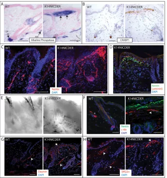
Epidermal Notch activity results in accumulation of dermal cells that express neuronal, muscle, dermal papilla and neural crest markers
To examine the effect of epidermal Notch activity on the composition of the underlying dermal compartment, we stained sections with markers to the various cell populations resident within the dermis. Alkaline phosphatase is a marker of both dermal papilla and arrector pili muscle (Fig. 3A) (Handjiski et al., 1994). In 4OHT-treated K14NICDER mice, alkaline phosphatase was additionally detected in dermal cells at the epidermal-dermal junction (Fig. 3A). The dermal papilla marker Crabp1 was also expressed by these cells (Fig. 3B) (Collins and Watt, 2008).
The intermediate filament protein nestin is expressed by peripheral nerves adjacent to the hair follicle bulge and by neural crest stem cells, melanocyte precursors and dermal papilla cells (Amoh et al., 2005; Dunn et al., 2000; Kruse et al., 2006; Li et al., 2003; Tiede et al., 2007). In K14NICDER mice treated with 4OHT
for 14 days, there was marked accumulation of nestin-positive cells at the epidermal-dermal boundary (Fig. 3C) and in the subepidermal blisters of 4OHT-treated K14NICDER skin (Fig. 3D). Cells in this region also expressed neurofilament protein, an intermediate filament protein that is highly expressed in neuronal cells, but were negative for two additional markers of skin peripheral neurons, Sox2 and NCAM (data not shown) (Botchkarev et al., 1997; Driskell et al., 2009; Lauria et al., 2004). There was also an increase in differentiated melanin-positive melanocytes (Fig. 3E). The melanocytes expressed Kit (Fig. 3F), and tended to lie below the nestin-positive cells at the epidermal/dermal boundary (Fig. 3F). When transgenic skin was treated with 4OHT for 21 days, the number of dermal Kit-positive cells increased approximately threefold (see Fig. S4A in the supplementary material).
Subepidermal dermal cells in 4OHT-treated K14NICDER skin expressed the smooth muscle markers desmin and SM22(Tagln – Mouse Genome Informatics) but did not express the vascular endothelial marker CD31 (Fig. 3G,H; data not shown) (Ya et al., 1997; Zhang et al., 2001).
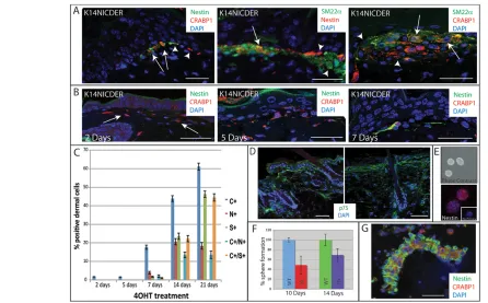
In wild-type adult back skin, nestin, Crabp1 and SM22are individual markers of peripheral nerves, dermal papilla cells and arrector pili muscle cells, respectively (Fig. 3B,C,H; wild-type panels). In back skin of 4OHT-treated K14NICDER mice, cells that were singly positive for each marker were also present (Fig. 4A, arrowheads; Fig. 4C). However, many cells co-expressed nestin and Crabp1 or Sm22and Crabp1 (Fig. 4A, arrows; Fig. 4C). Quantitation of double- and single-positive cells was performed both on skin sections (Fig. 4C) and on cytospin preparations of disaggregated dermis (see Fig. S4B in the supplementary material).
We detected a gradient in staining intensity for nestin and SM22, those cells immediately adjacent to the epidermis being most strongly labelled (Fig. 4A). By contrast, Crabp1 staining was equally strong, regardless of cell location (Fig. 4A). We conclude that the cells that accumulate at the epidermal-dermal boundary of 4OHT treated K14NICDER skin are heterogeneous and distinct from the cell types resident in wild-type dermis.
We next examined K14NICDER skin that had been treated with 4OHT for 0 to 21 days. Occasional Crabp1- and nestin-positive cells were detected after 2 days, with the majority of Crabp1-positive cells initially located in unblistered areas of the subepidermal dermis (Fig. 4B; data not shown). The number of Crabp1-positive cells increased progressively throughout the treatment period, whereas the number of nestin-positive cells was maximal by 14 days (Fig. 4B,C). Nestin positive cells often co-expressed Crabp1 (Fig. 4B,C). Sm22-positive cells were first detected at 7 days and increased in abundance thereafter (Fig. 4B,C). Notch-induced stromal cells tended to form stable intercellular adhesions that were not disrupted under conditions used to isolate single dermal cell suspensions (Fig. 4G).
[image:4.612.50.298.59.254.2]Origin of cells at the epidermal-dermal boundary During development, peripheral neurons, smooth muscle cells, facial dermal papilla cells and melanocytes are all derived from the embryonic neural crest (Le Douarin et al., 2008). The neural crest marker p75 was detected in cells at the epidermal/dermal junction in 4OHT-treated K14NICDER skin (Fig. 4D). Nestin- and Crabp1-positive cells were readily detected in embryonic and early postnatal dermis (see Fig. S5A,D in the supplementary material) (Collins and Watt, 2008). Thus, Notch-induced dermal cells expressed several markers of neural crest derivatives.
Fig. 2. Notch-induced skin inflammation.(A,B)Back skin sections of
4OHT-treated wild-type (WT) and K14NICDER littermates stained with antibodies to CD4 (A) and CD8 (B) (brown). Arrows indicate T cells in epidermis and dermis. (C)Macroscopic phenotype of K14NICDER transgenic mice treated with 4OHT or 4OHT + dexamethasone (4OHT/DEX). (D-F)Back skin sections of K14NICDER mice treated with acetone, 4OHT or 4OHT and dexamethasone (4OHT/DEX). Sections were stained with Haematoxylin and Eosin (D) or labeled with
antibodies to CD4 (E), 6 integrin subunit (red, F) and laminin 5 (green, F). Nuclei were counterstained with Haematoxylin (A,B,E) or DAPI (blue, F). Mice were injected with dexamethasone or saline for 24 days and treated with 4OHT or acetone for 21 days. Scale bars: 50mm in A,B,D,E; 10mm in F.
D
E
V
E
LO
P
M
E
N
The dermal papilla contains multipotent stem cells (skin-derived precursors; SKPs) that have similarities to neural crest stem cells and can form nestin-positive neurospheres in culture (Fig. 4E) (Fernandes et al., 2004; Toma et al., 2001; Wong et al., 2006). The percentage of sphere-forming cells was lower in 4OHT-treated K14NICDER transgenics than in control back skin, regardless of length of treatment (Fig. 4E,F; data not shown). Thus the appearance of dermal cells that expressed neural crest markers correlated with a reduction in the number of multipotent dermal stem cells.
Nestin-positive cells are not of epidermal origin During development, the neural crest arises from neuroectoderm and correlates with upregulation of the transcription factor Slug1 (Sechrist et al., 1995), a Notch target gene (Niessen et al., 2008). In wild-type skin, Slug1 was detected in some basal and suprabasal epidermal cells and scattered dermal cells (Fig. 5A) (Turner et al., 2006). In 4OHT-treated K14NICDER back skin, Slug1 was detected in all epidermal layers, and was strongly expressed in dermal cells at the epidermal/dermal junction (Fig. 5A).
In mammary epithelial cells, upregulation of Slug1 results in downregulation of E-cadherin and induces an epithelial to mesenchymal transition (Leong et al., 2007). However, in both 4OHT-treated K14NICDER and wild-type mice E-cadherin was strongly expressed in basal and suprabasal epidermal layers (Fig. 5B). To further investigate whether the dermal cells accumulating in response to Notch activation were derived from the epidermis,
we generated triple transgenic mice by crossing the following strains: K14NICDER, K14CreER and CAG-CAT-EGFP, which contains a flox-stop-flox GFP reporter (Estrach et al., 2006; Hong et al., 2004; Kawamoto et al., 2000). As the keratin 14 promoter is specifically active in basal epidermal cells, Cre-induced expression of GFP will only occur in cells of epidermal origin.
Seven-week-old K14CreER/CAG-CAT-EGFP (n10) and K14Cre/ER/CAG-CAT-EGFP/K14NICDER (n11) littermates were treated with 4OHT for 14 days to activate both the CreER and NICDER transgenes. In 4OHT-treated K14CreER/CAG-CAT-EGFP and K14CreER/CAG-CAT-K14CreER/CAG-CAT-EGFP/K14NICDER mice, GFP expression was patchy, as a result of incomplete epidermal recombination (Lopez-Róvira et al., 2005) (Fig. 5C). GFP-positive cells were only detected within the epidermal compartment, never the dermis, in 4OHT-treated mice, whether or not the mice expressed the K14NICDER transgene (Fig. 5C). This was confirmed by double labelling disaggregated epidermal and dermal cells for GFP and nestin (Fig. 5D). Therefore nestin-positive dermal cells do not arise via an epithelial to mesenchymal transition.
Epidermal Notch activation results in
[image:5.612.52.396.60.428.2]upregulation of jagged 1 in epidermis and dermis In developing skin, jagged 1 is detected both in the epidermis and dermis (see Fig. S6 in the supplementary material) (Powell et al., 1998), whereas in adult skin, jagged 1 is primarily expressed in the bulb of anagen follicles (Estrach et al., 2006).
Fig. 3. Epidermal Notch activity induces accumulation of stromal cells in the
upper dermis.(A-H)4mm (A,B,D,F-H) or
150mm (C,E) back skin sections of 4OHT-treated wild-type (WT) and K14NICDER littermates were analysed. (A)Alkaline phosphatase activity (blue). Locations of dermal papilla (DP) and arrector pili muscle (AM) are indicated with arrows. Arrows in the right-hand panel show stromal cell accumulation. (B-D,F-H) Immunolabelling with antibodies to Crabp1 (B, brown), nestin (C, red; D, green; F, green), laminin 5 (D, red), Kit (F, red), desmin (G, red) and SM22 (H, red). Arrowheads in G,H indicate arrector pili muscle; arrows in G,H indicate stromal cells at the epidermal/dermal junction. Asterisk in F indicates a Kit-positive mast cell. (E)Brightfield image showing dermal melanocytes in K14NICDER transgenic skin. Sections were counterstained with Fast Red (A), Haematoxylin (B) or DAPI (C,D,F-H, blue). Mice were treated with 4OHT for 14 days. Scale bars: 50mm.
D
E
V
E
LO
P
M
E
N
In 4OHT-treated K14NICDER skin jagged 1 was upregulated in the interfollicular epidermis (Fig. 6A), consistent with a previous report that Jag1is positively regulated by Notch signaling (Ross and Kadesch, 2004). Jagged 1 protein was also detected in the upper dermis of 4OHT-treated K14NICDER skin (Fig. 6A), correlating with dermal expression of Hes1 and HeyL (Fig. 1C; see Fig. S1 in the supplementary material). The increase in jagged 1 protein was confirmed by western blotting of total skin lysates (Fig. 6B). The Notch-induced increase in jagged 1 expression and accumulation of dermal cells expressing Crabp1, Sm22and other markers was also observed in transgenic mice treated with dexamethasone (see Fig. S6 in the supplementary material), indicating that they were independent of the Notch-induced inflammatory infiltrate.
We performed in situ hybridisation to determine whether the dermal accumulation of jagged 1 (Fig. 6A) was due to epidermal secretion (Aho, 2004) or jagged 1 transcription in dermal cells (Fig. 6C-E). Jag1mRNA was detected at low levels in wild-type telogen back skin (Fig. 6C) (Estrach et al., 2006; Ambler and Watt, 2007). When -catenin activity was induced in the epidermis by 4OHT treatment of K14⌬N-cateninER transgenic mice, Jag1 was upregulated in the hair follicles and interfollicular epidermis, but in the not dermis (Fig. 6E) (Estrach et al., 2006). By contrast, in 4OHT-treated K14NICDER back skin, Jag1 mRNA levels were increased in both epidermis and
dermis (Fig. 6D). We conclude that activation of Notch in the epidermal basal layer causes upregulation of jagged 1 expression in both epidermis and dermis.
Accumulation of cells at the epidermal/dermal junction is dependent on epidermal jagged 1 expression
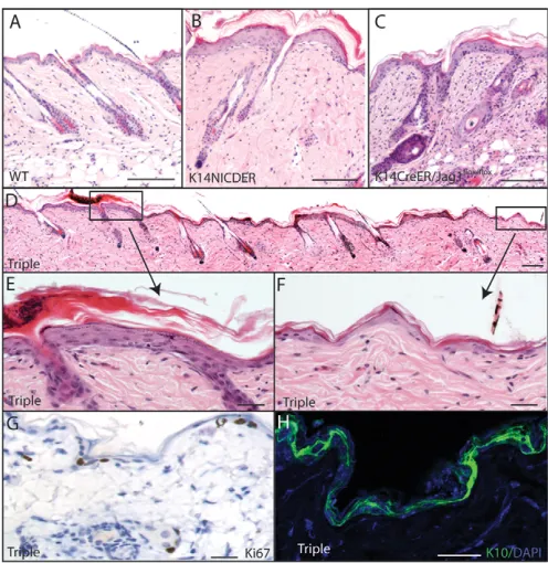
To examine the role of jagged 1 in the Notch-induced skin phenotype, we crossed K14NICDER, K14CreER and Jag1flox/flox strains of mice. In these mice, 4OHT treatment results in deletion of Jag1 in the same cells in which Notch is activated. K14NICDER/K14CreER/Jag1flox/flox(triple) transgenic mice were compared with untreated K14NICDER/K14CreER/Jag1flox/floxmice and littermates that were wild type for Notch. Treatment of K14CreER/Jag1flox/floxand K14NICDER mice with 4OHT induced minimal changes to the health of the mice (Estrach et al., 2006) (data not shown). However, all 4OHT-treated triple transgenic mice became sick (data not shown). Therefore, 7-week-old mice triple and control mice were treated for a maximum of 10 days.
[image:6.612.54.500.59.336.2]The back skin of treated K14NICDER mice had the expected phenotypic characteristics, including thickened, hyperproliferative epidermis, blistering and accumulation of dermal cells at the epidermal/dermal junction (Fig. 7A,B). In 4OHT-treated K14CreER/Jag1flox/floxmice, the epidermis was also thicker than wild-type controls, consistent previous data (Fig. 7C) (Estrach et
Fig. 4. Kinetics of appearance of Notch-induced stromal cells.(A,B,D) Back skin sections of 4OHT-treated K14NICDER mice stained with
antibodies to nestin (A, green or red; B, green), Crabp1 (A,B; red), Sm22(A, green) and p75 (D, green). (A)Arrows indicate double-labeled cells; arrowheads indicate single-labeled cells. (B)Arrows indicate Crabp1-positive cells. (C)Percentage Crabp1 (C), nestin (N), Sm22(S) single-positive cells and Crabp1/nestin and Crabp1/SM22double-positive cells at the epidermal-dermal boundary (n≥15 samples). (E)Skin-derived neurospheres 7 days after seeding, viewed by phase contrast (top) or anti-nestin labelling (bottom, red) and counterstained with DAPI (blue). Inset: labelling with secondary antibody alone. (F)Percentage sphere formation by cells from wild-type (WT) and K14NICDER (TG) littermates treated with 4OHT for 10 or 14 days. (G)Cytospin preparation of dermal cells isolated from 4OHT-treated K14NICDER skin stained with antibodies to nestin (green) and Crabp1 (red). Sections and cells were counterstained with DAPI (blue; A,B,D,E,G). Mice were treated with 4OHT for 2 days (B), 5 days (B), 7 days (B), 10 days (A, left panel; G) or 14 days (A, middle and right panels; D). Scale bars: 25mm in A,B,G; 50mm in D. Data are mean±s.e.m.
D
E
V
E
LO
P
M
E
N
al., 2006). In addition, there was a marked dermal infiltrate of CD3 positive T-cells (Fig. 7C and data not shown; Table 1) (Estrach et al., 2006).
Triple transgenic mice treated with 4OHT for 10 days developed two distinct back skin phenotypes (Fig. 7D-F). In some areas, the skin was similar to 4OHT-treated K14NICDER skin (Fig. 7E), whereas in other areas the epidermis was similar in thickness to wild type, but with a reduction in nucleated cells (Fig. 7F; Table 1). In areas of reduced epidermal thickness, the number of Ki67 positive, proliferating cells was similar to wild type and was markedly reduced compared with K14NICDER transgenic and K14CreER/Jagflox/floxmice (Fig. 1F; Fig. 7G and data not shown). In addition, keratin 10 was expressed in all suprabasal epidermal cells and a significant proportion of basal cells (Fig. 7H). Blistering was reduced in thin epidermis of triple transgenic mice, but present in thick epidermis (Fig. 7E,F; data not shown; Table 1).
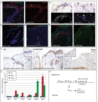
As reported previously, jagged 1 protein was undetectable in epidermis of 4OHT-treated K14CreER/Jag1flox/flox back skin (data not shown) (Estrach et al., 2006). However, in 4OHT-treated triple transgenics, jagged 1 deletion was incomplete. The
areas of thick epidermis that retained the K14NICDER Notch activation phenotype expressed jagged 1 protein and mRNA in both epidermis and adjacent dermis (Fig. 8A,B), whereas jagged 1 was absent in thin epidermis and underlying dermis (Fig. 8B,C). The incomplete deletion of Jag1is consistent with the partial expression of the CAG-CAT-eGFP reporter (Fig. 5C) and indicates that 4OHT treatment was not sufficient to efficiently activate two ER transgene fusion proteins simultaneously.
[image:7.612.51.381.61.211.2]Sections of 4OHT-treated K14NICDER and triple transgenic back skin were labeled with antibodies to jagged 1 and nestin, Crabp1, or SM22(Fig. 8A,C; data not shown). In K14NICDER dermis, nestin-, Crabp1- and SM22-positive cells co-labeled with jagged 1 (data not shown). In triple transgenic sections, positively stained cells at the epidermal/dermal boundary were detected only in the thick epidermal regions that retained jagged 1 in the epidermis and dermis (Fig. 8A). Dermal cells did not accumulate below thin, jagged 1-negative, regions in triple transgenics, nor in back skin of 4OHT-treated K14CreER/Jagflox/floxmice (Fig. 8C,D). Therefore epidermal jagged 1 is required for epidermal Notch-induced dermal jagged 1 expression and the accumulation of dermal cells that express neural crest markers.
Fig. 5. Nestin-positive dermal cells are not of
epidermal origin.(A,B)Back skin sections of
4OHT-treated wild-type (WT) and K14NICDER littermates stained with antibodies to Slug 1 (A, brown) and E-cadherin (B, green). (C,D)Back skin sections (150mm) (C) and cytospin preparations (D) of cells isolated from 4OHT-treated K14CreER/CAG-CAT-EGFP mice and K14CreER/CAG-CAT-EGFP/K14NICDER mice stained with antibodies to GFP (green) and nestin (red) with DAPI nuclear counterstain (blue). Mice were 4OHT-treated for 14 days (A-D). Scale bars: 20mm in D; 50mm in A-C.
Fig. 6. Notch induces Jag1in the epidermis and dermis.(A,C-E) Back skin of wild-type (A,C), K14NICDER (A,D) and K14-⌬NcateninER
(catER; E) mice. (A)Jagged 1 (red) immunolabelling with DAPI counterstain (blue). (B)Western blot of protein lysates from skin of wild-type (WT) and K14NICDER mice probed with anti-jagged 1. Each lane contains protein from a different mouse. Arrow indicates position of jagged 1 protein. Lower molecular mass bands are nonspecific and serve as loading controls. Molecular mass markers (kDa) are indicated. (C-E)RNA in situ
hybridisation using jagged 1 radiolabelled antisense probe. Corresponding brightfield (BF) and darkfield (DF) panels show same field. Red lines mark the epidermal/dermal boundary. The right-hand panels in D are higher magnification views of the boxed region in the left-hand panels. Mice were
treated with 4OHT for 21 days (A,B); 10 days (C,D) or 7 days (E). Scale bars: 50mm.
D
E
V
E
LO
P
M
E
N
[image:7.612.53.481.472.659.2]Jagged 1-dependent upregulation of TNF signalling
To investigate how epidermal Notch activation via jagged 1 led to changes in the underlying dermis, we performed gene expression profiling of epidermis, dermis and whole skin from transgenic and wild-type mice treated with 4OHT for 14 days. The microarray data were analysed using GeneSpring GX10 and Ingenuity Pathway Analysis software programmes after average signal intensities had been determined by MAS5-calculation. Data are deposited in the NIH GEO repository (Accession Number GSE23782: http://www.ncbi.nlm.nih.gov/geo/query/acc.cgi?accGSE23782).
Within the epidermis, Notch activation resulted in changes in many genes that are associated with barrier formation and integrity, including several metalloproteinases, S100A8, Sprr1b and filaggrin (see Table S1 in the supplementary material). In whole skin, markers of melanocytes (such as Tyrp1) and regulators of neural crest cell specification (such as Edn1) and glial and neuronal differentiation (e.g. Sox11) were strongly upregulated (see Table S1 in the supplementary material). These changes in gene expression are consistent with the epidermal and dermal phenotypes of 4OHT-treated K14NICDER skin.
To gain insights into potential mechanisms by which epidermal Notch activation could induce jagged 1 in the underlying dermis, we examined the microarrays for epidermal growth factors and cytokines. The list included three growth factors that were upregulated more than 10 fold: neuregulin 1, inhibin A, and
tumour necrosis factor (TNF) (see Table S1 in the supplementary material). The results were validated by quantitative RT-PCR of epidermal mRNA. All three factors were strongly upregulated in 4OHT-treated K14NICDER epidermis (Fig. 8G). Relative mRNA abundance was normalised to endogenous wild-type levels (designated as 1).
The upregulation of TNFwas of particular interest because it is linked to epidermal barrier disruption and skin inflammation (Incorvaia et al., 2008), and also to the Notch pathway. TNF activates the NF-B family of transcription factors that includes p65 (RelA). There is crosstalk between the Notch and NF-B pathways in many tissue types (Osipo et al., 2008) and in cultured human keratinocytes, exposure to jagged 1 activates the NF-B pathway (Nickoloff et al., 2002). To confirm that the NF-B pathway was activated, sections were stained with an antibody specific to active serine-phosphorylated (residue 276) p65. Occasional phospho-p65 positive nuclei were detected in wild-type epidermis but were largely absent from the dermis (Fig. 8F). In 4OHT-treated transgenic skin, phospho-p65 positive nuclei were detected in all epidermal layers and in the dermis (Fig. 8F). The majority of phospho-p65 positive dermal cells expressed Crabp1 (Fig. 8E).
To further examine the link between jagged 1 and p65, we used Ingenuity Pathway Analysis (IPA; see Fig. S7 in the supplementary material). Three factors that directly link p65 activity and jagged 1 expression in the epidermis and dermis of 4OHT-treated K14NICDER mice were identified: TNF, interferon and transforming growth factor beta (TGF). Interferon levels were increased 2.5-fold and TGF levels increased 3.0-fold in the epidermal microarrays. Quantitative (Q) PCR performed on epidermis treated with 4OHT for 10 days revealed an increase in TGFbut not in interferon-mRNA levels (Fig. 8G).
To examine whether upregulation of TNFand other secreted factors was dependent on epidermal jagged 1 expression, we performed Q-PCR of mRNA from 4OHT-treated K14NICDER/K14CreER/Jag1flox/flox(triple) transgenics (Fig. 8G). Levels of inhibin A and IFNdid not decline in response to Jag1 deletion; indeed inhibin A levels were further elevated. However, levels of TNF, TGF, Edn1 and Nrg1 were all decreased on Jag1 deletion. Furthermore, in areas of Jag1 deletion in 4OHT-treated K14NICDER/K14CreER/Jag1flox/floxmice, phospho-p65 was not detected in the epidermis or underlying dermis (Fig. 8F).
We conclude that the mechanism by which epidermal Notch activation exerts its pleiotropic effects on the skin (Fig. 8H) involves jagged 1-dependent induction of a number of secreted factors and their downstream pathways, including NF-B.
DISCUSSION
[image:8.612.51.299.59.314.2]We report that Notch activation in the basal layer of the epidermis not only results in thickening and blistering of the interfollicular epidermis (Estrach et al., 2006; Blanpain et al., 2006), but also
Table 1. Effect of epidermal Jag1deletion on Notch-induced
skin phenotype
Phenotype IFE thickening IFE blisters Stromal cells T cells
Wild type – – – –
Notch ++ ++ ++ ++
Jag1–/– + – – ++
Notch and Jag1–/– – + – –
[image:8.612.313.562.81.135.2]Notch, 4OHT-treated K14NICDER skin; Jag1–/–, 4OHT-treated skin of K14CreER ⫻ Jag1flox/floxmice; Notch and Jag1–/–, 4OHT-treated triple transgenics (K14CreER ⫻ Jag1flox/flox⫻K14NICDER); –, wild-type phenotype; +, detectable effect; ++, strong effect; IFE, interfollicular epidermis.
Fig. 7. Jag1is required for Notch-induced skin phenotype.
(A-F)Haematoxylin and Eosin-stained sections of back skin of wild type (A, WT), K14NICDER (B), K14CreER/Jag1flox/flox(C) and
K14CreER/Jag1flox/flox/K14NICDER (D-F, triple) mice treated with 4OHT for 10 days. In triple transgenics, areas of normal (F) and thickened (E) epidermis were found. (E,F)Higher magnifications views of boxed regions in D. (G,H)Back skin sections of triple transgenics in areas of normal thickness stained with antibodies to Ki67 (G, brown) and K10 (H, green). Sections were counterstained with Haematoxylin (G) or DAPI (H). Scale bars: 100mm in A-D; 25mm in E-H.
D
E
V
E
LO
P
M
E
N
causes remarkable changes in the dermis. These include a CD4-positive T infiltrate and accumulation of cells that express neural crest markers. Our data uncover a previously unappreciated role for Notch signalling in epidermal-dermal interactions.
It is interesting that both inhibition and activation of epidermal Notch leads to increased TSLP expression (see Table S1 in the supplementary material) and accumulation of CD4-positive T cells (Tournoy et al., 2004; Demehri et al., 2008). This suggests that the inflammatory changes are a consequence of epidermal barrier dysfunction rather than being caused by the direct effects of activating or inhibiting Notch activity. Inflammation was responsible for hair clumping, which parallels a human skin condition known as tufted hair folliculitis (Tong and Baden, 1989; Dalziel et al., 1990; Pranteda et al., 2004).
Epidermal Notch activation led to accumulation of cells that expressed markers of smooth muscle, peripheral nerve, neural crest and melanocytes, but not of endothelial cells. Notch activity has been linked to Sm22and nestin expression in other cell types (Kennard et al., 2008; Shih and Holland, 2006) and controls melanocyte survival (Moriyama et al., 2008). The markers are consistent with a neural crest origin, and the increase in p75 might indicate an expansion of neural crest stem cells (Stemple and Anderson, 1992). We excluded an epidermal origin for the dermal cells. Nevertheless, the contribution of neural crest to trunk skin is limited (Wong et al., 2006; Le Douarin et al., 2008) and we do not exclude the possibility of a mesenchymal origin of the Notch-induced cells. Notch-induced stromal cells could arise from a resident stem cell population, through transdifferentiation, or by both processes.
On epidermal Notch activation, jagged 1 was upregulated in both epidermis and dermis (Fig. 8H). Several additional factors that were upregulated in the epidermis are known to influence proliferation and differentiation of neural crest derivatives. These include endothelin 1 (Pla and Larue, 2003) and neuregulin 1 (Mei and Xiong, 2008), expression of both of which was partially dependent on epidermal jagged 1 (Fig. 8G). We have thus identified several secreted factors that are likely to mediate the increase in neural crest derivatives in response to epidermal Notch activation.
Gene expression profiling revealed one potential mechanism by which jagged 1 is induced in the dermis. Epidermal Notch activation resulted in induction of TNFin the epidermis and activation of NF-B in both epidermis and dermis. TNFinduces jagged 1 expression via NF-B (Johnston et al., 2009). As jagged 1 activates the NF-B pathway (Nickoloff et al., 2002) we envision a positive auto-regulatory loop involving TNF and jagged 1 expression in the skin. The upregulation of TNFfollowing Notch activation is also likely to contribute to the inflammation and barrier defects observed (Incorvaia et al., 2008).
In conclusion, our results identify jagged 1 as a key component of cell autonomous and non-cell autonomous Notch signalling in the skin. The mechanisms we have identified may explain some of the previously reported changes in the stromal compartment of epithelial tumours in which Notch is activated (Callahan and Egan, 2004; Collins and Watt, 2008; Demehri et al., 2009).
Acknowledgements
[image:9.612.52.383.59.403.2]This paper is dedicated to George Elia, Rob Rudling and their teams with our gratitude, admiration and affection. We thank Charlotte Collins and Ryan Driskell for advice, Pooja Seedhar for superb technical assistance, and everyone
Fig. 8. Relationship between jagged 1 expression and Notch-induced stromal cells.
(A-F)K14CreER/Jag1flox/flox/K14NICDER (triple) skin with either increased (thick) or normal (thin) epidermal thickness (A-C) and
K14CreER/Jag1flox/floxskin (D) were 4OHT-treated for 10 days. (E,F) Wild-type (WT), K14NICDER and triple (thin) skin was 4OHT-treated for 14 days. (A,C-F) Sections were stained with antibodies to nestin (A,C, green; D, red), jagged 1 (A,C, red), SM22(D, green), phosphorylated p65 (serine 276; E, green; F, brown) and Crabp1 (E, red) with DAPI (A,C-E, blue) or Haematoxylin (F) counterstain. Both panels in A and E show same field. Arrow in A marks jagged 1 staining. Insets in F show higher magnification views. (B)RNA in situ hybridisation using a radiolabelled jagged 1 antisense probe. Corresponding brightfield (BF, top) and darkfield (DF, bottom) panels show same field. In B, black arrows indicate interfollicular epidermis and the white arrows indicate hair follicle infundibulum. Scale bars: 50mm. (G)Quantitative polymerase chain reaction of cDNA from 4OHT-treated wild-type (WT), K14NICDER (NICD) and
K14CreER/Jag1flox/flox/K14NICDER (Triple) epidermis. Results (mean±s.e.m.) are expressed relative to wild type (1) for each TAQman probe. (H)Schematic summary of results. Epidermal Notch activation results in increased expression of jagged 1 in epidermis and dermis, which contributes to the epidermal changes and accumulation of cells in the dermis.
D
E
V
E
LO
P
M
E
N
who provided us with reagents. This work was funded by Cancer Research UK (F.M.W.), a Wellcome Trust VIP award (C.A.A.) and the European Skin Research Foundation (C.A.A.). C.A.A. was also supported by the National Institute of Health under Ruth L. Kirschstein National Researce Service Award number F32-AR049651. We gratefully acknowledge the support of Hutchison Whampoa and Cambridge University. Deposited in PMC for release after 6 months.
Competing interests statement
The authors declare no competing financial interests.
Supplementary material
Supplementary material for this article is available at
http://dev.biologists.org/lookup/suppl/doi:10.1242/dev.050310/-/DC1
References
Aho, S.(2004). Soluble form of Jagged1: unique product of epithelial
keratinocytes and a regulator of keratinocyte differentiation.J. Cell Biochem. 92, 1271-1281.
Ambler, C. A. and Watt, F. M.(2007). Expression of Notch pathway genes in mammalian epidermis and modulation by beta-catenin. Dev. Dyn. 236, 1595-1601.
Amoh, Y., Li, L., Katsuoka, K., Penman, S. and Hoffman, R. M.(2005). Multipotent Nestin-positive, keratin-negative hair-follicle bulge stem cells can form neurons. Proc. Natl. Acad. Sci. USA 102, 5530-5534.
Blanpain, C., Lowry, W. E., Pasolli, H. A. and Fuchs, E.(2006). Canonical notch signaling functions as a commitment switch in the epidermal lineage. Genes Dev. 20, 3022-3035.
Botchkarev, V. A., Eichmuller, S., Johansson, O. and Paus, R.(1997). Hair cycle-dependent plasticity of skin and hair follicle innervation in normal murine skin. J. Comp. Neurol. 386, 379-395.
Braun, K. M., Niemann, C., Jensen, U. B., Sundberg, J. P., Silva-Vargas, V. and Watt, F. M.(2003). Manipulation of stem cell proliferation and lineage commitment: visualisation of label-retaining cells in wholemounts of mouse epidermis. Development130, 5241-5255.
Brooker, R., Hozumi, K. and Lewis, J.(2006). Notch ligands with contrasting functions: Jagged1 and Delta1 in the mouse inner ear. Development 133, 1277-1286.
Byrne, C. and Hardman, M. J.(2005). Whole-mount assays for gene induction and barrier formation in the developing epidermis. Methods Mol Biol. 289, 127-136.
Callahan, R. and Egan, S. E.(2004). Notch signaling in mammary development and oncogenesis. J. Mamm. Gland Biol. Neoplasia 9, 145-163.
Collins, C. A. and Watt, F. M.(2008). Dynamic regulation of retinoic acid-binding proteins in developing, adult and neoplastic skin reveals roles for -catenin and Notch signalling. Dev. Biol. 324, 55-67.
Dalziel, K. L., Telfer, N. R., Wilson, C. L. and Dawber, R. P.(1990). Tufted folliculitis. A specific bacterial disease? Am. J. Dermatopathol. 12, 37-41.
Demehri, S., Liu, Z., Lee, J., Lin, M. H., Crosby, S. D., Roberts, C. J., Grigsby, P. W., Miner, J. H., Farr, A. G. and Kopan, R.(2008). Notch-deficient skin induces a lethal systemic B-lymphoproliferative disorder by secreting TSLP, a sentinel for epidermal integrity. PLoS Biol. 6, e123.
Demehri, S., Turkoz, A. and Kopan, R.(2009). Epidermal Notch1 loss promotes skin tumorigenesis by impacting the stromal microenvironment. Cancer Cell 16, 55-66.
Driskell, R. R., Giangreco, A., Jensen, K. B., Mulder, K. W. and Watt, F. M.
(2009). Sox2-positive dermal papilla cells specify hair follicle type in mammalian epidermis. Development 136, 2815-2823.
Duarte, M., Kolev, V., Kacer, D., Mouta-Bellum, C., Soldi, R., Graziani, I., Kirov, A., Friesel, R., Liaw, L., Small, D. et al.(2008). Novel cross-talk between three cardiovascular regulators: thrombin cleavage fragment of Jagged1 induces fibroblast growth factor 1 expression and release. Mol. Biol. Cell 19, 4863-4874.
Dunn, K. J., Williams, B. O., Li, Y. and Pavan, W. J.(2000). Neural crest-directed gene transfer demonstrates Wnt1 role in melanocyte expansion and
differentiation during mouse development. Proc. Natl. Acad. Sci. USA97, 10050-10055.
Estrach, S., Ambler, C. A., Lo Celso, C., Hozumi, K. and Watt, F. M.(2006). Jagged 1 is a beta-catenin target gene required for ectopic hair follicle formation in adult epidermis. Development 133, 4427-4438.
Estrach, S., Legg, J. and Watt, F. M.(2007). Syntenin mediates Delta1-induced cohesiveness of epidermal stem cells in culture. J. Cell Sci. 120, 2944-2952.
Estrach, S., Cordes, R., Hozumi, K., Gossler, A. and Watt, F. M.(2008). Role of the Notch ligand Delta1 in embryonic and adult mouse epidermis. J. Invest. Dermatol. 128, 825-832.
Fernandes, K. J., McKenzie, I. A., Mill, P., Smith, K. M., Akhavan, M., Barnabe-Heider, F., Biernaskie, J., Junek, A., Kobayashi, N. R., Toma, J. G. et al.(2004). A dermal niche for multipotent adult skin-derived precursor cells.
Nat. Cell Biol. 6, 1082-1093.
Fuchs, E. and Horsley, V.(2008). More than one way to skin… Genes Dev. 22, 976-985.
Handjiski, B. K., Eichmuller, S., Hofmann, U., Czarnetzki, B. M. and Paus, R.
(1994). Alkaline phosphatase activity and localization during the murine hair cycle. Br. J. Dermatol. 131, 303-310.
Hong, K. U., Reynolds, S. D., Watkins, S., Fuchs, E. and Stripp, B. R.(2004). Basal cells are a multipotent progenitor capable of renewing the bronchial epithelium. Am. J. Pathol. 164, 577-588.
Incorvaia, C., Frati, F., Verna, N., D’Alò, S., Motolese, A. and Pucci, S.(2008). Allergy and the skin. Clin. Exp. Immunol. 153, 27-29.
Jensen, K. B., Collins, C. A., Nascimento, E., Tan, D. W., Frye, M., Itami, S. and Watt, F. M.(2009). Lrig1 expression defines a distinct multipotent stem cell population in mammalian epidermis. Cell Stem Cell 4, 427-439.
Johnston, D. A., Dong, B. and Hughes, C. C.(2009). TNF induction of Jagged-1 in endothelial cells is NFB-dependent. Gene 435, 36-44.
Kawamoto, S., Niwa, H., Tashiro, F., Sano, S., Kondoh, G., Takeda, J., Tabayashi, K. and Miyazaki, J.(2000). A novel reporter mouse strain that expresses enhanced green fluorescent protein upon Cre-mediated recombination. FEBS Lett. 470, 263-268.
Kawano, M., Komi-Kuramochi, A., Asada, M., Suzuki, M., Oki, J., Jiang, J. and Imamura, T.(2005). Comprehensive analysis of FGF and FGFR expression in skin: FGF18 is highly expressed in hair follicles and capable of inducing anagen from telogen stage hair follicles. J. Invest. Dermatol.124, 877-885.
Kennard, S., Liu, H. and Lilly, B.(2008). Transforming growth factor-(TGF- 1) down-regulates Notch3 in fibroblasts to promote smooth muscle gene expression. J. Biol. Chem.283,1324-1333.
Kishimoto, J., Burgeson, R. E. and Morgan, B. A.(2000). Wnt signaling maintains the hair-inducing activity of the dermal papilla. Genes Dev. 14, 1181-1185.
Kopan, R. and Ilagan, M. X.(2009). The canonical Notch signaling pathway: unfolding the activation mechanism. Cell 137, 216-233.
Kruse, C., Bodo, E., Petschnik, A. E., Danner, S., Tiede, S. and Paus, R.(2006). Towards the development of a pragmatic technique for isolating and
differentiating Nestin-positive cells from human scalp skin into neuronal and glial cell populations: generating neurons from human skin? Exp. Dermatol. 15, 794-800.
Lauria, G., Borgna, M., Morbin, M., Lombardi, R., Mazzoleni, G., Sghirlanzoni, A. and Pareyson, D.(2004). Tubule and neurofilament immunoreactivity in human hairy skin: markers for intraepidermal nerve fibers.
Muscle Nerve 30, 310-316.
Le Douarin, N. M., Calloni, G. W. and Dupin, E.(2008). The stem cells of the neural crest. Cell Cycle7, 1013-1019.
Lee, J., Basak, J. M., Demehri, S. and Kopan, R.(2007). Bi-compartmental communication contributes to the opposite proliferative behavior of Notch1-deficient hair follicle and epidermal keratinocytes. Development 134, 2795-2806.
Lefort, K., Mandinova, A., Ostano, P., Kolev, V., Calpini, V., Kolfschoten, I., Devgan, V., Lieb, J., Raffoul, W., Hohl, D. et al.(2007). Notch1 is a p53 target gene involved in human keratinocyte tumor suppression through negative regulation of ROCK1/2 and MRCKalpha kinases. Genes Dev. 21, 562-577.
Leong, K. G., Niessen, K., Kulic, I., Raouf, A., Eaves, C., Pollet, I. and Karsan, A.(2007). Jagged1-mediated Notch activation induces
epithelial-to-mesenchymal transition through Slug-induced repression of E-cadherin. J. Exp. Med. 204, 2935-2948.
Li, L., Mignone, J., Yang, M., Matic, M., Penman, S., Enikolopov, G. and Hoffman, R. M.(2003). Nestin expression in hair follicle sheath progenitor cells.
Proc. Natl. Acad. Sci. USA 100, 9958-9961.
Lo Celso, C., Prowse, D. M. and Watt, F. M.(2004). Transient activation of beta-catenin signalling in adult mouse epidermis is sufficient to induce new hair follicles but continuous activation is required to maintain hair follicle tumours.
Development 131,1787-1799.
López-Rovira, T., Silva-Vargas, V. and Watt, F. M.(2005). Different
consequences of beta1 integrin deletion in neonatal and adult mouse epidermis reveal a context-dependent role of integrins in regulating proliferation, differentiation, and intercellular communication. J. Invest. Dermatol. 125, 1215-1227.
Lowell, S. and Watt, F. M.(2001). Delta regulates keratinocyte spreading and motility independently of differentiation. Mech. Dev. 107, 133-140.
Lowell, S., Jones, P., Le Roux, I., Dunne, J. and Watt, F. M.(2000). Stimulation of human epidermal differentiation by delta-notch signalling at the boundaries of stem-cell clusters.Curr. Biol. 10, 491-500.
McGowan, K. M. and Coulombe, P. A.(2000). Keratin 17 expression in the hard epithelial context of the hair and nail, and its relevance for the pachyonychia congenita phenotype. J. Invest. Dermatol. 114, 1101-1107.
Mei, L. and Xiong, W. C.(2008). Neuregulin 1 in neural development, synaptic plasticity and schizophrenia. Nat. Rev. Neurosci. 9, 437-452.
Moriyama, M., Durham, A. D., Moriyama, H., Hasegawa, K., Nishikawa, S., Radtke, F. and Osawa, M.(2008). Multiple roles of Notch signaling in the
regulation of epidermal development. Dev. Cell 14, 594-604.
D
Nickoloff, B. J., Qin, J. Z., Chaturvedi, V., Denning, M. F., Bonish, B. and Miele, L.(2002). Jagged-1 mediated activation of notch signaling induces complete maturation of human keratinocytes through NF-B and PPAR. Cell Death Differ. 9, 842-855.
Nicolas, M., Wolfer, A., Raj, K., Kummer, J. A., Mill, P., van Noort, M., Hui, C. C., Clevers, H., Dotto, G. P. and Radtke, F.(2003). Notch1 functions as a tumor suppressor in mouse skin. Nat. Genet. 33, 416-421.
Niessen, K., Fu, Y., Chang, L., Hoodless, P. A., McFadden, D. and Karsan, A.
(2008). Slug is a direct Notch target required for initiation of cardiac cushion cellularization.J. Cell Biol. 182, 315-325.
Nikopoulos, G. N., Duarte, M., Kubu, C. J., Bellum, S., Friesel, R., Maciag, T., Prudovsky, I. and Verdi, J. M.(2007). Soluble Jagged1 attenuates lateral inhibition, allowing for the clonal expansion of neural crest stem cells. Stem Cells 25, 3133-3142.
Osipo, C., Golde, T. E., Osborne, B. A. and Miele, L. A.(2008). Off the beaten pathway: the complex cross talk between Notch and NF-kB. Lab. Invest. 88, 11-17.
Pan, Y., Lin, M. H., Tian, X., Cheng, H. T., Gridley, T., Shen, J. and Kopan, R.
(2004). gamma-secretase functions through Notch signaling to maintain skin appendages but is not required for their patterning or initial morphogenesis.
Dev. Cell 7,731-743.
Pla, P. and Larue, L.(2003). Involvement of endothelin receptors in normal and pathological development of neural crest cells. Int. J. Dev. Biol. 47, 315-325.
Powell, B. C., Passmore, E. A., Nesci, A. and Dunn, S. M.(1998). The Notch signalling pathway in hair growth. Mech. Dev. 78, 189-192.
Pranteda, G., Grimaldi, M., Palese, E., Di Napoli, A. and Bottoni, U.(2004). Tufted hair folliculitis: complete enduring response after treatment with rifampicin. J. Dermatolog. Treat. 15,396-398.
Rendl, M., Lewis, L. and Fuchs, E.(2005). Molecular dissection of mesenchymal-epithelial interactions in the hair follicle. PLoS Biol. 3, e331.
Rendl, M., Polak, L. and Fuchs, E.(2008). BMP signaling in dermal papilla cells is required for their hair follicle-inductive properties. Genes Dev. 22, 543-557.
Ross, D. A. and Kadesch, T.(2004). Consequences of Notch-mediated induction of Jagged1. Exp. Cell Res.296, 173-182.
Schneider, M. R., Schmidt-Ullrich, R. and Paus, R.(2009). The hair follicle as a dynamic miniorgan.Curr. Biol. 19, R132-R142.
Sechrist, J., Nieto, M. A., Zamanian, R. T. and Bronner-Fraser, M.(1995). Regulative response of the cranial neural tube after neural fold ablation: spatiotemporal nature of neural crest regeneration and up-regulation of Slug.
Development 121, 4103-4115.
Shih, A. H. and Holland, E. C.(2006). Notch signaling enhances Nestin expression in gliomas. Neoplasia8, 1072-1082.
Stemple, D. L. and Anderson, D. J.(1992). Isolation of a stem cell for neurons and glia from the mammalian neural crest. Cell71,973-985.
Tiede, S., Kloepper, J. E., Bodò, E., Tiwan, S., Kruse, C. and Paus, R.(2007). Hair follicle stem cells: walking the maze. Eur. J. Cell Biol. 86, 355-376.
Toma, J. G., Akhavan, M., Fernandes, K. J., Barnabe-Heider, F., Sadikot, A., Kaplan, D. R. and Miller, F. D.(2001). Isolation of multipotent adult stem cells from the dermis of mammalian skin. Nat. Cell Biol. 3, 778-784.
Tong, A. K. and Baden, H. P.(1989). Tufted hair folliculitis. J. Am. Acad. Dermatol. 21, 1096-1099.
Tournoy, J., Bossuyt, X., Snellinx, A., Regent, M., Garmyn, M., Serneels, L., Saftig, P., Craessaerts, K., De Strooper, B. and Hartmann, D.(2004). Partial loss of presenilins causes seborrheic keratosis and autoimmune disease in mice.
Hum. Mol. Genet. 13,1321-1331.
Turner, F. E., Broad, S., Khanim, F. L., Jeanes, A., Talma, S., Hughes, S., Tselepis, C. and Hotchin, N. A.(2006). Slug regulates integrin expression and cell proliferation in human epidermal keratinocytes.J. Biol. Chem. 281, 21321-21331.
Urs, S., Roudabush, A., O’Neill, C. F., Pinz, I., Prudovsky, I., Kacer, D., Tang, Y., Liaw, L. and Small, D.(2008). Soluble forms of the Notch ligands Delta1 and Jagged1 promote in vivo tumorigenicity in NIH3T3 fibroblasts with distinct phenotypes. Am. J. Pathol. 173, 865-878.
Waters, J. M., Richardson, G. D. and Jahoda, C. A.(2007). Hair follicle stem cells. Semin. Cell Dev. Biol. 18, 245-254.
Watt, F. M., Lo Celso, C. and Silva-Vargas, V.(2006). Epidermal stem cells: an update. Curr. Opin. Genet. Dev. 16, 518-524.
Watt, F. M., Estrach, S. and Ambler, C. A.(2008). Epidermal Notch signalling: differentiation, cancer and adhesion.Curr. Opin. Cell Biol. 20, 171-179.
Wong, C. E., Paratore, C., Dours-Zimmermann, M. T., Rochat, A., Pietri, T., Suter, U., Zimmermann, D. R., Dufour, S., Thiery, J. P., Meijer, D. et al.
(2006). Neural crest-derived cells with stem cell features can be traced back to multiple lineages in the adult skin.J. Cell Biol. 175, 1005-1015.
Ya, J., Markman, M. W., Wagenaar, G. T., Blommaart, P. J., Moorman, A. F. and Lamers, W. H.(1997). Expression of the smooth-muscle proteins alpha-smooth-muscle actin and calponin, and of the intermediate filament protein desmin are parameters of cardiomyocyte maturation in the prenatal rat heart.
Anat. Rec. 249, 495-505.
Zhang, J. C., Kim, S., Helmke, B. P., Yu, W. W., Du, K. L., Lu, M. M., Strobeck, M., Yu, Q. and Parmacek, M. S.(2001). Analysis of SM22alpha-deficient mice reveals unanticipated insights into smooth muscle cell differentiation and function. Mol. Cell. Biol. 21, 1336-1344.
Zhang, Y., Andl, T., Yang, S. H., Teta, M., Liu, F., Seykora, J. T., Tobias, J. W., Piccolo, S., Schmidt-Ullrich, R., Nagy, A. et al.(2008). Activation of beta-catenin signaling programs embryonic epidermis to hair follicle fate.
Development 135, 2161-2172.