RESEARCH ARTICLE
Slow calcium waves mediate furrow microtubule reorganization
and germ plasm compaction in the early zebrafish embryo
Celeste Eno1, Timothy Gomez2, Diane C. Slusarski3and Francisco Pelegri1,*
ABSTRACT
Zebrafish germ plasm ribonucleoparticles (RNPs) become recruited to furrows of early zebrafish embryos through their association with astral microtubules ends. During the initiation of cytokinesis, microtubules are remodeled into a furrow microtubule array (FMA), which is thought to be analogous to the mammalian midbody involved in membrane abscission. During furrow maturation, RNPs and FMA tubules transition from their original distribution along the furrow to enrichments at the furrow distal ends, which facilitates germ plasm mass compaction. We show that nebel mutants exhibit reduced furrow-associated slow calcium waves (SCWs), caused at least in part by defective enrichment of calcium stores. RNP and FMA distal enrichment mirrors the medial-to-distal polarity of SCWs, and inhibition of calcium release or downstream mediators such as Calmodulin affects RNP and FMA distal enrichment. Blastomeres with reduced or lacking SCWs, such as early blastomeres innebel mutants and wild-type blastomeres at later stages, exhibit medially bundling microtubules similar to midbodies in other cell types. Our data indicate that SCWs provide medial-to-distal directionality along the furrow to facilitate germ plasm RNP enrichment at the furrow ends.
KEY WORDS: Calcium, Germ plasm, Ribonucleoparticles, Microtubules, F-actin, Zebrafish
INTRODUCTION
In the zebrafishDanio rerio, as in many animal species, primordial germ cell specification is mediated by the maternal inheritance of the germ plasm, a specialized cytoplasm containing specific RNAs and proteins (Wylie, 1999; Strome and Lehmann, 2007; Eno and Pelegri, 2013). Zebrafish germ plasm ribonucleoparticles (RNPs) are inherited as spherical units, typically of less than 1 μm in diameter, that are initially dispersed through the blastodisc. During the first several embryonic divisions, these units multimerize, gathering into clusters while appearing to retain their initial spherical structure (Theusch et al., 2006; Eno and Pelegri, 2013; Nair et al., 2013; Eno and Pelegri, 2018).
During the early embryonic cell divisions, RNP aggregates move to the forming furrows to generate typically four germ plasm masses (Yoon et al., 1997; Pelegri et al., 1999; Knaut et al., 2000). These aggregates subsequently become incorporated into primordial germ cells (Olsen et al., 1997; Yoon et al., 1997; Braat et al., 1999; Knaut
et al., 2000), where they are necessary and sufficient for germ cell specification (Hashimoto et al., 2004; Bontems et al., 2009; Miranda-Rodríguez et al., 2017).
The assembly of germ plasm masses at the furrows involves sequential steps. First, RNP multimers assemble even prior to furrow initiation ( pre-aggregation). Second, RNP pre-aggregates gather locally towards the forming furrows (furrow recruitment). Both of these processes appear to be mediated by the outward growth of astral microtubules, the tips of which are associated with germ plasm RNPs (Theusch et al., 2006; Eno and Pelegri, 2013; Nair et al., 2013). Germ plasm recruitment during furrow formation results in the accumulation of RNP aggregates collectively forming an elongated, rod-like structure along most of the length of the furrow. At the same time, microtubules form the furrow microtubule array (FMA) (Danilchik et al., 1998; Jesuthasan, 1998), which is thought to be homologous to the mammalian midbody involved in cell abscission (Otegui et al., 2005). FMA tubules are initially arranged in parallel along the length of the furrow, oriented perpendicular to the cleavage plane, with germ plasm RNP aggregates recruited along the furrow maintaining their association with microtubule ends. A third step in the germ plasm segregation process,‘distal compaction’, occurs during furrow maturation and involves coordinated changes in both RNP aggregates at the furrow and the FMA (Fig. 1). During this step, RNPs recruited at the furrow translocate towards the nearest furrow distal end, where they form compact RNP masses (Pelegri et al., 1999). At the same time, FMA tubules become enriched and undergo bundling in the distal furrow region, while their ends, which maintain their association with germ plasm RNPs, acquire a characteristic outward tilt. As the furrow is completed, FMA microtubules become disassembled leaving the compacted germ plasm mass at the furrow distal end (Pelegri et al., 1999).
Germ plasm pre-aggregation, furrow recruitment and distal compaction therefore achieve the purpose of gathering maternally inherited single RNPs into discrete structures that will be subsequently inherited by primordial germ cells. In the accompanying article (Eno and Pelegri, 2018), we show that mutations in the maternal effect gene mid1ip1l (also known as aura), which encodes the cytoskeletal regulator Mid1ip1l, result in defects in the recruitment of germ plasm RNPs to the furrows. This function is mediated through mid1ip1l-dependent cortical F-actin dynamics and contractions associated with germ plasm RNPs, which allow RNP retention at the cortex prior to furrow formation and likely also facilitate RNP multimerization during furrow maturation. Here, we focus on the process of distal compaction and document a calcium-dependent mechanism required for the enrichment of germ plasm RNPs, together with associated FMA, to the distal ends of the furrow.
In both zebrafish and medaka, waves of intracellular calcium increase are associated with zebrafish cytokinesis during the first Received 20 July 2017; Accepted 27 March 2018
1Laboratory of Genetics, University of Wisconsin–Madison, Madison, WI 53706,
USA.2Department of Neuroscience, University of Wisconsin–Madison,
Madison, WI 53705, USA.3Department of Biology, The University of Iowa, Iowa City,
IA 52242, USA.
*Author for correspondence (fjpelegri@wisc.edu)
T.G., 1515-8228; D.C.S., 0000-0003-3793-8636; F.P., 0000-0002-0464-7042
© 2018. Published by The Company of Biologists Ltd|Development (2018) 145, dev156604. doi:10.1242/dev.156604
DEVEL
O
several cell cleavage cycles (Fluck et al., 1991; Chang and Meng, 1995; Reinhard et al., 1995; Webb et al., 1997; Créton et al., 1998). These waves have been termed slow calcium waves (SCWs) (Fig. 1) owing to their relatively low speed of propagation (0.1-1 µm/s) (Jaffe, 1993). Two primary furrow-associated SCWs have been described. A superficial‘initiation wave’, generated just prior to the appearance of the furrow cleft, is required for furrow formation. Subsequently, a larger‘maturation wave’penetrates deeper into the cytoplasm and has a role in events associated with furrow maturation, such as vesicle exocytosis and the formation of F-actin-dependent lamellipodia (Lee et al., 2003; Li et al., 2008).
The intracellular calcium signaling network is complex and the release of calcium from internal stores is controlled by three primary sets of ligands and calcium channels: inositol trisphosphate (IP3)
and cyclic adenosine diphosphate ribose (cADPR) through IP3and
ryanodine receptors, respectively, both at the endoplasmic reticulum (ER), and nicotinic acid adenine dinucleotide phosphate (NAADP) through NAADP-sensitive channels on lysosome-like organelles (Berridge et al., 2003). Previous studies have shown the presence of calcium stores activated by IP3 and NAADP, but not cADPR, in
early zebrafish embryos, and that inhibition of IP3receptors affects
furrow formation in early blastomeres (Lee et al., 2003).
Embryos from homozygousnebelmutant females exhibit normal nuclear division and furrow initiation, but the forming furrows either fail to develop a fully adhesive membrane septum between daughter cells or undergo regression, revealing a defect in the completion of cytokinesis (Pelegri et al., 1999). In nebelmutant embryos, germ plasm RNP aggregates form compact masses and FMA tubule tips undergo bundling, but these events occur in medial, rather than distal, regions of the furrow.
Several factors are known to mediate RNP aggregate and FMA dynamics in early embryonic blastomeres. In myosin-inhibited embryos, the RNP aggregate and FMA are arrested in a conformation corresponding to furrow initiation, namely as an elongated RNP aggregate along the furrow with the FMA retaining its original parallel conformation (Urven et al., 2006). A similar reorganization defect occurs in mid1ip1l mutant embryos, likely because of the function of this gene in F-actin reorganization (Eno et al., 2016; Eno and Pelegri, 2018). Recent studies have shown that inhibition of the small GTPase RhoA and its effector ROCK result in aberrant enrichment of germ plasm in medial regions of the mature furrow, a defect similar to that observed in nebel mutants (Miranda-Rodríguez et al., 2017). However, mechanisms
underlying germ plasm and FMA reorganization, including cues conferring medial-to-distal directionality along the furrow plane, remain largely unknown.
Here, we show that during the early cleavage stages,nebelmutant embryos show reduced intensity and temporal dynamics of furrow-associated SCWs. Treatments that interfere with SCW formation also result in defects in germ plasm RNP and FMA distal enrichment. Under these conditions, FMA bundles inward, as in later blastomeres that lack SCWs. Germ plasm RNP aggregate compaction and FMA bundling direction correlate with the medial-to-distal directionality of SCWs and are dependent on Calmodulin activity. Our studies indicate that SCWs in the early embryo modify RNP distribution and microtubule reorganization during furrow maturation, in order to facilitate the formation of multiple germ plasm masses capable of inducing the germ cell fate.
RESULTS
nebelmutant embryos show defects in furrow-associated SCWs
Because of the proposed role for SCWs in early teleost embryo furrow formation (Chang and Meng, 1995; Webb et al., 1997; Créton et al., 1998; Li et al., 2008), we visualized SCWs in wild-type andnebelmutant embryos with intracellular calcium-sensitive dyes. We first used the ratiometric indicator Fura-2-Dextran, which allows for quantitation of intracellular calcium levels. The patterns of intracellular calcium concentration in wild type and nebel mutants were markedly different. As previously reported (Chang and Meng, 1995; Webb et al., 1997; Créton et al., 1998; Li et al., 2008), intracellular calcium increases appear in wild-type embryos immediately prior to furrow initiation and during furrow maturation, corresponding to initiation and maturation SCWs, with the maturation SCW showing calcium release of greater intensity and depth (Fig. 2A, Fig. S1A, Movie 1). By contrast,nebelmutants exhibit intracellular SCWs of reduced amplitude and frequency (see below) compared with wild type (Fig. 2B, Fig. S1B, Movie 2). Embryos mutants for mid1ip1l, another maternal effect mutation affecting late cytokinesis (Pelegri et al., 2004; Eno et al., 2016), exhibit normal furrow-associated SCWs (data not shown), indicating that the SCW defect in nebel is not a general consequence of failed completion of cytokinesis.
[image:2.612.51.397.54.252.2]To better study the relationship between SCWs and furrow development, we scored wild-type and mutant embryos for defects during furrow initiation and completion (Table S2). Nearly all Fig. 1. Germ plasm and FMA reorganization. During furrow formation, germ plasm
ribonucleoparticles (RNPs) and associated furrow microtubule array (FMA) become enriched to the furrow distal ends. Waves of intracellular calcium [slow calcium waves (SCWs)] travel in a medial-to-distal orientation, with the maturation wave occurring during furrow formation (shown offset from the furrow plane for clarity).
DEVEL
O
cleavingnebelmutant embryos (98%,n=255) underwent normal furrow initiation (Fig. 2D) at a time and extent similar to wild type (Fig. 2C). However, these mutant embryos invariably showed defects in furrow completion, such as a lack of formation of a cell adhesive wall or furrow regression (Fig. 2G,H, compared with 2F). Only in rare cases (2%) did blastomeres exhibit defects in furrow initiation, as reflected by lack of furrowing in one of two blastomeres in the 2-cell stage embryo (Fig. 2E). These observations confirm our previous conclusions thatnebellargely affects late stages of cytokinesis (Pelegri et al., 1999), and further suggest that nebel function is required to achieve high calcium release levels characteristic of the maturation SCW. As opposed to
the two primary SCWs found in wild type, with the maturation SCW lasting most of furrow formation, the SCWs of reduced intensity observed innebelmutants appeared to fluctuate, with up to four waves of calcium release observed in a single cycle and a relatively constant periodicity of∼7 min (Fig. 2B).
[image:3.612.80.532.55.495.2]The defects in intracellular calcium release were also observed using the calcium-sensitive dye Oregon Green 488 BAPTA-Dextran, which does not allow measurement of absolute intracellular calcium concentration but does allow improved visualization and quantification of SCWs. Imaging of wild-type embryos with this dye readily reveals SCWs at the furrow (Fig. 2I), with kymograph analysis showing an average time between peaks of Fig. 2. Reduced calcium release underlies furrow formation defects in zebrafishnebelmutants.(A,B) Fluorescence after injection of Fura-2 during second cleavage furrow formation, with calcium-saturated (340 nm) and calcium-free (380 nm) images converted to a pseudocolor ratio. Intracellular calcium increases are observed prior to and during furrow formation in wild-type embryos (A), whereasnebelmutants show repeated intracellular increases of reduced intensity (B). Animal views, with numbers indicating the lapsed fraction of the cell cycle. Arrows indicate sites of high intracellular calcium, corresponding to SCWs. (C-H) In
nebelmutants, furrow initiation appears normal [black arrowheads during the first (D,E versus C) and third (G,H versus F) cell division]. In rare cases, the furrow
fails to initiate (white arrowhead in E; insert depicts a clearer focal plane). Black arrow (F) indicates the adhesive septum in wild type, which fails to form in mutants (G,H, white arrows). (I-L) Ratiometric analysis using Oregon Green 488 BAPTA-Dextran and Rhodamine in wild-type (I),nebelmutant (J) and 3-NP-treated wild-type (K) embryos, with representative kymographs (I′-K′, white line in I-K) and average time in seconds between calcium pulses (L,n>6 furrows). Error bars indicate the s.e.m.; two-tailedP-values were assessed using an unpairedt-test. (M-P) Pseudocleavages in unfertilized wild-type eggs (M, arrow) do not form in
nebelmutant eggs (N) but are induced by CaCl2injection (O), an effect independent of the KCl co-injected to improve egg survival (P).
RESEARCH ARTICLE Development (2018) 145, dev156604. doi:10.1242/dev.156604
DEVEL
O
76 s (s.d.=16.9 s,n=38 peaks from 8 embryos; Fig. 2I′,L, Fig. S2). By contrast,nebelmutants exhibit a higher frequency of SCWs, with an average period between peaks of 27 s (s.d.=8.1 s, n=56 peaks from 8 embryos; Fig. 2J,J′,L, Fig. S2). As expected, exposure of embryos to the IP3receptor inhibitor 2-APB results in the rapid
cessation of calcium release at the furrow (Lee et al., 2003) (data not shown). Thus, our data show that nebelmutant embryos exhibit defects in furrow-associated SCWs, such as a reduction in magnitude and increase in frequency of intracellular calcium release.
Intracellular calcium rescues aspects of thenebelmutant phenotype
If the nebel mutant phenotype is caused by reduced intracellular calcium release, the furrow defect may be rescued by an increase in intracellular calcium. Initial experiments involving CaCl2injections
into cleaving embryos did not result in significant rescue of thenebel mutant cellularization defect (data not shown), possibly because of the need for precise spatiotemporal regulation of calcium release at the forming furrow. However, we took advantage of differences between unfertilized eggs from wild-type andnebelmutant females (Pelegri et al., 1999) to test whether calcium can rescue thenebel mutant phenotype (Table S3). After egg activation, unfertilized eggs from wild-type females typically exhibit asymmetric membrane ingression, also known as pseudocleavages (Fig. 2M) (Kane and Kimmel, 1993), possibly through the unregulated exocytosis of intracellular membrane (data not shown; see also Danilchik et al., 1998). In contrast to wild type, activated yet unfertilized eggs from nebelmutant females typically fail to form pseudocleavages (6%, n=125; Fig. 2N). Injection of 0.1 mM or 0.2 mM CaCl2into eggs
fromnebelmutant females significantly increased pseudocleavage formation [to 42% (n=111) or 54% (n=115), respectively; Fig. 2O], whereas control injection did not have this effect (6%, n=123; Fig. 2P). Thus, the absence of pseudocleavage formation in unfertilized nebel mutant eggs can be reversed by introducing calcium, consistent with a reduction in intracellular calcium as an underlying basis for thenebelmutant phenotype.
Mitochondrial function is required for SCW dynamics
Previous studies have demonstrated the ability of calcium buffering by mitochondria to influence the magnitude and patterns of intracellular calcium waves (Jouaville et al., 1995; Babcock et al., 1997). Specifically, mitochondrial activity is directly correlated to the amplitude and inversely correlated to the frequency of IP3
-induced propagating calcium waves in theXenopusoocyte cortex (Jouaville et al., 1995). Because these effects are reminiscent of those found in furrow-associated SCWs innebelmutants, we tested whether inhibition of mitochondrial activity leads to defects in SCWs in wild-type embryos. Indeed, wild-type embryos exposed during furrow formation to the mitochondrial respiratory Complex II inhibitor 3-nitropropionic acid (Alston et al., 1977) exhibited reduced intensity and increased frequency of SCWs (Fig. 2K,K′,L, Fig. S2). Thus, similar to propagating calcium waves inXenopus oocytes, furrow-associated SCWs in early zebrafish embryos require functional mitochondrial activity.
Calcium stores are present innebelmutants but fail to become enriched to the furrow
Previous studies have shown that IP3- and NAADP-dependent, but
not cADPR-dependent, calcium stores are present and active in the early zebrafish embryo (Lee et al., 2003). To test whethernebel mutants contain functional calcium stores, the calcium release
response to injection of IP3or NAADP was compared with that of
wild-type embryos (Fig. 3A). Agonist was injected into unfertilized embryos in order to reduce experimental variation caused by the uneven distribution of calcium stores at the vicinity of forming furrows (Berridge, 1993; Berridge et al., 2003; Lawrence and Mandato, 2013) (see also Fig. 4). As expected from previous studies (Lee et al., 2003), both IP3and NAADP injections (Fig. 3D,F), but
not mock injections (Fig. 3B), elicit a calcium release response in wild type. These two agonists (Fig. 3E,G), but not control injections (Fig. 3C), elicited a calcium release response similar in magnitude to that innebelmutants (Fig. 3H). Consistent with the proposed role of IP3 in long-range calcium wave propagation through
calcium-mediated calcium release (Finch et al., 1991; Galione et al., 1993), the spatial extent of calcium release after IP3injection expands into
most of the blastodisc (Fig. 3D,E), whereas it is comparatively confined after NAADP injection (Fig. 3F,G), with wild type and nebelmutants exhibiting a similar behavior (Fig. 3I). Together, our data indicate thatnebelmutant eggs are able to elicit an apparently normal calcium release response to IP3and NAADP, suggesting that
the IP3- and NAADP-sensitive calcium stores and the
corresponding receptors are present and functional in mutant eggs and embryos.
Sincenebelmutant embryos exhibit reduced SCWs, yet the eggs exhibit a robust response to calcium agonists, we tested whether the localization patterns of calcium stores are affected in mutant embryos. Labeling with DiOC6shows an enrichment of ER at the
furrows of early wild-type embryos (Fig. 4A). By contrast, the ER fails to become enriched at the furrows innebelmutants, being found instead in a diffuse pattern throughout the blastodisc (Fig. 4B). Using Lysotracker as a labeling dye, we find that lysosomes also become aggregated to the forming furrows in wild-type embryos (Fig. 4C). nebelmutant embryos, on the other hand, lack an enrichment of lysosomes at the furrow, which instead accumulate in peripheral regions of the embryo (Fig. 4D). Labeling with Mitotracker shows that mitochondria exhibit a similar pattern to that of ER, with enrichment at the furrows in wild type (Fig. 4E) but distributed in a diffuse pattern innebelmutants (Fig. 4F). Thus,nebelmutants show a general defect in the enrichment of calcium stores at the forming furrow, including the ER, lysosomes and mitochondria. Given the essential role for these stores and their localization in regulated calcium release (Jouaville et al., 1995; Babcock et al., 1997; Lee et al., 2003), this defect is likely to be a primary cause for the observed effects on furrow-associated SCWs in these mutants.
Calcium release is required for the reorganization of the FMA and associated germ plasm
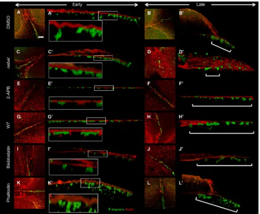
We tested whether inhibition of calcium release phenocopies the germ plasm segregation and FMA reorganization defects observed in nebelmutants, visualizing germ plasm RNPs with antibodies against serine 19 phospho-myosin light chain (P-myosin) (Nair et al., 2013), colabeled for microtubules, or byin situhybridization to detect the RNA for dead end (dnd) (Weidinger et al., 2003) (Fig. 5). In wild-type embryos, germ plasm RNPs exhibit distal compaction, involving the transition from a rod-like structure along the forming furrow to a compacted mass at its distal end (Pelegri et al., 1999; Eno and Pelegri, 2013; reviewed by Eno and Pelegri, 2016) (Fig. 5A,K), which is coupled to the outward tilting and distal enrichment of associated FMA (Jesuthasan, 1998; Pelegri et al., 1999; Urven et al., 2006). In nebel mutants, germ plasm RNPs become recruited normally along the furrow and the resulting aggregate typically transitions into a more compact mass, but this mass and associated FMA fail to become enriched in distal furrow
DEVEL
O
regions (Pelegri et al., 1999) (Fig. 5C,M). As expected considering the role of calcium in furrow initiation (Chang and Meng, 1995), inhibition of calcium release with an injection of the calcium chelator BAPTA results in a reduction in both furrow formation and germ plasm RNP furrow recruitment (Fig. 5E,O).
We next tested the effects on germ plasm and associated FMA in wild-type embryos of inhibitors predicted to interfere with SCWs (Lee et al., 2003). Exposure to the IP3-mediated calcium release
inhibitors 2-APB [an IP3 receptor inhibitor (Bilmen and
Michelangeli, 2002)] (Fig. 5G,Q) and U73122 [a Phospholipase C inhibitor that reduces IP3production (Macmillan and Mccarron,
2010)] (Fig. 5I,S) resulted in germ plasm RNPs and associated FMA retaining an arrangement characteristic of initiating furrows, namely a distribution of RNPs along the furrow without subsequent compaction, and an FMA perpendicular to the furrow without tilting or distal enrichment. Similar effects on RNPs and FMA were observed after exposure to the mitochondrial respiratory Complex II inhibitor 3-NP (Fig. 5B,L). Thus, treatments that interfere with SCWs in wild-type embryos lead to defects in enrichment at the
distal furrow of germ plasm RNPs and associated FMA–effects similar to those observed innebelmutants.
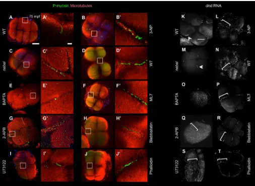
We further investigated FMA dynamics in live embryos under conditions that inhibit SCW formation using EMTB::EGFP transgenic fish (Fig. 6). In wild-type embryos at the 2- to 4-cell stage, the FMA translocates distally during furrow formation, where it subsequently undergoes disassembly (9 of 10 embryos, Fig. 6A) (Jesuthasan, 1998; Pelegri et al., 1999; Urven et al., 2006; Eno et al., 2016). By contrast, innebel mutant embryos the FMA becomes enriched in the medial regions of the furrow, either at a single site at the center of the blastodisc (3 of 10 embryos, Fig. 6B) or at two sites abutting the center of the blastodisc (6 of 10 embryos, not shown) (see also Pelegri et al., 1999).Similar to our observations in fixed embryos, in live embryos in which IP3receptors are inhibited with
[image:5.612.52.561.57.479.2]2-APB the FMA fails to become enriched in distal furrow regions, remaining instead in its original conformation along the length of the furrow (Fig. 6C). Similar effects are observed in live embryos after inhibition of mitochondrial function with the Complex II inhibitor 3-NP (data not shown).
Fig. 3.nebelmutants exhibit a normal response to calcium release agonists.(A) Experimental design. (B-G) IP3or NAADP triggers a normal calcium
release response innebelmutants as compared with wild type. Agonist injection (arrowhead) was at 15-30 mpf, with time relative to injection indicated in min:s. (H,I) Quantification of magnitude and spatial extent of calcium release. Error bars indicate s.e.m.P-values according to pairedt-test.
RESEARCH ARTICLE Development (2018) 145, dev156604. doi:10.1242/dev.156604
DEVEL
O
Furrow-associated SCWs are detected in early zebrafish embryos during the earliest cell cycles, leading to the 8-cell stage, but become undetectable during subsequent cell cycles (Chang and Meng, 1995). Such later cell cycles (leading to the 16-cell through 500-16-cell stages) continue to exhibit an FMA-like structure during early stages of cytokinesis. However, bundling of FMA tubules in wild type at these later stages occurs in medial regions of the furrow (Fig. 6D,E), similar to the behavior of coalescing midbodies in most cell types (Otegui et al., 2005; Steigemann and Gerlich, 2009). Thus, the presence of furrow-associated SCWs in the early wild-type embryo correlates with distally oriented reorganization of the FMA and associated germ plasm RNPs.
FMA reorganization mirrors the medial-to-distal polarity of SCWs
The requirement for intracellular calcium to implement the medial-to-distal reorganization of the FMA and germ plasm RNP aggregation is consistent with previous suggestions that SCWs originate in the center of the furrow and propagate towards its distal ends during furrow maturation (Chang and Meng, 1995; Webb et al., 1997; Créton et al., 1998), which we confirm using a transgenic line carrying the intracellular calcium reporter construct Tg[βactin2:GCaMP6s](Chen et al., 2017) (Fig. 7A, Fig. S3A,G). Analysis of microtubules in live wild-type embryos using the EMTB::EGFP transgene shows that the distally oriented calcium increase is mirrored by an increase in FMA intensity in distal regions at later stages of furrow formation (Fig. 7C, Fig. S3C,K). By contrast, similar analyses innebelmutant embryos show a lack of enrichment in the distal furrow for both intracellular calcium (Fig. 7B, Fig. S3B,H) and microtubules (Fig. 7D, Fig. S3D,L). Inhibition of SCWs in wild-type embryos using either the IP3
receptor inhibitor 2-APB (Fig. S3E,I,M) or the mitochondrial activity inhibitor 3-NP (Fig. S3F,J,N) also interferes with the distal enrichment of both intracellular calcium and FMA. These analyses provide further support for a medial-to-distal directionality of SCWs, and show that it is associated with the distally oriented reorganization of the FMA and germ plasm RNP aggregates during furrow formation.
Calmodulin function and actomyosin dynamics are required for germ plasm RNP and FMA distal enrichment
Our studies suggest that SCWs convey information to polarize the cytoskeleton during furrow formation. We therefore considered as potential mediators of furrow polarization two factors whose activity is known to be calcium dependent: non-muscle myosin (Scholey et al., 1980) and Calmodulin (Albert et al., 1984; Clapham, 2007). Treatment with either of two Calmodulin inhibitors, W7 and W5, results in the FMA failing to undergo distal reorganization and in RNP aggregates remaining distributed along medial regions of the furrow (Fig. 5D,N; data not shown). Labeling of wild-type embryos with anti-Calmodulin and anti-Calmodulin kinase (CamK) antibodies shows enrichment of these proteins at the furrow in wild-type embryos (Fig. 8A,C). Furrow localization for both factors is reduced innebelmutant embryos or 2-APB-treated embryos (Fig. 8B,D; data not shown), in which SCWs are inhibited. Inhibition of Rho activity by exposure to C3 exoenzyme also results in defects in RNP distal compaction (Fig. S4), as recently reported (Miranda-Rodríguez et al., 2017).
As previously reported (Urven et al., 2006), treatment of wild-type embryos with inhibitors of myosin activity, such as blebbistatin and ML7, results in a lack of FMA reorganization and germ plasm RNP distal compaction during furrow formation (Fig. 5F,H,P,R). Moreover, germ plasm RNPs are labeled with the P-myosin antibody (Nair et al., 2013), consistent with active myosin as one of their components. In contrast to Calmodulin and CamK, however, anti-P-myosin labeling is not reduced under conditions in which calcium is strongly reduced, as is also the case innebelmutants (Fig. 5C′) or in wild-type embryos treated with inhibitors of IP3
[image:6.612.48.383.59.287.2]release (2-APB, U73122; Fig. 5G′,I′) or of mitochondrial activity (3-NP; Fig. 5B′) (see Discussion). Treatment of wild-type embryos with phalloidin indicates that stabilization of furrow F-actin also interferes with the distal movement of germ plasm RNPs (Fig. 5J,T). Three-dimensional reconstructions show that, in control wild-type embryos, F-actin in the contractile ring forms indentations (Fig. 9A, inset), which during furrow maturation are progressively found in a more distal location, mirroring the enrichment of RNPs during distal compaction (Fig. 9B, bracket) (see Eno and Pelegri, 2018). Innebel mutants, in which germ plasm RNPs become recruited normally to the furrow (Fig. 9C) but remain in medial regions of the furrow after Fig. 4. Calcium stores are not properly enriched at the furrows innebelmutants.Furrow enrichment of (A,B) ER (5/5 wild type, 0/9nebel mutant), (C,D) lysosomes (6/6 wild type, 2/7nebel mutant) and (E,F) mitochondria (13/14 wild type,
2/18nebel). Images are from live (A-D) and fixed
(E,F) embryos. Scale bar: 100μm in A for A-F.
DEVEL
O
maturation (Fig. 9D, bracket), furrow F-actin indentations appear to be reduced (Fig. 9C, inset). Inhibition of calcium release by treatment of wild-type embryos with 2-APB also results in reduced furrow F-actin indentations (Fig. 9E, inset) and aberrant distal RNP enrichment (Fig. 9F, bracket). Similar effects are observed in embryos treated with Calmodulin or myosin inhibitors (Fig. 9G,I, insets; Fig. 9H,J, brackets). F-actin indentations continue to be apparent in phalloidin-treated embryos in spite of defects in germ plasm RNP distal movement (Fig. 9K-L′).
Altogether, these results and the accompanying manuscript (Eno and Pelegri, 2018) indicate that calcium- and myosin-dependent contractility, as well as F-actin dynamics, are essential for the distal reorganization of germ plasm RNPs and associated FMA.
DISCUSSION
In this study, we show thatnebelmutants exhibit changes in furrow-associated SCWs, specifically a reduction in calcium release levels and
an increase in release frequency. Our studies show that directional calcium release during the early mitotic cell cycles has a key role in the reorganization of furrow-associated microtubules and associated RNPs in the early embryo, leading to their enrichment at the furrow distal ends.
nebel-dependent furrow enrichment of calcium stores is required for SCW formation
[image:7.612.53.559.57.426.2]Wild-type embryos exhibit two primary SCW waves: an initiation wave prior to furrow indentation and a deeper and more intense wave during furrow maturation (Chang and Meng, 1995; Reinhard et al., 1995; Webb et al., 1997; Créton et al., 1998). By contrast,nebel mutants exhibit SCWs of comparatively low intensity. Intracellular calcium release is essential for both furrow initiation and maturation (Lee et al., 2003), butnebelmutant embryos are, in most cases, able to initiate furrow ingression (Pelegri et al., 1999; this report). These observations suggest that reduced SCWs in nebel mutants are sufficient for furrow initiation but not furrow maturation.
Fig. 5. Perturbation of calcium release inhibits germ plasm distal compaction.(A-J) RNPs labeled with anti-P-myosin and colabeled for microtubules. (A) In wild type, RNPs have underdone distal compaction in furrows for the first and second cell cycle (11/14 furrows). (C) Innebelmutants, RNPs fail to become distally enriched (2/15). (E) Injection after furrow initiation (35 mpf) of the calcium chelator BAPTA results in reduced and medially located RNPs (0/10). (B,D,F-J) Various inhibitors result in normal RNP furrow recruitment but aberrant RNP and FMA reorganization. (B) Mitochondrial activity inhibitor 3-NP (1/9). (D) Calmodulin inhibitors W7 (2/12) and W5 (not shown, 2/5). (F,H) Myosin light chain kinase inhibitor ML7 (F, 6/12) and active phospho-myosin inhibitor blebbistatin (H, 4/10). (G,I) IP3receptor inhibitor
2-APB (G, 0/9) and Phospholipase C inhibitor U73122 (I, 2/13). (J) F-actin dynamics inhibitor phalloidin (5/11). Boxed regions in A-J are magnified in A′-J′. (K-T) RNPs detected byin situhybridization fordndRNA. (K,M) Germ plasm undergoes compaction in distal furrow regions in wild type (K, arrowhead; 31/34) but not innebel mutants (M, arrowhead; 1/19), where compaction occurs in medial furrow regions. (O) BAPTA (0/20 normal). Other inhibitors result in defects in distal compaction, with germ plasm instead retaining an elongated structure (bracket): 2-APB (Q, 0/7), U73122 (S, 0/5), 3-NP (L, 0/9), W7 (N, 1/11) and W5 (not shown, 3/10), ML7 (P, 2/10), blebbistatin (R, 3/11) and phalloidin (T, 1/13). In all cases, the fraction is normally distally compacted germ plasm/total number furrows examined. Scale bars: 100μm in A for A-J and in K for K-T; 10μm in A′for A′-J′.
RESEARCH ARTICLE Development (2018) 145, dev156604. doi:10.1242/dev.156604
DEVEL
O
The calcium release response after injection of IP3and NAADP
receptor antagonists is robust in nebel mutants, indicating that calcium stores are present in the embryos and that IP3and NAADP
receptors are functional. However,nebelembryos show defects in the localization of calcium stores such as the ER and lysosomes (which contain IP3 and NAADP receptors, respectively). In wild
type, these calcium-rich organelles are enriched in the forming furrows, whereas innebelembryos they are distributed throughout the blastomeres. We also find that nebel embryos lack furrow enrichment of mitochondria, another organelle involved in calcium release and buffering (see below). Segregation analysis shows that nebel is located between markers z21679 and z14228 on chromosome 3 (our unpublished data), and we are currently attempting to unambiguously identify the affected gene and determine its molecular function.
Precise patterns of calcium release are known to depend on multiple cellular calcium stores often interacting in close proximity (Penny et al., 2015). Thus, it is likely that reduced proximity and density of calcium stores at the furrow results in the observed changes in SCWs ofnebelmutants (Fig. 10A). Our results highlight the importance of the spatial enrichment of calcium stores for the generation of SCWs.
Furrow mitochondria contribute to SCW dynamics
Previous studies have shown that the oocyte cortex exhibits autocatalytic properties characteristic of an excitable medium, where IP3-dependent calcium release above an activation threshold
results in further calcium release through calcium-induced calcium release (CICR) (Lechleiter and Clapham, 1992). In addition, calcium release channels are repressed by high concentrations of calcium, creating a negative-feedback loop. Together, CICR and
calcium-mediated channel inhibition are thought to mediate the spatial and temporal progression of calcium waves (Chang and Meng, 1995; Reinhard et al., 1995; Webb et al., 1997; Créton et al., 1998).
Mitochondria also accumulate and release calcium (DeLuca and Engstrom, 1961; Ichas et al., 1997). Calcium uptake into mitochondria is coupled to the proton electrochemical gradient across its inner membrane (Gunter and Pfeiffer, 1990) and raises the calcium concentration threshold for CICR excitation, resulting in calcium waves of higher amplitude and decreased frequency (Jouaville et al., 1995). Conversely, a decrease in mitochondrial activity results in calcium waves of reduced amplitude and increased frequency, as observed innebelmutants.
Because of the low sensitivity of mitochondria to calcium concentrations, the action of mitochondria has been proposed to rely strongly on the close proximity of mitochondria to IP3-dependent
receptors in the ER (Jouaville et al., 1995). Thus, the defective furrow enrichment of mitochondria is also likely to contribute to the SCW defects innebelmutants.
Calcium-dependent FMA reorganization allows distal enrichment of associated germ plasm RNPs
[image:8.612.52.560.59.325.2]The FMA in early vertebrate embryos such as Xenopus and zebrafish has been proposed to be homologous to the midbody (Danilchik et al., 1998; Jesuthasan, 1998), a microtubule-based structure found in other cell types essential for membrane abscission during the final stages of cytokinesis (Skop et al., 2004). Reorganization of the canonical midbody involves the entire set of midzone microtubules undergoing inward bundling, towards the center of the dividing cell (reviewed by Mierzwa and Gerlich, 2014). By contrast, the FMA in early zebrafish embryos undergoes Fig. 6. The FMA acts as a calcium-dependent modified midbody.(A-D) EMTB::EGFP transgenic fish. In wild type (A), the FMA initially aligns along the furrow (brackets) and becomes enriched distally (arrowheads). Innebelmutants (B), the FMA is initially normal but bundles in medial furrow regions. In 2-APB-treated wild-type embryos (C), the FMA forms normally but remains aligned along the furrow. During the early cleavage stages of wild-type EMTB::EGFP embryos (D), the FMA forms (bracket) but bundles medially (arrow). Time is in min. (E) Fixed wild-type 64-cell embryos stained for microtubules and for DNA (DAPI), showing inwardly bundling FMAs (arrowhead) and remnants (arrow). Scale bar: 75μm in E.
DEVEL
O
bundling of two populations, each representing half of its tubules, towards the distal furrow ends. We find that, in embryos with reduced SCWs, the FMA fails to undergo distally oriented bundling. An inwardly bundling FMA, resulting in a single medial structure, is also observed in wild-type blastomeres at later stages of development, when furrows are no longer associated with SCWs. These observations, together with the medial-to-distal directionality of SCWs along the furrow (Chang and Meng, 1995; Webb et al., 1997; Créton et al., 1998; this study), suggest a role for SCWs in modifying the FMA, a structure observed in large cells that is analogous to the midbody, into two distally bundling structures
(Fig. 10B). The distal FMA enrichment is coupled to the formation of large germ plasm masses at the furrow distal ends.
Germ plasm has been found to aggregate at cleavage furrows in other vertebrates, such as in the chick embryo (Tsunekawas et al., 2000). InXenopus, germ plasm also shows an association to furrow ends (Whittington and Dixon, 1975). Pluripotency shares common features with the germ cell state, such as a Blimp-1-positive stage (reviewed by Welling and Geijsen, 2013), and is associated with midbody inheritance (Ettinger et al., 2011; Kuo et al., 2011). Thus, studies on germ plasm segregation in zebrafish might provide valuable clues to aid in understanding the segregation during cell division of cell fate determinants associated with midbody-like structures.
Mechanism of germ plasm distal compaction
Germ plasm RNP segregation during furrow formation involves not only the reorganization of the germ plasm aggregate itself but also the reorganization of associated microtubule and F-actin networks (Fig. 10C) (see also Eno and Pelegri, 2018). Medial-to-distal SCW directionality is associated with the observed distally directed reorganization, suggesting that the direction of calcium release itself constitutes a polarizing cue along the furrow. SCWs are associated with the localization to the furrow of Calmodulin and its potential downstream regulators such as CamK (this report) and Rho (Miranda-Rodríguez et al., 2017; this report), enrichments that are likely to rely on the presence of binding partners and which are thought to allow subcellular functional specificity (Lloyd-Burton et al., 2007; Bielak-Zmijewska et al., 2008; Fok et al., 2008; Mikl et al., 2011; Abe et al., 2012; Liu and Murray, 2012; Braun and Olayioye, 2015).
[image:9.612.53.564.57.323.2]SCWs could provide a directional cue along the furrow by acting directly on F-actin. A potential candidate effector to mediate Fig. 8. Calmodulin/CaMK is recruited to the furrow in wild-type but not in
[image:9.612.94.255.520.693.2]nebelembryos.Wild-type furrows show localization of Calmodulin (A, 5/7) and CaMK (CaMK2γ1; C, 13/15).nebelmutants lack this localization (B, 0/15; D, 0/10). Arrows indicate furrows. Scale bars: 10μm in A for A-D.
Fig. 7. FMA distal enrichment correlates with SCW directionality.(A-B′) Calcium levels in singleTg[βactin2:GCaMP6s]transgenic embryos during furrow progression, with plotted profiles (A′,B′) corresponding to boxed areas. Wild-type embryos (A′), but notnebelmutants (B′), show increasing distal calcium levels. (C-D′) Microtubules in EMTB::EGFP transgenic embryos during furrow progression (C,D), with plotted profiles (C′,D′). Wild-type embryos (C′), but notnebel mutants (D′), show distal enrichment. Time points T1, T2 and T3 correspond, respectively, to frames within one-tenth of the total sequence of acquired images spanning a furrow formation cycle at the beginning, middle and end of that cycle. See Fig. S4 for additional profiles.
RESEARCH ARTICLE Development (2018) 145, dev156604. doi:10.1242/dev.156604
DEVEL
O
the effects of SCWs is non-muscle myosin II (reviewed by Conti and Adelstein, 2008). Our previous studies demonstrated a requirement for myosin function in distal compaction (Urven et al., 2006), and we determine that furrow F-actin indentations, associated with aggregating RNPs, depend on myosin activity. Antibodies that recognize the serine 19 phosphorylated form of myosin light chain, diagnostic of myosin-II activation (Ikebe and Hartshorne, 1985), label aggregating germ plasm RNPs (Nair et al., 2013). However, P-myosin labeling in RNPs remains at apparently normal levels in nebel mutants or calcium-inhibited wild-type embryos, indicating that this particular phosphorylation is unlikely on its own to mediate a directional response to SCWs. It is possible that SCWs, via Calmodulin, directly influence the F-actin cytoskeleton network, for example through the action of the Rho and ROCK pathway (Wolff et al., 1999; Harada et al.,
2000; Fransson et al., 2003; Okabe et al., 2003; Miranda-Rodríguez et al., 2017; this report) or other Calmodulin mediators (Zhang et al., 2012; Zhao et al., 2012).
[image:10.612.51.564.56.478.2]Directional reorganization may also be mediated by directional bundling of FMA tubules through processes analogous to those occurring during midbody formation in smaller cell types (Mierzwa and Gerlich, 2014), and indeed the integrity of FMA microtubules is important for the distal aggregation of RNPs (Pelegri et al., 1999). In neuronal synapses, activation of CamK by intracellular calcium promotes microtubule stabilization and bundling through the regulation of microtubule-associated proteins (MAPs) (reviewed by McVicker et al., 2015). It is possible that calcium may be similarly acting to promote distal stabilization of FMA tubules in the zebrafish embryo. Recent studies have shown a role for microtubule ends in promoting F-actin polymerization, by recruiting the mDia Fig. 9. Furrow F-actin contractions associated with germ plasm RNPs depend on calcium, Calmodulin and myosin.(A-L)z-projections from early and late furrows (second and first furrows at 60 mpf, respectively). (A′-L′) Lateral views showing F-actin contractions and RNPs. Control treated embryos exhibit F-actin indentations (A,A′, 5/7) and subsequent RNP distal enrichment (B,B′, 4/5).nebelmutants and 2-APB-treated wild-type embryos exhibit reduced F-actin contractions (C,C′, 2/10) and failed distal RNP enrichment (D,D′, 3/10). (E-J) Various inhibitors result in reduced F-actin indentations and RNP distal enrichment: IP3receptor inhibitor 2-APB (E-F′, 1/5, 0/4); Calmodulin inhibitor W7 (G-H′, 0/4, 0/4); myosin inhibitor blebbistatin (I-J′, 2/5, 2/7). Phalloidin-treated embryos
exhibit F-actin indentations (K,K′, 5/8) but late furrows exhibit incomplete RNP distal enrichment (L,L′, 0/14). Insets (A′,C′,E′,G′,I′,K′) are magnifications of the boxed regions. Brackets (B′,D′,F′,H′,J′,L′) highlight regions of RNP distribution. Scale bar: 10μm in A for A-L.
DEVEL
O
formins (McVicker et al., 2015; Henty-Ridilla et al., 2016), and it will be interesting to test a potential role for the FMA in modulating F-actin dynamics.
Zebrafish germ plasm RNPs are thought to interact via fluid phase properties mediated by intrinsically disordered proteins (Brangwynne et al., 2009, 2015), such as Bucky ball (Marlow and Mullins, 2008; Bontems et al., 2009; Heim et al., 2014; Riemer et al., 2015; Boke et al., 2016). It is possible that such forces also contribute to RNP reorganization and multimerization during furrow formation. Indeed, the transition of RNP aggregate arrangement from an elongated rod to a compact mass during furrow maturation is expected to involve an overall increase in RNP
neighbor-neighbor interactions, presumably towards a reduced energy state. The observation that germ plasm RNPs undergo compaction, albeit in medial regions, in embryos in which the microtubule network has been reduced through nocodazole treatment (Pelegri et al., 1999) further supports an intrinsic ability of RNPs to undergo reorganization through inter-particle interactions.
[image:11.612.54.557.54.512.2]Understanding the precise molecular mechanisms underlying this complex process, involving cellular mediator signals, F-actin, microtubules and RNPs, will provide insights into how organelles exhibiting fluid phase behavior reorganize in the context of complex networks.
Fig. 10. SCWs confer medial-to-distal directionality.(A) In wild-type embryos, intracellular organelles functioning in calcium release (ER, lysosomes) or buffering (mitochondria) become enriched to the forming furrows, with their close proximity facilitating high cytosolic calcium concentrations to generate SCWs.
Innebelmutants, calcium stores fail to become enriched at the furrow, resulting in defective SCWs. (B) In wild-type embryos, SCWs signal to split the FMA as
two distinct distal entities. Reduction of SCWs innebelmutants and calcium-inhibited embryos (IP3receptor inhibition by 2-APB; mitochondrial activity inhibition
by 3-NP) results in medially enriched or arrested FMAs. Later, wild-type blastomeres lack SCWs and also form a single medial FMA. (C) Events leading to RNP distal aggregation, with early furrows at left and late furrows at right, top views highlighting RNPs and FMA, and side views RNPs and F-actin. SCWs are proposed to impart directional cytoskeletal reorganization that facilitates RNP distal compaction. This reorganization additionally depends on myosin II function and F-actin dynamics (Urven et al., 2006; Eno and Pelegri, 2018).
RESEARCH ARTICLE Development (2018) 145, dev156604. doi:10.1242/dev.156604
DEVEL
O
In summary, we show that SCWs contribute to overriding the normal tendency of microtubules during late cytokinesis to bundle as a single unit in the center region of the furrow, facilitating instead the formation of two half-midbody equivalents, one at each of the distal furrow ends. This cytoskeletal reorganization allows the generation of multiple large germ plasm masses, each capable of inducing the primordial germ cell fate. Our studies provide insights into how processes related to the cell division machinery can be modified by overlying cellular signals to facilitate the directed segregation of cellular determinants.
MATERIALS AND METHODS
Fish maintenance
Stocks of wild-type AB andnebel(nebt023) mutant (Pelegri et al., 1999,
2004; Pelegri and Schulte-Merker, 1999) lines were raised and maintained under standard conditions at 28.5°C (Brand et al., 2002). Homozygous mutantnebelfish were identified by genotyping flanking SSLP markers z21679 and z14228. End-point genotyping (Integrated DNA Technologies) was used to maintain the line with custom-designed primers and probes (Table S1). Mutant embryos were obtained by crossing homozygousnebel females to AB males. Embryos were collected and allowed to develop in E3 embryonic medium (Pelegri and Schulte-Merker, 1999) and were staged according to time to egg activation/fertilization and standard morphological landmarks (Kimmel et al., 1995). Embryos in all experiments were at 26°C, with the exception of embryos for the morphological analysis of furrow initiation and completion, which were transferred within several minutes after fertilization to 22°C to increase phenotypic penetrance (Pelegri et al., 1999). Activated eggs were obtained through exposure of mature, extruded eggs to E3 medium in the absence of sperm.
Imaging with calcium-sensitive dyes
Analysis with calcium-sensitive dyes was performed by injecting either the ratiometric calcium-sensitive dye Fura-2-Dextran (Slusarski et al., 1997; Westfall et al., 2003) or the calcium indicator Oregon Green 488 BAPTA-Dextran (OGB) (Thermo Fisher O6812) at the 1-cell stage, followed by imaging and analysis. For Fura-2-Dextran, embryos were microinjected with ∼3 nl Fura-2-Dextran solution (Molecular Probes B6810), oriented in a glass-bottomed dish on a Zeiss Axiovert, and image pairs at 340 nm and 380 nm excitation wavelengths (510 nm emission) were collected at 15 s intervals, for a total of 300 image pairs. The ratio image – a pixel-by-pixel match of both excitation wavelengths – was calculated using Ratio Tool (Innovision) software. For OGB, embryos were injected with a mixture of 100 µM OGB and 40 µM Rhodamine (Ashworth et al., 2001) and imaged using a Zeiss Axioplan2 epifluorescence microscope. Embryos were imaged for OGB and Rhodamine at one focal plane every 3 s. For each time point, the Rhodamine channel was subtracted from the OGB channel using FIJI. Kymograph and Plot Profile (FIJI) were used to obtain values for analysis of calcium pulses using AxoGraph software.
Drug exposure and agonist injection
Drug exposure was initiated by exposure of dechorionated embryos in E3 medium within 15 min of fertilization, with the exception of BAPTA, which was microinjected (∼1 nl at 5 mM; Sigma A1076) after furrow initiation at 35 minutes postfertilization (mpf ) to avoid karyokinesis and/or furrow initiation defects (Chang and Meng, 1995). For drug exposure, inhibitor concentrations in E3 medium were: IP3receptor, 500μM 2-APB (Sigma D9754); mitochondrial Complex I inhibitor, 100μM rotenone (Sigma R8875); mitochondrial Complex II inhibitor, 10 mM 3-NP (Sigma N5636); Calmodulin, 400μM W7 (Tocris 0369); Phospholipase (C), 100μM U73122 (Tocris 1268); Rho, 20 µg/ml C3 exoenzyme (Calbiochem 341208). Control embryos were exposed to the same concentration of carrier solvent (DMSO).
For agonist injection into unfertilized eggs and embryos, solutions of IP3 (Sigma I9766) and NAADP (Sigma N5655) were injected at 10μM and 0.5μM by 15 mpf in∼0.25 nl. Injection of CaCl2(Sigma C1016) at 0.1 and
0.2 mM in∼1 nl was carried out together with 0.1 M KCl to prevent egg damage (Gilmour et al., 2002), with controls injected with KCl alone at 0.2 mM.
In situhybridization, immunofluorescence, fluorescence labeling and live imaging
In situhybridizations were carried out as described previously (Eno and Pelegri, 2013) with digoxigenin-labeled probes for germ plasm RNAsdnd (dnd1) (Weidinger et al., 2003) and vasa (ddx4) (Yoon et al., 1997). Immunofluorescence was carried out as previously described (Lindeman and Pelegri, 2012; Eno et al., 2016) with primary antibodies against phospho-myosin light chain (S19) (Cell Signaling Technology 3671L, 1:50) (Nair et al., 2013), Calmodulin (Millipore 05-173, 1:50) and alpha-tubulin (Sigma T5168, 1:2500). Percentage RNP distal enrichment (compaction) was determined using the number of furrows at the appropriate stage of formation (1-4 furrows per embryo where RNPs are detected). Imaging was acquired using either an Axioplan2 epifluorescence microscope or a Zeiss LSM510 confocal microscope. All images were analyzed using FIJI.
Fluorescent dyes, diluted in E3 medium, were Lysotracker Red (Thermo Fisher L7528, 1:100), MitotrackerCMXRos (Thermo Fisher M7512, 1:100) and DiOC6(Thermo Fisher D273, 1:2500). Exposure to dyes was initiated by 15 mpf and dyes were washed out with several changes of E3 medium before imaging at 30 mpf. Transgenic lines to visualize microtubules and intracellular calcium were EMTB-EGFP (Wühr et al., 2010) and Tg[βactin2:GCaMP6s] (Chen et al., 2017), respectively. Live imaging employed an Axioplan2 epifluorescence microscope. Image processing and analysis were carried with FIJI modules Z-project ( projections and 3D reconstructions), Reslice (kymographs) and Plot Profile (quantitation).
Acknowledgements
We thank members of our laboratories for advice during various stages of this work, as well as animal husbandry staff for the care of the aquatic facility. We thank Erica Weber for assistance with image acquisition. We are also grateful to Drs Liliana Solnica-Krezel (Washington University School of Medicine), Martin Wühr (Harvard University, Princeton University) and Tim Mitchison (Harvard University) for generously providing transgenic lines.
Competing interests
The authors declare no competing or financial interests.
Author contributions
Conceptualization: C.E., T.G., D.C.S., F.P.; Methodology: C.E., T.G., D.C.S.; Software: T.G., D.C.S.; Validation: C.E.; Formal analysis: C.E., T.G., D.C.S.; Investigation: C.E., D.C.S.; Writing - original draft: C.E.; Writing - review & editing: C.E., T.G., D.C.S., F.P.; Visualization: C.E.; Supervision: F.P.; Project
administration: F.P.; Funding acquisition: C.E., F.P.
Funding
Funding was provided by National Institutes of Health grants to C.E.
(F31GM108449), D.C.S. (CA112369) and F.P. (GM065303). Deposited in PMC for release after 12 months.
Supplementary information
Supplementary information available online at
http://dev.biologists.org/lookup/doi/10.1242/dev.156604.supplemental
References
Abe, M., Makino, A., Hullin-Matsuda, F., Kamijo, K., Ohno-Iwashita, Y., Hanada, K., Mizuno, H., Miyawaki, A. and Kobayashi, T.(2012). A role for sphingomyelin-rich lipid domains in the accumulation of phosphatidylinositol-4,5-bisphosphate to the cleavage furrow during cytokinesis.Mol. Cell. Biol.32, 1396-1407.
Albert, K. A., Wu, W. C., Nairn, A. C. and Greencard, P.(1984). Inhibition by calmodulin of calcium/phospholipid-dependent protein phosphorylation.Proc. Natl. Acad. Sci. USA81, 3622-3625.
Alston, T. A., Mela, L. and Bright, H. J. (1977). 3-Nitropropionate, the toxic substance of Indigofera, is a suicide inactivator of succinate dehydrogenase. Proc. Natl. Acad. Sci. USA74, 3767-3771.
Ashworth, R., Zimprich, F. and Bolsover, S. R.(2001). Buffering intracellular calcium disrupts motoneuron development in intact zebrafish embryos.Dev. Brain
Res.129, 169-179.
DEVEL
O
Babcock, D. F., Herrington, J., Goodwin, P. C., Park, Y. B. and Hille, B.(1997). Mitochondrial participation in the intracellular Ca2+
network.J. Cell Biol.136, 833-844.
Berridge, M. J.(1993). Inositol trisphosphate and calcium signalling.Nature361, 315-325.
Berridge, M. J., Bootman, M. D. and Roderick, H. L.(2003). Calcium signalling: dynamics, homeostasis and remodelling.Nat. Rev. Mol. Cell Biol.4, 517-529. Bielak-Zmijewska, A., Kolano, A., Szczepanska, K., Maleszewski, M. and
Borsuk, E. (2008). Cdc42 protein acts upstream of IQGAP1 and regulates cytokinesis in mouse oocytes and embryos.Dev. Biol.322, 21-32.
Bilmen, J. G. and Michelangeli, F.(2002). Inhibition of the type 1 inositol 1,4,5-trisphosphate receptor by 2-aminoethoxydiphenylborate. Cell. Signal. 14, 955-960.
Boke, E., Ruer, M., Wühr, M., Coughlin, M., Lemaitre, R., Gygi, S. P., Alberti, S., Drechsel, D., Hyman, A. A. and Mitchison, T. J. (2016). Amyloid-like self-assembly of a cellular compartment.Cell166, 637-650.
Bontems, F., Stein, A., Marlow, F., Lyautey, J., Gupta, T., Mullins, M. C. and Dosch, R.(2009). Bucky ball organizes germ plasm assembly in zebrafish.Curr. Biol.19, 414-422.
Braat, A. K., Zandbergen, T., van de Water, S., Goos, H. J. T. and Zivkovic, D. (1999). Characterization of zebrafish primordial germ cells: morphology and early distribution of vasa RNA.Dev. Dyn.216, 153-167.
Brand, M., Granato, M. and Nüsslein-Volhard, C.(2002). Keeping and raising zebrafish. InZebrafish - A Practical Approach, Vol. 261 (ed. C. Nüsslein-Volhard and R. Dahm), pp. 7-37. Oxford: Oxford University Press.
Brangwynne, C. P., Eckmann, C. R., Courson, D. S., Rybarska, A., Hoege, C., Gharakhani, J., Jülicher, F. and Hyman, A. A.(2009). Germline P granules are liquid droplets that localize by controlled dissolution/condensation.Science324, 1729-1732.
Brangwynne, C. P., Tompa, P. and Pappu, R. V.(2015). Polymer physics of intracellular phase transitions.Nat. Phys.11, 899-904.
Braun, A. C. and Olayioye, M. A.(2015). Rho regulation: DLC proteins in space and time.Cell. Signal.27, 1643-1651.
Chang, D. C. and Meng, C.(1995). A localized elevation of cytosolic free calcium is associated with cytokinesis in the zebrafish embryo.J. Cell Biol.131, 1539-1545. Chen, J., Xia, L., Bruchas, M. R. and Solnica-Krezel, L.(2017). Imaging early embryonic calcium activity with GCaMP6s transgenic zebrafish.Dev. Biol.430, 385-396.
Clapham, D. E.(2007). Calcium signaling.Cell131, 1047-1058.
Conti, M. A. and Adelstein, R. S.(2008). Nonmuscle myosin II moves in new directions.J. Cell Sci.121, 11-18.
Créton, R., Speksnijder, J. E. and Jaffe, L. F.(1998). Patterns of free calcium in zebrafish embryos.J. Cell Sci.111, 1613-1622.
Danilchik, M. V., Funk, W. C., Brown, E. E. and Larkin, K.(1998). Requirement for microtubules in new membrane formation during cytokinesis of Xenopus embryos.Dev. Biol.194, 47-60.
DeLuca, H. F. and Engstrom, G. W. (1961). Calcium uptake by rat kidney mitochondria.Proc. Natl. Acad. Sci. USA47, 1744-1750.
Eno, C. and Pelegri, F.(2013). Gradual recruitment and selective clearing generate germ plasm aggregates in the zebrafish embryo.Bioarchitecture3, 125-132. Eno, C. and Pelegri, F.(2016). Germ cell determinant transmission, segregation
and function in the zebrafish embryo. InInsights from Animal Reproduction(ed. R. P. Carreira), pp. 115-142. Rijeka, Croatia: InTech.
Eno, C. and Pelegri, F.(2018). Modulation of F-actin dynamics by maternal Mid1ip1l controls germ plasm aggregation and furrow recruitment in the zebrafish embryo.Development145, dev156596.
Eno, C., Solanki, B. and Pelegri, F. (2016). aura (mid1ip1l) regulates the cytoskeleton at the zebrafish egg-to-embryo transition. Development 143, 1585-1599.
Ettinger, A. W., Wilsch-Bräuninger, M., Marzesco, A.-M., Bickle, M., Lohmann, A., Maliga, Z., Karbanová, J., Corbeil, D., Hyman, A. A. and Huttner, W. B. (2011). Proliferating versus differentiating stem and cancer cells exhibit distinct midbody-release behaviour.Nat. Commun.2, 443-446.
Finch, E. A., Turner, T. J. and Goldin, S. M.(1991). Calcium as a coagonist of inositol 1,3,4-trisphosphate-inducing calcium release.Science252, 443-446. Fluck, R. A., Miller, A. L. and Jaffe, L. F.(1991). Slow calcium waves accompany
cytokinesis in medaka fish eggs.J. Cell Biol.115, 1259-1265.
Fok, A. K., Aihara, M. S., Ishida, M. and Allen, R. D. (2008). Calmodulin localization and its effect on endocytic and phagocytic membrane trafficking in Paramecium multimicronucleatum.J. Eukaryot. Microbiol.55, 481-491. Fransson, A., Ruusala, A. and Aspenström, P.(2003). Atypical Rho GTPases
have roles in mitochondrial homeostasis and apoptosis. J. Biol. Chem.278, 6495-6502.
Galione, A., McDougall, A., Busa, W. B., Willmott, N., Gillot, I. and Whitaker, M. (1993). Redundant mechanisms of calcium-induced calcium release underlying calcium waves during fertilization of sea urchin eggs.Science261, 348-352. Gilmour, D. T., Jessen, J. R. and Lin, S.(2002). Manipulating gene expression in
the zebrafish. InZebrafish: Practical Approach Series, Vol. 261 (ed. C. Nü sslein-Volhard and R. Dahm), pp. 121-143. Oxford: Oxford Press.
Gunter, T. E. and Pfeiffer, D. R.(1990). Mechanisms by which mitochondria transport calcium.Am. J. Physiol.258, C755-C786.
Harada, A., Furuta, B., Takeuchi, K.-I., Itakura, M., Takahashi, M. and Umeda, M. (2000). Nadrin, a novel neuron-specific GTPase-activating protein involved in regulated exocytosis.J. Biol. Chem.275, 36885-36891.
Hashimoto, Y., Maegawa, S., Nagai, T., Yamaha, E., Suzuki, H., Yasuda, K. and Inoue, K.(2004). Localized maternal factors are required for zebrafish germ cell formation.Dev. Biol.268, 152-161.
Heim, A. E., Hartung, O., Rothhämmel, S., Ferreira, E., Jenny, A. and Marlow, F. L. (2014). Oocyte polarity requires a Bucky ball-dependent feedback amplification loop.Development141, 842-854.
Henty-Ridilla, J. L., Rankova, A., Eskin, J. A., Kenny, K. and Goode, B. L.(2016). Accelerated actin filament polymerization from microtubule plus ends.Science 352, 1004-1009.
Ichas, F., Jouaville, L. S. and Mazat, J.-P.(1997). Mitochondria are excitable organlles capable of generating and conveying electrical and calcium signals.Cell 89, 1145-1153.
Ikebe, M. and Hartshorne, D. J.(1985). Phosphorylation of smooth muslce myosin at two distinct sites by myosin light chain kinase. J. Biol. Chem. 260, 10027-10031.
Jaffe, L. F.(1993). Classes and mechanisms of calcium waves.Cell Calcium14, 736-745.
Jesuthasan, S.(1998). Furrow-associated microtubule arrays are required for the cohesion of zebrafish blastomeres following cytokinesis. J. Cell Sci. 111, 3695-3703.
Jouaville, L. S., Ichas, F., Holmuhamedov, E. L., Camacho, P. and Lechleiter, J. D.(1995). Synchronization of calcium waves by mitochondrial substrates in Xenopus laevis oocytes.Nature377, 438-441.
Kane, D. A. and Kimmel, C. B. (1993). The zebrafish midblastula transition. Development119, 447-456.
Kimmel, C., Ballard, W. W., Kimmel, S. R., Ullmann, B. and Schilling, T. F. (1995). Stages of embryonic development of the zebrafish.Dev. Dyn. 203, 253-310.
Knaut, H., Pelegri, F., Bohmann, K., Schwarz, H. and Nüsslein-Volhard, C. (2000). ZebrafishvasaRNA but not its protein is a component of the germ plasm and segregates asymmetrically before Germline Specification.J. Cell Biol.149, 875-888.
Kuo, T.-C., Chen, C.-T., Baron, D., Onder, T. T., Loewer, S., Almeida, S., Weismann, C. M., Xu, P., Houghton, J.-M., Gao, F.-B. et al.(2011). Midbody accumulation through evasion of autophagy contributes to cellular reprogramming and tumorigenicity.Nat. Cell Biol.13, 1214-1223.
Lawrence, E. J. and Mandato, C. A.(2013). Mitochondria localize to the cleavage furrow in mammalian cytokinesis.PLoS ONE8, e72886.
Lechleiter, J. D. and Clapham, D. E.(1992). Molecular mechanisms of intracellular calcium excitability in X. laevis oocytes.Cell69, 283-294.
Lee, K. W., Webb, S. E. and Miller, A. L.(2003). Ca2+
released via IP3receptors is required for furrow deepening during cytokinesis in zebrafish embryos.Int. J. Dev. Biol.47, 411-421.
Li, W. M., Webb, S. E., Chan, C. M. and Miller, A. L.(2008). Multiple roles of the furrow deepening Ca2+
transient during cytokinesis in zebrafish embryos.Dev. Biol.316, 228-248.
Lindeman, R. E. and Pelegri, F.(2012). Localized products offutile cycle/lrmp promote centrosome-nucleus attachment in the zebrafish zygote.Curr. Biol.22, 843-851.
Liu, X.-B. and Murray, K. D.(2012). Neuronal excitability and calcium/calmodulin/ calmodulin-dependent protein kinase type II: location, location, location.Epilepsia 53Suppl. 1, 45-52.
Lloyd-Burton, S. M., Yu, J. C. H., Irvine, R. F. and Schell, M. J.(2007). Regulation of inositol 1,4,5-trisphosphate 3-kinases by calcium and localization in cells. J. Biol. Chem.282, 9526-9535.
Macmillan, D. and Mccarron, J. G.(2010). The phospholipase C inhibitor U-73122 inhibits Ca2+release from the intracellular sarcoplasmic reticulum Ca2+store by inhibiting Ca2+pumps in smooth muscle.Br. J. Pharmacol.160, 1295-1301.
Marlow, F. L. and Mullins, M. C.(2008). Bucky ball functions in Balbiani body assembly and animal-vegetal polarity in the oocyte and follicle cell layer in zebrafish.Dev. Biol.321, 40-50.
McVicker, D. P., Millette, M. M. and Dent, E. W.(2015). Signaling to the microtubule cytoskeleton: an unconventional role for CaMKII.Dev. Neurobiol.75, 423-434. Mierzwa, B. and Gerlich, D. W. (2014). Cytokinetic abscission: molecular
mechanisms and temporal control.Dev. Cell31, 525-538.
Mikl, M., Vendra, G. and Kiebler, M. A.(2011). Independent localization ofMAP2, CaMKII(alpha) and β-actin RNAs in low copy numbers. EMBO Rep. 12, 1077-1084.
Miranda-Rodrıguez, J. R., Salas-Vidal, E., Lomeĺ ı, H., Zurita, M. and Schnabel,́ D.(2017). RhoA/ROCK pathway activity is essential for the correct localization of the germ plasm mRNAs in zebrafish embryos.Dev. Biol.421, 27-42.
Nair, S., Marlow, F., Abrams, E., Kapp, L., Mullins, M. C. and Pelegri, F.(2013). The chromosomal passenger protein Birc5b organizes microfilaments and germ plasm in the zebrafish embryo.PLoS Genet.9, e1003448.
RESEARCH ARTICLE Development (2018) 145, dev156604. doi:10.1242/dev.156604
DEVEL
O
Okabe, T., Nakamura, T., Nishimura, Y. N., Kohu, K., Ohwada, S., Morishita, Y. and Akiyama, T.(2003). RICS, a novel GTPase-activating protein for Cdc42 and Rac1, is involved in theβ-catenin-N-cadherin andN-methyl-D-aspartate receptor signaling.J. Biol. Chem.278, 9920-9927.
Olsen, L. C., Aasland, R. and Fjose, A.(1997). A vasa-like gene in zebrafish identifies putative primordial germ cells.Mech. Dev.66, 95-105.
Otegui, M. S., Verbrugghe, K. J. and Skop, A. R. (2005). Midbodies and phragmoplasts: analogous structures involved in cytokinesis.Trends Cell Biol.15, 404-413.
Pelegri, F. and Schulte-Merker, S. (1999). A gynogenesis-based screen for maternal-effect genes in the zebrafish,Danio rerio. InThe Zebrafish: Genetics and Genomics, Vol. 60 (ed. W. Detrich, L. I. Zon and M. Westerfield), pp. 1-20. San Diego: Academic Press.
Pelegri, F., Knaut, H., Maischein, H.-M., Schulte-Merker, S. and Nü sslein-Volhard, C.(1999). A mutation in the zebrafish maternal-effect genenebelaffects furrow formation andvasaRNA localization.Curr. Biol.9, 1431-1440. Pelegri, F., Dekens, M. P. S., Schulte-Merker, S., Maischein, H.-M., Weiler, C.
and Nüsslein-Volhard, C. (2004). Identification of recessive maternal-effect mutations in the zebrafish using a gynogenesis-based method.Dev. Dyn.231, 324-335.
Penny, C. J., Kilpatrick, B. S., Eden, E. R. and Patel, S.(2015). Coupling acidic organelles with the ER through Ca2+microdomains at membrane contact sites. Cell Calcium58, 387-396.
Reinhard, E., Yokoe, H., Niebling, K. R., Allbritton, N. L., Kuhn, M. A. and Meyer, T.(1995). Localized calcium signals in early zebrafish development.Dev. Biol. 170, 50-61.
Riemer, S., Bontems, F., Krishnakumar, P., Gömann, J. and Dosch, R.(2015). A functional Bucky ball-GFP transgene visualizes germ plasm in living zebrafish. Gene Exp. Patterns18, 44-52.
Scholey, J. M., Taylor, K. A. and Kendrick-Jones, J.(1980). Regulation of non-muscle myosin assembly by calmodulin-dependent light chain kinase.Nature 287, 233-235.
Skop, A. R., Liu, H., Yates, J., III, Meyer, B. J. and Heald, R.(2004). Dissection of the mammalian midbody proteome reveals conserved cytokinesis mechanisms. Science305, 61-66.
Slusarski, D. C., Yang-Snyder, J., Busa, W. B. and Moon, R. T. (1997). Modulation of embryonic intracellular Ca2+
signaling byWnt-5A.Dev. Biol.182, 114-120.
Steigemann, P. and Gerlich, D. W. (2009). Cytokinetic abscission: cellular dynamics at the midbody.Trends Cell Biol.19, 606-616.
Strome, S. and Lehmann, R.(2007). Germ versus soma decisions: lessons from flies and worms.Science316, 392-393.
Theusch, E. V., Brown, K. J. and Pelegri, F.(2006). Separate pathways of RNA recruitment lead to the compartmentalization of the zebrafish germ plasm.Dev. Biol.292, 129-141.
Tsunekawas, N., Noito, M., Sakai, Y., Nishida, T. and Noce, T.(2000). Isolation of chickenvasahomolog gene and tracing the origin of primordial germ cells. Development127, 2741-2750.
Urven, L. E., Yabe, T. and Pelegri, F.(2006). A role for non-muscle myosin II function in furrow maturation in the early zebrafish embryo.J. Cell Sci.119, 4342-4352.
Webb, S. E., Lee, K. W., Karplus, E. and Miller, A. L.(1997). Localized calcium transients accompany furrow positioning, propagation, and deepening during the early cleavage period of zebrafish embryos.Dev. Biol.192, 78-92.
Weidinger, G., Stebler, J., Slanchev, K., Dumstrei, K., Wise, C., Lovell-Badge, R., Thisse, C., Thisse, B. and Raz, E.(2003).dead end, a novel vertebrate germ plasm component, is required for zebrafish primordial germ cell migration and survival.Curr. Biol.13, 1429-1434.
Welling, M. and Geijsen, N.(2013). Uncovering the true identity of naïve pluripotent stem cells.Trends Cell Biol.23, 442-448.
Westfall, T. A., Hjertos, B. and Slusarski, D. C. (2003). Requirement for intracellular calcium modulation in zebrafish dorsal-ventral patterning.Dev. Biol. 259, 380-391.
Whittington, P. M. and Dixon, K. E.(1975). Quantitative studies of germ plasm and germ cells during early embryogenesis ofXenopus laevis.J. Embryol. Exp. Morph.33, 57-74.
Wolff, A. M., Litske Petersen, J. G., Nilsson-Tillgren, T. and Din, N.(1999). The open reading frame of YAL048c affects the secretion of proteinase A in S. cerevisiae.Yeast15, 427-434.
Wühr, M., Tan, E. S., Parker, S. K., Detrich, H. W. I. and Mitchison, T. J.(2010). A model for cleavage plane determination in early amphibian and fish embryos. Curr. Biol.20, 2040-2045.
Wylie, C.(1999). Germ cells.Cell96, 165-174.
Yoon, C., Kawakami, K. and Hopkins, N.(1997). Zebrafishvasahomologue RNA is localized to the cleavage planes of 2- and 4-cell-stage embryos and is expressed in the primordial germ cells.Development124, 3157-3165. Zhang, X.-F., Hyland, C., Van Goor, D. and Forscher, P.(2012).
Calcineurin-dependent cofilin activation and increased retrograde actin flow drive 5-HT-dependent neurite outgrowth inAplysiabag cell neurons.Mol. Biol. Cell 23, 4833-4848.
Zhao, J.-W., Gao, Z.-L., Ji, Q.-Y., Wang, H., Zhang, H.-Y., Yang, Y.-D., Xing, F.-J., Meng, L.-J. and Wang, Y.(2012). Regulation of cofilin activity by CaMKII and Calcineurin.Am. J. Med. Sci.344, 462-472.


![Fig. 7. FMA distal enrichment correlates with SCW directionality. (A-B(C-D′) Calcium levels in single Tg[βactin2:GCaMP6s] transgenic embryos during furrowprogression, with plotted profiles (A′,B′) corresponding to boxed areas](https://thumb-us.123doks.com/thumbv2/123dok_us/8888224.949468/9.612.94.255.520.693/enrichment-correlates-directionality-calcium-transgenic-furrowprogression-profiles-corresponding.webp)