IJPSR (2013), Vol. 4, Issue 6 (Research Article)
Received on 14 February, 2013; received in revised form, 23 March, 2013; accepted, 28 May, 2013
PREPARATION AND CHARACTERIZATION OF NYSTATIN-LOADED SOLID LIPID
NANOPARTICLES FOR TOPICAL DELIVERY
Rawia Khalil*1, Mahfouz Kassem 1, Ahmed A. Elbary 2, Mohamed El Ridi 1 and Mona AbouSamra 1
Department of Pharmaceutical Technology, National Research Centre, Dokki, Cairo, Egypt
Faculty of Pharmacy, Cairo University 2, Cairo, Egypt
ABSTRACT: Solid lipid nanoparticle proved appropriate as prolonged release formulation for lipophilic drugs. The present work dealt with the preparation of the antifungal lipophilic drug nystatin into solid lipid nanoparticles ((Nyst-SLNs).) formulation for topical delivery with the aim to prolong its release and effectiveness. Hot homogenization and ultrasonication were applied in the production of (Nyst-SLNs). Different matrix material and stabilizers were tested. All the formulations were subjected to particle size analysis, zeta potential, drug entrapment efficiency and in-vitro release studies. Transmission electron microscopy was conducted to investigate the morphology of nystatin loaded nanoparticles. The differential scanning calorimetry visualized the dispersed amorphous state of nystatin in the SLN. In addition, the present study addresses the compositions of different formulation on the physicochemical properties and drug release profile of Nyst-SLNs.
INTRODUCTION: Solid lipid nanoparticles (SLNs) are the new generation of nanoparticulate active substance vehicles which attract major attention as novel colloidal drug carriers for topical use. It improves the penetration and transport active substances particularly lipophilic agents and thus intensifies the concentration of these agents in the skin 1.
Nystatin is a polyene lipophilic antifungal drug, that has been used in treatment of cutaneous, vaginal and
oral fungal infection since 1950 2. It is often used in
large dose which varies from 100,000 units (for oral infections) and 1 million (for intestinal ones) at risk for fungal infections such as AIDS patients and patients receiving chemotherapy.
QUICK RESPONSE CODE
DOI:
10.13040/IJPSR.0975-8232.4(6).2292-00
Article can be accessed online on: www.ijpsr.com
Trial to improve its effectiveness or decrease its dose is going. Recently topical formulation of nystatin
was developed, Quinones et al., prepared nystatin gel
for topical delivery of nystatin 3.
The aim of current study is to develop lipid nanoparticulate formulations of nystatin in attempt to increase its efficacy for topical application. Its formulation in solid lipid nanoparticles can promote its lethal effect as its mode of action depend on its binds to ergosterol, a major component of the fungal cell membrane forming pores in the membrane that lead to potassium leakage and death of the fungus.
MATERIAL AND METHODS:
Materials: Nystatin was kindly supplied by Rameda Pharmaceutical, Sixth of October, Egypt. Precirol ATO5 (glyceryl palmitostearate), Compritol 888 ATO (glyceryl behenate), Geleol (glyceryl mono-stearate 40-55), Gelucire (Lauroyl macrogol-32 glycerides EP) and Monosteol (propylene glycol monopalmitostearate) was kindly donated by
Keywords:
Physicochemical properties, Skin targeting, Solid lipid nanoparticles,
Topical delivery
Correspondence to Author: Rawia Khalil
Department of Pharmaceutical
Technology, National Research Centre, Dokki, Cairo, Egypt
Gattefossé, France. Poloxamer 188 (Pluronic F68; a triblock copolymer of polyoxyethylene- polyoxy propylene), Tween 80 (polysorbate 80), PVA (polyvinyl alcohol), Dialysis tubing cellulose membrane (molecular weight cut-off 12,000-14,000 g/mole) and Methanol were purchased from Sigma-Aldrich Chemical Company., St. Louis, USA. Potassium dihydrogen orthophosphate and citric acid were of analytical grade.
Methods:
1. Measurement of Nystatin Solubility in Lipids:
As equilibrium solubility study was not possible due to the solid nature of the lipids, another method was adopted to measure solubility of
drug in the solid lipids 4. Briefly 10 mg nystatin
was weighted accurately and placed in a screw capped glass bottle covered with aluminium foil. About 200 mg of lipid was added in the bottle and heated at 80ºC under continuous stirring. Then additional lipid was added in portions under continuous stirring and heating at 80ºC until a clear solution was obtained. Total amount of lipid added to get a clear solution was recorded.
2. Preparation of Nystatin Solid Lipid Nanoparticles (Nyst-SLNs): Solid lipid nanoparticles were prepared by the modified high shear homogenization and ultrasonication
method 5, 6, 7. Lipid was melted to approximately
5ºC above its melting point, nystatin was dispersed in melted Compritol 888 ATO .An aqueous phase was prepared by dissolving the surfactant in distilled water and heated up to the same temperature of the molten lipid. The hot lipid phase was poured on the aqueous phase and homogenization was carried out at 25000 rpm for 5 minutes using Heidolph homogenizer. The resulted O/W emulsion was sonicated for 30 minutes. The dispersion thus obtained was allowed to cool to room temperature, forming lipid nanoparticles by recrystallization of the
dispersed lipid 8. The produced nystatin SLNs
were kept at 4oC for 24 hours before
centrifugation and separation.
3. Determination of Nystatin Entrapment efficiency (E.E.): The entrapment efficiency was determined indirectly by measuring the concentration of drug in the supernatant after
centrifugation. The unentrapped nystatin was determined by adding 1ml of nystatin loaded nanoparticles to 9 ml methanol and then this dispersion was centrifuged at 9000 rpm for 30 minutes at -4ºC, and washed 3 times with methanol. The supernatant was collected, filtered through millipore membrane filter (0.2µm) then diluted with methanol and
measured spectrophoto-metrically. The
entrapment efficiency was calculated using the
following equation 9, 10:
E.E%= Winitial drug - Wfree drug x 100
Winitial drug
Where “Winitial drug” is the mass of initial drug used
and the “Wfree drug” is the mass of free drug detected
in the supernatant after centrifugation of the aqueous dispersion.
4. Measurement of Size and Zeta Potential of SLN: Size and zeta potential of SLN were measured using a Zetasizer at 25ºC. Samples were diluted appropriately with the aqueous phase of the formulation for the measurements.
5. Transmission Electron Microscopy (TEM):
The morphology of the SLN was examined by TEM. One drop of diluted sample was stained with 2 % (W/V) phosphotungstic acid for 30 s and placed on copper grids with films for viewing.
6. Differential Scanning Calorimetry (DSC):
The thermal characteristics of selected batches of lipid nanoparticles were determined by
differential scanning calorimetry (DSC).
Samples containing 10 mg nanoparticle
dispersions were weighted accurately into standard aluminium pans using an empty pan as a reference. DSC scans were recorded at a heating and cooling rate of 10 ºC/min. The samples were heated from 30-300 ºC and cooled from 300-30ºC under liquid nitrogen.
7. In-vitro Release Kinetics of Nystatin from SLN: In-vitro release of nystatin from different SLNs was evaluated by the dialysis bag
diffusion technique reported by Yang et al 11.
Nystatin-loaded SLN equivalent to 2 mg of nystatin were suspended in distilled water (donor compartment) and placed in a dialysis bag and sealed at both ends. The dialysis bag was
immersed in the receptor compartment
containing 50ml of dissolution medium, which was stirred at 100 rpm and maintained at 32±2ºC. The receptor compartment was covered to prevent evaporation of the dissolution medium.
Samples (2 ml) were taken from the receptor compartment and same amount of fresh dissolution medium was added to keep a constant volume at fixed time intervals (0.5, 1, 2, 3, 4, 5, 6, 7, 8 and 24 h). Nystatin in the samples was measured spectrophotometrically at 305.4 nm. The release studies were carried out in triplicate for all formulations and the results were expressed as the mean values ±SD. The release efficiency (R.E.%) for nystatin solid lipid
nanoparticles was calculated at 24 hr (Fig. 4 &
5).
8. Stability studies: Selected SLN were stored in the sealed amber colored glass vials at 4ºC under dark environment. The formulations were analyzed with respect to particle size, drug entrapment efficiency after 4 months of storage and compared with fresh formulations. In addition, the drug release study of the SLNs and NLCs stored for 4 months were performed and
compared with fresh formulations. The
experiments were performed in triplicate.
RESULTS AND DISCUSSIONS:
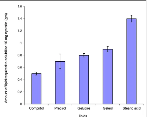
Solubility of Nystatin in Lipids: Solubility of drug in lipid is one of the most important factors for determining drug loading capacity of the SLNs. Solubility of nystatin was tested in 5 lipids, precirol ATO5, Geleol, Gelucire, Compritol 888 ATO and
stearic acid (Fig. 1). The amount of lipid required to
solubilize 10 mg drug was 0.5±0.025, 0.7±0.12, 0.9±0.44, 0.8±0.03 and 1.4±0.055 gm for compritol 888ATO, precirol, geleol, gelucire and stearic acid respectively. From the entire lipid used, this result indicated that nystatin loading capacity of compritol might be more than other lipids and it was chosen for the preparation of SLN.
FIG. 3: SOLUBILITY PROFILE OF NYSTATIN IN THE SOLID LIPIDS
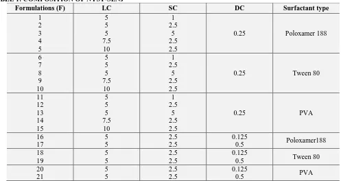
Preparation of Nyst-SLNs: Nystatin loaded SLNs dispersions were composed of Compritol with different concentrations 5, 7.5 & 10%(w/w). The lipid based carrier systems were stabilized by 1, 2.5 & 5%(W/W) surfactant (poloxamer 188 or tween 80 or PVA).Nystatin was incorporated with different concentrations 0.125, 0.25 & 0.5% (W/W). Twenty one different formulations were prepared; the composition of the investigated Nyst-SLNs is shown in table 1.
Effect of Formulation Variables: The effect of
different formulation variables as lipid
concentrations (LC), surfactant types (ST) and surfactant concentrations (SC) as well as drug concentrations (DC) on nystatin loaded SLNs were investigated. Effects of different formulation variables on particle size (PS), zeta potential (ZP), and entrapment efficiency (E.E.) are discussed in the
following section and presented in table 2.
1. Surfactant Concentration: Concentration of surfactants has a great influence on the drug E.E%. Table 2 shows that the entrapment efficiency increases with increasing SC in case of poloxamer 188 and PVA while no significant increase in E.E% was observed using tween 80
table 2. The possible reason for this observed
increase in the E.E% might be due to that at this concentration, surfactant provided sufficient covering to the lipid core so as to minimize possible leaching of the drug which means preventing drug diffusion to the external phase
[image:3.612.318.574.42.246.2]As the percentage of surfactant increased, part of the nystatin was incorporated in the surfactant layer at the surface of the SLN, leading to a high
entrapment efficacy 13. This may be also due to
the increase in the solubility of the drug in the lipid on increasing the concentration of the
surfactant 14-15. However , in case of tween 80 ,
further increase in concentration doesn’t has a significant increase in the E.E% could suggest that an optimum concentration of the surfactant
was reached to cover the surface of
nanoparticles effectively. Similar observations
were reported by other researchers 16. There was
a gradual decrease in particle size with an increase in surfactant concentrations. This observation was noticed for all formulations except for F7, F8 and F9 where an initial increase in surfactant concentration from 2.5%(w/w) to 5%(w/w) doesn’t result in change in particle size.
The decrease in nanoparticles size at high surfactant concentrations is due to effective reduction in interfacial tension between the aqueous and lipid phase, leading to the formation of emulsion droplets of smaller size, which on cooling results in smaller nanoparticles
15
. As depicted from table 2, all formulations were negatively charged, the zeta potential varied from -18.9mV for F16 to -38.8 mV for F21 indicating a relatively good stability and dispersion quality. It was clear that as the
amount of surfactant increased in the
formulation, the zeta potential became more negative.
2. Surfactant Type: As shown in table 2, a significant increase in E.E. was observed when poloxamer 188 or PVA was used. However, E.E. was significantly low when using tween 80 which mean that tween 80 was unsuitable for the preparation of Nyst-SLNs. It is evident from the above data that using PVA gave rise to nanoparticles of larger size compared to that using poloxamer 188 and tween 80. On the other hand, the influence of surfactant type is not well-defined on the zeta potential.
3. Lipid Concentration: As expected, table 2
shows that upon increasing the concentration of compritol a gradual increase in the entrapment efficiencies of nystatin SLNs in case of
poloxamer 188 & PVA was observed. A possible explanation for this observation is that the increase in lipid content can afford more space to encapsulate more drug and thus
reducing drug partition in the outer phase 16-17.
This also may probably be due to increase in the viscosity of the medium resulting in faster solidification of nanoparticles. This would further prevent the drug diffusion to the external
phase of the medium 12. Particle size
significantly increased with increasing lipid concentration (LC). It was noticeable that F4, F5, F14 and F15 show larger particle size at 7.5%(w/w) and 10%(w/w) concentration. At higher lipid contents, the efficiency of
homogenization decreases due to higher
viscosity of the sample, resulting in larger particles. Also, a high particle concentration at high lipid contents increases the probability of
particle contact and subsequent aggregation 18.
4. Drug Concentration: Increasing the amount of drug while keeping the emulsifier level constant, as shown in table 2, would decrease the E.E%. As lipid has certain drug loading capacity, addition of excess drug led to increase of unencapsulated drug (i.e. decrease in E.E.). Furthermore, after reaching maximum drug loading capacity E.E. cannot increase, which signify that E.E. reach its maximum point at
0.125% drug concentration 19.
Drug concentration has also an observed role in particle size. The results show that the particle size increase with increasing drug concentration from 0.125 %(w/w) to 0.5%(w/w). This can be explained as before lipid has certain loading capacity, addition of excess drug may aggregate giving larger particle size.
Transmission Electron Microscopy (TEM):
Electron micrographs indicate that the prepared SLNs had nanometer-size with no drug crystal formation. Most of the particles were round and uniform in size with smooth surface morphology (Fig. 2). Figure 2(D) illustrates the presence of a layer surrounding the particles of nystatin SLNs. During the lipid solidification process, lipid molecules solidify first to form a lipid core, which results in a high concentration of drug crystallizing on the surface of the particles which explain the
TABLE 1: COMPOSITION OF NYST-SLNs
Formulations (F) LC SC DC Surfactant type
1 2 3 4 5 5 5 5 7.5 10 1 2.5 5 2.5 2.5
0.25 Poloxamer 188
6 7 8 9 10 5 5 5 7.5 10 1 2.5 5 2.5 2.5
0.25 Tween 80
11 12 13 14 15 5 5 5 7.5 10 1 2.5 5 2.5 2.5
0.25 PVA
16 17 5 5 2.5 2.5 0.125
0.5 Poloxamer188 18 19 5 5 2.5 2.5 0.125
0.5 Tween 80
20 21 5 5 2.5 2.5 0.125
[image:5.612.59.560.325.754.2]0.5 PVA
TABLE 2: ENTRAPMENT EFFICIENCY (E.E.), PARTICLE SIZE (PS), ZETA POTENTIAL (ZP) OF THE NYST-SLNs Formulations (F) Entrapment Efficiency (%) Particle Size (nm) Zeta Potential (-mv)
FIG. 2: TRANSMISSION ELECTRON MICROGRAPHS OF (A) NYSTATIN-LOADED SLN DISPERSION F19, (B) NYSTATIN-LOADED SLN DISPERSION F6, (C) NYSTATIN-LOADED SLN DISPERSION F1, (D) SLN MICROGRAPH REPRESENTING THE CORE AND SHELL THEORY
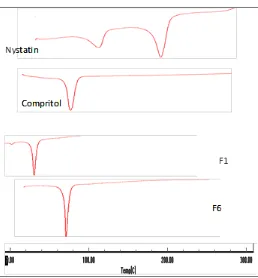
Differential Scanning Calorimetry (DSC):
Thermal behavior of the pure drug, compritol compared with the thermograms of different lyophilized SLNs formulae in the range of 25 to
350ºC is shown in Fig. 3. Crystalline nystatin
demonstrated a sharp endothermic peak at 167.11ºC corresponds to melting temperature of nystatin while sharp endothermic peak at 74ºC was observed for compritol. Absence of the peak at167.11ºC in nystatin loaded SLNs indicates either formation of amorphous dispersion of nystatin in lipid matrix or solubilization of nystatin in lipid matrix upon
heating. Similar results were obtained by Das et al.,
in studying the thermal analysis of tretinoin loaded SLNs and proving the amorphous dispersion and
[image:6.612.102.517.43.188.2]solubilization of tretinoin upon heating 19.
FIG. 3: DSC SCANS OF NYSTATIN, COMPRITOL AND DIFFERENT FORMULATION OF NYST-SLNS
In-vitro Drug Release: The dialysis bag diffusion technique was adopted to evaluate the release of nystatin from the investigated formulations. The dialysis membrane retains the nanoparticles and permitted the transfer of the drug directly into the receiver compartment. Soluble drug was partitioned from the nanoparticles into the liquid phase, and then it diffused into the receptor medium. The data clearly shows that the release of the drug can be influenced by the type and concentration of surfactant, in addition the concentration of lipid matrix used, and
[image:6.612.44.302.474.752.2]FIG. 4: RELEASE EFFICIENCY OF NYSTATIN FROM DIFFERENT FORMULATIONS USING DIFFERENT SURFACTANTS
In order to compare the drug release profile from the prepared SLN formulations, the release efficiency (R.E. %) after 24 hours was studied. The figures clearly show that the release of the drug can be influenced by the type and concentration of surfactant and the concentration of nystatin used (Fig. 5). The results show that upon increasing the concentration of surfactants, the percentage of nystatin released decrease as well as the release efficiency (Fig. 4), which was noticeable for all three surfactants. The decrease in drug release at higher surfactant concentration as obtained is most likely due to retardation of the drug release caused by micellar entrapment of the drug. The slow release of nystatin from all the formulae suggests homogenous
entrapment of the drug throughout the systems 21.
The increase in the lipid concentration is accompanied with decrease of the percentage of nystatin released (F4, F5, F9, F10, F14 and F15).
FIG. 5: RELEASE EFFICIENCY OF NYSTATIN FROM DIFFERENT FORMULATIONS USING DIFFERENT DRUG CONCENTRATIONS
This decrease can be attributed to the higher lipid content encapsulating the drug may cause that the diffusion distance for drug to diffuse out from Solid
Lipid Nanoparticles was increased 22. Nystatin is
released more quickly when using in lower concentration because of the drug-enriched shell
model 23-24. According to the drug-enriched shell
model of drug incorporation, a solid lipid core forms when the recrystallization temperature of the lipid is reached. On reducing the temperature of the dispersion, the drug concentrates in the still liquid outer shell of the SLN.
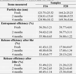
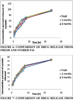
Stability studies: The stability study was done for 2
formulations: F16 and F20. From table 3, it was
found that the particle size of SLNs dispersion show no significant increase , the same was observed for the entrapment efficiency which show also a no significant decrease. Although sustained drug release was observed from both formulations after 2 and 4 months of storage, release was relatively faster from
SLNs stored than fresh SLNs (Fig. 6 & 7). This
faster release might be due to rearrangement of crystal lattice in the SLN lipid matrix over time, which pushes some of the drug molecules migrate
[image:7.612.43.300.43.225.2]towards nanoparticle surface 19.
TABLE 3: STABILITY PROFILE OF SLNS Items measured Samples
F16 F20
Particle size (nm)
Fresh 123.75±25.52 164.2±25.23 2 months 124.81±15.61 168.06±9.55 4 months 124.90±10.12 169.34±8.66
Entrapment efficiency (%)
Fresh 56.58±3.21 58.77±4.01 2 months 54.62±2.10 56.77±3.11 4 months 55.90±0.65 56.80±1.22
Release efficiency after 8hr (%)
Fresh 41.43±1.22 17.86±0.67 2 months 48.88±0.56 17.68±1.34 4 months 53.43±0.77 20.13±2.01
Release efficiency after 24hr (%)
[image:7.612.319.587.420.679.2] [image:7.612.43.300.550.714.2]FIGURE 6: COMPARISON OF DRUG RELEASE FROM FRESH AND STORED F16
FIGURE 7: COMPARISON OF DRUG RELEASE FROM FRESH AND STORED F20
CONCLUSION: Homogenization followed by ultrasonication method is suitable to produce Nyst-SLNs of 83.26-955 nm size ranges. Lipophilic drug like nystatin can be loaded in the compritol using poloxamer 188 or PVA as surfactants. Tween 80 was unsuitable for the preparation of Nyst-SLNs. The loading of nystatin explain the drug-enriched-shell theory. DSC analysis showed amorphous state of nystatin. Nystatin is released more quickly when using in lower concentration. Stability studies proved that the 2 formulae suggested are stable upon storage.
REFERENCES:
1. U. Malike and Y.Gugun,” Importance of solid lipid nanoparticles (SLN) in various administration routes and future perspectives”, Int.J.Nanomedecine 2007; Vol. 2: 289-300.
2. H.R. Race and S.I.Schantz,” Nystatin (Mycostatin) in the treatment of monilial and non monilial Vaginitis”, JAMA 1956; Vol. 162: 268-271.
3. D. Quinones, MS. Evone, S. Ghaly, “Formulation and characterization of nystatin gel”, PRHSJ, Vol. 27(1), 61-67, 2008.
4. M. Joshi, and V.Patravale, Drug Dev. Ind. Pharm., Vol. 32, 911, 2006
5. W. Mehnert and K. Mader, “Solid lipid nanoparticles: Production, Characterization and applications”, Adv. Drug Deliv. Rev. 2001; Vol. 47: 165.
6. V. Venkateswarlu, and K. Manjunath, “Preparation, characterization and in vitro release kinetics of clozapine solid lipid nanoparticles”, J. Control. Release 2004; Vol. 95: 627.
7. V. Venkateswarlu, and K. Manjunath, “Pharmacokinetics, tissue distribution and bioavailability of clozapine solid lipid nanoparticles after intravenous and intraduodenal administration” J. Control. Release 2005; Vol. 107: 215.
8. SH. Gohla, and A. Dingler,” Scaling up feasibility of the production of solid lipid nanoparticles (SLN)”, Pharmazie 2001; Vol. 56: 61-63.
9. D., Hou, C. Xie, K. Huang and C. Zhu,” The production and characteristics of solid lipid nanoparticles (SLNs)”, Biomaterials. 2003; Vol. 24: 1781.
10. .B. Souto, S.A. Wissing, C.M. Barbosa and R.H. Muller, “Development of a controlled release formulation based on SLN and NLC for topical clotrimazole delivery”, Int.J.Pharm 2004; Vol. 278: 71. 11. S.C. Yang, L.F. Lu, Y., Cai, J.B., Zhu, B.W. Liang and C.Z. Yang, “Body distribution in mice of intravenously injected camptothecin solid lipid nanoparticles and targeting effect on brain”, J. Control.Release 1999; Vol. 59: 299.
12. Y.Yang, T.Chung, X. Bai and W. Chan,” Effect of preparation conditions on morphology and release profiles of biodegradable polymeric microspheres containing protein fabricated by double emulsion method”,Chem. Eng. Sci. 2000; Vol. 55: 2223-2236. 13. H. Jifu, F. Xinsheng, Z. Yanfang, W. Jianzhu, G.
Fengguang, L. Fei, and P. Xinsheng, “Development and optimization of solid lipid nanoparticle formulation for ophthalmic delivery of chloramphenicol using a box-Behnken design”, Int. J.Nanomedicine 2001; Vol. 6: 687.
14. P. Ekambaram and S.A. Hasan,” Formulation and evaluation of solid lipid nanoparticles of ramipril”, J. Young Pharm. 2001; Vol. 3: 216-220.
15. G. Abdelbary and RH. Fahmy,” Diazepam loaded solid lipid nanoparticles: design and characterization”, AAPS Pharm. Sci. Tech. 2009; Vol. 10: 213-215. 16. Q. Lva, A. Yua, Y.Xia, H. Lia , Z. Songa, J. Cuic, F.
Caoa and G. Zhaia,” Development and evaluation of penciclovir-loaded solid lipid nanoparticles for topical delivery”, Int. J. Pharm. 2009; Vol. 372: 191.
17. K.A. Shah, A.A. Date, M.D. Joshi and V.B. Patravale,” Solid lipid nanoparticles (SLN) of tretinoin: Potential in topical delivery”, Int.J.Pharm. 2007; Vol. 345: 163.
18. C. Freitas and R.H. Muller,” Effect of light and temperature on zeta potential and physical stability in solid lipid nanoparticle (SLN) dispersions”, Int.J.Pharm. 1998; Vol. 168: 221.
variables”, Colloids Surf. B. Biointerfaces. 2011; Vol. 88: 483-489.
20. M. Uner and G. Yener,” Importance of solid lipid nanoparticles (SLN) in various administration routes and future perspectives”, Int J Nanomedicine 2007; Vol. 2: 289–300.
21. F.A. Oladimiji, S.I. Omoruyl, and C.O. Onyeji, “Preparation and in vitro evaluation of suppositories of halofantrine hydrochloride”, African J. Biotechnology 2006; Vol. 5: 1775-1780.
22. P. Pravin, K.S., Anil, C.D. Subhash, and K.S. Vijay,” Development of solid lipid nanoparticles of
lamivudine for brain targeting”, Int. J. Drug Deliv.Tech. 2009; Vol. 1: 36-38.
23. H. Heiati, R. Tawashi, and R.R. Shivers,” Solid lipid nanoparticles as drug carriers I. Incorporation and retention of the lipophilic prodrug3’- azido-3’deaxythymidine palmitate”, Int. J. Pharm. 1997; Vol. 146: 123-31.
24. R.H. Muller, M. Radtke, and S.A. Wissing,” Solid lipid nanoparticles (SLN) and nanostructured lipid carriers (NLC) in cosmetic and dermatological preparations”, Adv. Drug Deliv. Rev. 2002; Vol. 1, 131-55.
All © 2013 are reserved by International Journal of Pharmaceutical Sciences and Research. This Journal licensed under a Creative Commons Attribution-NonCommercial-ShareAlike 3.0 Unported License.
How to cite this article: